Emerging Technologies in Breast Imaging
|
|
|
- May Sanders
- 5 years ago
- Views:
Transcription

1 June 30, 2016 Emerging Technologies in Breast Imaging Jay A. Baker, M.D. Division of Breast Imaging
2 New Technologies in Breast Imaging Full Field Digital Mammography (FFDM) X-ray/Mammography Computer-Aided Detection (CAD) Contrast Enhanced Spectral Mammography Ultrasound Digital Breast Tomosynthesis (DBT) Contrast Enhanced Tomosynthesis MRI Dedicated Breast CT Tissue Harmonic Imaging (THI) Nuclear Medicine Compound Imaging Elastography Extended Field of View Imaging (EFOV) Other Automated Breast Ultrasound (ABUS) Diffusion Weighted Imaging (DWI) Diffusion Tensor Imaging Gamma Imaging (BSGI/MBI) Positron Emission Mammography (PEM) Radioactive Seed Localization Optical Imaging
3 New Technologies in Breast Imaging Full Field Digital Mammography (FFDM) X-ray/Mammography Computer-Aided Detection (CAD) Contrast Enhanced Spectral Mammography Ultrasound Digital Breast Tomosynthesis (DBT) Contrast Enhanced Tomosynthesis MRI Dedicated Breast CT Tissue Harmonic Imaging (THI) Nuclear Medicine Compound Imaging Elastography Extended Field of View Imaging (EFOV) Other Automated Breast Ultrasound (ABUS) Diffusion Weighted Imaging (DWI) Diffusion Tensor Imaging Gamma Imaging (BSGI/MBI) Positron Emission Mammography (PEM) Radioactive Seed Localization Optical Imaging
4 Lecture Outline Contrast Enhanced Mammography Gamma Imaging courtesy of Dr. Andrew Maidment Radionuclide seed localization
5 Lecture Outline Contrast Enhanced Mammography Gamma Imaging courtesy of Dr. Andrew Maidment Radionuclide seed localization


6 GE Medical Systems, Inc. Contrast Enhanced Digital Mammography CEDM General approach: modified digital mammography system iodinated contrast material
7 Contrast Enhanced Digital Mammography CEDM General approach: modified digital mammography system iodinated contrast material + Iodine Iodine Sub two images subtract one image from another iodine-only image
8 Contrast Enhanced Digital Mammography Modifications Contrast Enhanced Digital Mammography kvp copper filter rapid filter change Temporal Subtraction k-edge of iodine Dual Energy
9 Contrast Enhanced Digital Mammography Contrast Enhanced Digital Mammography Temporal Subtraction Dual Energy
10 Contrast Enhanced Digital Mammography Contrast Enhanced Digital Mammography Temporal Subtraction Dual Energy
11 Temporal Subtraction Before & After inject contrast position patient and light compress baseline image (mask) at kvp inject iodine contrast ml/sec repeat images q1 minute over 3-5 min subtract mask from subsequent images all images high energy: kvp
12 Temporal Subtraction post pre-con subtract (mask)
13 Jong RA, Yaffe M, et al. Radiology 2003
14 Temporal Subtraction Advantages high energy = low dose kinetic curves (MRI) low cost vs MRI Disadvantages long exam time motion misregistration one projection only (can t move) kinetics not robust compressed breast longer time to inject miss sec window

15 Jong RA, Yaffe M, et al. Radiology 2003
16 Contrast Enhanced Digital Mammography Contrast Enhanced Digital Mammography Temporal Subtraction Dual Energy
17 Contrast Enhanced Digital Mammography Contrast Enhanced Digital Mammography Temporal Subtraction Dual Energy
18 Dual Energy Subtraction CESM = Contrast Enhanced Spectral Mammography dual energy technique both images after contrast one sees contrast; other doesn t
 low absorption at low energy (26-32 kvp) k-edge of")
19 Dual Energy Subtraction x-ray absorption of iodine at dif energies high absorption at high energy (45-49 kvp) low absorption at low energy (26-32 kvp) k-edge of iodine
20 Dual Energy Subtraction x-ray absorption of iodine at dif energies high absorption at high energy low absorption at low energy inject 1.5 ml/kg 3cc/sec wait 2 min position & compress in MLO; hi/lo E images hi/lo E images in all 4 projections (5 min)
21 Dual Energy Subtraction Advantages Disadvantages no motion issues shorter compresion multiple projections


22 Case 1 79 yo w palpable mass, left breast standard mammogram GE Healthcare, Inc images courtesy of Mikawa Breast Center, Mikawa,


23 Invasive Ductal Carcinoma 2min Low Energy Subtraction 4min Subtraction Low Energy GE Healthcare, Inc images courtesy of Mikawa Breast Center, Mikawa,


24 Case 2 Invasive Ductal Carcinoma 2min Low Energy Subtraction 4min Subtraction Low Energy GE Healthcare, Inc images courtesy of Mikawa Breast Center, Mikawa,


25 Case 3 Invasive Lobular Carcinoma 2min Low Energy Subtraction 4min Subtraction Low Energy GE Healthcare, Inc images courtesy of Mikawa Breast Center, Mikawa,
26 Dual Energy Subtraction Advantages no motion issues shorter compresion multiple projections Disadvantages dose
27 Contrast Enhanced Digital Mammography Dose dual energy 1.2x routine mammo high energy = 0.2x mmg low energy = 1.0 mmg 4 views 4.8x mmg temporal subtraction 0.2x mmg pre post images total 1.0 mmg
28 CEDM Clinical Studies Thibault Eur Radiol 2011 Methods prototype dual energy (spectral) n=142 lesions (80 malignant) compare: mammo alone mammo + US mammo + CESM
29 CEDM Clinical Studies Thibault Eur Radiol 2011 Methods prototype dual energy (spectral) n=142 lesions (80 malignant) Sensitivity Specificity ROC multifocal lesions detected mammo mammo + US mammo + CEDM 0.93 * 0.56 * 0.88 * 23
30 CEDM Clinical Studies Jochelson MS (MSK) Radiology 2013 Methods prototype dual energy (spectral) n=52 women; bx-proven malignancy MRI vs dual energy CESM
31 CEDM Clinical Studies Jochelson MS (MSK) Radiology 2013 Methods prototype dual energy (spectral) n=52 women; bx-proven malignancy mammo MRI CESM Index lesion (n=52) 81% 96% 96% Multifocal/centric (n=25) 28% 88% 56% Contralateral CA (n=1) False positives 13 2
32 Pros/Cons CEDM Advantages cost accessibility false positives Disadvantages contrast reaction contrast nephropathy patient acceptibility lack of image-guided bx
33 CEDM State of the Art imaging system Health Canada and FDA-approved (GE) software upgrade to existing units (2010) generator already capable of kvp copper filter billing HCPCS G0204 bilat digit mammo CPT diagnostic injection contrast material

34
35
36 Lecture Outline Contrast Enhanced Mammography Gamma Imaging Radionuclide seed localization

 cardiac perfusion")
37 In the beginning Cardiolite Tc-99m sestamibi (20 mci) (2-methoxy isobutyl isonitrile) cardiac perfusion imaging itonline.net itonline.net
 (2-methoxy isobutyl isonitrile) cardiac perfusion")
38 In the beginning Cardiolite Tc-99m sestamibi (20 mci) (2-methoxy isobutyl isonitrile) cardiac perfusion heart thyroid submandibular glands liver breast cancer S Usmani BJR, 83, 2010
39 Why does sestamibi concentrate in breast cancer?
40 Why does sestamibi concentrate in breast cancer? unclear 90% concentrates in mitochondria depends on regional blood flow angiogenesis tissue metabolism courtesy of Dr. Andrew Maidment
41 Small Field of View Gamma Cameras


42 Dedicated Breast Gamma Cameras Courtesy of GE Healthcare Courtesy of Gamma Medica Courtesy of Dilon Technologies BSGI = Breast Specific Gamma Imaging (analog) MBI = Molecular Breast Imaging (digital, CZT detector)
43 Small FOV Camera high resolution (<2mm) scatter from liver, heart mild compression detector-to-breast narrow dead zone at edge visibility medially visibility posteriorly Courtesy of GE Healthcare same projections as mammo
44 Imaging Technique Tc-99m sestamibi dose = mci <20 mci is off-label wait 5-15 min 10 min per projection (4 total 60min) light compression CC and MLO positions match mammo


45 50 yo High risk screening Courtesy of R. Brem, GWU invasive ductal carcinoma
46 Clinical Trials
47 BSGI Accuracy Brem RF, Radiology 247, 2008 retrospective review of BSGI n =146 women (variety of indications) n =167 lesions (83 malignant) 30 mci Tc-99m-sestamibi (CC and MLO) Results Sensitivity Specificity PPV NPV BSGI 96% 60% 69% 94% BSGI 1cm 85%

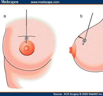
")


48 MBI Screening Rhodes DJ. Radiology 258, 2010 (Mayo) MBI for screening n=936 women hetero or extremely dense tissue
49 MBI Screening Rhodes DJ. Radiology 258, 2010 (Mayo) MBI for screening n=936 women hetero or extremely dense tissue moderate-high risk 20 mci Tc-99m sestamibi Risk Factors BRCA1 or BRCA2 Lifetime risk 20% 5-yr risk % Mantel or axilla radiation Personal hx of breast CA Hx of ADH, ALH, LCIS, etc Family Hx of breast cancer
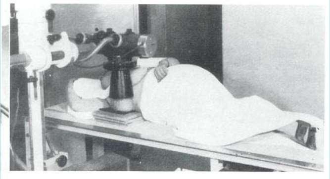
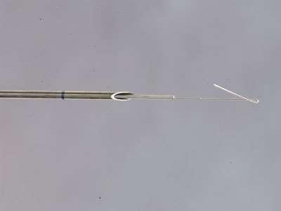
50 MBI Screening Rhodes DJ. Radiology 258, 2010 (Mayo) MBI for screening n=936 women hetero or extremely dense tissue moderate-high risk 20 mci Tc-99m sestamibi Results Cancers Mammo 1 MBI 7 Both 2 Neither 1 TOTAL 11
 MBI for screening n=936 women hetero or extremely dense tissue moderate-high")
51 MBI Screening Rhodes DJ. Radiology 258, 2010 (Mayo) MBI for screening n=936 women hetero or extremely dense tissue moderate-high risk 20 mci Tc-99m sestamibi Results Mammo MBI Both CA/ p=0.016

52 Courtesy of GE Healthcare High risk screening, Normal mammogram Bx = 5mm invasive ductal CA
53 Gamma Imaging Limitations false positives NPV < 98% (ie., cannot rule-out cancer) availability fat necrosis dose FDA = mci req d = 2-4 mci courtesy of R. Brem, GWU
 MBI for screening n=1585 women hetero or extremely dense tissue improved collimator; wider")
54 Low Dose MBI Screening Rhodes DJ. AJR 204, 2015 (Mayo) MBI for screening n=1585 women hetero or extremely dense tissue improved collimator; wider energy window 8 mci Tc-99m sestamibi Results Mammo MMG/MBI CA/ p<0.001
55
56 Lecture Outline Contrast Enhanced Mammography Gamma Imaging Radionuclide seed localization
57 Disclosure Off label usage: Breast localization with an I-125 radioactive seed is off label use of this FDA-approved product.
58 Traditional Wire Localization Wire placed using mammo or ultrasound guidance Wire skewers lesion Surgeon uses wire as roadmap to lesion


59
60 Traditional Wire Localization Pros easy placement accurate excision standard-of-care Cons uncomfortable procedure wire in place lengthens day for patient can t be first case for O.R. limits surgical approach wire kinks send surgeon in wrong direction localization must be done in same facility as surgery


61 Radioactive Seed Localization I-125 seed, sealed titanium source prostate brachytherapy seed but lower dose Seed placed using mammo or ultrasound guidance Seed placed within the lesion and/or near biopsy marking clip Surgeon uses handheld gamma probe to detect seed and dissect down to seed

62


63

64

65


66
67 Accuracy McGahn LJ, et al. Ann Surg Onc Oct RSL procedures 97% negative margin on 1 st excision 9% re-excision for close margin
68 Accuracy Lovrics PJ, et al. Ann Surg Onc Nov 2011 >300 cases: half RSL / half wire loc WIRE LOC SEED LOC Positive Margins 11.5% 10.8% Operative Time 22.2 min 19.4 min Surgeon Preference p=0.008 Patient Pain p=0.04 p=ns p<0.001
69 Radioactive Seed Localization Pros easy, comfortable procedure no wire in place done 1-5 days before O.R. shortens day for patient any surgical approach can be first case of day same or better positive margin rate (seed placed at lesion; seeds don t migrate) seed placement disconnected from O.R. location Cons uncomfortable procedure radioactivity bracketing is challenging seed cannot be repositioned billing issues for Radiology losing a seed creates substantial problems

70 Because of it s benign physical properties, Iodine- 125 is not THIS complicated to deal with...


71 Proper Handling of Seeds This Way......NOT This Way!
72 Seed Program Challenges Start up cooperation: Radiology Surgery Pathology Radiation Safety Learning curve Administrative oversight Reimbursement issues
73

74
75 Conclusions Contrast Enhanced Mammography potential alternative to MRI Gamma Imaging (BSGI or MBI) limited utility for work-ups screening requires dose reduction Radioactive Seed Localization advantages for patient, surgeon, radiol requires cooperation and oversight

76
77 THANK YOU
78
79
80
81
82
83


84

85 Courtesy of GE Medical Systems, Inc. Metabolic
86 now near future


87 CESM Case 3 CESM Image Rt CC 2 min. CESM Image Rt MLO 4 min. Images courtesy of Dr Mizutani Mikawa Breast Cancer Clinic Miakawa-anjo, JAPAN


 and")


.")
88 Thibault F. EJR Sept 2012 Fig. 2. Same patient as in Fig. 1. Preoperative bilateral contrast enhanced spectral mammography. Post-contrast lowenergy (a-b) and combined images (c-d). Contrast uptake is highlighted in both index lesion and anterior focus (arrows). No significant enhancement of breast tissue is seen otherwise. Note similar lesion visibility on CESM and


89 History of Breast Imaging 1895 Wilhelm Roentgen discovers the x- ray 1913 Albert Salomon images mastx specimens 1930 Stafford Warren invents mammogram
90 History of Breast Imaging 1895 Wilhelm Roentgen discovers the x-ray 1913 Albert Salomon images mastx specimens 1930 Stafford Warren invents mammogram 1950 s Robert Egan, dedicated mammo film 1966 first dedicated mammo machine 1970s xeromammography 1980s plain film analog mammography 2000 first digital mammography system
91 History of Breast Imaging 1895 Wilhelm Roentgen discovers the x-ray 1913 Albert Salomon images mastx specimens 1930 Stafford Warren invents steroscopic technique for imaging breast in vivo 1950 s Robert Egan, reproducible, low kvp technique 1966 first dedicated mammo machine (CGR Senographe, France) 1971 xeromammography 1972 first dedicated mammo film/screen combination (DuPont) 1980s plain film analog mammography 2000 first digital mammography system (GE) 2011 digital breast tomosynthesis (Hologic)


92 osa.edu

93
94 History of Breast Imaging 1895 Wilhelm Roentgen discovers the x-ray 1913 Albert Salomon images mastx specimens 1930 Stafford Warren invents steroscopic technique for imaging breast in vivo 1950 s Robert Egan, reproducible, low kvp technique 1966 first dedicated mammo machine (CGR Senographe, France) 1971 xeromammography 1972 first dedicated mammo film/screen combination (DuPont) 1980s plain film analog mammography 2000 first digital mammography system (GE) 2011 digital breast tomosynthesis (Hologic)
95 What Will It Take? Out-side-the-box concepts and engineering Compelling empiric data Screening vs Diagnostic vs Extent of Disease Consecutive case series Multi-institutional data Convenience (prone, water bath, etc) Cost Reimbursement Biopsy capability (eg., ARIA)
96 FFDM CAD dual energy contrast enhanced CESM tomo contrast tomo tomo CAD
97 asdf Review emerging technologies Discuss potential role in patient care Problem solving? What is NPV? Compare claims to empiric data

98 Tomosynthesis

99 Case 4 Invasive Ductal Carcinoma


100

101 F ½ by 1 ½


102
103
104

105

106

107
108 Contrast Enhanced Digital Mammography Equipment modified mammo gantry generator/tube: kvp fast kv switching: avoid motion (dual sub) copper filter power injector

109
110 Disclosure Siemens Medical Systems tomosynthesis grant
Contrast-Enhanced Digital Mammography
 2015 ARRS Breast Symposium Contrast-Enhanced Digital Mammography John Lewin, M.D. Diversified Radiology of Colorado CEDM - Outline History Technique Literature Review / Cases Clinical Status Inexpensive,
What s New in Breast Imaging. Jennifer A. Harvey, M.D., FACR Professor of Radiology University of Virginia
 What s New in Breast Imaging Jennifer A. Harvey, M.D., FACR Professor of Radiology University of Virginia Disclosure Hologic, Inc. Shareholder and research agreement. Volpara Solutions, Ltd. Shareholder
High Risk Screening: A Multimodality Approach
 High Risk Screening: A Multimodality Approach John Lewin, M.D., FACR, FSBI The Women s Imaging Center Denver, Colorado Disclosures Consultant to Hologic Previously received research funds from Hologic
Emerging Techniques in Breast Imaging: Contrast-Enhanced Mammography and Fast MRI
 Emerging Techniques in Breast Imaging: Contrast-Enhanced Mammography and Fast MRI Lilian Wang, M.D. Breast Imaging Section Department of Radiology Northwestern Medicine Overview Rationale for new imaging
Molecular Breast Imaging: History and Recent Developments
 Molecular Breast Imaging: History and Recent Developments Associate Professor, Department of Imaging Physics The University of Texas MD Anderson Cancer Center, Houston, Texas Educational Objectives 1.
Contrast Enhanced Spectral Mammography (CESM) Updates
 Updates") Contrast Enhanced Spectral Mammography (CESM) Updates Georgeta Mihai, PhD, DABR Medical Physicist, BIDMC, Boston Assistant Professor, Harvard Medical School, Boston Disclosures None Acknowledgments: Da
Contrast Enhanced Spectral Mammography (CESM) Updates Georgeta Mihai, PhD, DABR Medical Physicist, BIDMC, Boston Assistant Professor, Harvard Medical School, Boston Disclosures None Acknowledgments: Da
New Imaging Modalities for better Screening and Diagnosis
 New Imaging Modalities for better Screening and Diagnosis Miri Sklair-Levy, MD Department of Diagnostic Imaging Sheba Medical Center, Sackler School of Medicine, Tel Aviv University Department of Diagnostic
2/14/2019. Advances in Breast Imaging. Outline. Jiali Wang, Ph.D., DABR Medical Physicist. February 23, 2019
 Advances in Breast Imaging SCPMG Medical Imaging Technology & Informatics Jiali Wang, Ph.D., DABR Medical Physicist February 23, 2019 Outline History of breast imaging Advances in breast imaging a) Full-Field
Current Status of Supplementary Screening With Breast Ultrasound
 Current Status of Supplementary Screening With Breast Ultrasound Stephen A. Feig, M.D., FACR Fong and Jean Tsai Professor of Women s Imaging Department of Radiologic Sciences University of California,
Physics of MBI (~10 slides)
") Physics of MBI (~10 slides) Molecular Breast Imaging (MBI) physics and performance testing JW Hugg, BR Simrak, PD Smith, BE Patt Gamma Medica, Inc., Northridge, CA Molecular Breast Imaging (MBI) is an
Physics of MBI (~10 slides) Molecular Breast Imaging (MBI) physics and performance testing JW Hugg, BR Simrak, PD Smith, BE Patt Gamma Medica, Inc., Northridge, CA Molecular Breast Imaging (MBI) is an
Low Dose Molecular Breast Imaging
 Low Dose Molecular Breast Imaging Dr. M.K. O Connor Conflict of Interest Royalties - Gamma Medica Research funding GE Healthcare Research support MTTI Michael O Connor, Ph.D Dept. of Radiology Mayo Clinic
Contrast-Enhanced Spectral Mammography
 Contrast-Enhanced Spectral Mammography Illuminating Breast Cancer Detection SenoBright HD TM gehealthcare.com/senobright Mammography is the most reliable imaging technique for breasts, but limitations
Mammographic imaging of nonpalpable breast lesions. Malai Muttarak, MD Department of Radiology Chiang Mai University Chiang Mai, Thailand
 Mammographic imaging of nonpalpable breast lesions Malai Muttarak, MD Department of Radiology Chiang Mai University Chiang Mai, Thailand Introduction Contents Mammographic signs of nonpalpable breast cancer
CZT Detectors for Medical Imaging and Plant Biology
 Small Business, High Tech Development CZT Detectors for Medical Imaging and Plant Biology Haris Kudrolli Inc. Workshop 2018 January 11, 2018 Outline Scientific Section CZT detectors Plant Imaging Applications
Update of Digital Breast Tomosynthesis. Susan Orel Roth, MD
 Update of Digital Breast Tomosynthesis Susan Orel Roth, MD NCI estimates that : Why DBT? Approximately 20% of breast cancers are missed at mammography screening Average recall rates approximately 10%
Breast Cancer Imaging
 Breast Cancer Imaging I. Policy University Health Alliance (UHA) will cover breast imaging when such services meet the medical criteria guidelines (subject to limitations and exclusions) indicated below.
Contrast-Enhanced Spectral Mammography helps to improve breast cancer diagnostics
 GE Healthcare Contrast-Enhanced Spectral Mammography helps to improve breast cancer diagnostics SenoBright TM mammography, Midwest U.S. hospital Simple test benefits clinicians and patients A hospital
Updates in Mammography. Dr. Yang Faridah A. Aziz Department of Biomedical Imaging University Malaya Medical Centre
 Updates in Mammography Dr. Yang Faridah A. Aziz Department of Biomedical Imaging University Malaya Medical Centre Updates in Mammography Breast Imaging Dr. Yang Faridah A. Aziz Department of Biomedical
SCREENING FOR BREAST CANCER BREAST IMAGING
 SCREENING FOR BREAST CANCER BREAST IMAGING Liane Philpotts, MD, FSBI, FACR Professor, Radiology and Biomedical Imaging Division Chief, Breast Imaging Dec. 5, 2017 Warner, E. NEJM 2011 Screening for
Breast Imaging Update: Old Dog New Tricks
 Breast Imaging Update: Old Dog New Tricks Claire McKay, DO M&S Imaging Assoc. San Antonio, TX cmckayhart@juno.com Goals Describe modalities available, old and new Provide understanding of pros and cons
Outline. Digital Breast Tomosynthesis: Update and Pearls for Implementation. Tomosynthesis Dataset: 2D/3D (Hologic Combo Acquisition)
") Outline Digital Breast Tomosynthesis (DBT) the new standard of care Digital Breast Tomosynthesis: Update and Pearls for Implementation Emily F. Conant, M.D. Professor, Chief of Breast Imaging Department
Outline Digital Breast Tomosynthesis (DBT) the new standard of care Digital Breast Tomosynthesis: Update and Pearls for Implementation Emily F. Conant, M.D. Professor, Chief of Breast Imaging Department
Screening Options in Dense Breasts. Donna Plecha, M.D. Co-Director UHCMC Breast Centers Associate Professor of Radiology Director of Breast Imaging
 Screening Options in Dense Breasts Donna Plecha, M.D. Co-Director UHCMC Breast Centers Associate Professor of Radiology Director of Breast Imaging Dense Breasted Women Decreased sensitivity of mammography
Here are examples of bilateral analog mammograms from the same patient including CC and MLO projections.
 Good afternoon. It s my pleasure to be discussing Diagnostic Breast Imaging over the next half hour. I m Wei Yang, Professor of Diagnostic Radiology and Chief, the Section of Breast Imaging as well as
Dense Breasts. A Breast Cancer Risk Factor and Imaging Challenge
 Dense Breasts A Breast Cancer Risk Factor and Imaging Challenge Renee Pinsky, MD University of Michigan Department of Radiology Division of Breast Imaging No Disclosures QUIZ: ARE YOU DENSE? a. Breast
Molecular Breast Imaging
 Molecular Breast Imaging Development of a Low-Dose Screening Test for Dense Breasts Conflict of Interest Royalties for technologies licensed to Gamma Medica Ideas Carrie B. Hruska, Ph.D. Department of
Digital Breast Tomosynthesis from a first idea to clinical routine
 International Master Programm Biomedical Engineering Digital Breast Tomosynthesis from a first idea to clinical routine Historical background 2D imaging of 3D objects has important limitations Jörg Barkhausen
Since its introduction in 2000, digital mammography has become
 Review Article Smith A, PhD email : Andrew.smith@hologic.com Since its introduction in 2000, digital mammography has become an accepted standard of care in breast cancer screening and has paved the way
SenoBright Contrast Enhanced Spectral Mammography Technology. Ann-Katherine Carton Sylvie Saab-Puong Matt Suminski
 SenoBright Contrast Enhanced Spectral Mammography Technology Ann-Katherine Carton Sylvie Saab-Puong Matt Suminski White Paper October 2012 SenoBright Contrast Enhanced Spectral Mammography Technology Ann-Katherine
Pitfalls and Limitations of Breast MRI. Susan Orel Roth, MD Professor of Radiology University of Pennsylvania
 Pitfalls and Limitations of Breast MRI Susan Orel Roth, MD Professor of Radiology University of Pennsylvania Objectives Review the etiologies of false negative breast MRI examinations Discuss the limitations
Newly Diagnosed Breast Cancer: Preoperative Imaging and Localization
 Newly Diagnosed Breast Cancer: Preoperative Imaging and Localization Debra Monticciolo, MD Professor of Radiology Texas A&M University no disclosures Debra Monticciolo, MD Professor of Radiology Texas
BREAST DENSITY WHAT IS IT? WHY IS IT IMPORTANT? & What IOWA SF250 Means to Patients and Providers
 BREAST DENSITY WHAT IS IT? WHY IS IT IMPORTANT? & What IOWA SF250 Means to Patients and Providers Arnold Honick, MD Radiology Consultants of Iowa, PLC ahonick@rciowa.com BREAST DENSITY LEGISLATION Nancy
Medical Policy. MP Scintimammography and Gamma Imaging of the Breast and Axilla
 Medical Policy MP 6.01.18 BCBSA Ref. Policy: 6.01.18 Last Review: 09/19/2018 Effective Date: 09/19/2018 Section: Radiology Related Policies 9.01.502 Experimental / Investigational Services DISCLAIMER Our
Financial Disclosures
 Financial Disclosures 3D Mammography: The Latest Developments in the Breast Imaging Arena I have no financial disclosures Dr. Katharine Lampen-Sachar Breast and Body Radiologist Radiology Associates of
Standard Breast Imaging Modalities. Lilian Wang, M.D. Breast Imaging Section Department of Radiology Northwestern Medicine
 Standard Breast Imaging Modalities Lilian Wang, M.D. Breast Imaging Section Department of Radiology Northwestern Medicine Overview Standard breast imaging modalities Mammography Ultrasound MRI Imaging
Hacia la imagenología tomográfica de mama
 Hacia la imagenología tomográfica de mama Futuro y presente Ioannis Sechopoulos, Ph.D., DABR Advanced X ray Tomographic Imaging (AXTI) Lab Department of Radiology and Nuclear Medicine Radboud University
Scintimammography and Gamma Imaging of the Breast and Axilla
 Scintimammography and Gamma Imaging of the Breast and Axilla Policy Number: 6.01.18 Last Review: 9/2017 Origination: 9/2006 Next Review: 9/2018 Policy Blue Cross and Blue Shield of Kansas City (Blue KC)
Detailed Program of the second BREAST IMAGING AND INTERVENTIONS PROGRAM am am : Clinician s requirements from breast imaging
 Detailed Program of the second BREAST IMAGING AND INTERVENTIONS PROGRAM 2012 Day one, 2 nd November BREAST IMAGING AND INTERVENTIONS PROGRAM 2012 9.00 AM 9.10 am Introduction 9.10 am - 9.30 am : Clinician
Detection to Prediction: Imaging Markers of Breast Cancer Risk
 Detection to Prediction: Imaging Markers of Breast Cancer Risk Carrie B. Hruska, PhD, DABR Associate Professor of Medical Physics Mayo Clinic, Rochester, MN 2017 MFMER slide-1 Disclosure Per agreement
Courtesy of Dan Kopans, M.D.
 Molecular Breast Imaging New Modality for Diagnosing i Breast Cancer Don t we have enough tools for diagnosing i Breast Cancer??? By Lillian H. Stern, M.D. Current Modalities Mammograms (analogue v. digital)
Medical Policy An independent licensee of the Blue Cross Blue Shield Association
 Scintimammography and Gamma Imaging of the Breast and Axilla Page 1 of 28 Medical Policy An independent licensee of the Blue Cross Blue Shield Association Title: Scintimammography and Gamma Imaging of
Contrast enhanced spectral mammography: A literature review
 Contrast enhanced spectral mammography: A literature review Poster No.: R-0172 Congress: RANZCR-AOCR 2012 Type: Authors: Keywords: DOI: Educational Exhibit S. Buzynski, D. Taylor; Perth/AU Breast, Digital
Breast Cancer Screening and High Risk
 Breast Cancer Screening and High Risk Mary Freyvogel, DO Breast Surgeon Clinical Assistant Professor of Surgery University Hospitals Case Medical Center St. John Medical Center / Elyria Medical Center
Disclosures. Outline. Learning Objectives. Introduction. Introduction. Stereotactic Breast Biopsy vs Mammography: Image Quality and Dose.
 Disclosures Stereotactic Biopsy vs Mammography: and Dose None Vikas Patel, PhD, DABR Upstate Medical Physics 2014 Annual Meeting The American Association of Physicists in Medicine Austin, TX Learning Objectives
Contrast Enhanced Spectral Mammography (CESM) Initial UK Experience. Dr Sarah L Tennant BMedSci, BMBS, MRCP, FRCR
 Initial UK Experience. Dr Sarah L Tennant BMedSci, BMBS, MRCP, FRCR") Contrast Enhanced Spectral Mammography (CESM) Initial UK Experience Dr Sarah L Tennant BMedSci, BMBS, MRCP, FRCR Vote Now Your experience of CESM 1. No experience of CESM 44% 2. I ve seen some cases in
Contrast Enhanced Spectral Mammography (CESM) Initial UK Experience Dr Sarah L Tennant BMedSci, BMBS, MRCP, FRCR Vote Now Your experience of CESM 1. No experience of CESM 44% 2. I ve seen some cases in
Breast Cancer Screening and Diagnosis
 Breast Cancer Screening and Diagnosis Priya Thomas, MD Assistant Professor Clinical Cancer Prevention and Breast Medical Oncology University of Texas MD Anderson Cancer Center Disclosures Dr. Thomas has
2/9/2015. If you do, you ve been working way too hard and too long! 1953 Uruguay
 IN THE BEGINNING Mammography technology has come a long way since the first machine specifically designed for producing mammograms was introduced in 1966. The first mammography system was essentially a
TOMOSYNTHESIS: WORTH ALL THE HYPE?
 X-Ray Associates of New Mexico, P.C. TOMOSYNTHESIS: WORTH ALL THE HYPE? MICHAEL N. LINVER, MD, FACR MAMMOGRAPHY: THE GOOD, THE PRETTY GOOD, & THE NOT SO GOOD MAMMOGRAPHY: THE GOOD, THE PRETTY GOOD, & THE
BREAST IMAGING and NEW IMAGING MODALITIES- A Surgeons view
 BREAST IMAGING and NEW IMAGING MODALITIES- A Surgeons view DR CHANTEL THORNTON SPECIALIST BREAST CANCER SURGEON BMSc (hons) MBBS (hons) FRACS Epworth Hospital, Richmond- Agora Centre for Women s Health
Role of positron emission mammography (PEM) for assessment of axillary lymph node status in patients with breast cancer
 for assessment of axillary lymph node status in patients with breast cancer") Role of positron emission mammography (PEM) for assessment of axillary lymph node status in patients with breast cancer Poster No.: C-1260 Congress: ECR 2011 Type: Scientific Paper Authors: K. M. Kulkarni,
Role of positron emission mammography (PEM) for assessment of axillary lymph node status in patients with breast cancer Poster No.: C-1260 Congress: ECR 2011 Type: Scientific Paper Authors: K. M. Kulkarni,
Radioactive Seed Localization of Nonpalpable Breast Lesions
 Subject: Radioactive Seed Localization of Page: 1 of 7 Last Review Status/Date: March 2017 Radioactive Seed Localization of Description Radioactive seed localization is used to detect nonpalpable breast
8/3/2016. DBT Physics Basic to Advanced: Primer On Tomosynthesis. Tomosynthesis Pedigree
 DBT Physics Basic to Advanced: Primer On Tomosynthesis Andrew D. A. Maidment, Ph.D. University of Pennsylvania Department of Radiology Acknowledgements of Support Research support from the Komen Foundation,
11/1/2014. Radiologic incidentalomas Ordering pitfalls Newer technology and applications
 Bilal Tahir, MD Gitasree Borthakur, MD Indiana University School of Medicine Department of Radiology & Imaging Sciences October 31, 2014 ACP 2014 Dr. V. Aaron Nuclear (vaaron@iupui.edu) Dr. S. Westphal
Breast tomosynthesis reduces radiologist performance variability compared to digital mammography
 Breast tomosynthesis reduces radiologist performance variability compared to digital mammography Andrew Smith 1, Elizabeth Rafferty 2, Loren Niklason 1 1 Hologic, Inc., Bedford MA, USA 2 Massachusetts
Contrast-enhanced spectral mammography (CESM) in a large scale breast cancer screening program. Preliminary clinical experience.
 in a large scale breast cancer screening program. Preliminary clinical experience.") Contrast-enhanced spectral mammography (CESM) in a large scale breast cancer screening program. Preliminary clinical experience. Poster No.: C-1651 Congress: ECR 2012 Type: Educational Exhibit Authors:
Contrast-enhanced spectral mammography (CESM) in a large scale breast cancer screening program. Preliminary clinical experience. Poster No.: C-1651 Congress: ECR 2012 Type: Educational Exhibit Authors:
CURRENTLY FDA APPROVED ARE FULL FIELD DIGITAL MAMMOGRAPHY SYSTEMS AND FILM SCREEN STILL BEING USED AT SOME INSTITUTIONS
 ABBY DUROJAYE,M.D CURRENTLY FDA APPROVED ARE FULL FIELD DIGITAL MAMMOGRAPHY SYSTEMS AND FILM SCREEN STILL BEING USED AT SOME INSTITUTIONS BOTH HAVE BEEN SHOWN TO BE EFFECTIVE TOOLS EARLY DETECTION OF BREAST
What is Molecular Breast Imaging?
 What is Molecular Breast Imaging? The basics of Functional Imaging Functional imaging is the detection of breast abnormality based on the altered characteristic of the tissue, rather than its altered morphology
EARLY DETECTION: MAMMOGRAPHY AND SONOGRAPHY
 EARLY DETECTION: MAMMOGRAPHY AND SONOGRAPHY Elizabeth A. Rafferty, M.D. Avon Comprehensive Breast Center Massachusetts General Hospital Harvard Medical School Breast Cancer Screening Early detection of
MANAGEMENT OF DENSE BREASTS. Nichole K Ingalls, MD, MPH NW Surgical Specialists September 25, 2015
 MANAGEMENT OF DENSE BREASTS Nichole K Ingalls, MD, MPH NW Surgical Specialists September 25, 2015 No financial disclosures National Cancer Institute National Cancer Institute Increased Cancer Risk... DENSITY
BREAST CANCER SCREENING IS A CHOICE
 BREAST CANCER SCREENING IS A CHOICE by ELAINE SCHATTNER, MD no financial disclosures (ES) American Association for Cancer Research Typical headlines focus on controversy 2 Data: Breast Cancer Incidence
Mammography. Background and Perspective. Mammography Evolution. Background and Perspective. T.R. Nelson, Ph.D. x41433
 - 2015 Background and Perspective 2005 (in US) Women Men Mammography Invasive Breast Cancer Diagnosed 211,240 1,690 Noninvasive Breast Cancer Diagnosed 58,940 Deaths from Breast Cancer 40,410 460 T.R.
Screening mammograms in women <50 years of age: Low risk is NOT protective
 Screening mammograms in women
Opportunities and Innovations in Digital Mammography John M. Sandrik, Ph.D. GE Healthcare Milwaukee, WI
 Opportunities and Innovations in Digital Mammography John M. Sandrik, Ph.D. GE Healthcare Milwaukee, WI john.sandrik@med.ge.com with many thanks to Vince Polkus, Advanced Applications Product Mgr. 1 Content
EARLY DETECTION: MAMMOGRAPHY AND SONOGRAPHY
 EARLY DETECTION: MAMMOGRAPHY AND SONOGRAPHY Elizabeth A. Rafferty, M.D. Avon Comprehensive Breast Center Massachusetts General Hospital Harvard Medical School Breast Cancer Screening Early detection of
#46: DIGITAL TOMOSYNTHESIS: What is the Data Really Showing? TERMS (AKA) WHAT IS TOMOSYNTHESIS? 3/3/2014. Digital breast tomosynthesis =
 WHAT IS TOMOSYNTHESIS? 3/3/2014. Digital breast tomosynthesis =") #46: DIGITAL TOMOSYNTHESIS: What is the Data Really Showing? January K. Lopez, MD Hoag Breast Care Center Newport Beach, CA Disclosures: None TERMS (AKA) Digital breast tomosynthesis = DBT Tomo 3D Full
#46: DIGITAL TOMOSYNTHESIS: What is the Data Really Showing? January K. Lopez, MD Hoag Breast Care Center Newport Beach, CA Disclosures: None TERMS (AKA) Digital breast tomosynthesis = DBT Tomo 3D Full
Supplemental Screening for Dense Breasts. Reagan Leverett, MD, MS
 Supplemental Screening for Dense Breasts Reagan Leverett, MD, MS Outline Anatomy and Density Risk of dense breasts Theory of Supplemental Screening Options for supplemental screening Tomosynthesis Ultrasound
BCCCNP Service CPT Code FY 2019 Rate Oct 1, 2018 Dec 31, 2018
 1 Screening Mammogram (Bilateral); including CAD Service CPT Code 77067 77067-TC 77067-26 $111.40 $81.32 $30.08 $131.51 $93.70 $37.82 * Note: Breast tomosynthesis, unilateral (77061) and bilateral (77062)
PURPOSE IMAGE-GUIDANCE MODALITIES IMAGE-GUIDED BREAST BIOPSY. US-Techniques. Ultrasound. US guided NLOBB. TH. Helbich
 IMAGE-GUIDED BREAST BIOPSY PURPOSE TH. Helbich Department of Radiology Division of Molecular & Gender Imaging Medical University of Vienna Imaging techniques Interventional procedures Quality management
Tomosynthesis and breast imaging update. Dr Michael J Michell Consultant Radiologist King's College Hospital NHS Foundation Trust
 Tomosynthesis and breast imaging update Dr Michael J Michell Consultant Radiologist King's College Hospital NHS Foundation Trust Breast imaging new technology BREAST CANCER FLT PET shows different grades
Ge elastography cpt codes
 Ge elastography cpt codes Aetna considers digital mammography a medically necessary acceptable alternative to film mammography. Currently, there are no guideline recommendations from leading medical professional
Breast density: imaging, risks and recommendations
 Breast density: imaging, risks and recommendations Maureen Baxter, MD Radiologist Director of Ruth J. Spear Breast Center Providence St. Vincent Medical Center Alison Conlin, MD/MPH Medical Oncologist
Update in Breast Cancer Screening
 Disclosure information: Update in Breast Cancer Screening Karla Kerlikowske, MDDis Update in Breast Cancer Screening Grant/Research support from: National Cancer Institute and Grail - and - Karla Kerlikowske,
Mammography. What is Mammography?
 Scan for mobile link. Mammography Mammography is a specific type of breast imaging that uses low-dose x-rays to detect cancer early before women experience symptoms when it is most treatable. Tell your
BCCCNP Service CPT Code FY 2019 Rate Oct 1, 2018 Dec 31, 2018
 1 Screening Mammogram (Bilateral); including CAD 2 Screening Breast Tomosynthesis (Bilateral) 3D Mammogram ** Can only be paid w/ screening mammography (77067))** 3 Diagnostic Mammogram (Unilateral); including
Update in Breast Cancer Screening
 Disclosure information: Update in Breast Cancer Screening Karla Kerlikowske, MDDis Update in Breast Cancer Screening Grant/Research support from: National Cancer Institute - and - Karla Kerlikowske, MD
Scintimammography. What is scintimammography?
 Scan for mobile link. Scintimammography Scintimammography uses small amounts of radioactive materials called radiotracers, a special camera and a computer to help investigate an abnormality discovered
Initial Experience with Contrast Enhanced Digital Mammography (SenoBright) In a Comprehensive Clinical Breast Center
 In a Comprehensive Clinical Breast Center") Journal of Cancer Therapy, 2017, 8, 146-154 http://www.scirp.org/journal/jct ISSN Online: 2151-1942 ISSN Print: 2151-1934 Initial Experience with Contrast Enhanced Digital Mammography (SenoBright) In a
Journal of Cancer Therapy, 2017, 8, 146-154 http://www.scirp.org/journal/jct ISSN Online: 2151-1942 ISSN Print: 2151-1934 Initial Experience with Contrast Enhanced Digital Mammography (SenoBright) In a
Women s Imaging Original Research
 Women s Imaging Original Research Brandt et al. DBT for Screening Recalls Without Calcifications Women s Imaging Original Research FOCUS ON: Kathleen R. Brandt 1 Daniel A. Craig 1 Tanya L. Hoskins 2 Tara
Role of PEM in Breast Cancer Management. Judy Kalinyak, MD, PhD Chief Medical Officer Naviscan, Inc (San Diego, CA)
") Role of PEM in Breast Cancer Management Judy Kalinyak, MD, PhD Chief Medical Officer Naviscan, Inc (San Diego, CA) Role of PEM in Breast Cancer Management Introduction to Positron Emission Mammography
Role of PEM in Breast Cancer Management Judy Kalinyak, MD, PhD Chief Medical Officer Naviscan, Inc (San Diego, CA) Role of PEM in Breast Cancer Management Introduction to Positron Emission Mammography
CURRENT METHODS IN IMAGE GUIDED BREAST BIOPSY
 CURRENT METHODS IN IMAGE GUIDED BREAST BIOPSY Stuart Silver April 24, 2004 OBJECTIVES Review development of current techniques Discuss stereotactic breast biopsy Discuss US guided breast biopsy 1 OBJECTIVES
Successful Breast MRI Program : The ingredients
 Successful Breast MRI Program : The ingredients Dr. Smriti Hari Associate Professor Deptt. Of Radiology All India Institute of Medical Sciences New Delhi How to perform Breast MRI Breast MRI descriptors
Angela Gilliam, MD University of Colorado Surgical Grand Rounds November 3, 2008
 Angela Gilliam, MD University of Colorado Surgical Grand Rounds November 3, 2008 Breast Cancer Most common cancer in American women 180,000 new cases per year Second most common cause of cancer death 44,000
Corporate Medical Policy
 Corporate Medical Policy File Name: Origination: Last CAP Review: Next CAP Review: Last Review: digital_breast_tomosynthesis 3/2011 6/2016 6/2017 11/2016 Description of Procedure or Service Conventional
Screening with New Modalities: Breast Ultrasound
 Screening with New Modalities: Breast Ultrasound Wendie A. Berg, MD, PhD Professor of Radiology Magee-Womens Hospital of UPMC University of Pittsburgh School of Medicine Disclosures No personal financial
arxiv: v2 [cs.cv] 8 Mar 2018
![arxiv: v2 [cs.cv] 8 Mar 2018](/thumbs/87/97094636.jpg "arxiv: v2 [cs.cv] 8 Mar 2018") Automated soft tissue lesion detection and segmentation in digital mammography using a u-net deep learning network Timothy de Moor a, Alejandro Rodriguez-Ruiz a, Albert Gubern Mérida a, Ritse Mann a, and
Automated soft tissue lesion detection and segmentation in digital mammography using a u-net deep learning network Timothy de Moor a, Alejandro Rodriguez-Ruiz a, Albert Gubern Mérida a, Ritse Mann a, and
Contrast-Enhanced Breast Tomosynthesis
 Contrast-Enhanced Breast Tomosynthesis AIP Industrial Physics Forum 2009 Andrew D. A. Maidment, Ph.D. Chief, Physics Section Department of Radiology University of Pennsylvania Acknowledgements of Support
FY16 BCCS Reimbursement Rates and Billing Guidelines Appendix B 2
 FY16 BCCS Reimbursement Rates and Billing Guidelines Appendix B 2 77053 Mammary ductogram or galactogram, single duct, Global Fee $59.05 May be billed with 77055, G0206, 77056, G0204, 76641, 76642 Billable
Screening Mammography: The Controversy, Risk Assessment and Individualized Screening recommendations. Jonathan T. Sims MD, MBA
 Screening Mammography: The Controversy, Risk Assessment and Individualized Screening recommendations. Jonathan T. Sims MD, MBA I have no relevant Financial Disclosures Agenda Discuss the recent studies
Radiation Dosimetry in Digital Breast Tomosynthesis. March, 2015 William J. O Connel, Dr. Ph, Senior Medical Physicist
 Radiation Dosimetry in Digital Breast Tomosynthesis March, 2015 William J. O Connel, Dr. Ph, Senior Medical Physicist Imagination at work. Syllabus 1. Introduction 2. Dosimetry in Mammography 3. Dosimetry
Related Policies None
 Medical Policy MP 6.01.57 BCBSA Ref. Policy: 6.01.57 Last Review: 09/19/2018 Effective Date: 09/19/2018 Section: Radiology Related Policies None DISCLAIMER Our medical policies are designed for informational
The Cure Starts Here 2/9/2015. You can get up and use the restroom anytime, but be discreet please. Objectives
 Objectives The Cure Starts Here Deborah Thames R.T. (R)(M)(QM) Staging a Patient through Imaging Modalities Knowing the Choices with BI-RADs and the Lexicon Treatment Options and Managing Patient Care
Moderne mammadiagnostikk hvor står vi og hvor går vi?
 NBCG Oslo 15. juni 2018 Moderne mammadiagnostikk hvor står vi og hvor går vi? Professor dr.med. emeritus Per Skaane Oslo University Hospital Ullevaal Breast Imaging Center Oslo / Norway PERSKA@ous-hf.no
Contrast-Enhanced Breast Tomosynthesis: Combining the Best of Both Worlds for Better Breast-Cancer Diagnosis
 Contrast-Enhanced Breast Tomosynthesis: Combining the Best of Both Worlds for Better Breast-Cancer Diagnosis T Wu (twu2@partners.org), E Rafferty, R Moore, D Kopans, Massachusetts General Hospital, Boston,
Diagnostic Medical Physicist Via Christi Hospitals Wichita, Wichita, KS
 Digital Breast Tomosynthesis SWAAPM Meeting 30 Mar 2012 Jerry A. Thomas, MS, FAAPM, DABR, CHP, DABSNM Diagnostic Medical Physicist Via Christi Hospitals Wichita, Wichita, KS Talk Overview Breast Cancer
Breast Tomosynthesis An additional screening tool in the fight against breast cancer
 What to Expect Breast Tomosynthesis An additional screening tool in the fight against breast cancer Every woman over 40 should be examined for breast cancer once a year. American Cancer Society What to
FY 2017 BCCCNP Unit Cost Reimbursement Rate Schedule
 1 Screening Mammogram (Bilateral) 1a. ** NEW **- 01/01/2017- ** Replaces 77057** Screening Mammogram (Bilateral) 2 Digital Screening Mammogram (Bilateral) Service CPT Code 2a. Screening Breast Tomosynthesis
Breast Imaging & You
 Breast Imaging & You What s Inside: Breast Imaging... 2 Digital Breast Tomosynthesis (DBT) mammograms... 4 Breast cancer screening... 6 Dense breast tissue... 8 Automated Breast Ultrasound (ABUS)... 9
WHAT TO EXPECT. Breast Tomosynthesis An additional screening tool in the fight against breast cancer HOLOGIC. The Women's Health Company
 WHAT TO EXPECT Breast Tomosynthesis An additional screening tool in the fight against breast cancer HOLOGIC The Women's Health Company ...,. Screening for breast cancer Doctors and scientists agree that
Breast MRI Update. Jeffrey C. Weinreb, MD, FACR Yale University School of Medicine
 Breast MRI Update Jeffrey C. Weinreb, MD, FACR jeffrey.weinreb@yale.edu Yale University School of Medicine I disclose the following financial relationships with relevant commercial interests: Bracco Bayer
Breast Tomosynthesis. What is breast tomosynthesis?
 Scan for mobile link. Breast Tomosynthesis Breast tomosynthesis is an advanced form of mammography, a specific type of breast imaging that uses low-dose x-rays to detect cancer early when it is most treatable.
Armed Forces Institute of Pathology.
 Armed Forces Institute of Pathology www.radpath.com Armed Forces Institute of Pathology Breast Disease www.radpath.org Armed Forces Institute of Pathology Interpretation of Breast MRI Leonard M. Glassman