11/1/2014. Radiologic incidentalomas Ordering pitfalls Newer technology and applications
|
|
|
- Melinda Singleton
- 5 years ago
- Views:
Transcription
 Dr. K. Sandrasegaran Abdomen (ksandras@iupui.edu) Dr. K. Walker Breast (khwalker@iupui.edu) Dr. H. Kipfer Breast (hdkipfer@iupui.edu) Dr. E. Beckley (ebeckley@iupui.")
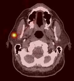
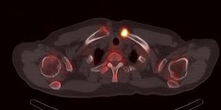
1 Bilal Tahir, MD Gitasree Borthakur, MD Indiana University School of Medicine Department of Radiology & Imaging Sciences October 31, 2014 ACP 2014 Dr. V. Aaron Nuclear Dr. S. Westphal Nuclear Dr. K. Sandrasegaran Abdomen Dr. K. Walker Breast Dr. H. Kipfer Breast Dr. E. Beckley Radiologic incidentalomas Ordering pitfalls Newer technology and applications Adrenal nodules Low density liver lesions Pancreatic cysts Renal cysts 1

:601 10. 3. Song JH, et al. AJR 008;190(5):1163 8.")

 a functioning adenoma = Conn syndrome")
 had adrenal gland involvement at presentation Only 4 (0.")
 with 1049 AI on CT 2 1-year imaging follow up or 2-yer clinical")
 likelihood of malignancy 1.")


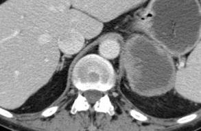
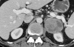
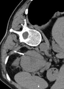
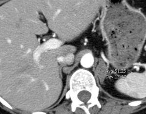
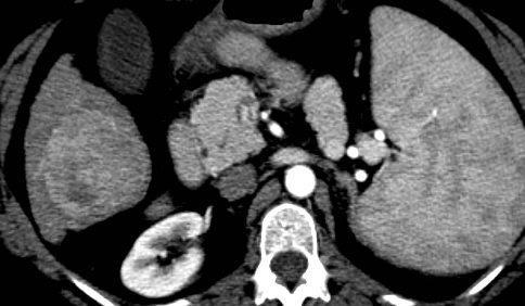
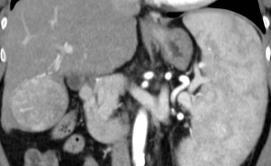
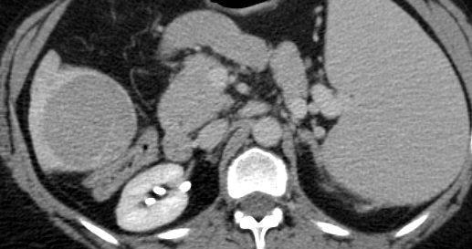
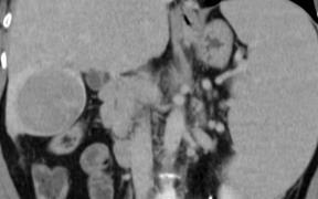
2 6% of CTs (10% in > 70- years age) 1 show adrenal incidentaloma (AI) 3-5% of AI are hormonally active 2 78% of AI are likely to be adenomas 3 70% of AI are lipid rich 1. Kloos RT, et alendocr Rev 1995;16(4): Young WF Jr. N Engl J Med 2007;356(6): Song JH, et al. AJR 008;190(5): Noncontrast CT for? renal calculi a Rt AI Density = 1 HU a lipid-rich adenoma Retrospective review of 1639 patients with suspected carcinoma of unknown primary 1 6 month later hypertension + low serum K + Underwent sampling (Lt, Rt adrenal veins) a functioning adenoma = Conn syndrome CT does not assess functionality of adenoma 95 (5.8%) had adrenal gland involvement at presentation Only 4 (0.2%) had isolated adrenal met (all > 6 cm size) Retrospective review of 973 patients (not known to have malignancy) with 1049 AI on CT 2 1-year imaging follow up or 2-yer clinical follow-up on all No adrenal malignancies seen Bottom line: < 4 cm nodule without known cancer or other metastatic lesions has very small (< 0.2%) likelihood of malignancy 1. Lee JE. Surgery 1998; 124(6): Song JH, et al. AJR 2008;190(5): May 2008 Jun 2008 Jun 2012 Guidelines for Managing AI Berland LL, et al. J Am Coll Radiol 2010;7(10): Left AI with high density (41 HU on Pre). No known cancer. Should have had follow-up unenhanced CT or MRI. Instead had biopsy which was not indicated without h/o cancer. Pre Enh Del CT 4 years later shows no change in size. Bladder cancer and bone met 5 cm left adrenal nodule. Likelihood of met is high. Should have had PET- CT or biopsy Instead had nonindicated triple-phase CT with showed indeterminate washout. AI less than 1 cm may be ignored When 1-4 cm, risk of malignant AI in noncancer patient is very low (< 0.2%) Lesion > 4 cm, require biopsy, PET-CT or resection 2


 Low density definitely benign features: no follow up Low")

3 Low risk < 40 years No malignancy No risk factors or history of chronic liver disease No liver-related symptoms Average risk: > 40years, o/w same as low risk High risk: Malignancy with known ability to metastasize to liver Berland L, et al. Managing Incidental Findings on Abdominal CT: White Paper of the ACR Incidental Findings Committee. J Am Coll Radiol 2010;7: If low or average risk: no further follow up If high risk: CT or MRI in 3 to 6 months If any of these features worrisome Ill-defined margin HU > 20 units Size change from prior Heterogenous enchancement No worrisome features No follow up (all risks) Any worrisome feature Follow up (typically MRI in 3-6 months) Low density definitely benign features: no follow up Low density suspicious features Low risk: 6-month follow up or MRI Average risk: Immediate MRI High risk: Biopsy or MRI Guidelines for managing incidental liver lesions depends on patient's risk for malignancy Most small lesions < 1.5 cm in low or intermediate risk patients do not need follow up 3
 Nonenhancing mural nodule High")
 No")
 imperceptible wall rounded")
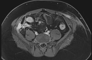
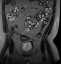
4 Low risk: Asymptomatic cyst < 2 cm Intermediate risk Cysts > 3 cm Main PD dilation of 5-9 mm Thick septa (> 2 mm) Nonenhancing mural nodule High risk Pancreatic head cyst with CBD obstruction Main PD 10 mm Enhancing mural nodule If cyst < 2 cm: follow up in 1 2 years (preferably MRI) No growth = stop surveillance If > 5 mm growth = treat as intermediate risk Jan 2008 Mar 2010 Simple cyst (Bosniak Type I) imperceptible wall rounded Work up: none 4


5 Mildly complex (Bosniak type 2) Thin septa Thin calcification Work up: none Confusion over when to order a screening mammogram vs. diagnostic mammogram Screen Women with no complaints Typically doctor order NOT needed, doctor name needed Order needed if history of breast cancer Order needed if younger than 35 Implants considered screen Diagnostic Clinical concern Lump, thickening, skin changes, nipple retraction, clear or bloody nipple discharge, FOCAL pain Must have a doctor order Radiologist on site and will completely work-up abnormality and give results at that time Ordering diagnostic mammograms for common breast complaints 5
6 Diagnostic mammogram not needed for Galactorrhea Diffuse breast pain Cyclical breast pain History of benign breast biopsies Skin lesions Confusion over when to order a breast US vs diagnostic mammogram ACR appropriateness criteria: Women 30 and older always get a mammogram first then a focused ultrasound if indicated Women under 30 get a FOCUSED ultrasound first Confusion over when to order a screening breast MRI Screening for High-risk patients ACS: High risk women Greater than 20% lifetime risk should get an MRI and a mammogram every year Women at moderately increased risk 15% to 20% lifetime risk should talk with their doctors about the benefits and limitations of adding MRI screening to their yearly mammogram Low to average risk Less than 15% lifetime risk should NOT get screening MRI Ordering a CT without contrast in addition to CT with contrast when not needed 6
7 Metastatic cancer work-up / follow-up Trauma (head & C/T/L-spine without) Abdominal / pelvic / chest pain, weight loss, infection, inflammatory processes, most other indications For arterial stenosis, CTA Some indications require multiple post contrast phases (HCC screening triple phase, pancreatic dual phase) Forgetting to order a CT without when needed in addition to a with contrast CT Aortic dissection Adrenal mass workup (may only need noncontrast) Renal mass workup HCC follow-up status post non-surgical treatment AAA status post endovascular stent graft repair CT Urogram CT Cystogram Ordering wrong protocol CT or excluding body part For aortic dissections/aneurysms CTA chest ordered but forget to order CTA abdomen/pelvis CT renal protocol renal mass vs renal stone Ordering CT stone protocol when meant to order CT urogram For abdominal pain forgetting to order CT pelvis in addition to abdomen For PE. Ordering a CTA chest Confusion over CT IV contrast administration and chronic renal insufficiency 7
8 On hemodialysis --- full IV contrast dose For GFR > 45, full IV contrast dose For GFR of and not on hemodialysis, IV hydration (250 ml NS prior and 250 ml after), decrease IV contrast dose by 20-25% For GFR of 30 or less and not on hemodialysis, no IV contrast Confusion over when to order a post contrast spine (C, T or L-spine) MRI Malignancy Infection Prior spine surgery Confusion over when to give enteric contrast for CT Gastrografin --- if pt < 150 lbs, recent GI surgery/enteric leak Water --- pancreatic protocol Volumen --- enterography 8


9 FDA approval 2011 Screening and diagnostic modality Same breast positioning and standard compression Acquires images at several angles in a short amount of time and reconstructs the images into thin, high-resolution slices Removes challenges from overlapping tissue Increases interpretation time Increases dose Images acquired at multiple angles of a compressed breast (as little as 4 seconds) as the detector and anode move around the breast Information is then reconstructed into slices Not widely available Expensive technology Increase interpretation time No reimbursement Insurance companies consider tomo investigational No CPT code January,
10 No consensus/recommendation All screening patients Subset of screening patients Dense High-risk Diagnostic add-views Tomosynthesis Increase Cancer Detection Rate Decrease Recall Rate Decrease patient anxiety Decrease biopsy rate Increase positive predictive value Increase cost-effectiveness Risk assessment Role for PCP High-risk clinics Molecular Breast Imaging (MBI) Single and Double detector systems commercially available Breast Specific Gamma Imaging (BSGI) refers to single detector studies Not effected by breast density Smaller dose may be possible with double-detector systems Systemic ionizing radiation exposure still main concern for use in screening Positron Emission Mammography (PEM) Similar efficacy with MRI in biopsy proven cancer: Sensitivity: PEM = 85%; MRI = 98% Specificity: PEM = 74%; MRI = 48% Systemic ionizing radiation still a concern May be a good alternative for patients with contraindications to MRI SPECT obtains biologic activity in 3D plane Conventional nuclear medicine images in 2D plane CT provides anatomic imaging SPECT-CT combines both imaging modalities Cardiac Parathyroid Bone Thyroid Cancer Lymphoscintigraphy 10

11 11
12 12
13 13

14 Calcifications Type II endoleak Use radiologist as a consultant rather than lab service. Radiologists can add value to patient care. Incidental adrenal nodules under 1 cm can be ignored. Those 1-4 cm may be followed up in 1 year. Most small low density liver lesions < 1.5 cm in low or intermediate risk patients do not need follow up. Asymptomatic pancreatic cysts < 2 cm require MRI follow-up to demonstrate 1-2 yr stability. Most renal cysts are simple or mildly complex (thin septa or calcification) and require no further imaging. Be aware of common pitfalls when ordering radiology exams Constant growth in radiologic technology and applications Feel free to with questions: btahir@iupui.edu hmtkko 14
Effective Utilization of Imaging. John V. Roberts, M.D. Premier Radiology Abdominal Imaging
 Effective Utilization of Imaging John V. Roberts, M.D. Premier Radiology Abdominal Imaging Safety Contrast and Radiation What to order Abdomen/Pelvis Brain/Spine Chest Musculoskeletal Ob/Gyn Head and Neck
Newcastle HPB MDM updated radiology imaging protocol recommendations. Author Dr John Scott. Consultant Radiologist Freeman Hospital
 Newcastle HPB MDM updated radiology imaging protocol recommendations Author Dr John Scott. Consultant Radiologist Freeman Hospital This document is intended as a guide to aid radiologists and clinicians
11/17/2013. Dr. Kumar Sandrasegaran Dr. Curtis Wright Beth Ward. Increased utilization of medical imaging:
 Bilal Tahir, MD Gitasree Borthakur, MD Indiana University School of Medicine Department of Radiology & Imaging Sciences Dr. Kumar Sandrasegaran Dr. Curtis Wright Beth Ward November 15, 2013 ACP 2013 Increased
ESUR 2018, Sept. 13 th.-16 th., 2018 Barcelona, Spain
 ESUR 2018, Sept. 13 th.-16 th., 2018 Barcelona, Spain OUR APPROACH Incidental adrenal nodule/mass Isaac R Francis, M.B;B.S University of Michigan, Ann Arbor, Michigan Disclosures None (in memory) M Korobkin,
Breast Imaging & You
 Breast Imaging & You What s Inside: Breast Imaging... 2 Digital Breast Tomosynthesis (DBT) mammograms... 4 Breast cancer screening... 6 Dense breast tissue... 8 Automated Breast Ultrasound (ABUS)... 9
Breast Cancer Screening and Diagnosis
 Breast Cancer Screening and Diagnosis Priya Thomas, MD Assistant Professor Clinical Cancer Prevention and Breast Medical Oncology University of Texas MD Anderson Cancer Center Disclosures Dr. Thomas has
Breast Imaging & You
 Breast Imaging & You What s Inside: Breast Imaging... 2 Digital Breast Tomosynthesis (DBT) mammograms... 4 Breast cancer screening... 6 Dense breast tissue... 8 Automated breast ultrasound (ABUS)... 9
Breast Cancer Imaging
 Breast Cancer Imaging I. Policy University Health Alliance (UHA) will cover breast imaging when such services meet the medical criteria guidelines (subject to limitations and exclusions) indicated below.
Breast Imaging Update: Old Dog New Tricks
 Breast Imaging Update: Old Dog New Tricks Claire McKay, DO M&S Imaging Assoc. San Antonio, TX cmckayhart@juno.com Goals Describe modalities available, old and new Provide understanding of pros and cons
STANDARDIZED MANAGEMENT RECOMMENDATIONS FOR ADRENAL NODULES: EVIDENCE-BASED CONSENSUS POWERSCRIBE MACROS FROM AN ACADEMIC/PRIVATE PRACTICE
 STANDARDIZED MANAGEMENT RECOMMENDATIONS FOR ADRENAL NODULES: EVIDENCE-BASED CONSENSUS POWERSCRIBE MACROS FROM AN ACADEMIC/PRIVATE PRACTICE COLLABORATIVE Pamela Johnson 1, Darcy Wolfman 2, Upma Rawal 3,
Imaging the Symptomatic Patient. Avice M.O Connell MD,FACR,FSBI Professor of Imaging Sciences Director, Women s Imaging University of Rochester
 Imaging the Symptomatic Patient Avice M.O Connell MD,FACR,FSBI Professor of Imaging Sciences Director, Women s Imaging University of Rochester The four most common symptoms Mass Pain Discharge Infection
EARLY DETECTION: MAMMOGRAPHY AND SONOGRAPHY
 EARLY DETECTION: MAMMOGRAPHY AND SONOGRAPHY Elizabeth A. Rafferty, M.D. Avon Comprehensive Breast Center Massachusetts General Hospital Harvard Medical School Breast Cancer Screening Early detection of
New Imaging Modalities for better Screening and Diagnosis
 New Imaging Modalities for better Screening and Diagnosis Miri Sklair-Levy, MD Department of Diagnostic Imaging Sheba Medical Center, Sackler School of Medicine, Tel Aviv University Department of Diagnostic
Contributors. Thanks to Peter Miller, MD; LCDR Kevin Preston, MD; and Keith Newbrough, MD for their generous contribution of images:
 Contributors Thanks to Peter Miller, MD; LCDR Kevin Preston, MD; and Keith Newbrough, MD for their generous contribution of images: Peter Miller, MD, Indiana University School of Medicine Chapter 1: Figure
Breast Imaging! Ravi Adhikary, MD!
 Breast Imaging! Ravi Adhikary, MD! ACS Estimated Cancers Statistics 2014! Breast! New Cases in Women! 232,670 (+67,570 in situ)! Deaths in Women! 40,000! Colon! 48,380! 24,040! Cervical! 12,360! 4,020!
MANAGEMENT RECOMMENDATIONS
 1 MANAGEMENT RECOMMENDATIONS 1. Adrenal masses!!!!!!! page 2 2. Liver Masses!!!!!!! page 3 3. Obstetric US Soft Markers for Aneuploidy!! pages 4-6 4. Ovarian and Adnexal Cysts!!!!! pages 7-10 5. Pancreatic
Epworth Healthcare Benign Breast Disease Symposium. Sat Nov 12 th 2016
 Epworth Healthcare Benign Breast Disease Symposium Breast cancer is common Sat Nov 12 th 2016 Benign breast disease is commoner, and anxiety about breast disease commoner still Breast Care Campaign UK
Q: Why is breast cancer a big deal?
 I hate breast cancer. As a radiologist who specializes in breast imaging, my career is devoted to the detection and diagnosis of breast cancer. I am passionate about women s health and my goal is to find
Clinical indications for positron emission tomography
 Clinical indications for positron emission tomography Oncology applications Brain and spinal cord Parotid Suspected tumour recurrence when anatomical imaging is difficult or equivocal and management will
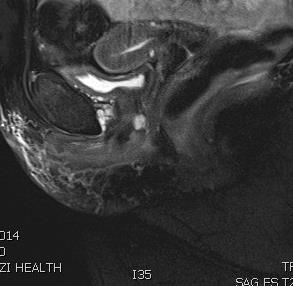
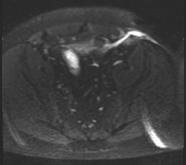
Endocrine MR. Jan 30, 2015 Michael LaFata, MD
 Endocrine MR Jan 30, 2015 Michael LaFata, MD Brief case 55-year-old female in ED PMH: HTN, DM2, HLD, GERD CC: Epigastric/LUQ abdominal pain, N/V x2 days AF, HR 103, BP 155/85, room air CMP: Na 133, K 3.6,
Melissa Hartman, DO Women s Health Orlando VA Medical Center
 Melissa Hartman, DO Women s Health Orlando VA Medical Center Most common non-skin cancer and Second deadliest cancer in women Majority are diagnosed by abnormal screening study An approach to breast cancer
Screening Mammograms: Questions and Answers
 CANCER FACTS N a t i o n a l C a n c e r I n s t i t u t e N a t i o n a l I n s t i t u t e s o f H e a l t h D e p a r t m e n t o f H e a l t h a n d H u m a n S e r v i c e s Screening Mammograms:
EARLY DETECTION: MAMMOGRAPHY AND SONOGRAPHY
 EARLY DETECTION: MAMMOGRAPHY AND SONOGRAPHY Elizabeth A. Rafferty, M.D. Avon Comprehensive Breast Center Massachusetts General Hospital Harvard Medical School Breast Cancer Screening Early detection of
Armed Forces Institute of Pathology.
 Armed Forces Institute of Pathology www.radpath.com Armed Forces Institute of Pathology Breast Disease www.radpath.org Armed Forces Institute of Pathology Interpretation of Breast MRI Leonard M. Glassman
Standard Breast Imaging Modalities. Lilian Wang, M.D. Breast Imaging Section Department of Radiology Northwestern Medicine
 Standard Breast Imaging Modalities Lilian Wang, M.D. Breast Imaging Section Department of Radiology Northwestern Medicine Overview Standard breast imaging modalities Mammography Ultrasound MRI Imaging
ADRENAL INCIDENTALOMA. Jamii St. Julien
 ADRENAL INCIDENTALOMA Jamii St. Julien Outline Definition Differential Evaluation Treatment Follow up Questions Case Definition The phenomenon of detecting an otherwise unsuspected adrenal mass on radiologic
What s New in Breast Imaging. Jennifer A. Harvey, M.D., FACR Professor of Radiology University of Virginia
 What s New in Breast Imaging Jennifer A. Harvey, M.D., FACR Professor of Radiology University of Virginia Disclosure Hologic, Inc. Shareholder and research agreement. Volpara Solutions, Ltd. Shareholder
INTERDISCIPLINARY DISCUSSIONS IN LOCALISED RCC DIAGNOSIS AND SURGICAL STRATEGIES FOR ATYPICAL RENAL CYSTIC LESIONS. Maria Cova
 INTERDISCIPLINARY DISCUSSIONS IN LOCALISED RCC DIAGNOSIS AND SURGICAL STRATEGIES FOR ATYPICAL RENAL CYSTIC LESIONS Maria Cova Radiology Department University of Trieste (IT) Eleventh European International
Radiology Update: Balancing Diagnostic Needs with Cost Effectiveness
 Radiology Update: Balancing Diagnostic Needs with Cost Effectiveness October 2018 Ramin Khorasani, MD, MPH Professor of Radiology, Harvard Medical School Director, Center for Evidence-Based Imaging Vice
Current Status of Supplementary Screening With Breast Ultrasound
 Current Status of Supplementary Screening With Breast Ultrasound Stephen A. Feig, M.D., FACR Fong and Jean Tsai Professor of Women s Imaging Department of Radiologic Sciences University of California,
Ryan Niederkohr, M.D. Slides are not to be reproduced without permission of author
 Ryan Niederkohr, M.D. CMS: PET/CT CPT CODES 78814 Limited Area (e.g., head/neck only; chest only) 78815 78816 Regional (skull base to mid-thighs) True Whole Body (skull vertex to feet) SELECTING FIELD
Radiological Tests: Which One is Most Appropriate for My Patient?
 Radiological Tests: Which One is Most Appropriate for My Patient? Robert Hartman, M.D. 2016 MFMER slide-1 Disclosures No Disclosures 2016 MFMER slide-2 Objectives Discuss tools to aid in the ordering of
POSITRON EMISSION TOMOGRAPHY (PET)
") Status Active Medical and Behavioral Health Policy Section: Radiology Policy Number: V-27 Effective Date: 08/27/2014 Blue Cross and Blue Shield of Minnesota medical policies do not imply that members should
Status Active Medical and Behavioral Health Policy Section: Radiology Policy Number: V-27 Effective Date: 08/27/2014 Blue Cross and Blue Shield of Minnesota medical policies do not imply that members should
THYROID NODULES: THE ROLE OF ULTRASOUND
 THYROID NODULES: THE ROLE OF ULTRASOUND NOVEMBER 2017 DR. DEAN DURANT DEFINITION Thyroid nodule: Focal area within the thyroid gland with echogenicity different from surrounding parenchyma. THYROID NODULES
REVIEW. Distinguishing benign from malignant adrenal masses
 Cancer Imaging (2003) 3, 102 110 DOI: 10.1102/1470-7330.2003.0006 CI REVIEW Distinguishing benign from malignant adrenal masses Isaac R Francis Professor of Radiology, Department of Radiology, University
Mammography. What is Mammography?
 Scan for mobile link. Mammography Mammography is a specific type of breast imaging that uses low-dose x-rays to detect cancer early before women experience symptoms when it is most treatable. Tell your
Disclosures. Breast Cancer. Breast Imaging Modalities. Breast Cancer Screening. Breast Cancer 6/4/2014
 : Information for the Primary Care Physician Disclosures No financial relationships with commercial entities producing health care products/services. Roxsann Roberts, MD Section Chief, MRI Erlanger/EmCare
Traumatic and Non Traumatic Adrenal Emergencies
 Traumatic and Non Traumatic Adrenal Emergencies Michael N. Patlas, MD, FRCPC (1), Christine O. Menias, MD (2), Douglas S. Katz, MD, FACR (3), Ania Z. Kielar, MD, FRCPC (4), Alla M. Rozenblit, MD (5), Jorge
Contrast Enhanced Ultrasound of Parenchymal Masses in Children
 Contrast Enhanced Ultrasound of Parenchymal Masses in Children Sue C Kaste, DO On behalf of Beth McCarville, MD St. Jude Children s Research Hospital Memphis, TN Overview Share St. Jude experience with
ROLE OF MRI IN SCREENING, DIAGNOSIS AND MANAGEMENT OF BREAST CANCER. B.Zandi Professor of Radiology
 ROLE OF MRI IN SCREENING, DIAGNOSIS AND MANAGEMENT OF BREAST CANCER B.Zandi Professor of Radiology Introduction In the USA, Breast Cancer is : The Most Common Non-Skin Cancer The Second Leading cause of
Breast Tomosynthesis. What is breast tomosynthesis?
 Scan for mobile link. Breast Tomosynthesis Breast tomosynthesis is an advanced form of mammography, a specific type of breast imaging that uses low-dose x-rays to detect cancer early when it is most treatable.
objectives Pitfalls and Pearls in PET/CT imaging Kevin Robinson, DO Assistant Professor Department of Radiology Michigan State University
 objectives Pitfalls and Pearls in PET/CT imaging Kevin Robinson, DO Assistant Professor Department of Radiology Michigan State University To determine the regions of physiologic activity To understand
Job Task Analysis for ARDMS Abdomen Data Collected: June 30, 2011
 Job Task Analysis for ARDMS Abdomen Data Collected: June 30, 2011 Reported: Analysis Summary for: Abdomen Examination Survey Dates 06/13/2011-06/26/2011 Invited Respondents 6,000 Surveys with Demographics
The Incidental Renal lesion
 The Incidental Renal lesion BACKGROUND Increase in abdominal CT/US in last 15 years Resulted in detection of many (small) renal lesions 50% > 50yrs has at least 1 lesion majority simple cysts Renal lesions
Renal masses - the role of diagnostic imaging
 Renal masses - the role of diagnostic imaging Poster No.: C-2471 Congress: ECR 2015 Type: Educational Exhibit Authors: V. Rai#; Bjelovar/HR Keywords: Cysts, Cancer, Structured reporting, Ultrasound, MR,
Foundational funding sources allow BCCHP to screen and diagnose women outside of the CDC guidelines under specific circumstances in Washington State.
 Program Description The Breast, Cervical and Colon Health Program (BCCHP) screens qualifying clients for breast cancer. The program is funded through a grant from the Centers for Disease Control and Prevention
Index. radiologic.theclinics.com. Note: Page numbers of article titles are in boldface type.
 Index Note: Page numbers of article titles are in boldface type. A ACC. See Adrenal cortical carcinoma. Acromegaly and the pituitary gland, 551 Acute suppurative thyroiditis, 405, 406 Addison, Thomas and
CT PROCEDURE REFERENCE GUIDE 2017
 Head CT PROCEDURE REFERENCE GUIDE 2017 Procedure Contrast Scan Field Preparatio n Base of Skull to Vertex Sinuses Orbits Mastoids/IAC/ Temporal Bones Facial Bones ST Neck Low Dose Lung Screening Routine
Amammography report is a key component of the breast
 Review Article Writing a Mammography Report Amammography report is a key component of the breast cancer diagnostic process. Although mammographic findings were not clearly differentiated between benign
Looking Outside the Box: Incidental Extracardiac Finding in Echo
 Looking Outside the Box: Incidental Extracardiac Finding in Echo Dr. Aijaz Shah Head of Division, Adult Echocardiography Laboratory Prince Sultan Cardiac Centre Riyadh Case 1 17 year old boy presented
(Non-EKG Gated) CTA Thoracic Aorta = CTA Chest
 CTA Thoracic Aorta = CTA Chest") (Non-EKG Gated) CTA Thoracic Aorta = CTA Chest Reviewed By: Dan Verdini, MD, Rachael Edwards, MD Last Reviewed: January 2019 Contact: (866) 761-4200, Option 1 In accordance with the ALARA principle, TRA
(Non-EKG Gated) CTA Thoracic Aorta = CTA Chest Reviewed By: Dan Verdini, MD, Rachael Edwards, MD Last Reviewed: January 2019 Contact: (866) 761-4200, Option 1 In accordance with the ALARA principle, TRA
Guidelines for the Management of Incidental Findings on MRI, CT and Ultrasound Thomas J. Gilbert, M.D., M.P.P 3/3/2015
 Guidelines for the Management of Incidental Findings on MRI, CT and Ultrasound Thomas J. Gilbert, M.D., M.P.P 3/3/2015 Introduction: This document is a compilation of ACR and professional society guidelines
CASE 1 11/1/2016 HEPATOBILIARY IMAGING CASE PRESENTATIONS DECLARATION. Dr. Chirag Patel ORGAN IMAGING yr old lady
 HEPATOBILIARY IMAGING CASE PRESENTATIONS DECLARATION No financial disclosures or affiliations with commercial organisations No discussion of investigational or off-label use of medical devices, products
Imaging in breast cancer. Mammography and Ultrasound Donya Farrokh.MD Radiologist Mashhad University of Medical Since
 Imaging in breast cancer Mammography and Ultrasound Donya Farrokh.MD Radiologist Mashhad University of Medical Since A mammogram report is a key component of the breast cancer diagnostic process. A mammogram
Evaluation of Thyroid Nodules
 Evaluation of Thyroid Nodules Stephan Kowalyk, MD January 25 28, 2018 1 Primary goal Exclude malignancy Incidental thyroid nodules If found on CT, MRI, PET scan, carotid Doppler ULTRASOUND!! January 25
Imaging of liver and pancreas
 Imaging of liver and pancreas.. Disease of the liver Focal liver disease Diffusion liver disease Focal liver disease Benign Cyst Abscess Hemangioma FNH Hepatic adenoma HCC Malignant Fibrolamellar carcinoma
Body CT Protocols General tips:
 Body CT Protocols General tips: -Check the list of protocol types on Intrad -Look at prior studies for help -Look up clinical info in Epic or Powerchart -Call ordering clinician if you have unanswered
Here are examples of bilateral analog mammograms from the same patient including CC and MLO projections.
 Good afternoon. It s my pleasure to be discussing Diagnostic Breast Imaging over the next half hour. I m Wei Yang, Professor of Diagnostic Radiology and Chief, the Section of Breast Imaging as well as
Customizing Contrast Injection for Body MDCT: Algorithmic Approach
 Customizing Contrast Injection for Body MDCT: Algorithmic Approach Lincoln L. Berland, M.D., F.A.C.R. University of Alabama at Birmingham Before Contrast Prep and Hydration Hydration single most important
Since its introduction in 2000, digital mammography has become
 Review Article Smith A, PhD email : Andrew.smith@hologic.com Since its introduction in 2000, digital mammography has become an accepted standard of care in breast cancer screening and has paved the way
Appropriate Imaging Tests Lead to Meaningful Results. Dr. Richard Wasley May 2011
 Appropriate Imaging Tests Lead to Meaningful Results Dr. Richard Wasley May 2011 Summarize the advantages and limitations of specific imaging tests and why clinical information is so important to radiologists
PREAMBLE GENERAL DIAGNOSTIC RADIOLOGY
 PREAMBLE The General Diagnostic Radiology category is intended to cover the body of knowledge a practicing board certified Diagnostic Radiologist should know. Since the range of content relevant to the
Emerging Techniques in Breast Imaging: Contrast-Enhanced Mammography and Fast MRI
 Emerging Techniques in Breast Imaging: Contrast-Enhanced Mammography and Fast MRI Lilian Wang, M.D. Breast Imaging Section Department of Radiology Northwestern Medicine Overview Rationale for new imaging
Current issues and controversies in breast imaging. Kate Brown, South GP CME 2015
 Current issues and controversies in breast imaging Kate Brown, South GP CME 2015 JUDICIOUS USE OF RESOURCES IN REFERRALS FOR BREAST IMAGING THE DILEMMA How do target referrals for breast imaging? Want
High Risk Screening: A Multimodality Approach
 High Risk Screening: A Multimodality Approach John Lewin, M.D., FACR, FSBI The Women s Imaging Center Denver, Colorado Disclosures Consultant to Hologic Previously received research funds from Hologic
ABSITE Review. RTC Conference Christina Bailey January 15, 2009
 ABSITE Review RTC Conference Christina Bailey January 15, 2009 How It s Broken Down? 220 questions Junior level (PGY 1 and 2) Exam 60% Basic Science 40% Clinical Management Senior Level (PGY 3-5) exam
Contrast-Enhanced Spectral Mammography
 Contrast-Enhanced Spectral Mammography Illuminating Breast Cancer Detection SenoBright HD TM gehealthcare.com/senobright Mammography is the most reliable imaging technique for breasts, but limitations
Contrast-Enhanced Spectral Mammography helps to improve breast cancer diagnostics
 GE Healthcare Contrast-Enhanced Spectral Mammography helps to improve breast cancer diagnostics SenoBright TM mammography, Midwest U.S. hospital Simple test benefits clinicians and patients A hospital
SCBT-MR 2015 Incidentaloma on Chest CT
 SCBT-MR 2015 Incidentaloma on Chest CT Reginald F. Munden MD, DMD, MBA I have no conflicts of interest to report Incidentaloma Pulmonary Nodule Mediastinal Lymph Node Coronary Artery Calcium Incidental
Breast Cancer. What is breast cancer?
 Scan for mobile link. Breast Cancer Breast cancer is a malignant tumor in or around breast tissue. It usually begins as a lump or calcium deposit that develops from abnormal cell growth. Most breast lumps
Breast Health and Imaging Glossary
 Contact: Lorna Vaughan HerSpace Breast Imaging & Biopsy Associates 300 State Route 35 South W. Long Branch, NJ 07764 732-571-9100, ext. 104 lorna@breast-imaging.com Breast Health and Imaging Glossary Women
BREAST DENSITY WHAT IS IT? WHY IS IT IMPORTANT? & What IOWA SF250 Means to Patients and Providers
 BREAST DENSITY WHAT IS IT? WHY IS IT IMPORTANT? & What IOWA SF250 Means to Patients and Providers Arnold Honick, MD Radiology Consultants of Iowa, PLC ahonick@rciowa.com BREAST DENSITY LEGISLATION Nancy
CT Laser Mammography CTLM
 CT Laser Mammography CTLM Scanning for life IMAGINE...A breast imaging method that is painless, non-invasive, does not touch the breast, uses no harmful ionizing radiation, can be repeated as often as
ADRENAL MR: PEARLS AND PITFALLS
 ADRENAL MR: PEARLS AND PITFALLS Frank Miller, M.D. Lee F. Rogers MD Professor of Medical Education Chief, Body Imaging Section and Fellowship Medical Director, MR Imaging Professor of Radiology Northwestern
HEALTHFIRST 2011 RADIOLOGY PROGRAM CODE LIST
 HEALTHFIRST 2011 RADIOLOGY PROGRAM CODE LIST Outpatient Radiology utilization call Carecore at 1-877-773-6964 Modality CPT CODE Description CT SCANS 70450 CT HEAD/BRAIN W/O CONTRAST CT SCANS 70460 CT HEAD/BRAIN
Dr Claire Smith, Consultant Radiologist St James University Hospital Leeds
 Dr Claire Smith, Consultant Radiologist St James University Hospital Leeds Imaging in jaundice and 2ww pathway Image protocol Staging Limitations Pancreatic cancer 1.2.4 Refer people using a suspected
MEDICAL IMAGING AND BREAST DISEASE HOW CAN WE HELP YOU?
 MEDICAL IMAGING AND BREAST DISEASE HOW CAN WE HELP YOU? Barbara M. Preston, M.D. SCREENING MAMMOGRAPHY AVERAGE RISK PATIENTS KAISER RECOMMENDATION: ALL WOMEN (INCLUDING TRANSGENDER FEMALES) Every 1-21
BREAST IMAGING and NEW IMAGING MODALITIES- A Surgeons view
 BREAST IMAGING and NEW IMAGING MODALITIES- A Surgeons view DR CHANTEL THORNTON SPECIALIST BREAST CANCER SURGEON BMSc (hons) MBBS (hons) FRACS Epworth Hospital, Richmond- Agora Centre for Women s Health
Incidental findings: A retrospective analysis of management
 Incidental findings: A retrospective analysis of management Authors and disclosures Authors: Steven Boe, Dana Boe, Jeffrey Kaye, Anu Bansal, Marc Glickstein Disclosures: None Purpose Determine if appropriate
Breast Cancer Screening
 Scan for mobile link. Breast Cancer Screening What is breast cancer screening? Screening examinations are tests performed to find disease before symptoms begin. The goal of screening is to detect disease
The Radiology Aspects
 REQUIREMENTS FOR INTERNATIONAL ACCREDITATION OF BREAST CENTERS/UNITS The Radiology Aspects Miri Sklair-Levy, Israel RADIOLOGY GUIDELINES FOR QUALITY ASSURANCE IN BREAST CANCER SCREENING AND DIAGNOSIS Radiologists
Imaging in the Evaluation of Coronary Artery Disease and Abdominal Aortic Aneurysm
 Imaging in the Evaluation of Coronary Artery Disease and Abdominal Aortic Aneurysm Mark J. Sands, MD Vice Chairman, Imaging Institute Clinical Operations and Quality Objectives Review of available radiologic
Breast Cancer. What is breast cancer?
 Scan for mobile link. Breast Cancer Breast cancer is a malignant tumor in or around breast tissue. It usually begins as a lump or calcium deposit that develops from abnormal cell growth. Most breast lumps
Case Scenario 1. 4/19/13 Bone Scan: No scintigraphic findings to suggest skeletal metastases.
 Case Scenario 1 3/8/13 H&P 68 YR W/M presents w/elevated PSA. Patient is a non-smoker, current alcohol use. Physical Exam: On digital rectal exam the sphincter tone is normal and there is a 1 cm nodule
Measure #405: Appropriate Follow-up Imaging for Incidental Abdominal Lesions National Quality Strategy Domain: Effective Clinical Care
 Measure #405: Appropriate Follow-up Imaging for Incidental Abdominal Lesions National Quality Strategy Domain: Effective Clinical Care 2016 PQRS OPTIONS FOR INDIVIDUAL MEASURES: CLAIMS, REGISTRY DESCRIPTION:
Management of the Incidental Renal Mass on CT: A White Paper of the ACR Incidental Findings Committee
 ORIGINAL ARTICLE Management of the Incidental Renal Mass on CT: A White Paper of the ACR Incidental Findings Committee Brian R. Herts, MD a, Stuart G. Silverman, MD b, Nicole M. Hindman, MD c, Robert G.
Hepatobiliary and Pancreatic Malignancies
 Hepatobiliary and Pancreatic Malignancies Gareth Eeson MD MSc FRCSC Surgical Oncologist and General Surgeon Kelowna General Hospital Interior Health Consultant, Surgical Oncology BC Cancer Agency Centre
Imaging Appropriateness: What s out there
 Imaging Appropriateness: What s out there Anish Kirpalani, MD FRCPC Staff Radiologist & Co-Director of MRI Department of Medical Imaging St. Michael's Hospital and University of Toronto Outline Why are
ADRENAL MEDULLARY DISORDERS: PHAEOCHROMOCYTOMAS AND MORE
 ADRENAL MEDULLARY DISORDERS: PHAEOCHROMOCYTOMAS AND MORE DR ANJU SAHDEV READER AND CONSULTANT RADIOLOGIST QUEEN MARY UNIVERSITY AND ST BARTHOLOMEW S HOSPITAL BARTS HEALTH, LONDON, UK DISCLOSURE OF CONFLICT
Breast Health. Doug Martin M.D. Medical Director Dept of Radiology CRMC Quantum Radiology
 Breast Health Doug Martin M.D. Medical Director Dept of Radiology CRMC Quantum Radiology Breast Cancer Statistics (2015 USA Data) 2 nd Highest Diagnosed cancer in women (Skin) 30% of all newly dx cancers
Genitourinary. Common Clinical Scenarios Protocoling Module. Patty Ojeda & Mariam Shehata
 The following training module was developed as a quality improvement project to serve as an educational tool for junior radiology residents. The following diagnostic radiology protocoling modules were
Mammographic imaging of nonpalpable breast lesions. Malai Muttarak, MD Department of Radiology Chiang Mai University Chiang Mai, Thailand
 Mammographic imaging of nonpalpable breast lesions Malai Muttarak, MD Department of Radiology Chiang Mai University Chiang Mai, Thailand Introduction Contents Mammographic signs of nonpalpable breast cancer
Lesion Imaging Characteristics Mass, Favoring Benign Circumscribed Margins Intramammary Lymph Node
 Lesion Imaging Characteristics Mass, Favoring Benign Circumscribed Margins Intramammary Lymph Node Oil Cyst Mass, Intermediate Concern Microlobulated Margins Obscured Margins Mass, Favoring Malignant Indistinct
Thyroid nodules. Most thyroid nodules are benign
 Thyroid nodules Postgraduate Course in General Surgery Jessica E. Gosnell MD Assistant Professor March 22, 2011 Most thyroid nodules are benign thyroid nodules occur in 77% of the world s population palpable
Breast Cancer Diagnosis, Treatment and Follow-up
 Breast Cancer Diagnosis, Treatment and Follow-up What is breast cancer? Each of the body s organs, including the breast, is made up of many types of cells. Normally, healthy cells grow and divide to produce
Digital Breast Tomosynthesis from a first idea to clinical routine
 International Master Programm Biomedical Engineering Digital Breast Tomosynthesis from a first idea to clinical routine Historical background 2D imaging of 3D objects has important limitations Jörg Barkhausen
Fusion Ultrasound: Characterization of Abdominal Masses with MR, CT, PET, and Contrast Ultrasound
 Fusion Ultrasound: Characterization of Abdominal Masses with MR, CT, PET, and Contrast Ultrasound Mollie Rashid, MD Corinne Deurdulian, MD Hisham Tchelepi, MD Keck School of Medicine, University of Southern
ADI Procedure Codes. August 2016 Revised April 2017 Page 1 of 7 ADI Procedure Codes
 Code Description 70450 CT Head without contrast 70460 CT Head with contrast 70470 CT Head with & without contrast 70480 CT Orbit, et al without contrast 70481 CT Orbit, et al with contrast 70482 CT Orbit,
5/18/2013. Most thyroid nodules are benign. Thyroid nodules: new techniques in evaluation
 Most thyroid nodules are benign Thyroid nodules: new techniques in evaluation Incidence Etiology Risk factors Diagnosis Gene classification system Treatment Postgraduate Course in General Surgery Jessica