BREAST IMAGING and NEW IMAGING MODALITIES- A Surgeons view
|
|
|
- Theresa O’Connor’
- 6 years ago
- Views:
Transcription
1 BREAST IMAGING and NEW IMAGING MODALITIES- A Surgeons view DR CHANTEL THORNTON SPECIALIST BREAST CANCER SURGEON BMSc (hons) MBBS (hons) FRACS Epworth Hospital, Richmond- Agora Centre for Women s Health Victorian Comprehensive Cancer Centre, Peter MacCallum Cancer Centre Breast Imaging Women with breast symptoms must be referred for diagnostic assessment, not to breast screening services Women < 35 Years- bilateral ultrasound is the initial imaging modality Women > 35 Years bilateral mammogram and bilateral US 1

2 PRESENTATION ULTRASOUND MAMMOGRAPHY DIGITAL BREAST TOMOSYNTHESIS MRI BREAST ULTRASOUND Less sensitive then mammography More useful in ladies with dense breasts Not for screening Well established for triple assessment Operator dependent Diagnosis of malignancy 75 % sensitive and 97% specific Differentiation of solid from cystic lesions 2


3 Simple cysts Localised collection of fluid Peak incidence years Very common: 7% women will present with a palpable cyst in their lifetime Single or multiple, hormone responsive Usually disappear after menopause unless using HRT 3

 Complex cyst with nodule Fluid requires")
4 Complex cysts Sonographic diagnosis 5 % of cysts are complex Rate of malignancy is 0.3% Internal echoes blood or septae (Category 2) Intra-cystic nodule or thickened wall (Category 3) Complex cyst with nodule Fluid requires ultrasound guided aspiration Solid component requires ultrasound guided core biopsy 4

5 1/12/2016 Fibroadenoma 12% of all symptomatic breast masses Peak incidence: year age group Cause unknown:? hormonal Overgrowth of benign breast elements - both stroma and epithelium Solitary or multiple Round or oval, firm, smooth, mobile, may be tender on clinical examination More common Asian / African women ea st D is ea se Sy m po si um Fibroadenoma Be ni gn Br Management: Surgical Excision vs Surveillance Age Risk Profile Patient anxiety Increase in size of lesion Atypical features on pathology Atypical features on imaging Size > 2.5cm PHYLLODES TUMORS 10 5

6 BREAST CANCER MAMMOGRAPHY BREAST SCREENING WOMEN >40 YEARS ( TARGET YO) FULL FIELD (2D) DIGITAL MAMMOGRAPHY FOR > 10 YEARS CANCER DETECTION RATES IMPROVED DESPITE THIS 10 % OF WOMEN ARE RECALLED FOR FURTHER INVESTIGATIONS 6

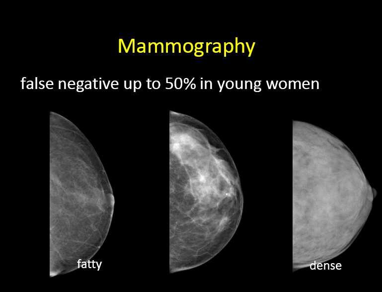
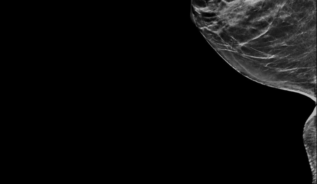
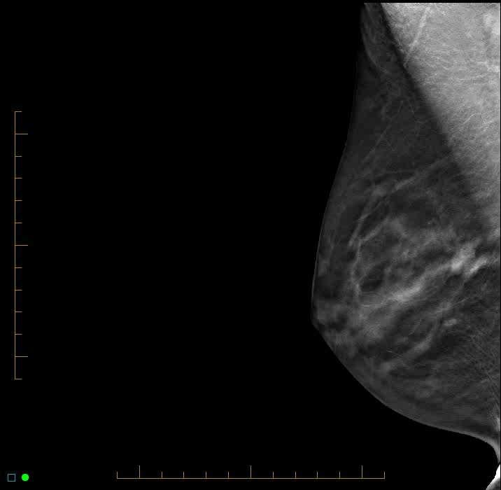
7 HOW DENSE ARE YOU? 7
.")
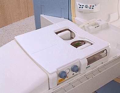
8 DIGITAL MAMMOGRAPHY WITH 3D TOMOSYNTHESIS (DBT) A sequence of successive images are acquired while the X-ray source travels along an arc, allowing multiple slices to be examined ( mm thin image slices). 3D RECONSTRUCTED IMAGES ( and a 2D synthesised mammogram can be digitally reconstructed) 8


9 DBT- the Xray tube moves in an arc across the breast 15/30 degrees- low dose xrays are then reconstructed to give depth information 9

10 /12/2016 D is ea se Sy m po si um 3D MAMMOGRAPHY WAS RANKED AS TIME MAGAZINE S 11TH MOST REMARKABLE HEALTH ADVANCE IN 2014 Be ni gn Br ea st DIGITAL TOMOSYNTHESIS 10


11 Right Breast -CC + MLO views C VIEWS Tomosynthesis Movie CC and MLO 11


12 MLO C VIEW + Tomosynthesis movie EFFICACY OF DBT Reduces false positives- Normal overlapping tissue, on standard digital mammogram can mimic breast cancer, DBT adds depth resolution to prevent this problem. Reduces false negatives- in digital mammogram overlapping breast tissue can obscure and mask cancers. BONUS - LESS COMPRESSION REQUIRED!!!! 12

13 WHAT ABOUT THE RADIATION DOSE? 2D AND DBT increases radiation exposure TWO-FOLD This is still BELOW SAFETY LIMIT BACKGROUND RADIATION IN AUSTRALIA millisieverts/year Digital Mammogram millisieverts Digital Mammogram and DBT millisieverts Adding DBT is equivalent to up to 6 months of annual background radiation (BUT with C view reformatted DBT DOSE= 4 MONTHS) Radiation Doses 13


14 REFORMATING THE DIGITAL MAMMOGRAM REDUCES THE RADIATION DOSE OF DBT TO ALMOST THAT OF A STANDARD DIGITAL MAMMOGRAM NEED TO CHECK WITH RADIOLOGY COMPANY, LOOK ON THE FILMS IS IT A C VIEW? EXCEPTION DBT CAN EXAGERATE MICROCALCIFICATIONS STANDARD MAG VIEWS WILL BE REQUIRED HAVE WE BEEN TRUMPED? We re gonna make this Country great again IN THE USA DBT HAS BEEN SHOWN TO - Improve cancer detection rates Reduces patient recalls Approved for screening 14


15 BREAST SCREEN AUSTRALIA POSITION STATEMENT, November 2014 TWO VIEW MAMMOGRAPHY CONTINUES TO BE THE MOST EFFECTIVE POPULATION PRIMARY SCREENING TEST FOR BREAST CANCER THERE IS EVIDENCE THAT DBT CAN BE OF BENFIT IN AN ASSESSMENT SETTING LESS SUPPORTING EVIDENCE FOR THE BENEFIT OF DBT AS THE SCREENING TEST FOR POPULATION SCREENING OF WELL WOMEN. 15
16 ISSUES WITH DBT An increase in cancer detection rates do not indicate a MORTALITY benefit. Still uncertain if DBT results in a REDUCTION in interval cancer rates. CONFLICTING EVIDENCE ON WHETHER BREAST DENISTY IMPACTS ON ACCURACY OF DBT MOST BENEFIT OF DBT REDUCE FALSE POSITIVE RECALLS AND MINIMISE OVER INVESTIGATION NEW MODALITY- Contrast Mammography NOT WIDELY AVAILABLE SPECIALIST REFERRAL ONLY IODINE CONTRAST OVER 2 MINS- CAN MISS VERY HIGH GRADE TUMORS RELIES ON NEOVASCULARISATION may miss low grade tumors/dcis SPECILAIST RADIOLOGIST REPORTING WATCH THIS SPACE CONTRAST MAMMOGRAPHY = POOR MANS MRI 16
17 BREAST MRI USES MAGNETIC FIELDS TO CREATE AN IMAGE OF THE BREAST TISSUE, USING HUNDREDS OF IMAGES TAKEN VERY QUICKLY NO RADIATION No mortality/survival data No RCTs NO evidence it is more effective than mammography and US in the screening of women over the age of 50 NO evidence of survival benefit with the use of MRI BREAST MRI is not a substitute for mammogram Breast MRI Very high sensitivity for the detection of breast cancer >90% IDC/ILC DCIS % Implant Rupture- 94% Vast majority of malignant lesions enhance 1-Dec

")
18 Breast MRI Lower specificity ~ 30 60% PPV Some benign lesions enhance Normal breast parenchyma may enhance, (day 6-16 cycle is best) Early + late in cycle benign hormonal enhancement 1-Dec Magnetic Resonance Imaging (MRI ) 1-Dec


19 Patient Positioning 1-Dec Dec

20 Medicare MBS eligibility <50 yrs of age To be eligible for rebate MUST be ordered by breast specialist Breast or ovarian ca 3 or more 1st / 2nd deg relatives, same side family Breast or ovarian ca 2 or more 1st / 2nd deg on same side family plus 1 or more of: bilateral breast ca onset breast ca < 40 yrs onset ovarian ca < 50 yrs both breast and ovarian ca in one relative Ashkenazi Jewish ancestry male breast cancer relative Breast cancer onset < 45 yrs 1 1st / 2nd deg relative plus 1 bone / ST sarcoma < 45 yrs 1st / 2nd deg same side family Genetic mutation BRCA1, BRCA2 or P53 NB. Not included: Radiation treatment to the chest for Hodgkin's Disease 20
21 DIAGNOSTIC MRI Extent of Disease- ipsilateral and contralateral- synchronous,multifocal or multicentric disease Positive margins Response to neoadjuvant chemotherapy Metastatic Axillary Lymphadenopathy of unknown primary ( 75-80%) Chest wall invasion Recurrent breast cancer / scar changes USA 5% WOMEN > 30 HAVE HAD AN MRI BREAST Limitations and Problems with MRI Contra indications- METAL Patient claustrophobia/noisy/ obesity / prone position Pregnancy gadolinium contrast contra indicated (no RCT) Breast feeding cease for 24hrs interpretation difficult- esp.for lobular CA + DCIS diffuse enhancement not circumscribed IV line - midazolam / accompanied by an adult Anaphylaxis rate 0.03% Time consuming- 25 minutes Scheduling difficulties (day 6-16 menstrual cycle) Mass < 5mm too small to characterise Limited trained technologists- expert, experienced interpretation MR guided biopsy time consuming/ EXPENSIVE 1-Dec
22 FALSE NEGATIVES with MRI TECHNICAL CAUSES- Breast tissue not included in the coil, motion, inadequate contrast, too much compression Marked Background enhancement CAUTION IF MAMMOGRAPHY OR ULTRASOUND IS POSITIVE OR PALPABLE FINDINGS NEED TO TREAT/ BIOPSY / EXCISE DESPITE NEGATIVE MRI! ALL THAT ENHANCES ON MRI ISN T CANCER DUCTAL ENHANCEMENT MALIGNANT CAUSES- DCIS / INVASIVE CA BENIGN HIGH RISK LESIONS- ADH, LCIS BENIGN FIBROSIS, DUCTAL HYPERPLASIA, FIBROCYSTIC CHANGE 22


23 Juvenile FA Lobulated FA Pre contrast show presence of fat in a lesion 23


24 CYSTS show up as a filling defect SPICULATED MASS > 80% chance malignant 24


25 CAD- Computer Aided Detection - KINETIC EVALUATIONlarge invasive cancer LEFT BREAST CANCER 25


26 INVASIVE CANCER with linear DCIS RIGHT AXILLARY METASTASIS 26


27 Diagnosis Mammogram + US > 35 years, (Ultrasound< 35 )? MRI- young/ dense/ lobular CA/occult mammogram-specialist REFERRAL Biopsy Staging Investigations The BREAST radiologist is your friend! Dr. Anthony Felber 27


28 Breast Imaging is a TEAM SPORT - MULTIDISCIPLINARY CARE 28
Epworth Healthcare Benign Breast Disease Symposium. Sat Nov 12 th 2016
 Epworth Healthcare Benign Breast Disease Symposium Breast cancer is common Sat Nov 12 th 2016 Benign breast disease is commoner, and anxiety about breast disease commoner still Breast Care Campaign UK
Mammographic imaging of nonpalpable breast lesions. Malai Muttarak, MD Department of Radiology Chiang Mai University Chiang Mai, Thailand
 Mammographic imaging of nonpalpable breast lesions Malai Muttarak, MD Department of Radiology Chiang Mai University Chiang Mai, Thailand Introduction Contents Mammographic signs of nonpalpable breast cancer
Armed Forces Institute of Pathology.
 Armed Forces Institute of Pathology www.radpath.com Armed Forces Institute of Pathology Breast Disease www.radpath.org Armed Forces Institute of Pathology Interpretation of Breast MRI Leonard M. Glassman
Breast Cancer Screening and High Risk
 Breast Cancer Screening and High Risk Mary Freyvogel, DO Breast Surgeon Clinical Assistant Professor of Surgery University Hospitals Case Medical Center St. John Medical Center / Elyria Medical Center
Breast Cancer Imaging
 Breast Cancer Imaging I. Policy University Health Alliance (UHA) will cover breast imaging when such services meet the medical criteria guidelines (subject to limitations and exclusions) indicated below.
Breast Cancer Screening and Treatment Mrs Belinda Scott Breast Surgeon Breast Associates Auckland
 Breast Cancer Screening and Treatment 2009 Mrs Belinda Scott Breast Surgeon Breast Associates Auckland BREAST CANCER THE PROBLEM 1.1 million women per year 410,000 deaths each year Increasing incidence
Imaging in breast cancer. Mammography and Ultrasound Donya Farrokh.MD Radiologist Mashhad University of Medical Since
 Imaging in breast cancer Mammography and Ultrasound Donya Farrokh.MD Radiologist Mashhad University of Medical Since A mammogram report is a key component of the breast cancer diagnostic process. A mammogram
Standard Breast Imaging Modalities. Lilian Wang, M.D. Breast Imaging Section Department of Radiology Northwestern Medicine
 Standard Breast Imaging Modalities Lilian Wang, M.D. Breast Imaging Section Department of Radiology Northwestern Medicine Overview Standard breast imaging modalities Mammography Ultrasound MRI Imaging
EARLY DETECTION: MAMMOGRAPHY AND SONOGRAPHY
 EARLY DETECTION: MAMMOGRAPHY AND SONOGRAPHY Elizabeth A. Rafferty, M.D. Avon Comprehensive Breast Center Massachusetts General Hospital Harvard Medical School Breast Cancer Screening Early detection of
Breast Evaluation & Management Guidelines
 Breast Evaluation & Management Guidelines Pamela L. Kurtzhals, M.D. F.A.C.S. Head, Dept. of General Surgery Scripps Clinic, La Jolla Objective Review screening & diagnostic guidelines Focused patient complaints
Breast Imaging & You
 Breast Imaging & You What s Inside: Breast Imaging... 2 Digital Breast Tomosynthesis (DBT) mammograms... 4 Breast cancer screening... 6 Dense breast tissue... 8 Automated Breast Ultrasound (ABUS)... 9
EARLY DETECTION: MAMMOGRAPHY AND SONOGRAPHY
 EARLY DETECTION: MAMMOGRAPHY AND SONOGRAPHY Elizabeth A. Rafferty, M.D. Avon Comprehensive Breast Center Massachusetts General Hospital Harvard Medical School Breast Cancer Screening Early detection of
Screening Mammograms: Questions and Answers
 CANCER FACTS N a t i o n a l C a n c e r I n s t i t u t e N a t i o n a l I n s t i t u t e s o f H e a l t h D e p a r t m e n t o f H e a l t h a n d H u m a n S e r v i c e s Screening Mammograms:
Breast Imaging Update: Old Dog New Tricks
 Breast Imaging Update: Old Dog New Tricks Claire McKay, DO M&S Imaging Assoc. San Antonio, TX cmckayhart@juno.com Goals Describe modalities available, old and new Provide understanding of pros and cons
Breast Imaging & You
 Breast Imaging & You What s Inside: Breast Imaging... 2 Digital Breast Tomosynthesis (DBT) mammograms... 4 Breast cancer screening... 6 Dense breast tissue... 8 Automated breast ultrasound (ABUS)... 9
Pitfalls and Limitations of Breast MRI. Susan Orel Roth, MD Professor of Radiology University of Pennsylvania
 Pitfalls and Limitations of Breast MRI Susan Orel Roth, MD Professor of Radiology University of Pennsylvania Objectives Review the etiologies of false negative breast MRI examinations Discuss the limitations
Current Status of Supplementary Screening With Breast Ultrasound
 Current Status of Supplementary Screening With Breast Ultrasound Stephen A. Feig, M.D., FACR Fong and Jean Tsai Professor of Women s Imaging Department of Radiologic Sciences University of California,
BREAST MRI. VASILIKI FILIPPI RADIOLOGIST CT MRI & PET/CT Departments Hygeia Hospital, Athens, Greece
 BREAST MRI VASILIKI FILIPPI RADIOLOGIST CT MRI & PET/CT Departments Hygeia Hospital, Athens, Greece Breast ΜR Imaging (MRM) Breast MR imaging is an extremely powerful diagnostic tool, that when used in
MANAGEMENT OF DENSE BREASTS. Nichole K Ingalls, MD, MPH NW Surgical Specialists September 25, 2015
 MANAGEMENT OF DENSE BREASTS Nichole K Ingalls, MD, MPH NW Surgical Specialists September 25, 2015 No financial disclosures National Cancer Institute National Cancer Institute Increased Cancer Risk... DENSITY
Detailed Program of the second BREAST IMAGING AND INTERVENTIONS PROGRAM am am : Clinician s requirements from breast imaging
 Detailed Program of the second BREAST IMAGING AND INTERVENTIONS PROGRAM 2012 Day one, 2 nd November BREAST IMAGING AND INTERVENTIONS PROGRAM 2012 9.00 AM 9.10 am Introduction 9.10 am - 9.30 am : Clinician
Melissa Hartman, DO Women s Health Orlando VA Medical Center
 Melissa Hartman, DO Women s Health Orlando VA Medical Center Most common non-skin cancer and Second deadliest cancer in women Majority are diagnosed by abnormal screening study An approach to breast cancer
1. Screening, Diagnosis and Surgical Management of Breast Cancer
 1. Screening, Diagnosis and Surgical Management of Breast Cancer Dr Melanie Walker, MBBS, FRACS (Breast Surgeon) Oncoplastic Breast Surgery Combination of optimal cancer surgery with plastic surgical techniques
Breast imaging in general practice
 Breast series CLINICAL PRACTICE Breast imaging in general practice Nehmat Houssami, MBBS, FAFPHM, FASBP, PhD, is Associate Clinical Director, NSW Breast Cancer Institute, Westmead Hospital, Honorary Senior
Non-mass Enhancement on Breast MRI. Aditi A. Desai, MD Margaret Ann Mays, MD
 Non-mass Enhancement on Breast MRI Aditi A. Desai, MD Margaret Ann Mays, MD Breast MRI Important screening and diagnostic tool, given its high sensitivity for breast cancer detection Breast MRI - Indications
What s New in Breast Imaging. Jennifer A. Harvey, M.D., FACR Professor of Radiology University of Virginia
 What s New in Breast Imaging Jennifer A. Harvey, M.D., FACR Professor of Radiology University of Virginia Disclosure Hologic, Inc. Shareholder and research agreement. Volpara Solutions, Ltd. Shareholder
MEDICAL IMAGING AND BREAST DISEASE HOW CAN WE HELP YOU?
 MEDICAL IMAGING AND BREAST DISEASE HOW CAN WE HELP YOU? Barbara M. Preston, M.D. SCREENING MAMMOGRAPHY AVERAGE RISK PATIENTS KAISER RECOMMENDATION: ALL WOMEN (INCLUDING TRANSGENDER FEMALES) Every 1-21
Imaging Guidelines for Breast Cancer Screening
 Imaging Guidelines for Breast Cancer Screening Sarah Colwick, MD Dr. Sarah Colwick was born and raised in Sikeston, MO. She attended college and medical school at the University of Missouri-Kansas City
FIBROEPITHELIAL LESIONS
 DEFINITIONS FIBROEPITHELIAL LESIONS Suzanne Moore FIBROADENOMA- A discrete benign tumour showing evidence of connective tissue and epithelial proliferation- WHO Fibrous stromal element of these tumours
BREAST MRI. Elizabeth A. Rafferty, M.D. Avon Comprehensive Breast Center Massachusetts General Hospital Harvard Medical School
 BREAST MRI Elizabeth A. Rafferty, M.D. Avon Comprehensive Breast Center Massachusetts General Hospital Harvard Medical School BREAST MRI Any assessment of the breast parenchyma requires the administration
Imaging the Symptomatic Patient. Avice M.O Connell MD,FACR,FSBI Professor of Imaging Sciences Director, Women s Imaging University of Rochester
 Imaging the Symptomatic Patient Avice M.O Connell MD,FACR,FSBI Professor of Imaging Sciences Director, Women s Imaging University of Rochester The four most common symptoms Mass Pain Discharge Infection
BREAST MRI. Elizabeth A. Rafferty, M.D. Avon Comprehensive Breast Center Massachusetts General Hospital Harvard Medical School
 BREAST MRI Elizabeth A. Rafferty, M.D. Avon Comprehensive Breast Center Massachusetts General Hospital Harvard Medical School BREAST MRI Any assessment of the breast parenchyma requires the administration
Contrast-enhanced Breast MRI RSSA 2013
 Contrast-enhanced Breast MRI RSSA 2013 Prof. dr. Maurice van den Bosch University Medical Center Utrecht, the Netherlands Index 1) Breast cancer 2) Why MRI of the breast 3) Technique 4) Interpretation
A GP S APPROACH TO BREAST LUMPS AND SYMPTOMS DR KK CHEUNG GPGC WORKSHOP
 A GP S APPROACH TO BREAST LUMPS AND SYMPTOMS DR KK CHEUNG GPGC WORKSHOP 18.08.18 HAVE A SYSTEM HISTORY EXAMINATION INVESTIGATION FOLLOW UP BREAST SYMPTOMS HISTORY DON T FORGET SKIN CHANGES AND NIPPLE CHANGES
Breast Cancer Screening and Diagnosis
 Breast Cancer Screening and Diagnosis Priya Thomas, MD Assistant Professor Clinical Cancer Prevention and Breast Medical Oncology University of Texas MD Anderson Cancer Center Disclosures Dr. Thomas has
Mousa. Israa Ayed. Abdullah AlZibdeh. 0 P a g e
 1 Mousa Israa Ayed Abdullah AlZibdeh 0 P a g e Breast pathology The basic histological units of the breast are called lobules, which are composed of glandular epithelial cells (luminal cells) resting on
Lesion Imaging Characteristics Mass, Favoring Benign Circumscribed Margins Intramammary Lymph Node
 Lesion Imaging Characteristics Mass, Favoring Benign Circumscribed Margins Intramammary Lymph Node Oil Cyst Mass, Intermediate Concern Microlobulated Margins Obscured Margins Mass, Favoring Malignant Indistinct
Emerging Techniques in Breast Imaging: Contrast-Enhanced Mammography and Fast MRI
 Emerging Techniques in Breast Imaging: Contrast-Enhanced Mammography and Fast MRI Lilian Wang, M.D. Breast Imaging Section Department of Radiology Northwestern Medicine Overview Rationale for new imaging
Contrast Enhanced Spectral Mammography (CESM) Initial UK Experience. Dr Sarah L Tennant BMedSci, BMBS, MRCP, FRCR
 Initial UK Experience. Dr Sarah L Tennant BMedSci, BMBS, MRCP, FRCR") Contrast Enhanced Spectral Mammography (CESM) Initial UK Experience Dr Sarah L Tennant BMedSci, BMBS, MRCP, FRCR Vote Now Your experience of CESM 1. No experience of CESM 44% 2. I ve seen some cases in
Contrast Enhanced Spectral Mammography (CESM) Initial UK Experience Dr Sarah L Tennant BMedSci, BMBS, MRCP, FRCR Vote Now Your experience of CESM 1. No experience of CESM 44% 2. I ve seen some cases in
Breast Cancer Screening
 Scan for mobile link. Breast Cancer Screening What is breast cancer screening? Screening examinations are tests performed to find disease before symptoms begin. The goal of screening is to detect disease
BREAST DENSITY WHAT IS IT? WHY IS IT IMPORTANT? & What IOWA SF250 Means to Patients and Providers
 BREAST DENSITY WHAT IS IT? WHY IS IT IMPORTANT? & What IOWA SF250 Means to Patients and Providers Arnold Honick, MD Radiology Consultants of Iowa, PLC ahonick@rciowa.com BREAST DENSITY LEGISLATION Nancy
Case 1. BREAST CANCER From Diagnosis to Treatment: The Role of Primary Care
 BREAST CANCER From Diagnosis to Treatment: The Role of Primary Care Leah Karliner, MD MAS University of California San Francisco Primary Care Medicine Update 2009 April 2009 Case 1 AR, a 60 year old African
ROLE OF MRI IN SCREENING, DIAGNOSIS AND MANAGEMENT OF BREAST CANCER. B.Zandi Professor of Radiology
 ROLE OF MRI IN SCREENING, DIAGNOSIS AND MANAGEMENT OF BREAST CANCER B.Zandi Professor of Radiology Introduction In the USA, Breast Cancer is : The Most Common Non-Skin Cancer The Second Leading cause of
Here are examples of bilateral analog mammograms from the same patient including CC and MLO projections.
 Good afternoon. It s my pleasure to be discussing Diagnostic Breast Imaging over the next half hour. I m Wei Yang, Professor of Diagnostic Radiology and Chief, the Section of Breast Imaging as well as
Breast Cancer: Selected Topics for the Primary Care Clinician
 Breast Cancer: Selected Topics for the Primary Care Clinician Leah Karliner, MD MAS October 2009 Primary Care Medicine: Principles and Practice OUTLINE Incidence and Mortality Risk Factors and Risk Reduction/Prevention
National Diagnostic Imaging Symposium 2013 SAM - Breast MRI 1
 National Diagnostic Imaging Symposium 2013 December 8-12, 2013 Disney s Yacht Club Resort Lake Buena Vista, Florida Self Assessment Module Questions, Answers and References Day SAM Title - Each SAM title
DR AISHA A UMAR CHIEF CONSULTANT RADIOLOGIST NATIONAL HOSPITAL ABUJA.
 DR AISHA A UMAR CHIEF CONSULTANT RADIOLOGIST NATIONAL HOSPITAL ABUJA. OUTLINE WHY DO WE IMAGE WHOM TO IMAGE WHEN TO IMAGE HOW TO IMAGE WHAT TO IMAGE WITH PERSONAL EXPERIENCE CONCLUSION/RECOMMENDATIONS
Wellness Along the Cancer Journey: Cancer Types Revised October 2015 Chapter 2: Breast Cancer
 Wellness Along the Cancer Journey: Cancer Types Revised October 2015 Chapter 2: Breast Cancer Cancer Types Rev. 10.20.15 Page 19 Breast Cancer Group Discussion True False Not Sure 1. Breast cancer is not
Breast Cancer Diagnosis, Treatment and Follow-up
 Breast Cancer Diagnosis, Treatment and Follow-up What is breast cancer? Each of the body s organs, including the breast, is made up of many types of cells. Normally, healthy cells grow and divide to produce
Breast Magnetic Resonance Imaging (MRI) Westmead Breast Cancer Institute
 Westmead Breast Cancer Institute") Breast Magnetic Resonance Imaging (MRI) Westmead Breast Cancer Institute What is breast MRI? Breast MRI is a technique that uses a magnetic field to create an image of the breast tissue, using hundreds
Breast Magnetic Resonance Imaging (MRI) Westmead Breast Cancer Institute What is breast MRI? Breast MRI is a technique that uses a magnetic field to create an image of the breast tissue, using hundreds
Amammography report is a key component of the breast
 Review Article Writing a Mammography Report Amammography report is a key component of the breast cancer diagnostic process. Although mammographic findings were not clearly differentiated between benign
Financial Disclosures
 Financial Disclosures 3D Mammography: The Latest Developments in the Breast Imaging Arena I have no financial disclosures Dr. Katharine Lampen-Sachar Breast and Body Radiologist Radiology Associates of
Diseases of the breast (1 of 2)
") Diseases of the breast (1 of 2) Introduction A histology introduction Normal ducts and lobules of the breast are lined by two layers of cells a layer of luminal cells overlying a second layer of myoepithelial
Diseases of the breast (1 of 2) Introduction A histology introduction Normal ducts and lobules of the breast are lined by two layers of cells a layer of luminal cells overlying a second layer of myoepithelial
Ultrasound of the Breast BASICS FOR THE ORDERING CLINICIAN
 Ultrasound of the Breast BASICS FOR THE ORDERING CLINICIAN Breast Ultrasound Anatomy Skin Breast Parenchyma Pectoralis Fascia Pectoralis Breast Ultrasound Anatomy Indications for Breast Ultrasound Palpable
Breast Tomosynthesis. What is breast tomosynthesis?
 Scan for mobile link. Breast Tomosynthesis Breast tomosynthesis is an advanced form of mammography, a specific type of breast imaging that uses low-dose x-rays to detect cancer early when it is most treatable.
Breast Cancer. Most common cancer among women in the US. 2nd leading cause of death in women. Mortality rates though have declined
 Breast Cancer Most common cancer among women in the US 2nd leading cause of death in women Mortality rates though have declined 1 in 8 women will develop breast cancer Breast Cancer Breast cancer increases
Index. C Calcifications fat necrosis 1, 61 fat necrosis 4, 69 nipple/peri-areolar involvement 1, 165
 A ADH. See Atypical ductal hyperplasia (ADH) American College of Radiology (ACR), BI-RADS background parenchymal enhancement, 8, 9, 81, 82 fibroglandular tissue guidelines, 6 American Joint Committee on
Breast Cancer. Saima Saeed MD
 Breast Cancer Saima Saeed MD Breast Cancer Most common cancer among women in the US 2nd leading cause of death in women 1 in 8 women will develop breast cancer Incidence/mortality rates have declined Breast
RADIOLOGIC EVALUATION OF BREAST CANCER
 RADIOLOGIC EVALUATION OF BREAST CANCER Orsolya Farkas, Gabriella Bodrogi and Gábor Szalai Department of Radiology, Pécs University Orsifarkas@yahoo.com Complex evaluation of the breast Patient history
Mammography. What is Mammography?
 Scan for mobile link. Mammography Mammography is a specific type of breast imaging that uses low-dose x-rays to detect cancer early before women experience symptoms when it is most treatable. Tell your
Policy Library Clinical Advantages of Digital Breast Tomosynthesis in Symptomatic Patients
 Policy Library Clinical Advantages of Digital Breast Tomosynthesis in Symptomatic Patients Version: 1 Approved by: Faculty of Clinical Radiology Council Date of approval: Click and type: day month and
MR sin plass i brystkreftdiagnostikk, dagens anbefalinger og fremtidsperspektiver
 MR sin plass i brystkreftdiagnostikk, dagens anbefalinger og fremtidsperspektiver Kathinka Kurz, MD, PhD, seksjonsoverlege SUS, kathinka.dehli.kurz@sus.no Technique - Subtraction Without contrast agent
Mammography and Other Screening Tests. for Breast Problems
 301.681.3400 OBGYNCWC.COM Mammography and Other Screening Tests What is a screening test? for Breast Problems A screening test is used to find diseases, such as cancer, in people who do not have signs
Breast Cancer. Common kinds of breast cancer are
 Breast Cancer A breast is made up of three main parts: glands, ducts, and connective tissue. The glands produce milk. The ducts are passages that carry milk to the nipple. The connective tissue (which
Breast Cancer Screening Clinical Practice Guideline. Kaiser Permanente National Breast Cancer Screening Guideline Development Team
 NATIONAL CLINICAL PRACTICE GUIDELINE Breast Cancer Screening Clinical Practice Guideline Kaiser Permanente National Breast Cancer Screening Guideline Development Team This guideline is informational only.
BI-RADS and Breast MRI. Kathy Borovicka, M.D. Thursday February 15, 2018
 BI-RADS and Breast MRI Kathy Borovicka, M.D. Thursday February 15, 2018 Learning Objectives Be familiar with the Breast Imaging Reporting and Data System (BI-RADS) Understand the components of a breast
Tomosynthesis and breast imaging update. Dr Michael J Michell Consultant Radiologist King's College Hospital NHS Foundation Trust
 Tomosynthesis and breast imaging update Dr Michael J Michell Consultant Radiologist King's College Hospital NHS Foundation Trust Breast imaging new technology BREAST CANCER FLT PET shows different grades
Angela Gilliam, MD University of Colorado Surgical Grand Rounds November 3, 2008
 Angela Gilliam, MD University of Colorado Surgical Grand Rounds November 3, 2008 Breast Cancer Most common cancer in American women 180,000 new cases per year Second most common cause of cancer death 44,000
Mary Smania, DNP, FNP-BC Clinical Practice Champion Assistant Professor Michigan State University College of Nursing
 Mary Smania, DNP, FNP-BC Clinical Practice Champion Assistant Professor Michigan State University College of Nursing Identify evidence based routine screening guidelines for women of all ages Identify
Breast MRI: Friend or Foe?
 Breast MRI: Friend or Foe? UCSF Postgraduate Course May 18, 2013 Cheryl Ewing, MD Clinical Professor of Surgery UCSF Department of Surgery APPLEGATE HAS DOUBLE MASTECTOMY IN CANCER SCARE DIAGNOSED WITH
WHICH INDICATION FOR BREAST MRI?
 WHICH INDICATION FOR BREAST MRI? Dr. P. De Visschere, Prof. Dr. G. Villeirs Genitourinary Radiology and Mammography University Hospital Gent Symposium Belgian Menopause Society 13/03/2010 Which Indication
Breast Health and Imaging Glossary
 Contact: Lorna Vaughan HerSpace Breast Imaging & Biopsy Associates 300 State Route 35 South W. Long Branch, NJ 07764 732-571-9100, ext. 104 lorna@breast-imaging.com Breast Health and Imaging Glossary Women
CPC 4 Breast Cancer. Rochelle Harwood, a 35 year old sales assistant, presents to her GP because she has noticed a painless lump in her left breast.
 CPC 4 Breast Cancer Rochelle Harwood, a 35 year old sales assistant, presents to her GP because she has noticed a painless lump in her left breast. 1. What are the most likely diagnoses of this lump? Fibroadenoma
Evaluations & CE Credits
 Evaluations & CE Credits Nursing Contact Hours, CME and CHES credits are available. Please visit www.phlive.org to fill out your evaluation and complete the post-test. 1 Breast Density and Breast Cancer
SAMPLE. Do Not Reproduce. Breast Lumps & Breast Cancer. Breast Lumps. Breast Cancer. Treatment. Signs, Symptoms, and Causes. Signs and Symptoms
 Breast Lumps & Breast Cancer Feeling a lump in a breast can be scary. For a lot of women, the first thought is cancer. The good news is that 80% to 90% of breast lumps are not cancer. If you feel a lump
Ultrasonography. Methods. Brief Description. Indications. Device-related Prerequisites. Technical Requirements. Evaluation Criteria
 1 Ultrasonography Brief Description Imaging modality using sound waves Tissue-specific wave reflection. Indications Evaluation of palpable breast nodules Evaluation of clinically occult mammographic findings
Breast Health. Learning Objectives. Breast Anatomy. Poll Question. Breast Anatomy
 Learning Objectives Describe breast anatomy to a patient Breast Health Answer questions about causes of breast pain and masses Explain breast cancer screening/diagnostic modalities Appropriately triage
BREAST PATHOLOGY. Fibrocystic Changes
 BREAST PATHOLOGY Lesions of the breast are very common, and they present as palpable, sometimes painful, nodules or masses. Most of these lesions are benign. Breast cancer is the 2 nd most common cause
Jeddah Breast Cancer Pilot Screening Program, KSA
 Jeddah Breast Cancer Pilot Screening Program, KSA 7 th Global Summit on Cancer Therapy, Oct 5-7, 2015 Dubai, Crown Plaza Hotel Muna Baslaim, MD Consultant Surgeon Head of the Breast Unit, King Fahd General
Certified Breast Care Nurse (CBCN ) Test Content Outline (Effective 2018)
 Test Content Outline (Effective 2018)") Certified Breast Care Nurse (CBCN ) Test Content Outline (Effective 2018) I. Coordination of Care - 26% A. Breast health, screening, early detection, risk assessment and reduction 1. Issues related to
Certified Breast Care Nurse (CBCN ) Test Content Outline (Effective 2018) I. Coordination of Care - 26% A. Breast health, screening, early detection, risk assessment and reduction 1. Issues related to
Breast Cancer. American Cancer Society
 Breast Cancer American Cancer Society Reviewed February 2017 What we ll be talking about How common is breast cancer? What is breast cancer? What causes it? What are the risk factors? Can breast cancer
Case study 1. Rie Horii, M.D., Ph.D. Division of Pathology Cancer Institute Hospital, Japanese Foundation for Cancer Research
 NCCN/JCCNB Seminar in Japan April 15, 2012 Case study 1 Rie Horii, M.D., Ph.D. Division of Pathology Cancer Institute Hospital, Japanese Foundation for Cancer Research Present illness: A 50y.o.premenopausal
BreastScreen Victoria Annual Statistical Report
 BreastScreen Victoria Annual Statistical Report 2010 BREASTSCREEN VICTORIA: ANNUAL STATISTICAL REPORT, 2010 Produced by: BreastScreen Victoria Coordination Unit Level 1, 31 Pelham Street, Carlton South
MEDICAL POLICY SUBJECT: MAGNETIC RESONANCE IMAGING (MRI) OF THE BREAST. POLICY NUMBER: CATEGORY: Technology Assessment
 OF THE BREAST. POLICY NUMBER: CATEGORY: Technology Assessment") MEDICAL POLICY SUBJECT: MAGNETIC RESONANCE IMAGING (MRI) OF THE BREAST PAGE: 1 OF: 9 If the member's subscriber contract excludes coverage for a specific service it is not covered under that contract.
MEDICAL POLICY SUBJECT: MAGNETIC RESONANCE IMAGING (MRI) OF THE BREAST PAGE: 1 OF: 9 If the member's subscriber contract excludes coverage for a specific service it is not covered under that contract.
SIGNIFICANT OTHERS. Miscellaneous Benign Breast Conditions
 SIGNIFICANT OTHERS Miscellaneous Benign Breast Conditions Epworth HealthCare 1 FAT NECROSIS TRAUMATIC Cell rupture Seat-Belt injury Blunt trauma Iatrogenic injury Surgery, Flaps, Radiotherapy Pathology
Diagnostic Dilemmas of Breast Imaging
 Diagnostic Dilemmas of Breast Imaging Common Causes of Error in Breast Cancer Detection By: Jason Cord, M.D. Mammography: Initial Imaging The standard for detection of breast cancer Screening mammography
Risk Assessment and Risk Management
 Risk Assessment and Risk Management Epworth Benign Breast Disease Symposium Dr Laura Chin-Lenn 12 November 2016 Why identify those at increased risk of breast cancer? Should I be worried? 1 Why identify
Jose A Torres, MD 1/12/2017
 Jose A Torres, MD 1/12/2017 Background Globally leading cause of cancer related death in women ~249,000 Americans diagnosed with invasive breast cancer ~40,890 will die of their disease Breast cancer risk
CLINICAL SIGNIFICANCE OF BENIGN EPITHELIAL CHANGES
 Papillomas. Papillomas are composed of multiple branching fibrovascular cores, each having a connective tissue axis lined by luminal and myoepithelial cells ( Fig. 23-11 ). Growth occurs within a dilated
ORIGINAL ARTICLE EVALUATION OF BREAST LESIONS USING X-RAY MAMMOGRAM WITH HISTOPATHOLOGICAL CORRELATION
 Available online at www.journalijmrr.com INTERNATIONAL JOURNAL OF MODERN RESEARCH AND REVIEWS IJMRR ISSN: 2347-8314 Int. J. Modn. Res. Revs. Volume 3, Issue 10, pp 807-814, October, 2015 ORIGINAL ARTICLE
Screening Mammography: Who, what, where, when, why and how?
 Screening Mammography: Who, what, where, when, why and how? Jillian Lloyd, MD, MPH Breast Surgical Oncologist University Surgical Oncology Department of Surgery University of Tennessee Medical Center Disclosures
Benign Breast Conditions. Dr. Kim Kelly, CCFP, FCFP Breast Expert, CBCP February, 2015
 Benign Breast Conditions Dr. Kim Kelly, CCFP, FCFP Breast Expert, CBCP February, 2015 Kim Kelly M.D. U of A Family Medicine 1996 2011 Special Interest in Breast Medicine designation, CP&S 2012 15, Breast
University of Washington Radiology Review Course: Strange and Specific Diagnoses. Case #1
 University of Washington Radiology Review Course: Strange and Specific Diagnoses Katherine E. Dee, MD Seattle Breast Center Via Radiology 2014 Case #1 37 year old presents with bilateral palpable lumps.
Digital Breast Tomosynthesis from a first idea to clinical routine
 International Master Programm Biomedical Engineering Digital Breast Tomosynthesis from a first idea to clinical routine Historical background 2D imaging of 3D objects has important limitations Jörg Barkhausen
New Imaging Modalities for better Screening and Diagnosis
 New Imaging Modalities for better Screening and Diagnosis Miri Sklair-Levy, MD Department of Diagnostic Imaging Sheba Medical Center, Sackler School of Medicine, Tel Aviv University Department of Diagnostic
International Day of Radiology 2016 Interview on Breast Imaging Australia / Dr. Michelle Reintals. Breast imaging in Australia
 International Day of Radiology 2016 Interview on Breast Imaging Australia / Dr. Michelle Reintals Breast imaging in Australia An interview with Dr. Michelle Reintals, Director of Breast at IMED Queensland
Breast MRI: Friend or Foe?
 Breast : Friend or Foe? APPLEGATE HAS DOUBLE MASTECTOMY IN CANCER SCARE DIAGNOSED WITH CANCER IN ONE BREAST Comments: 0 ASSOCIATED PRESS 8/19/2008 UCSF Postgraduate Course March 19, 2009 E. Shelley Hwang
Breast Cancer Early Detection
 Breast Cancer Early Detection The importance of finding breast cancer early The goal of screening exams for breast cancer is to find cancers before they start to cause symptoms (like a lump that can be
Intracystic papillary carcinoma of the breast
 Intracystic papillary carcinoma of the breast Poster No.: C-1932 Congress: ECR 2011 Type: Educational Exhibit Authors: V. Dimarelos, F. TZIKOS, N. Kotziamani, G. Rodokalakis, 1 2 3 1 1 1 2 T. MALKOTSI
Radiologic and pathologic correlation of non-mass like breast lesions on US and MRI: Benign, high risk, versus malignant
 Radiologic and pathologic correlation of non-mass like breast lesions on US and MRI: Benign, high risk, versus malignant Poster No.: C-1161 Congress: ECR 2013 Type: Educational Exhibit Authors: J. Kwak,