A Case of Primary Signet-Ring Cell/Histiocytoid Carcinoma of the Eyelid:
|
|
|
- Bonnie Francis
- 5 years ago
- Views:
Transcription
1 A Case of Primary Signet-Ring Cell/Histiocytoid Carcinoma of the Eyelid: Immunohistochemical Comparison with the Normal Sweat Gland and Review of the Literature Mai Iwaya, MD 1, Takeshi Uehara, MD, PhD 1, Akihiko Yoshizawa, MD, PhD 1, Yukihiro Kobayashi, PhD 1, Masanobu Momose, PhD 1, Takayuki Honda, MD, PhD 2 and Hiroyoshi Ota, MD, PhD 3 1 Department of Laboratory Medicine, Shinshu University Hospital, Matsumoto, Nagano , Japan 2 Department of Laboratory Medicine, Shinshu University School of Medicine, Matsumoto, Nagano , Japan 3 Department of Biomedical Sciences, School of Health Sciences, Shinshu University School of Medicine, Matsumoto, Nagano , Japan Address correspondence to: Hiroyoshi Ota, Department of Biomedical Sciences, School of Health Sciences, Shinshu University School of Medicine, Asahi, Matsumoto , Japan Tel&Fax: hohta@shinshu-u.ac.jp This work was supported by a Grant-in-Aid for Scientific Research C (to HO) from the Ministry of Education, Culture, Sports, Science and Technology, Japan. Disclosure: There are no conflicts of interest to declare. Abstract Primary signet-ring cell/histiocytoid carcinomas of the eyelid are extremely rare tumors considered to originate from sweat glands. Here, we report the case of a 72-year-old man diagnosed with primary signet-ring cell/histiocytoid carcinoma of the eyelid and present immunohistochemical analyses of the eyelid apocrine gland
2 (Moll gland) and apocrine and eccrine sweat glands of perineum and axilla. Widespread infiltration of tumor cells with signet-ring cell or histiocytoid appearance was observed in his left eyelid, orbit, and periocular lesion. Tumor cells expressed mucins and showed immunoreactivity that was similar to that of the Moll gland: MUC6(+), GlcNAcα1 4Gal R(-), MUC2(-), MUC5AC(-), GCDFP15(+), CD15(+), S100(-), CK7(+), CK20(-), ER(+), PgR (+), HER2(-), E-cadherin(+), p63(-), PSA (-), and TTF-1(-). The tumor cells differed from those of perineal and axillary apocrine and eccrine sweat glands, which were MUC6(-). The Moll gland was ER(-) and PgR(-), whereas perineal and axillar apocrine sweat glands were ER(+) and PgR(+), and perineal and axillary eccrine sweat glands were ER(+) and PgR(-).The tumor showed characteristics similar to that of the eyelid Moll gland, which is demonstrated to be an apocrine gland with a protein expression distinct from that of other apocrine glands. MUC6 and GCDFP15 expression are useful in identifying the Moll's gland immunophenotype and GCDFP15, ER and PgR expression are useful in distinguishing primary eyelid signet ring/histocytoid carcinoma from gastrointestinal malignancies. Keywords: eyelid, immunohistochemistry, mucin, sweat gland carcinoma
3 Introduction Primary signet-ring cell/histiocytoid carcinoma of the eyelid is a rare tumor, and to date, only 28 cases have been reported. 1,2 The tumor develops mostly in elderly men, and the typical clinical manifestation is a progressive painless swelling of the affected eyelid, leading to a monocle-like appearance. The tumor cells show signet-ring cell or histiocytoid appearance and diffuse infiltration into the dermis and subcutis, without epidermal involvement. The tumor is considered to originate from sweat glands in the eyelid: 1-5 eccrine and apocrine sweat glands (Moll gland); however, there is much debate on whether the tumor expresses the phenotype of eccrine or apocrine sweat glands. The tumor mimics metastasizing signet ring cell adenocarcinoma, especially from the gastrointestinal tract, breast, and urinary bladder. 1-5 Here, we report a patient with primary signet-ring cell/histiocytoid carcinoma of the eyelid who showed a phenotype of protein expression similar to that of Moll gland and investigate its histogenesis by immunohistochemical comparison of the tumor with normal apocrine and eccrine sweat glands of the perineal and axillary region. Case report A 72-year-old Japanese man presented to our hospital with swelling of his left eyelid (Fig. 1A) that had developed over the past 6 7 months. Physical examination showed a diffuse induration of his left eyelid and focal involvement of the periorbital skin. His medical history was unremarkable, except for diabetes and hyperlipidemia. Maxillary and facial computed tomography and magnetic resonance imaging were performed, and they showed diffuse infiltration of the left orbit and periocular region (Fig. 1B). A biopsy of the periorbital skin revealed infiltration of signet-ring cells and histiocyte-like cells. Gastrointestinal endoscopic examination showed multiple gastric ulcers. Biopsy specimens of the lesions revealed reactive epithelial changes and inflammatory cell infiltrates. The patient s serum carcinoembryonic antigen and carbohydrate antigen 19-9 levels were within the normal range. 18 F-fluorodeoxy glucose positron emission tomography showed a marked accumulation of the glucose radiotracer in his left orbit and periocular lesion, and there were no other significant sites of accumulation to indicate an occult primary tumor or
4 metastatic lesions. Left orbital exenteration was performed, and the patient underwent adjuvant radiotherapy (a total of 70 Gy) and chemotherapy with TS-1that contains tegafur, gimeracil and oteracil potassium. Within 6 months, the tumor recurred as multiple cervical lymph-node metastases. One and a half years after the left orbital exenteration, the patient is alive with the disease. Materials and Methods The tissue specimens were fixed in 10% buffered formalin and embedded in paraffin. Sections of 3-μm thickness were cut and stained with hematoxylin-eosin and diastase digestion-alcian blue (ph2.5)-periodic acid-schiff (Di AB-PAS) and immunohistochemically studied by using the immunoenzyme polymer method (Histofine Simple Stain MAX PO Multi, Nichirei Biosciences, Tokyo, Japan) or by using an automated stainer for immunostaining for HER2, ER, and PgR (Ventana Medical Systems, Tucson, AZ) with 3, 3 -diaminobenzidine as the chromogen. The commercially available antibodies are listed in Table 1. We used 4 cases of resected eyelid specimens containing Moll gland and 5 cases of resected perineal skin and 3 cases of resected axillary skin containing histologically normal apocrine sweat glands and eccrine sweat glands as control specimens. These control specimens were retrieved from the pathology files at Shinshu University Hospital. The results of immunostaining were evaluated as the percentage of positively stained cells. The degree of positive staining was graded into 4 categories as follows: (i), negative, no positive cells; (ii) +, focal expression, less than one third of the cells were positive; (iii) ++, one third to two thirds of the cells were positive; and (iv) +++, strong diffuse expression, more than two thirds of the cells were positive. Acknowledgements This work was supported by a Grant-in-Aid for Scientific Research C (to HO) from the Ministry of Education, Culture, Sports, Science and Technology, Japan.
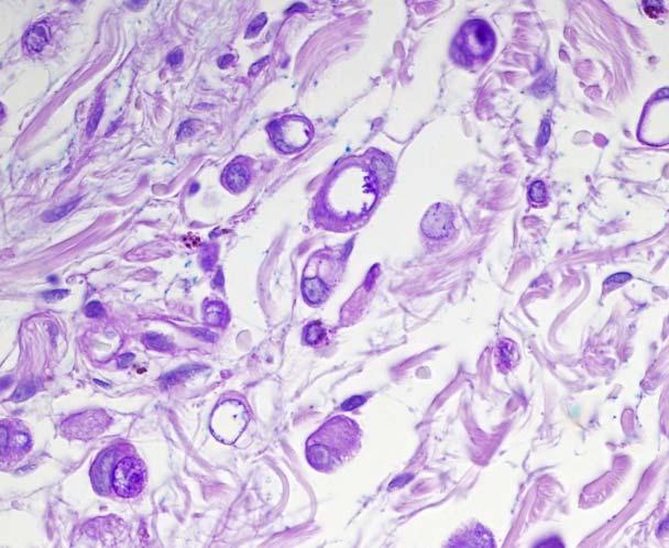
5 Results Tumor of the eyelid Histopathologic findings Microscopic examination of the resected specimen revealed widespread infiltration of the tumor. The tumor extended across the upper and lower eyelid, periorbital skin, and orbit. Infiltrative growth from the upper dermis into subcutaneous tissue, skeletal muscle, and orbital soft tissue was detected, but the epidermis and conjunctival epithelium were not involved. In the dermis and subcutis, the tumor cells were scattered among the collagen fibers as single cells (Fig. 2). The cells were atypical, with abundant clear or eosinophilic cytoplasm, and resembled histiocytes (Fig. 2). Some cells contained single vacuoles and had a characteristic signet-ring appearance (Fig. 2). In the orbital soft tissue, the tumor cells formed a solid diffuse growth and had a monomorphic appearance. The cells were larger than those in the dermis and subcutis and had relatively large nuclei and abundant amphophilic cytoplasm resembling that of histiocytes (Fig. 3). Histochemical and immunohistochemical findings Table 2 summarizes the immunohistochemical profiles observed in the tumor of the eyelid. The intracytoplasmic vacuoles showed positive results for staining with Di-AB/PAS (Fig. 2B). The tumor cells showed diffusely positive results for CK7 (Fig. 2C) and CD15 and focally positive results for GCDFP-15 (Fig. 2D), MUC6, (Fig. 2E), E-cadherin, ER (Fig. 3B), and PgR. CK20, S-100, HER2, GlcNAcα1 4Gal R (Fig. 2F), MUC2, MUC5AC, p63, PSA, and TTF-1 were all negative. Normal Moll glands, perineal and axillary apocrine sweat glands, and eccrine sweat glands Histochemical and immunohistochemical findings Moll glands and perineal and axillary sweat glands (Fig. 4) revealed a positive reaction with Di-AB-PAS at the luminal surfaces of the acinal cells (Fig. 4B). Table 2 summarizes the immunohistochemical reactivity of normal Moll glands, perineal and axillary apocrine/eccrine sweat glands.
6 Briefly, CK7 was expressed in Moll glands (Fig. 4C1), and perineal and axillary apocrine (Fig. 4C2)/eccrine glands (Fig. 4C3), but CK20 was not expressed in these glands. GCDFP-15 and CD15 were expressed in Moll glands (Fig. 4D1), and perineal and axillary apocrine (Fig. 4D2)/eccrine sweat glands (Fig. 4D3). Acinal cells and myoepithelial cell of perineal and axillary eccrine glands showed positive results for S100. E-cadherin was expressed in the sweat glands. ER was expressed in the perineal and axillary apocrine (Fig. 4E2) /eccrine sweat glands (Fig. 4E3) but not in the Moll glands (Fig. 4E1). PgR was expressed only in perineal and axillary apocrine sweat glands, and HER2 was expressed only in perineal and axillary eccrine sweat glands. MUC6 was expressed in the Moll gland (Fig. 5F1), but not in perineal and axillary apocrine/eccrine sweat glands. GlcNAcα1 4Gal R (Fig. 5G1), MUC2, and MUC5AC were all negative. p63 was expressed in myoepithelial cells but not in acinal cells.
7 Discussion Histologically, primary signet-ring cell/histiocytoid carcinoma of the eyelid may be indistinguishable from signet-ring cell carcinoma of the gastrointestinal tract and histiocytoid lobular carcinoma of the breast; these are the most common origins for secondary metastatic tumors of the eyelid 6 and may be detected prior to the primary tumor. 1-3 Thus, before making a diagnosis of primary signet-ring cell/histiocytoid carcinoma of the eyelid, close clinicopathological correlation and a systemic work-up should be performed to exclude a metastatic primary malignancy origin. The immunohistochemical analysis showed that the tumor cells exhibited positive results for CK7, GCDFP-15, CD15, ER, and PgR. Immunoreactivity for E-cadherin was weak. In addition, the tumor cells showed negative results for S-100 protein, CK20, p63, MUC2, and MUC5AC. These results are consistent with those of earlier reports. A novel finding of the present study was that, in the present case, positive results were observed for MUC6, whereas negative results were observed for HIK1083-reactive mucin, which is co-expressed with MUC6 in the gastric-gland mucous cells, Brunner gland, and accessory glands around the bile duct and pancreatic duct. This mucin expression pattern is identical to that observed in the Moll gland and to that previously reported in normal breast and breast carcinoma specimens. 7 Therefore, no conclusions about the primary or metastatic nature of the eyelid neoplasms can be made from this mucin immunoprofile. Significantly, GCDFP-15 has been reported to be expressed in the apocrine and eccrine sweat glands, but not in the normal gastrointestinal tract. 8 Furthermore, gastric adenocarcinomas show negative results for ER and PgR. 8 This expression pattern of GCDFP-15, ER, and PgR in the present case can exclude the possibility of metastasis of gastrointestinal cancer to the eyelid. There is much debate on the histogenesis of primary signet-ring cell/histiocytoid carcinoma of the eyelid. It has been postulated to originate from the apocrine sweat gland 3,9 or the eccrine sweat gland. 4,5,10,11 Primary cutaneous carcinoma with features of signet-ring cell/histiocytoid carcinoma has been reported in the axilla 12 as well as in the eyelid. The most common sites of occurrence for this tumor are the apocrine sweat glands, supporting an apocrine differentiation of this tumor. In addition, our patient expressed MUC6 that was detected in the Moll gland but not in the eccrine sweat gland, suggesting that the tumor cells expressed the phenotype of
8 the Moll gland. The present case revealed typical clinical features. The clinical course of the primary signet-ring cell/histiocytoid carcinoma of the eyelid is indolent, but it extends to the adjacent eyelid, involves the adjacent orbit, and often recurs over many years with metastasis to the regional lymph node 1,10,11 and internal viscera. 1,2,5 13, 14 Furthermore, there have been reports of patients who have died of widespread metastasis of this disease. From an evolutionary perspective, 2 different scenarios have been proposed for the evolution of the apocrine sweat glands; 15 the first scenario is that apocrine sweat glands primarily function in reproduction and sexual behavior, and the second scenario is that they are primarily a defense against microorganisms. Indeed, 2 different phenotypes of apocrine sweat glands corresponding to these 2 proposed functions exist. Glands thought to have evolved to function in reproduction and sexual behavior express ER and develop during puberty and become atrophic in old age (for example, the apocrine axillary glands). Glands proposed to have a primary antimicrobial function develop in infants and do not express ER (for example, the Moll gland in the eyelid and the ceruminous glands in the ear canal). In this study, we also detected ER in the perineal and axillary apocrine gland but not in the Moll gland. In addition, we observed MUC6 expression in the Moll gland but not in the perineal and axillary apocrine gland. These differences could be related to the different functions of these 2 types of apocrine glands. In conclusion, we have reported a case of primary signet-ring cell/histiocytoid carcinoma of the eyelid. Histologic and immunohistochemical analyses indicated that the tumor showed characteristics similar to that of the Moll gland, which is demonstrated to be an apocrine gland with a protein expression distinct from that of other apocrine glands. GCDFP15 and MUC6 expression are useful in identifying the Moll's gland immunophenotype and GCDFP15, ER and PgR expression are useful in distinguishing primary eyelid signet ring/histocytoid carcinoma from gastrointestinal malignancies.
9 Figure legends Figure 1 A 72-year-old man with a diffuse induration of his left eyelid and focal involvement of the periorbital skin that produced a monocle-like appearance (A). Magnetic resonance image showing diffuse infiltration of the left orbit and periocular region (B). Figure 2 Histology, histochemistry, and immunostaining of signet-ring cell/histiocytoid carcinoma of the eyelid in the dermis and subcutis. Single tumor cells were scattered among the collagen fibers. The cells were atypical, with eosinophilic cytoplasm, and resembled histiocytes. Some cells contained single vacuoles and exhibited a signet-ring cell appearance (A). The intracytoplasmic vacuoles showed positive results for staining with diastase digestion-alcian blue (ph 2.5) and periodic acid Schiff reaction (diab-pas) (B). The tumor cells showed positive results for CK7 (C), GCDFP-15 (D), and MUC6 (E), but not for GlcNAcα1 4Gal R, as defined by the absence of immunoreactivity with antibody HIK1083 (F). Figure 3 Histology and immunostaining of signet-ring cell/histiocytoid carcinoma of the eyelid in the orbital soft tissue. The tumor cells were larger than those in the dermis and subcutis and had relatively large nuclei and abundant amphophilic cytoplasm, causing them to resemble histiocytes (A), and showed positive results for ER (B, immunostaining for ER). Figure 4 Histology, histochemistry, and immunostaining of Moll glands, perineal apocrine sweat glands, and perineal sweat glands. Hematoxylin and eosin staining (HE), diastase digestion-alcian blue (ph 2.5) and periodic acid Schiff reaction (diab-pas), and immunostaining of normal sweat glands. Each column of panels represents normal sweat glands, in the order from left to right, as follows: Moll gland, perineal apocrine sweat gland, and perineal eccrine sweat gland. Each row of panels represents an immunostaining result, from top to bottom, as follows: HE (A),
10 diab-pas(b), cytokeratin 7 (CK7) (C), gross cystic disease fluid protein-15 (GCDFP15) (D), estrogen receptor (ER) (E), MUC6 (F), and GlcNAcα1 4Gal R (G) Acinal cells of sweat glands revealed a positive reaction with AB-PAS at the luminal surface (B), CK (C), and GCDFP-15 (D). Perineal sweat glands (E2 and E3), but not (E1), showed positive results for ER. MUC6 was expressed only in the Moll glands (F1). GlcNAcα1 4Gal R was not expressed in the Moll glands (G1) or the perineal glands (G2 and G3).
11 References 1. Requena L, Prieto VG, Requena C, et al. Primary signet-ring cell/histiocytoid carcinoma of the eyelid: a clinicopathologic study of 5 cases and review of the literature. Am J Surg Pathol. 2011;35: Mortensen AL, Heegaard S, Clemmensen O, et al. Signet ring cell carcinoma of the eyelid - the monocle tumour. APMIS. 2008;116: Langel DJ, Yeatts RP, White WL. Primary signet ring cell carcinoma of the eyelid: report of a case demonstrating further analogy to lobular carcinoma of the breast with a literature review. Am J Dermatopathol. 2001;23: Swinson B, Ryan F, Barrett AW, et al. Histiocytoid eccrine sweat gland carcinoma of the eyelid: report of a case. Clin Exp Dermatol. 2006;31: Kim YM, Kim JW, Oh DE. A case of histiocytoid variant eccrine sweat gland carcinoma of the orbit. Korean journal of ophthalmology : KJO. 2011;25: Steinbrecher JS, Silverberg SG. Signet-ring cell carcinoma of the breast. The mucinous variant of infiltrating lobular carcinoma? Cancer. 1976;37: Nakajima K, Ota H, Zhang MX, et al. Expression of gastric gland mucous cell-type mucin in normal and neoplastic human tissues. J Histochem Cytochem. 2003;51: Park SY, Kim BH, Kim JH, et al. Panels of immunohistochemical markers help determine primary sites of metastatic adenocarcinoma. Arch Pathol Lab Med. 2007;131: Jakobiec FA, Austin P, Iwamoto T, et al. Primary infiltrating signet ring carcinoma of the eyelids. Ophthalmology. 1983;90:
12 10. Wollensak G, Witschel H, Bohm N. Signet ring cell carcinoma of the eccrine sweat glands in the eyelid. Ophthalmology. 1996;103: Kramer TR, Grossniklaus HE, McLean IW, et al. Histiocytoid variant of eccrine sweat gland carcinoma of the eyelid and orbit: report of five cases. Ophthalmology. 2002;109: Misago N, Shinoda Y, Okawa T, et al. Histiocytoid and signet-ring cell carcinoma of the axilla: a type of cutaneous apocrine carcinoma equivalent to histiocytoid lobular carcinoma of the breast? Clin Exp Dermatol. 2011;36: Bellman B, Gregory NA, Silvers D, et al. Sweat gland carcinoma with metastases to the skin: response to 5-fluorouracil chemotherapy. Cutis. 1995;55: Thomas JW, Fu YS, Levine MR. Primary mucinous sweat gland carcinoma of the eyelid simulating metastatic carcinoma. Am J Ophthalmol. 1979;87: Stoeckelhuber M, Stoeckelhuber BM, Welsch U. Human glands of Moll: histochemical and ultrastructural characterization of the glands of Moll in the human eyelid. J Invest Dermatol. 2003;121:28-36.
13 Table 1. List of primary antibodies used in this study Antigen Clone Source Dilution Retrieval CK7 OV TL 12/30 DAKO 1:30 Microwave (Carpinteria, CA) CK20 Ks20.8 DAKO 1:30 Microwave GCDFP-15 D6 COVANCE 1:30 Microwave (Princeton, NJ) CD15 MMA BD Biosciences 1:30 Microwave (Franklin Lakes, NJ) S-100 Rabbit polyclonal DAKO 1:800 Microwave E-cadherin 36 BD Biosciences 1:2000 Microwave ER SP1 Ventana Medical Systems 1:1 Microwave (Tucson, AZ) PgR 1E2 Ventana 1:1 Microwave HER2/ neu 4B5 Ventana 1:1 Microwave MUC2 Ccp58 Novocastra 1:100 Microwave (Newcastle-upon-Tyne, UK) MUC5AC CLH2 Novocastra 1:100 Microwave MUC6 CLH5 Novocastra 1:100 Microwave GlcNAcα1 4Gal R HIK1083 Kanto Reagents 1:10 Microwave (Tokyo, Japan) P63 4A4 Nichirei Bioscience Inc. 1:1 Microwave (Tokyo, Japan) PSA 35H9 Novocastra 1:40 Microwave TTF-1 8G7G3/1 DAKO 1:100 Microwave
14 Table 2. Immunohistochemical expression patterns observed in primary signet-ring cell/histiocytoid carcinoma of the eyelid and acinal cells of normal Moll glands, and perineal and axillary apocrine/eccrine sweat glands Antigen Primary Signet-ring Cell/Histiocytoid Moll Glands Apocrine Sweat Glands Eccrine Sweat Glands Carcinoma of the Eyelid CK CK GCDFP-15 +(Focal) CD (Focal) S E-cadherin +(Focal) ER +(Focal) 0 +(Focal) +(Focal) PgR +(Focal) 0 +(Focal) 0 HER (Focal) MUC6 ++(Focal) GlcNAcα1 4Gal R MUC MUC5AC p PSA 0 ND ND ND TTF1 0 ND ND ND The intensity of immunostaining was scored as follows: 0, negative; +, weak; ++, moderate; +++, strong; and ND, not done.


15 A Fig 1 B


16 A B C D Fig 2 E F Fig 3 A B


17 Moll gland Apocrine gland Eccrine gland HE A1 A2 A3 AB-PAS B1 B2 B3 CK7 C1 C2 C3 GCDFP15 D1 D2 D3 ER E1 E2 E3 MUC6 F1 F2 F3 Fig 4 HIK1086 G1 G2 G3
Primary enteric adenocarcinoma with predominantly signet ring features of the lung: A case report with clinicopathological and molecular findings
 CASE REPORT Primary enteric adenocarcinoma with predominantly signet ring features of the lung: A case report with clinicopathological and molecular findings Makoto Nagashima 1, Ayako Moriyama 1, Yasuo
Coordinate Expression of Cytokeratins 7 and 20 in Prostate Adenocarcinoma and Bladder Urothelial Carcinoma
 Anatomic Pathology / CYTOKERATINS 7 AND 20 IN PROSTATE AND BLADDER CARCINOMAS Coordinate Expression of Cytokeratins 7 and 20 in Prostate Adenocarcinoma and Bladder Urothelial Carcinoma Nader H. Bassily,
Cluster designation 5 staining of normal and non-lymphoid neoplastic skin*
 J Cutan Pathol 2005: 32: 50 54 Copyright # Blackwell Munksgaard 2005 Blackwell Munksgaard. Printed in Denmark Journal of Cutaneous Pathology Cluster designation 5 staining of normal and non-lymphoid neoplastic
Acantholytic Anaplastic Extramammary Paget s Disease: A Case Report and Review of the Literature
 Ann Dermatol Vol. 23, Suppl. 2, 2011 http://dx.doi.org/10.5021/ad.2011.23.s2.s226 CASE REPORT Acantholytic Anaplastic Extramammary Paget s Disease: A Case Report and Review of the Literature Yu-Jin Oh,
Cancers of unknown primary : Knowing the unknown. Prof. Ahmed Hossain Professor of Medicine SSMC
 Cancers of unknown primary : Knowing the unknown Prof. Ahmed Hossain Professor of Medicine SSMC Definition Cancers of unknown primary site (CUPs) Represent a heterogeneous group of metastatic tumours,
Primary Cutaneous Apocrine Carcinoma of Sweat Glands: A Rare Case Report
 This is an Open Access article licensed under the terms of the Creative Commons Attribution-NonCommercial-NoDerivs 3.0 License (www.karger.com/oa-license), applicable to the online version of the article
Case Report Aggressive invasive micropapillary salivary duct carcinoma of the parotid gland
 Pathology International 2008; 58: 322 326 doi:10.1111/j.1440-1827.2008.02231.x Case Report Aggressive invasive micropapillary salivary duct carcinoma of the parotid gland Hidetaka Yamamoto, 1 Hideoki Uryu,
BSD 2015 Case 19. Female 21. Nodule on forehead. The best diagnosis is:
 BSD 2015 Case 19 Female 21. Nodule on forehead. The best diagnosis is: A. mixed tumour of skin B. porocarcinoma C. nodular hidradenoma D. metastatic adenocarcinoma BSD 2015 Case 19 Female 21 Nodule on
CASE REPORT GASTRIC ADENOCARCINOMA METASTASIS TO THE BREAST- A DIFFERENTIAL DIAGNOSIS WITH PRIMARY BREAST ADENOCARCINOMA AND REVIEW OF LITERATURE.
 GASTRIC ADENOCARCINOMA METASTASIS TO THE BREAST- A DIFFERENTIAL DIAGNOSIS WITH PRIMARY BREAST ADENOCARCINOMA AND REVIEW OF LITERATURE. Ashwin Hebbar.K 1, Shashidar. K 2, Kamal Kishor 3, Manjunath 4, Shivakumar.H.
WT1, Estrogen Receptor, and Progesterone Receptor as Markers for Breast or Ovarian Primary Sites in Metastatic Adenocarcinoma to Body Fluids
 Anatomic Pathology / WT1, ESTROGEN RECEPTOR, AND PROGESTERONE RECEPTOR IN CYTOLOGY OF BODY FLUIDS WT1, Estrogen Receptor, and Progesterone Receptor as Markers for Breast or Ovarian Primary Sites in Metastatic
Pruritus Ani in an Elderly Man
 Pruritus Ani in an Elderly Man Pedro Redondo, MD; Michel Idoate, MD; Agustín España, MD; Emilio Quintanilla, MD; University of Navarra, Pamplona, Spain REPORT OF A CASE A 78-year-old man presented with
American Journal of. Cancer Case Reports
 American Journals of Cancer Case Reports http://ivyunion.org/index.php/ajccr/index Cancer Case Reports AK H et al. American Journal of Cancer Case Reports 2013, 1:93-97 Vol. 1, Article ID Page 201300216,
A 53 year-old woman with a lung mass, right hilar mass and mediastinal adenopathy.
 November 2015 Case of the Month A 53 year-old woman with a lung mass, right hilar mass and mediastinal adenopathy. Contributed by: Rasha Salama, M.D., IU Department of Pathology and Laboratory Medicine
Case history: Figure 1. H&E, 5x. Figure 2. H&E, 20x.
 1 Case history: A 49 year-old female presented with a 5 year history of chronic anal fissure. The patient s past medical history is otherwise unremarkable. On digital rectal examination there was a very
Presentation material is for education purposes only. All rights reserved URMC Radiology Page 1 of 98
 Presentation material is for education purposes only. All rights reserved. 2011 URMC Radiology Page 1 of 98 Radiology / Pathology Conference February 2011 Brooke Koltz, Cytopathology Resident Presentation
Metastatic Carcinoma to Subcutaneous Tissue and Skeletal Muscle: Clinicopathological
 Metastatic Carcinoma to Subcutaneous Tissue and Skeletal Muscle: Clinicopathological Features in 11 Cases Yasuo Yoshimura MD 1, Kenichi Isobe MD 1, Takeshi Koike MD 2, Hideki Arai MD 1, Kaoru Aoki MD 1,
Case Report Five-Year Survival after Surgery for Invasive Micropapillary Carcinoma of the Stomach
 Case Reports in Surgery Volume 2013, Article ID 560712, 4 pages http://dx.doi.org/10.1155/2013/560712 Case Report Five-Year Survival after Surgery for Invasive Micropapillary Carcinoma of the Stomach Shigeo
Synchronous squamous cell carcinoma of the breast. and invasive lobular carcinoma
 Sentani K et al. 1 Letter to the editor Synchronous squamous cell carcinoma of the breast and invasive lobular carcinoma Kazuhiro Sentani, 1 Takashi Tashiro, 2 Naohide Oue, 1 Wataru Yasui 1 1 Department
Case Report Apocrine Adenocarcinoma of the Vulva: A Case Report and Review of the Literature
 Case Reports in Obstetrics and Gynecology Volume 2016, Article ID 1712404, 4 pages http://dx.doi.org/10.1155/2016/1712404 Case Report Apocrine Adenocarcinoma of the Vulva: A Case Report and Review of the
Gastric Carcinoma with Lymphoid Stroma: Association with Epstein Virus Genome demonstrated by PCR
 Gastric Carcinoma with Lymphoid Stroma: Association with Epstein Virus Genome demonstrated by PCR Pages with reference to book, From 305 To 307 Irshad N. Soomro,Samina Noorali,Syed Abdul Aziz,Suhail Muzaffar,Shahid
Whitney A. High, MD, JD, MEng
 ADS Dermatopathology Meeting 2014 Selected Adnexal Tumors Whitney A. High, MD, JD, MEng Associate Professor, Dermatology & Pathology Director of Dermatopathology (Dermatology) University of Colorado School
Select problems in cystic pancreatic lesions
 Disclosure Select problems in cystic pancreatic lesions Five Prime Therapeutics shareholder Adicet Bio shareholder Bristol-Meyer Squibb advisory board grace.kim@ucsf.edu Pancreatic cystic lesions Intraductal
Primary Pulmonary Colloid Adenocarcinoma: How Can We Obtain a Precise Diagnosis?
 doi: 10.2169/internalmedicine.1153-18 Intern Med 57: 3637-3641, 2018 http://internmed.jp CASE REPORT Primary Pulmonary Colloid Adenocarcinoma: How Can We Obtain a Precise Diagnosis? Shinsuke Ogusu 1, Koichiro
Cytokeratin 20 (CK20) is a low-molecularweight
 is a low-molecularweight") CASE REPORTS Primary cutaneous signet ring cell carcinoma expressing cytokeratin 20 immunoreactivity Takahiro Kiyohara, MD, a Masanobu Kumakiri, MD, a Sachio Kouraba, MD, a Atsushi Tokuriki, MD, a and
CASE REPORTS Primary cutaneous signet ring cell carcinoma expressing cytokeratin 20 immunoreactivity Takahiro Kiyohara, MD, a Masanobu Kumakiri, MD, a Sachio Kouraba, MD, a Atsushi Tokuriki, MD, a and
Introduction. Results. Discussion. Histopathologic and immunohistochemical findings. Results. conclusions,
 1/5 2/5 Carcinoma distinctive carcinoma. form erysipeloides (CE), metastasis. which clinically Itfrom has resembles been termed erysipelas, is an uncommon, but may extend It164 toclassically back, presents
Cutaneous metastases. Thaddeus Mully. University of California, San Francisco Professor, Departments of Pathology and Dermatology
 Cutaneous metastases Thaddeus Mully University of California, San Francisco Professor, Departments of Pathology and Dermatology DISCLOSURE OF RELATIONSHIPS WITH INDUSTRY Thaddeus Mully Course C005 Essential
Mammary analogue secretory carcinoma of salivary gland A case report of new entity
 Case Report Mammary analogue secretory carcinoma of salivary gland A case report of new entity Vaibhav Bhika Bari 1*, Sandhya Unmesh Bholay 2 1 Assistant Professor, 2 Associate Professor Rajiv Gandhi Medical
Glycogen-rich adenocarcinoma in the lower lip: report of a case with particular emphasis on differential diagnosis
 Open Journal of Stomatology, 2011, 1, 109-113 doi:10.4236/ojst.2011.13017 Published Online September 2011 (http://www.scirp.org/journal//). Glycogen-rich adenocarcinoma in the lower lip: report of a case
PRELIMINARY CYTOLOGIC DIAGNOSIS: Suspicious for Acinic Cell Carcinoma. Cell Block: Immunohistochemical Studies CYTOLOGIC DIAGNOSIS:
 1 PRELIMINARY CYTOLOGIC DIAGNOSIS: Suspicious for Acinic Cell Carcinoma. Cell Block: Immunohistochemical Studies GCDFP-15 S-100 CYTOLOGIC DIAGNOSIS: Consistent with mammary analogue secretory carcinoma.
DOCTORAL THESIS (SUMMARY)
") Translation from Romanian UNIVERSITY OF MEDICINE AND PHARMACY CRAIOVA THE FACULTY OF MEDICINE DOCTORAL THESIS (SUMMARY) Scientific coordinator Prof. Dr. Laurentiu MOGOANTA PhD student, Dr. Madalin IONILA
Translation from Romanian UNIVERSITY OF MEDICINE AND PHARMACY CRAIOVA THE FACULTY OF MEDICINE DOCTORAL THESIS (SUMMARY) Scientific coordinator Prof. Dr. Laurentiu MOGOANTA PhD student, Dr. Madalin IONILA
Basement membrane in lobule.
 Bahram Memar, MD Basement membrane in lobule. Normal lobule-luteal phase Normal lobule-follicular phase Lactating breast Greater than 95% are adenocarcinomas in situ carcinomas and invasive carcinomas.
Puja R. Kathrotiya, MD; Andrew T. Bridge, MD; Simon J. Warren, MBBS; Ha Do, MD; Alison S. Klenk, MD; Lisa Y. Xu, MD; Anubhav N.
 Primary Apocrine Adenocarcinoma of the Axilla Puja R. Kathrotiya, MD; Andrew T. Bridge, MD; Simon J. Warren, MBBS; Ha Do, MD; Alison S. Klenk, MD; Lisa Y. Xu, MD; Anubhav N. Mathur, MD, PhD Practice Points
Synonyms. Nephrogenic metaplasia Mesonephric adenoma
 Nephrogenic Adenoma Synonyms Nephrogenic metaplasia Mesonephric adenoma Definition Benign epithelial lesion of urinary tract with tubular, glandular, papillary growth pattern Most frequently in the urinary
Case Report Polypoid Adenomyoma of Endocervical Type
 , Article ID 275421, 4 pages http://dx.doi.org/10.1155/2014/275421 Case Report Polypoid Adenomyoma of Endocervical Type Yuka Takeda, 1,2 Daiju Araki, 1,2 Toru Arase, 3 and Yutaka Tsutsumi 2 1 Fujita Health
University of Zurich. Histology and Immunophenotype of Invasive Lobular Breast Cancer. Daily Practice and Pitfalls. Zurich Open Repository and Archive
 University of Zurich Zurich Open Repository and Archive Winterthurerstr. 190 CH-8057 Zurich http://www.zora.uzh.ch Year: 2009 Histology and Immunophenotype of Invasive Lobular Breast Cancer. Daily Practice
Panels of Immunohistochemical Markers Help Determine Primary Sites of Metastatic Adenocarcinoma
 Panels of Immunohistochemical Markers Help Determine Primary Sites of Metastatic Adenocarcinoma Seog-Yun Park, MD; Baek-Hee Kim, MD; Jung-Ho Kim, MD; Sun Lee, MD; Gyeong Hoon Kang, MD Context. Although
Urinary Bladder: WHO Classification and AJCC Staging Update 2017
 Urinary Bladder: WHO Classification and AJCC Staging Update 2017 Houston Society of Clinical Pathologists 58 th Annual Spring Symposium Houston, TX April 8, 2017 Jesse K. McKenney, MD Classification
Case Report Histiocytoid breast carcinoma: a case report showing immunohistochemical profiles
 Int J Clin Exp Pathol 2013;6(11):2609-2614 www.ijcep.com /ISSN:1936-2625/IJCEP1308031 Case Report Histiocytoid breast carcinoma: a case report showing immunohistochemical profiles Peifeng Li 1*, Jinfeng
Oncocytic carcinoma: A rare malignancy of the parotid gland
 ISPUB.COM The Internet Journal of Pathology Volume 8 Number 2 Oncocytic carcinoma: A rare malignancy of the parotid gland K Mardi, J Sharma Citation K Mardi, J Sharma.. The Internet Journal of Pathology.
Characterization and significance of MUC1 and c-myc expression in elderly patients with papillary thyroid carcinoma
 Characterization and significance of MUC1 and c-myc expression in elderly patients with papillary thyroid carcinoma Y.-J. Hu 1, X.-Y. Luo 2, Y. Yang 3, C.-Y. Chen 1, Z.-Y. Zhang 4 and X. Guo 1 1 Department
Oncocytic-Appearing Salivary Gland Tumors. Oncocytic, Cystic, Mucinous, and High Grade Salivary Gland Tumors SALIVARY GLAND FNA: PART II
 William C. Faquin, MD, PhD Professor of Pathology Harvard Medical School Director of Head and Neck Pathology Massachusetts Eye and Ear Massachusetts General Hospital SALIVARY GLAND FNA: PART II Oncocytic,
FORELIMB SWEAT GLAND ADENOCARCINOMA IN A CAT
 I: 2047-2051 ISSN: 2277 4998 FORELIMB SWEAT GLAND ADENOCARCINOMA IN A CAT ABEDI G 1, HESARAKI S 2, ASGHARI A 1* 1: Department of Clinical Science, Science and Research branch, Islamic Azad University,
Central Poorly Differentiated Adenocarcinoma of the Maxilla: Report of a Case
 Kobe J. Med. Sci., Vol. 49, No. 2, pp. 45-49, 2003 Central Poorly Differentiated Adenocarcinoma of the Maxilla: Report of a Case MASAHIRO UMEDA 1), SATOSHI YOKOO 1), YASUYUKI SHIBUYA 1), TAKAHIDE KOMORI
Case Presentation. Maha Akkawi, MD, Fatima Obeidat, MD, Tariq Aladily, MD. Department of Pathology Jordan University Hospital Amman, Jordan
 Case Presentation Maha Akkawi, MD, Fatima Obeidat, MD, Tariq Aladily, MD Department of Pathology Jordan University Hospital Amman, Jordan The 25th Annual Congress of the ADIAP The 8/11/2013 1 5th International
Immunohistochemical Evaluation of Necrotic Malignant Melanomas
 Anatomic Pathology / EVALUATION OF NECROTIC MALIGNANT MELANOMAS Immunohistochemical Evaluation of Necrotic Malignant Melanomas Daisuke Nonaka, MD, Jordan Laser, MD, Rachel Tucker, HTL(ASCP), and Jonathan
DIAGNOSTIC DILEMMA. Case Reports Clinical history. Materials and Methods
 DIAGNOSTIC DILEMMA A Metastatic Renal Carcinoid Tumor Presenting as Breast Mass: A Diagnostic Dilemma Farnaz Hasteh, M.D., 1 Robert Pu, M.D., Ph.D., 2 and Claire W. Michael, M.D. 2 * We present clinicopathological
CASE 4 21/07/2017. Ectopic Prostatic Tissue in Cervix. Female 31. LLETZ for borderline nuclear abnormalities
 Female 31 CASE 4 LLETZ for borderline nuclear abnormalities PSA Ectopic Prostatic Tissue in Cervix AJSP 2006;30;209-215 usually incidental microscopic finding usually in ectocervical stroma? developmental
Mucin-producing urothelial-type adenocarcinoma of prostate: report of two cases of a rare and diagnostically challenging entity
 & 2005 USCAP, Inc All rights reserved 0893-3952/05 $30.00 www.modernpathology.org Mucin-producing urothelial-type adenocarcinoma of prostate: report of two cases of a rare and diagnostically challenging
Papillary Lesions of the Breast A Practical Approach to Diagnosis. (Arch Pathol Lab Med. 2016;140: ; doi: /arpa.
 Papillary Lesions of the Breast A Practical Approach to Diagnosis (Arch Pathol Lab Med. 2016;140:1052 1059; doi: 10.5858/arpa.2016-0219-RA) Papillary lesions of the breast Span the spectrum of benign,
Enterprise Interest None
 Enterprise Interest None B3 lesions of the breast What are they at surgery? Case 4 Edi Brogi MD PhD Attending Pathologist - Director of Breast Pathology Memorial Sloan Kettering Cancer Center New York
Case Report Intramucosal Signet Ring Cell Gastric Cancer Diagnosed 15 Months after the Initial Endoscopic Examination
 Hindawi Publishing Corporation Case Reports in Medicine Volume 2015, Article ID 479625, 5 pages http://dx.doi.org/10.1155/2015/479625 Case Report Intramucosal Signet Ring Cell Gastric Cancer Diagnosed
Immunohistochemical Characterization of Signet-Ring Cell Carcinomas of the Stomach, Breast, and Colon
 Anatomic Pathology / SIGNET-RING CELL CARCINOMAS OF THE STOMACH, BREAST, AND COLON Immunohistochemical Characterization of Signet-Ring Cell Carcinomas of the Stomach, Breast, and Colon Peiguo G. Chu, MD,
Case 18. M75. Excision of mass on scalp. Clinically SCC. The best diagnosis is:
 Case 18 M75. Excision of mass on scalp. Clinically SCC. The best diagnosis is: A. Pilomatrical carcinoma B. Adnexal carcinoma NOS C. Metastatic squamous cell carcinoma D.Primary squamous cell carcinoma
Case Report Primary Neuroendocrine Carcinoma of Ocular Adnexa
 Volume 2013, Article ID 281351, 4 pages http://dx.doi.org/10.1155/2013/281351 Case Report Primary Neuroendocrine Carcinoma of Ocular Adnexa Daisuke Yamanouchi, 1 Toshiyuki Oshitari, 1 Yosuke Nakamura,
Case Report Synchronous Bilateral Solid Papillary Carcinomas of the Breast
 Case Reports in Surgery Volume 2013, Article ID 812129, 4 pages http://dx.doi.org/10.1155/2013/812129 Case Report Synchronous Bilateral Solid Papillary Carcinomas of the Breast Noriko Yoshimura, 1 Shigeru
Epithelial tumors. Dr. F.F. Khuzin, PhD Dr. M.O. Mavlikeev
 Epithelial tumors Dr. F.F. Khuzin, PhD Dr. M.O. Mavlikeev Epithelial tumors Tumors from the epithelium are the most frequent among tumors. There are 2 group features of these tumors: The presence in most
Original Articles Primary orbital melanoma in association with cellular blue nevus
 Original Articles Primary orbital melanoma in association with cellular blue nevus Tarek El-Sawy, MD, PhD, a Mathieu F. Bakhoum, PhD, a Michael Tetzlaff, MD, PhD, b Qasiem J. Nasser, MD, a Victor G. Prieto,
Applications of IHC. Determination of the primary site in metastatic tumors of unknown origin
 Applications of IHC Determination of the primary site in metastatic tumors of unknown origin Classification of tumors that appear 'undifferentiated' by standard light microscopy Precise classification
Mucous and ciliated cell metaplasia in epithelial linings of odontogenic inflammatory and developmental cysts
 77 Journal of Oral Science, Vol. 47, No. 2, 77-81, 2005 Original Mucous and ciliated cell metaplasia in epithelial linings of odontogenic inflammatory and developmental cysts Yasunori Takeda, Yuko Oikawa,
Metachronous anterior urethral metastasis of prostatic ductal adenocarcinoma
 http://dx.doi.org/10.7180/kmj.2016.31.1.66 KMJ Case Report Metachronous anterior urethral metastasis of prostatic ductal adenocarcinoma Jeong Hyun Oh 1, Taek Sang Kim 1, Hyun Yul Rhew 1, Bong Kwon Chun
Title malignancy. Issue Date Right 209, 12, (2013)
") NAOSITE: Nagasaki University's Ac Title Author(s) A case of intracystic apocrine papi malignancy Hayashi, Hiroko; Ohtani, Hiroshi; Y Citation Pathology - Research and Practice, Issue Date 2013-12 URL Right
NAOSITE: Nagasaki University's Ac Title Author(s) A case of intracystic apocrine papi malignancy Hayashi, Hiroko; Ohtani, Hiroshi; Y Citation Pathology - Research and Practice, Issue Date 2013-12 URL Right
# Best Practices for IHC Detection and Interpretation of ER, PR, and HER2 Protein Overexpression in Breast Cancer
 #1034 - Best Practices for IHC Detection and Interpretation of ER, PR, and HER2 Protein Overexpression in Breast Cancer Richard W. Cartun, MS, PhD Andrew Ricci, Jr, MD Department of Pathology Hartford
Case Report Pigmented adenoid cystic carcinoma of the ear skin arising from the epidermis: a case report with immunohistochemical
 Int J Clin Exp Pathol 2012;5(3):254-259 www.ijcep.com /ISSN: 1936-2625/IJCEP1110004 Case Report Pigmented adenoid cystic carcinoma of the ear skin arising from the epidermis: a case report with immunohistochemical
University Journal of Pre and Para Clinical Sciences
 ISSN 2455 2879 Volume 2 Issue 1 2016 Metaplastic carcinoma breast a rare case report Abstract : Metaplastic carcinoma of the breast is a rare malignancy with two distinct cell lines described as a breast
How to Recognize Gynecologic Cancer Cells from Pelvic Washing and Ascetic Specimens
 How to Recognize Gynecologic Cancer Cells from Pelvic Washing and Ascetic Specimens Wenxin Zheng, M.D. Professor of Pathology and Gynecology University of Arizona zhengw@email.arizona.edu http://www.zheng.gynpath.medicine.arizona.edu/index.html
Anal gland adenocarcinoma in situ with pagetoid spread: a case report
 Ishioka et al. Surgical Case Reports (2018) 4:63 https://doi.org/10.1186/s40792-018-0469-5 CASE REPORT Anal gland adenocarcinoma in situ with pagetoid spread: a case report Open Access Kohei Ishioka 1*,
A Canine Case of Complex Carcinoma of the Mammary Gland with Metastasis to the Axillary Lymph Node
 A Canine Case of Complex Carcinoma of the Mammary Gland with Metastasis to the Axillary Lymph Node Kenjiro Hashimoto 1), Atsushi Kawabata 1), Tamio Ohmuro 2), Kinji Shirota 1, 3) * 1) Research Institute
Author(s) Segawa, Takehiko; Kakehi, Yoshiyuki. Citation 泌尿器科紀要 (1993), 39(6):
 Segawa, Takehiko; Kakehi, Yoshiyuki. Citation 泌尿器科紀要 (1993), 39(6):") Title Primary signet ring cell adenocarci report and literature review Author(s) Segawa, Takehiko; Kakehi, Yoshiyuki Citation 泌尿器科紀要 (1993), 39(6): 565-568 Issue Date 1993-06 URL http://hdl.handle.net/2433/117859
Title Primary signet ring cell adenocarci report and literature review Author(s) Segawa, Takehiko; Kakehi, Yoshiyuki Citation 泌尿器科紀要 (1993), 39(6): 565-568 Issue Date 1993-06 URL http://hdl.handle.net/2433/117859
Muco-epidermoid tumours of the anal canal
 J. clin. Path. (1963), 16, 200 Muco-epidermoid tumours of the anal canal B. C. MORSON AND H. VOLKSTADT From the Research Department, St. Mark's Hospital, London SYNOPSIS The pathology of 21 cases of muco-epidermoid
Update in Salivary Gland Pathology. Benjamin L. Witt University of Utah/ARUP Laboratories February 9, 2016
 Update in Salivary Gland Pathology Benjamin L. Witt University of Utah/ARUP Laboratories February 9, 2016 Objectives Review the different appearances of a selection of salivary gland tumor types Establish
Review and Updates of Immunohistochemistry in Selected Salivary Gland and Head and Neck Tumors
 Review and Updates of Immunohistochemistry in Selected Salivary Gland and Head and Neck Tumors. Monophasic tumors : myoepithelioma, acinic cell carcinoma, and salivary duct carcinoma. Biphasic tumors includes
Breast cancer: IHC classification. Mogens Vyberg Professor of Clinical Pathology Director of NordiQC Aalborg University Hospital, Aalborg, Denmark
 Breast cancer: IHC classification Mogens Vyberg Professor of Clinical Pathology Director of NordiQC Aalborg University Hospital, Aalborg, Denmark http://upload.wikimedia.org/wikipedia/commons/1/1a/breast.svg
Imaging in gastric cancer
 Imaging in gastric cancer Gastric cancer remains a deadly disease because of late diagnosis. Adenocarcinoma represents 90% of malignant tumors. Diagnosis is based on endoscopic examination with biopsies.
Case 1. ACCME/Disclosure. Clinical History. Dr. Mulligan has nothing to disclose
 Breast Evening Specialty Conference USCAP, 2016 Case 1 Anna Marie Mulligan University Health Network, Toronto University of Toronto ACCME/Disclosure Dr. Mulligan has nothing to disclose Clinical History
Immunohistochemistry on Fluid Specimens: Technical Considerations
 Immunohistochemistry on Fluid Specimens: Technical Considerations Blake Gilks Dept of Pathology University of British Columbia, Vancouver, BC, Canada Disclosures None Learning Objectives At the end of
Proliferative Epithelial lesions of the Breast. Sami Shousha, MD, FRCPath Charing Cross Hospital & Imperial College, London
 Proliferative Epithelial lesions of the Breast Sami Shousha, MD, FRCPath Charing Cross Hospital & Imperial College, London Amman, November2013 Proliferative Epithelial Lesions of the Breast Usual type
Signet-Ring Cell Change in Benign Prostatic Hyperplasia - A Rare Case Report
 Article ID: WMC00688 ISSN 2046-1690 Signet-Ring Cell Change in Benign Prostatic Hyperplasia - A Rare Case Report Author(s):Dr. Pallavi Bhuyan, Dr. Smita Mahapatra, Dr. Sujata Pujari, Dr. Jayasree Rath
Slide seminar. Asist. Prof. Jože Pižem, MD, PhD Institute of Pathology Medical Faculty, University of Ljubljana
 Slide seminar Asist. Prof. Jože Pižem, MD, PhD Institute of Pathology Medical Faculty, University of Ljubljana Case 5 A 57-year-old man with a dermal/subcutaneous lesion on the scalp, which was interpreted
Recent advances in breast cancers
 Recent advances in breast cancers Breast cancer is a hetrogenous disease due to distinct genetic alterations. Similar morphological subtypes show variation in clinical behaviour especially in response
Metastatic mechanism of spermatic cord tumor from stomach cancer
 Int Canc Conf J (2013) 2:191 195 DOI 10.1007/s13691-013-0-9 CANCER BOARD CONFERENCE Metastatic mechanism of spermatic cord tumor from stomach cancer Masahiro Seike Yoshikazu Kanazawa Ryuji Ohashi Tadashi
Case Report A Case of Primary Submandibular Gland Oncocytic Carcinoma
 Case Reports in Otolaryngology Volume 2013, Article ID 384238, 4 pages http://dx.doi.org/10.1155/2013/384238 Case Report A Case of Primary Submandibular Gland Oncocytic Carcinoma Kunihiko Tokashiki, Kiyoaki
Assessment Run CK19
 Assessment Run 29 200 CK9 The slide to be stained for CK9 comprised:. Appendix, 2. Thyroid gland, 3. Pancreas, 4. Ductal breast carcinoma, 5. Esophagus, 6. Papillary thyroid carcinoma. All tissues were
Disorders of Cell Growth & Neoplasia. Histopathology Lab
 Disorders of Cell Growth & Neoplasia Histopathology Lab Paul Hanna April 2010 Case #84 Clinical History: 5 yr-old, West Highland White terrier. skin mass from axillary region. has been present for the
Neuroendocrine Tumor of Unknown Primary Accompanied with Stomach Adenocarcinoma
 J Gastric Cancer 2011;11(4):234-238 http://dx.doi.org/10.5230/jgc.2011.11.4.234 Case Report Neuroendocrine Tumor of Unknown Primary Accompanied with Stomach Adenocarcinoma Ho-Yeun Kim, Sung-Il Choi 1,
Enterprise Interest None
 Enterprise Interest None What are triple negative breast cancers? A synopsis of their histological patterns Ian Ellis Molecular Medical Sciences, University of Nottingham Department of Histopathology,
Supplemental materials
 Supplemental materials 1 Supplemental Fig. 1 Immunogram This immunogram summarizes patient clinical data and immune parameters at corresponding time points for Patient IMF-32. The top panel illustrates
An Effect of Letrozole on Gastric Cancer?
 J Gastric Cancer 2011;11(3):180-184 http://dx.doi.org/10.5230/jgc.2011.11.3.180 Case Report Ahmed EL Hadi, Hazem AL-Momani, and Paul Edwards Department of General Surgery, Aneurin Bevan Healthcare NHS
Gross appearance of nodular hyperplasia in material obtained from suprapubic prostatectomy. Note the multinodular appearance and the admixture of
 Tiền liệt tuyến Tiền liệt tuyến Gross appearance of nodular hyperplasia in material obtained from suprapubic prostatectomy. Note the multinodular appearance and the admixture of solid and microcystic areas.
Intrahepatic Sarcomatoid Cholangiocarcinoma with Portal Vein Thrombosis: A Case Report 1
 Intrahepatic Sarcomatoid Cholangiocarcinoma with Portal Vein Thrombosis: A Case Report 1 Jae-Hoon Lim, M.D., Jin Woong Kim, M.D., Suk Hee Heo, M.D., Yong Yeon Jeong, M.D., Heoung Keun Kang, M.D. A 53-year-old
Disclosure of Relevant Financial Relationships
 Squamous entities of the thyroid: Reactive to Neoplastic Michelle D. Williams Associate Professor Dept of Pathology, Head & Neck Section University of Texas MD Anderson Cancer Center Disclosure of Relevant
Salivary Glands 3/7/2017
 Salivary Glands 3/7/2017 Goals and objectives Focus on the entities unique to H&N Common board type facts Information for your future practice Salivary Glands Salivary Glands Major gland. Paratid. Submandibular.
Among the benign intraepithelial melanocytic proliferations, Inflamed Conjunctival Nevi. Histopathological Criteria. Resident Short Reviews
 Resident Short Reviews Inflamed conjunctival nevi (ICN) may suggest malignancy because of their rapid growth and atypical histology. The objective of this study was to characterize the diagnostic features
Case Report Endocrine Mucin-Producing Sweat Gland Carcinoma, a Histological Challenge
 Hindawi Volume 2017, Article ID 6343709, 4 pages https://doi.org/10.1155/2017/6343709 Case Report Endocrine Mucin-Producing Sweat Gland Carcinoma, a Histological Challenge Mary Anne Brett, Samih Salama,
Rare Presentation Of Adenoidcystic Carcinoma Of External Auditory Canal With Subcutaneous Metastasis In Temporal Region
 ISPUB.COM The Internet Journal of Otorhinolaryngology Volume 13 Number 2 Rare Presentation Of Adenoidcystic Carcinoma Of External Auditory Canal With Subcutaneous Metastasis In Temporal Region S Kaushik,
Value of antimesothelioma HBME 1 in the diagnosis of inflammatory and malignant pleural effusions
 Romanian Journal of Morphology and Embryology 2006, 47(4):351 355 ORIGINAL PAPER Value of antimesothelioma HBME 1 in the diagnosis of inflammatory and malignant pleural effusions LILIANA MOCANU 1), ANCA
4/12/2018. MUSC Pathology Symposium Kiawah Island April 18, Jesse K. McKenney, MD
 MUSC Pathology Symposium Kiawah Island April 18, 2018 Jesse K. McKenney, MD 1 Urothelial Carcinoma with Alternative Differentiation 2 Urothelial Carcinoma with Alternative Differentiation Recognition as
ACCME/Disclosures. Case History 4/13/2016. USCAP GU Specialty Conference Case 3. Ann Arbor, MI
 USCAP GU Specialty Conference Case 3 March 2016 L. Priya Kunju, M.D. University of Michigan Health System Ann Arbor, MI University of Michigan Health System ACCME/Disclosures The USCAP requires that anyone
Diplomate of the American Board of Pathology in Anatomic and Clinical Pathology
 A 33-year-old male with a left lower leg mass. Contributed by Shaoxiong Chen, MD, PhD Assistant Professor Indiana University School of Medicine/ IU Health Partners Department of Pathology and Laboratory
Sonic hedgehog expression in gastric cancer and gastric adenoma
 ONCOLOGY REPORTS 17: 1051-1055, 2007 Sonic hedgehog expression in gastric cancer and gastric adenoma SUN-YOUNG LEE 1, HYE SEUNG HAN 2, KYUNG YUNG LEE 3, TAE SOOK HWANG 2, JEONG HWAN KIM 1, IN-KYUNG SUNG
Table of Contents. Preface xi. Acknowledgments xiii. Part I Overview of the Diagnostic Process 1. 1 Overview of Grading and Staging 3
 Table of Contents Preface xi Acknowledgments xiii Part I Overview of the Diagnostic Process 1 1 Overview of Grading and Staging 3 Identification of the process 3 Identification of tumor types 5 Grading