Atlas of Stains. Special Stains on Artisan Link Pro
|
|
|
- Andrew Crawford
- 5 years ago
- Views:
Transcription


1 Atlas of Stains Special Stains on Artisan Link Pro
2 Intended use Routinely processed samples (paraffin-embedded) may be used. The preferred fixative is neutral buffered formalin. The clinical interpretation of any staining, or its absence, should be complemented by morphological studies and proper controls, and should be evaluated within the context of the patient s clinical history and other diagnostic tests by a qualified pathologist. All reagents have been optimally prepared for use on the Artisan Staining System and require no mixing or diluting. Storage Each component should be stored at the temperature indicated on the label. Do not use after expiration date. If reagents are stored under any conditions other than those specified, the conditions must be verified by the user. There are no obvious signs to indicate instability of this product. Therefore, appropriate tissue must be run as controls for such verification. If unexpected staining is observed which cannot be explained by variations in laboratory procedures or improper storage, contact Agilent Technical Support. Time per Slide *Actual Time per Slide varies depending on a number of factors, including but not limited to: placement on carousel, quantity of slides on instrument, staining combination and staining protocol. Using onboard drying and deparaffinization will add additional time to the throughput. Note: This document is a general overview. For specific information on individual products, please consult that specific instruction for use. 2
3 Index AR162 Acid-Fast Bacteria (AFB)... 4 AR362 Acid-Fast Bacteria (AFB) Light Green... 6 AR169 Alcian Blue/PAS... 8 AR169 + AR171 Alcian Blue/PAS with Alpha-Amylase...10 AR178 Alcian Blue/PAS/Hematoxylin...12 AR160 Alcian Blue ph AR307 Colloidal Iron...16 AR161 Congo Red...18 AR163 Elastic...20 AR164 Giemsa...22 AR167 Gomori s Blue Trichrome...24 AR166 Gomori s Green Trichrome...26 AR175 Gram...28 AR306 Gram Yellow...30 AR176 Grocott s Methenamine Silver (GMS)...32 AR376 Grocott s Methenamine Silver (GMS) Eosin AR158 Iron...36 AR180 Jones Basement Membrane (PAS-M)...40 AR480 Jones Basement Membrane (PAS-M) H&E...42 AR380 Jones Basement Membrane (PAS-M) Light Green...44 AR173 Masson s Trichrome...46 AR168 Mucicarmine...48 AR313 Orcein...50 AR165 Periodic Acid-Schiff (PAS)...52 AR165 + AR171 Periodic Acid-Schiff (PAS) with Alpha-Amylase...54 AR172 Periodic Acid-Schiff (PAS) Green...56 AR182 Reticulin/No Counterstain...58 AR179 Reticulin/Nuclear Fast Red...60 AR181 Warthin-Starry

4 4 Acid-Fast Bacteria Varying shades of red
5 Acid-Fast Bacteria (AFB) Intended for laboratory use to aid the identification, by light microscopy, of acid-fast bacteria, such as Mycobacterium, in tissue samples. Product Code AR162 Staining Interpretation: Acid-fast bacteria: Varying shades of red Background: Light blue Section Thickness : Control Tissue: 4 μm Tissue infected with acid-fast bacteria Time per Slide*: 0:12:43 5

6 6 Acid-Fast Bacteria Red
7 Acid-Fast Bacteria (AFB) Light Green Intended for laboratory use to aid the identification, by light microscopy, of acid-fast bacteria, such as Mycobacterium, in tissue samples. Product Code AR362 Staining Interpretation: Acid-fast bacteria: Varying shades of red Background: Light green Section Thickness : Control Tissue: 4 μm Tissue infected with acid-fast bacteria Time per Slide*: 0:12:43 7

8 Acidic Mucins Blue Glycogen 8
9 Alcian Blue/PAS Intended for laboratory use to aid the identification, by light microscopy, of acidic and neutral mucins in tissue samples. Product Code AR169 Staining Interpretation: Neutral mucins: Magenta Acidic mucins: Blue Mixtures of acidic and neutral mucins: Color depends on the dominant entity and will range from blue-purple or purple through violet or mauve Section Thickness : Control Tissue: 4 μm Neutral mucin: Stomach, prostate Acidic mucin: Skin, umbilical cord Weakly sulfated epithelial mucin: Large intestine Time per Slide*: 0:22:18 9

10 Acidic Mucins Blue Digested Glycogen 10
11 Alcian Blue/PAS with Alpha-Amylase Intended for laboratory use to aid the identification, by light microscopy, of acidic and neutral mucins in tissue samples. Alpha-amylase digestion removes any glycogen in the tissue. Product Code AR169 (with AR171) Staining Interpretation: Neutral mucins: Magenta Acidic mucins: Blue Mixtures of acidic and neutral mucins: Color depends on the dominant entity and will range from blue-purple to violet-mauve Section Thickness : Control Tissue: 4 μm Stomach, colon or umbilical cord Time per Slide*: 0:37:45 11

12 Nuclei Black to blue Acidic Mucins Blue Neutral Mucins Magenta 12
13 Alcian Blue/PAS/Hematoxylin Intended for laboratory use to aid the identification, by light microscopy, of acidic and neutral mucins in tissue samples. Product Code AR178 Staining Interpretation: Neutral mucins: Magenta Acidic mucins: Blue Mixtures of acidic and neutral mucins: Color depends on the dominant entity and will range from green or purple through violet or mauve Nuclei: Black to blue Section Thickness : Control Tissue: 4 μm Neutral mucin: Stomach, prostate Acidic mucin: Skin, umbilical cord Weakly sulfated epithelial mucin: Large intestine Time per Slide*: 0:36:29 13

14 14 Nuclei: Red to pink Cytoplasm: Pale pink Weakly sulfated acidic mucopolysaccharides, sulfomucins, hyaluronic acid, sialomucins: Dark blue
15 Alcian Blue ph 2.5 Intended for laboratory use to aid the identification, by light microscopy, of acidic and neutral mucins in tissue samples. Product Code AR160 Staining Interpretation: Weakly sulfated acidic mucopolysaccharides, sulfomucins, hyaluronic acid, sialomucins: Dark blue Nuclei: Red to pink Cytoplasm: Pale pink Section Thickness : Control Tissue: 4 μm Small intestine, umbilical cord or colon Time per Slide*: 0:26:03 15

16 Cytoplasm: Light pink Nuclei: Light pink Carboxylated and Sulfated Mucopolysaccharides and Glycoprotein Mucin: Dark blue 16
17 Colloidal Iron Intended for laboratory use to aid the identification, by light microscopy, of carboxylated and sulfated mucopolysaccharides and glycoprotein mucin in tissue samples. Product Code AR307 Staining Interpretation: Carboxylated and sulfated mucopolysaccharides and glycoprotein mucin: Dark blue Nuclei: Light pink Cytoplasm: Light pink Section Thickness : Control Tissue: 4 μm Small intestine Time per Slide*: 0:40:16 17


18 Nuclei: Blue Amyloid, collagen, other fibrous materials: Pale pink/salmon Amyloid: Apple green 18
19 Congo Red Intended for laboratory use to aid the identification, by polarized light microscopy, of amyloid in tissue samples. Product Code Staining Interpretation: Section Thickness : Control Tissue: AR161 Polarized Light Microscopy Amyloid: Apple green Light Microscopy Amyloid, collagen, other fibrous materials: Pale Pink/salmon Nuclei: Blue 8 μm Tissue with amyloid or heart tissue Time per Slide*: 0:31:16 19

20 Elastic Fibers: Black Nuclei: Blue to black Collagen: Red 20
21 Elastic Intended for laboratory use to aid the identification, by light microscopy, of elastic fibers in tissue samples. Product Code AR163 Staining Interpretation: Elastin fibers: Black Nuclei: Blue to black Collagen: Red Other tissue elements: Yellow Section Thickness : Control Tissue: 4 μm Cross section of artery, appendix and skin Time per Slide*: 0:19:20 21

22 Eosinophils: Bright pink Lymphocytes: Blue 22
23 Giemsa Intended for laboratory use to aid the identification, by light microscopy, of hematopoietic cells (i.e. mast cells, basophils, polymorpho-nuclear leukocytes etc), in tissue samples. Product Code AR164 Staining Interpretation: Mast cell granules: Purple Basophils: Purple Eosinophils: Bright pink Lymphocytes: Blue Section Thickness : Control Tissue: 4 μm Tissue containing mast cells Time per Slide*: 01:01:48 23

24 Nuclei: Blue or black Cytoplasm: Red Collagen: Blue 24
25 Gomori s Blue Trichrome Intended for laboratory use to aid the identification, by light microscopy, of connective tissue in tissue samples. Product Code AR167 Staining Interpretation: Cytoplasm: Red Fibrin: Pink Collagen: Blue Nuclei: Blue or black Erythrocytes: Red Section Thickness : Control Tissue: 4 μm Uterus, kidney and liver Time per Slide*: 0:45:02 25

26 Cytoplasm: Red Fibrin: Pink Nuclei: Blue or black Collagen: Green 26
27 Gomori s Green Trichrome Intended for laboratory use to aid the identification, by light microscopy, of connective tissue in tissue samples. Product Code AR166 Staining Interpretation: Cytoplasm: Red Fibrin: Pink Collagen: Green Nuclei: Blue or black Erythrocytes: Red Section Thickness : Control Tissue: 4 μm Uterus, small intestine, liver, appendix and fallopian tube. Time per Slide*: 0:29:13 27

28 Gram-negative Bacteria: Red Gram-positive Bacteria: Blue 28
29 Gram Intended for laboratory use to aid the identification, by light microscopy, of Gram-positive and Gram-negative microorganisms in tissue samples. Product Code AR175 Staining Interpretation: Gram-positive bacteria: Blue Gram-negative bacteria: Red Background: Varying shades of blue/green Section Thickness : Control Tissue: 4 μm Tissue containing both Gram-positive and Gram- negative bacteria. Time per Slide*: 0:24:00 29

30 Gram-positive Bacteria: Dark blue Gram-negative Bacteria: Light pink to magenta 30
31 Gram Yellow Intended for laboratory use to aid the identification, by light microscopy, of Gram-positive and Gram-negative microorganisms in tissue samples. Product Code AR306 Staining Interpretation: Gram-positive bacteria: Dark blue Gram-negative bacteria: Light pink to magenta Background: Yellow Section Thickness : Control Tissue: 4 μm Tissue containing both Gram-positive and Gram-negative bacteria. Time per Slide*: 0:41:01 31

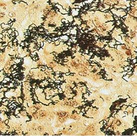
32 32 Pneumocystis Jirovecii: Black
33 Grocott s Methenamine Silver (GMS) Intended for laboratory use to aid the identification, by light microscopy, of fungal organisms and Pneumocystis jirovecii (formerly known as carinii) in tissue samples. Product Code AR176 Staining Interpretation: Fungi: Black Pneumocystis: Black Background: Light green Section Thickness : Control Tissue: 4 μm Tissue with Aspergillus, Candida or Pneumocystis Time per Slide*: 0:50:19 33

34 34 Fungi: Black
35 Grocott s Methenamine Silver (GMS) Eosin Intended for laboratory use to aid the identification, by light microscopy, of fungal organisms and Pneumocystis jirovecii (formerly known as carinii) in tissue samples. Product Code AR376 Staining Interpretation: Fungi: Black Pneumocystis: Black Background: Pink Section Thickness : Control Tissue: 4 μm Tissue with Aspergillus, Candida or Pneumocystis Time per Slide*: 0:56:17 35

36 Nuclei: Red Ferric Iron: Blue 36
37 Iron Intended for laboratory use to aid the identification, by light microscopy, of ferric iron deposits in tissue samples. Product Code AR158 Staining Interpretation: Ferric iron: Blue Nuclei: Red Background: Pink Section Thickness : Control Tissue: 4 μm Spleen or liver with hemosiderosis Time per Slide*: 0:09:58 37

38 Basement Membrane: Black Nuclei: Red 38
39 Jones Basement Membrane (PAS-M) Intended for laboratory use to aid the identification, by light microscopy, of basement membranes in tissue samples. Product Code AR180 Staining Interpretation: Basement membrane: Black Nuclei: Red Background: Pink Section Thickness : Control Tissue: 2 μm Kidney with glomeruli Time per Slide*: 0:52:53 39

40 Basement Membrane: Black Nuclei: Blue 40
41 Jones Basement Membrane (PAS-M) H&E Intended for laboratory use to aid the identification, by light microscopy, of basement membranes in tissue samples. Product Code AR480 Staining Interpretation: Basement membrane: Black Nuclei: Blue Background: Pink Section Thickness : Control Tissue: 2 μm Kidney with glomeruli Time per Slide*: 0:45:21 41

42 42 Basement Membrane: Black
43 Jones Basement Membrane (PAS-M) Light Green Intended for laboratory use to aid the identification, by light microscopy, of basement membranes in tissue samples cut at 2 μm. Product Code AR380 Staining Interpretation: Basement membrane: Black Background: Light green Section Thickness : Control Tissue: 2 μm Kidney with glomeruli Time per Slide*: 0:42:55 43

44 Muscle: Red Collagen: Blue 44
45 Masson s Trichrome Intended for laboratory use to aid the identification, by light microscopy, of connective tissue and muscle in tissue samples. Product Code AR173 Staining Interpretation: Muscle fibers: Red Fibrin: Pink Collagen: Blue Nuclei: Blue or black Erythrocytes: Red Section Thickness : Control Tissue: 4 μm Uterus, small intestine, liver, appendix or fallopian tube Time per Slide*: 0:57:15 45

46 Nuclei: Black Mucin: Pink 46
47 Mucicarmine Intended for laboratory use to aid the identification, by light microscopy, of epithelial mucins in tissue samples. Product Code AR168 Staining Interpretation: Mucin: Pink Nuclei: Black Background: Yellow Section Thickness : Control Tissue: 4 μm Small intestine, colon or appendix Time per Slide*: 0:50:54 47


48 Copper: Dark red-purple Elastin Fibers: Dark reddish brown HBsAG: Dark reddish brown 48
49 Orcein Intended for laboratory use to aid the identification, by light microscopy, of the viral inclusion bodies of hepatitis B surface antigen (HBsAG) and Copperassociated proteins in tissue samples. Product Code AR313 Staining Interpretation: HBsAG/elastin fibers: Dark reddish brown Copper-associated protein: Dark red-purple Background: Pale pink to pink Section Thickness : Control Tissue: 4 μm Liver tissue containing positive HBsAG. Fetal liver can be used for copper or liver with Wilson s disease. A multi-block containing positive liver for HBsAG and copper with normal liver is recommended. Time per Slide*: 0:15:44 49

50 PAS-positive structures: Magenta Nuclei: Blue 50
51 Periodic Acid-Schiff (PAS) Intended for laboratory use to aid the identification, by light microscopy, of glycogen and mucopolysaccharide components in tissue samples. Product Code AR165 Staining Interpretation: PAS-positive structures: Magenta Nuclei: Blue Background: Pink Section Thickness : Control Tissue: 4 μm Kidney for basement membrane Liver with glycogen Time per Slide*: 0:59:59 51

52 52 Nuclei: Blue
53 Periodic Acid-Schiff (PAS) with Alpha-Amylase Intended for laboratory use to aid the identification, by light microscopy, of glycogen and mucopolysaccharide components in tissue samples. Alphaamylase digestion removes any glycogen in the tissue. Product Code AR165 (with AR171) Staining Interpretation: Nuclei: Blue Background: Pink Section Thickness : Control Tissue: 4 μm Liver containing glycogen Time per Slide*: 01:14:02 53

54 54 Fungi: Magenta
55 Periodic Acid-Schiff (PAS) Green Intended for laboratory use to aid the identification, by light microscopy, of fungi in tissue samples. Product Code AR172 Staining Interpretation: Fungi: Magenta Background: Blue - green Section Thickness : Control Tissue: 4 μm Skin tissue with Candida albicans Time per Slide*: 0:24:12 55

56 56 Reticulin Fibers: Black
57 Reticulin/No Counterstain Intended for laboratory use to aid the identification, by light microscopy, of reticulin fibers in tissue samples. Product Code AR182 Staining Interpretation: Reticulin fibers: Black Section Thickness : Control Tissue: 4 μm Liver, spleen and lymph node Time per Slide*: 00:38:57 57

58 58 Reticulin Fibers: Black
59 Reticulin/Nuclear Fast Red Intended for laboratory use to aid the identification, by light microscopy, of reticulin fibers in tissue samples. Product Code AR179 Staining Interpretation: Reticulin fibers: Black Background: Red Section Thickness : Control Tissue: 4 μm Liver, spleen or lymph node Time per Slide*: 01:02:59 59

60 Spirochetes: Black Helicobacter Pylori: Black 60
61 Warthin-Starry Intended for laboratory use to aid the identification, by light microscopy, of Helicobacter pylori and spirochete microorganisms in tissue samples. Product Code AR181 Staining Interpretation: Helicobactor pylori: Black Spirochetes: Black Background: Golden yellow Section Thickness : Control Tissue: 4 μm Tissue with helicobacter and spirochetes Time per Slide*: 00:07:38 61
62 62
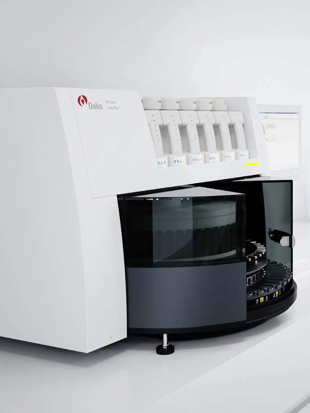
63 Artisan Link Pro Solution The consistent, safe and easy choice for special stains. Designed for multi staining of up to 14 different stains simultaneously 48 slide capacity for optimal productivity and overnight run Fully automated with on-board drying and deparaffinization capabilities 27 high-quality ready-to-use automated stain kits 29 optimized, validated protocols that are editable for easy yet flexible results Proven consistent staining quality Waste separation into four separate streams DakoLink Software with LIS and LAN/WAN connectivity enables full lab integration Complete data and staining transparency enabling optimized performance and full traceability Dedicated Service & Support provides fast, professional support when hands-on assistance is needed For additional information on Artisan Link Pro or other Dako solutions, contact your local Agilent representative or visit 63
64 This information is subject to change without notice. Agilent Technologies, Inc Published in the USA, June 4, JUN04
Fixation... Questions 1 Answers 16. Processing... Questions 25 Answers 36. Safety... Questions 67 Answers 73
 Table of Contents Fixation... Questions 1 Answers 16 Processing... Questions 25 Answers 36 Instrumentation... Questions 43 Answers 58 Safety... Questions 67 Answers 73 Laboratory Mathematics & Solution
Preface 1. Fixation and Processing 1
 Contents Preface xi 1. Fixation and Processing 1 Fixation 1 Processing 2 What Should Be Seen in a Well-Fixed, Well-Processed Specimen Stained with Hematoxylin and Eosin 3 Problems Encountered With Fixation
The Oral Histology Series Series 5 Special Stains
 The Oral Histology Series Series 5 Special Stains DAVID E. KLINGMAN, Lt Col, USAF, DC Views and opinions expressed are those of the author(s) and do not reflect official policy or position of the United
BenchMark Special Stains. Product guide
 BenchMark Special Stains Product guide Quick reference table Product name Ordering code Catalog number Tissue thickness Tests per kit Total vials in kit package *Not all vials in the kit are used at one
Schedule of Accreditation issued by United Kingdom Accreditation Service 2 Pine Trees, Chertsey Lane, Staines-upon-Thames, TW18 3HR, UK
 2 Pine Trees, Chertsey Lane, Staines-upon-Thames, TW18 3HR, UK Cellular Pathology Department University College London Hospital Rockefeller Building University Street London WC1E 6JJ Contact: Gavyn Barrett
BLIZARD INSTITUTE CORE PATHOLOGY ATLAS OF TINCTORIAL STAINS
 BLIZARD INSTITUTE CORE PATHOLOGY ATLAS OF TINCTORIAL STAINS Contents Introduction... 3 Background to Tinctorial Stains... 3 Haematoxylin and Eosin Stain (H&E)... 3 Connective Tissue Stains... 4 Nucleic
PATHOPHYSIOLOGY. DEFINED Involves the study of function that results from disease processes.
 DEFINED Involves the study of function that results from disease processes. What is pathology? Pathology is the branch of medical sciences that treats the essential nature of disease, especially the changes
ABOUT US WORLDWIDE DISTRIBUTION ICON KEY STAIN LINE PRODUCT LIST
 ABOUT US WORLDWIDE DISTRIBUTION ICON KEY STAIN LINE PRODUCT LIST BQCKit Company is one of the three differentiated business lines of Bioquochem S.L. We are a Spanish biotechnology company specialized
HISTOLOGY Lecture TWO DR. ASHRAF SAID
 HISTOLOGY Lecture TWO DR. ASHRAF SAID Start Of this lecture TISSUES TISSUE: A DEFINITION A group of connected and interdependent cells that cooperate to perform a specific function CONNECTIVE TISSUE The
Special Staining (I)
") Special Staining (I) Carbohydrates 1- PERIODIC ACID SCHIFF'S (PAS ) Purpose: Glycogen is present in liver, kidney, skeletal and cardiac muscle. The PAS stain is used to demonstrate neutral polysaccharides
Special Staining (I) Carbohydrates 1- PERIODIC ACID SCHIFF'S (PAS ) Purpose: Glycogen is present in liver, kidney, skeletal and cardiac muscle. The PAS stain is used to demonstrate neutral polysaccharides
Carbohydrates and mucins. Dr Phil Bryant, Wales, UK
 Carbohydrates and mucins Dr Phil Bryant, Wales, UK Carbohydrates Provide cells with energy by converting to glucose Excess glucose stored in liver and muscle as glycogen Residual unstored glycogen is turned
3. Staining solutions for histology and cytology Mounting and embedding media... 60
 INDEX 1. Hematoxylin stains... 4 2. Eosin... 6 3. Staining solutions for histology and cytology... 8 4. Reagents I... 48 5. Reagents II... 55 6. Decalcifying solutions... 58 7. Fixatives... 59 8. Formalins...
Special Stains. General Reference Guide
 Special Stains General Reference Guide Table of Contents AFB 4 Technical notes and references 6 Gram 32 Technical notes and references 34 Steiner 64 Technical notes and references 66 Alcian Blue 8 Technical
Schedule of Accreditation issued by United Kingdom Accreditation Service 2 Pine Trees, Chertsey Lane, Staines-upon-Thames, TW18 3HR, UK
 2 Pine Trees, Chertsey Lane, Staines-upon-Thames, TW18 3HR, UK 05/09/2018 Cellular Pathology Level 2 Esher Wing Galsworthy Road Kingston upon Thames KT2 7QB Contact: Dr Sussan Gharaie Tel: +44 (0)208 934
Schedule of Accreditation issued by United Kingdom Accreditation Service 2 Pine Trees, Chertsey Lane, Staines-upon-Thames, TW18 3HR, UK
 Schedule of Accreditation 2 Pine Trees, Chertsey Lane, Staines-upon-Thames, TW18 3HR, UK Department of Eye Pathology 1 st Floor, Cayton Street Building UCL Institute of Ophthalmology 11-43 Bath Street
2014 CURRENT ISSUES IN PATHOLOGY
 2014 CURRENT ISSUES IN PATHOLOGY SPECIAL STAINS IN LIVER BIOPSY PATHOLOGY Sanjay Kakar, MD University of California, San Francisco Trichrome stain : (1) Assess degree of fibrosis. H&E stain is not reliable
PRODUCT INFORMATION. New Reagents for Dako CoverStainer. Choose the H&E staining intensity you want.
 PRODUCT INFORMATION Primary Staining Dako CoverStainer Reagents New Reagents for Dako CoverStainer. Choose the H&E staining intensity you want. New flexible staining intensities to fit y Dako H&E Staining
Schedule of Accreditation issued by United Kingdom Accreditation Service 2 Pine Trees, Chertsey Lane, Staines-upon-Thames, TW18 3HR, UK
 2 Pine Trees, Chertsey Lane, Staines-upon-Thames, TW18 3HR, UK Blizard Institute Core Pathology Pathology and Pharmacy Building Second Floor 80 Newark Street London E1 2ES Contact: Pauline Levey Tel: +44
UNDERSTANDING SPECIAL STAINS
 Vet Times The website for the veterinary profession https://www.vettimes.co.uk UNDERSTANDING SPECIAL STAINS Author : MELANIE DOBROMYLSKYJ Categories : Vets Date : April 14, 2014 MELANIE DOBROMYLSKYJ explains
Practical Histology. Lab 3: Connective tissue
 Practical Histology Lab 3: Connective tissue Connective tissues Connective tissue provides structural support for the body by binding cells and tissues together to form organs. It also provides metabolic
Schedule of Accreditation issued by United Kingdom Accreditation Service 2 Pine Trees, Chertsey Lane, Staines-upon-Thames, TW18 3HR, UK
 2 Pine Trees, Chertsey Lane, Staines-upon-Thames, TW18 3HR, UK Laboratory locations: Department of Histopathology Royal United Hospitals NHS Foundation Trust Combe Park Bath BA1 3NG Contact: Lesley Shipway
Special Stains in Dermatopathology
 Application Special Stains in Dermatopathology Jameel Ahmad Brown, MD Bruce R. Smoller, MD Fellow: Division of Dermatopathology University of Arkansas for Medical Sciences 4301 W. Markham St. Little Rock,
HISTOPATHOLOGY. Introduction
 HISTOPATHOLOGY Introduction Contacts Services offered Pathology tissue request Laboratory hours Special instructions Histopathology reports List of specimens Introduction The Histopathology section of
LIST OF ORGANS FOR HISTOPATHOLOGICAL ANALYSIS:!! Neural!!!!!!Respiratory:! Brain : Cerebrum,!!! Lungs and trachea! Olfactory, Cerebellum!!!!Other:!
 LIST OF ORGANS FOR HISTOPATHOLOGICAL ANALYSIS:!! Neural!!!!!!Respiratory:! Brain : Cerebrum,!!! Lungs and trachea! Olfactory, Cerebellum!!!!Other:! Spinal cord and peripheral nerves! Eyes, Inner ear, nasal
CONNECTIVE TISSUE (C.T.)
") CONNECTIVE TISSUE (C.T.) Objectives: By the end of this lecture, the student should be able to: 1. Enumerate the general characteristics of C.T. 2. Classify C.T into C.T. proper and special types of C.T.
CONNECTIVE TISSUE (C.T.) Objectives: By the end of this lecture, the student should be able to: 1. Enumerate the general characteristics of C.T. 2. Classify C.T into C.T. proper and special types of C.T.
Presented by: Dr. Giuseppe Molinaro Dr. Davide De Biase
 Presented by: Dr. Giuseppe Molinaro Dr. Davide De Biase Dog Spayed Female LABRADOR RETRIEVER 3 Years old VACCINATIONS ANTIPARASITIC COMMERCIAL DIET VOMITING FOR A MONTH DULLNESS WEIGHT LOSS INAPPETANCE
Schedule of Accreditation issued by United Kingdom Accreditation Service 2 Pine Trees, Chertsey Lane, Staines-upon-Thames, TW18 3HR, UK
 2 Pine Trees, Chertsey Lane, Staines-upon-Thames, TW18 3HR, UK Lister Hospital Contact: Heather Taylor Coreys Mill Lane Tel: +44 (0) 01438 285086 Stevenage E-Mail: Heathertaylor3@nhs.net SG1 4AB Website:
Schedule of Accreditation issued by United Kingdom Accreditation Service 2 Pine Trees, Chertsey Lane, Staines-upon-Thames, TW18 3HR, UK
 2 Pine Trees, Chertsey Lane, Staines-upon-Thames, TW18 3HR, UK Calderdale and Huddersfield NHS Foundation Trust Calderdale Royal Hospital Salterhebble Halifax HX3 0PW United Kingdom Contact: Dr Richard
Blood: Functions. Liquid connective tissue 3 general functions 1. Transportation. 2. Regulation. 3. Protection
 Blood Elements Lecture Objectives List blood components. Classify formed elements of blood. Discuss the scientific basis of the above classification. Describe the basic structure of erythrocytes and criteria
Sheet #9. Dr. Heba Kalbouneh. Dr. Heba Kalbouneh. Dr. Heba Kalbouneh
 Sheet #9 Dr. Heba Kalbouneh Dr. Heba Kalbouneh Dr. Heba Kalbouneh Elastic fibers The main function of elastic fibers is to provide elasticity. In other words these fibers are able to restore the original
A Histochemical Study of Epithelial Mucin
 A Histochemical Study of Epithelial Mucin in the Chick Chorioallantois JAMES L. CONKLIN Department of Anatomy, The University of Michigan, Ann Arbor, Michigun ABSTRACT Histochemical methods demonstrate
BLOOD RUNS THROUGH YOUR BODY
 BLOOD RUNS THROUGH YOUR BODY WORKSHEET A Your heart and blood vessels make up your blood system. At the centre of your blood system is your heart. Its job is to pump the blood around your body. The rest
The goal of teaching:
 The goal of teaching: 1. Principles of LDG of SFI: Hair Nail Skin Mucosal Eye Ear 3. Methods for fungal identification Test methods for fungal susceptibility testing Diffusion Dilution Etest 2. Principles
Autopsy findings in 51 year-old man with mantle cell lymphoma
 Autopsy findings in 51 year-old man with mantle cell lymphoma Bobbi S. Pritt, MD, MSc Professor of Laboratory Medicine and Pathology Mayo Clinic Disclosure of Relevant Financial Relationships USCAP requires
Schedule of Accreditation issued by United Kingdom Accreditation Service 2 Pine Trees, Chertsey Lane, Staines-upon-Thames, TW18 3HR, UK
 2 Pine Trees, Chertsey Lane, Staines-upon-Thames, TW18 3HR, UK Spire Portsmouth Hospital Bartons Road Havant PO9 5NP United Kingdom Contact: Natalie Peck E-Mail: natalie.peck@spirehealthcare.com Website:
Schedule of Accreditation issued by United Kingdom Accreditation Service 2 Pine Trees, Chertsey Lane, Staines-upon-Thames, TW18 3HR, UK
 2 Pine Trees, Chertsey Lane, Staines-upon-Thames, TW18 3HR, UK The Princess Alexandra Hospital NHS Trust The Michael Letcher Department of Cellular Pathology The Princess Alexandra Hospital NHS Trust Hamstel
Dr. heba kalbouneh. Dr. heba kalbouneh
 7 Dr. heba kalbouneh Dr. heba kalbouneh Clinical applications: In surgical incision site, the site of injury is filled by collagen fibers synthesized by fibroblasts (the fibrocytes in the connective tissue
Compara've Medicine Animal Histology Services. Glossary of Terms
 Compara've Medicine Animal Histology Services Glossary of Terms About this guide This guide is intended to provide clarifica'on on standard histologic terminology. Included are examples of different sample
Individual cells Extracellular matrix
 Connective Tissue Connective Tissue Elements Individual cells Extracellular matrix»fibers» Collagen» Elastic» Reticular»Ground Substance» PG (proteoglycans)» GAG (glycosaminoglycan)» GP (glycoprotein)
EDUCATIONAL COMMENTARY MORPHOLOGIC CHANGES IN PERIPHERAL BLOOD CELLS
 EDUCATIONAL COMMENTARY MORPHOLOGIC CHANGES IN PERIPHERAL BLOOD CELLS Educational commentary is provided through our affiliation with the American Society for Clinical Pathology (ASCP). To obtain FREE CME/CMLE
Special stains in liver pathology
 Current Issues in Surgical Pathology 2014 Special stains in liver pathology Which, why, how Really? Sanjay Kakar, MD University of California, San Francisco Outline Which stains Why the stain is done How
Schedule of Accreditation issued by United Kingdom Accreditation Service 2 Pine Trees, Chertsey Lane, Staines-upon-Thames, TW18 3HR, UK
 2 Pine Trees, Chertsey Lane, Staines-upon-Thames, TW18 3HR, UK Department of Cellular Pathology Dorset County Hospital Williams Avenue Dorchester DT1 2JY Contact: Sharon Wood Tel: +44 (0)1305 254326 E-Mail:
Connective Tissue. Red: important. Black: in male female slides. Gray: notes. Editing File
 Connective Tissue Red: important. Black: in male female slides. Gray: notes. Editing File OBJECTIVES Enumerate the general characteristics of C.T Classify C.T into C.T. proper and special types of C.T
CHAPTER. V SUMMARY AND CONCLUSIONS. during postnatal period and to provide elasticity during prenatal and early
 CHAPTER. V SUMMARY AND CONCLUSIONS The present study was conducted on 40 samples, each of thymus and Spleen of goat from prenatal to four months and above age. The small pieces from each thymus and spleen
BioSciences. Peripheral Blood Smear Preparation. Blood Smear Preparation Materials
 PolyFacts Vol. 5 No. 1 BioSciences Use the Wright Stain High Quality StainRITE Ready-to-Use Stains for Hematology Peripheral blood smear (peripheral blood film) is a glass microscope slide coated with
TEST MENU BY SPECIALTY
 1 TEST MENU BY SPECIALTY Breast Pathology Surgical excisions, needle core biopsies, and plastic surgery accepted from all sites Assessment of margins Axillary lymph node dissections Sentinel lymph nodes
Schedule of Accreditation issued by United Kingdom Accreditation Service 2 Pine Trees, Chertsey Lane, Staines-upon-Thames, TW18 3HR, UK
 United Kingdom Accreditation Service 2 Pine Trees, Chertsey Lane, Staines-upon-Thames, TW18 3HR, UK North Tyneside General Hospital Rake Lane North Shields Tyne & Wear NE29 8NH Contact: Ian Taylor Tel:
Tissues. Tissues - Overview. Bio211 Laboratory 2. Epithelial and Connective Tissues
 Bio211 Laboratory 2 Epithelial and Connective Tissues 1 Tissues Tissues to be examined under the microscope Epithelial Tissue (p. 79 Lab Manual) [TODAY] Connective Tissue (p. 93 Lab Manual) [TODAY] Muscle/Nervous
Unit I Problem 9 Histology: Basic Tissues of The Body
 Unit I Problem 9 Histology: Basic Tissues of The Body - What is the difference between cytology and histology? Cytology: it is the study of the structure and functions of cells and their contents. Histology:
I. Concepts: Fill in the following sections with information from the text and lecture.
 Name: Period: 10 Blood Study Guide I. Concepts: Fill in the following sections with information from the text and lecture. 1. Composition and Function of Blood: 2. Hematopoiesis: 1 Miss School, Miss Out
HER2 CISH pharmdx TM Kit Interpretation Guide Breast Cancer
 P A T H O L O G Y HER2 CISH pharmdx TM Kit Interpretation Guide Breast Cancer FROM CERTAINTY COMES TRUST For in vitro diagnostic use HER2 CISH pharmdx Kit HER2 CISH pharmdx Kit is intended for dual-color
Dako IT S ABOUT TIME. Interpretation Guide. Agilent Pathology Solutions. ALK, ROS1 and RET IQFISH probes (Dako Omnis) MET IQFISH probe (Dako Omnis)
 MET IQFISH probe (Dako Omnis)") INTERPRETATION Dako Agilent Pathology Solutions IQFISH Interpretation Guide ALK, ROS1 and RET IQFISH probes (Dako Omnis) MET IQFISH probe (Dako Omnis) IT S ABOUT TIME For In Vitro Diagnostic Use ALK, ROS1,
INTERPRETATION Dako Agilent Pathology Solutions IQFISH Interpretation Guide ALK, ROS1 and RET IQFISH probes (Dako Omnis) MET IQFISH probe (Dako Omnis) IT S ABOUT TIME For In Vitro Diagnostic Use ALK, ROS1,
Schedule of Accreditation issued by United Kingdom Accreditation Service 2 Pine Trees, Chertsey Lane, Staines-upon-Thames, TW18 3HR, UK
 2 Pine Trees, Chertsey Lane, Staines-upon-Thames, TW18 3HR, UK 142-144 New Cavendish Street W1W 6YF Contact: Mr Tel: +44 (0) 20 7299 4490 Fax: +44 (0)20 7299 4491 E-Mail: Neil.Barrett@unilabs.com Website:
Blood & Blood Formation
 Module IB Blood & Blood Formation Histology and Embryology Martin Špaček, MD (m.spacek@centrum.cz) http://www.lf3.cuni.cz/histologie Approximately 7% of a person's weight is blood (about 5 L) Blood consists
Connective Tissue Part-2. Dr. Heba Kalbouneh Assistant Professor of Anatomy and Histology
 Connective Tissue Part-2 Dr. Heba Kalbouneh Assistant Professor of Anatomy and Histology 1 Features Composed of cells, fibers and extracellular matrix. Highly vascular Variable regenerative power Originates
Histo lab 7. Special connective tissue is derived from the mesoderm (mesenchyme).
.") Histo lab 7 Special connective tissue is derived from the mesoderm (mesenchyme). If we have high density of fibers, we call it dense connective tissue. (Fibers are more than the ground substance). If we
Histo lab 7 Special connective tissue is derived from the mesoderm (mesenchyme). If we have high density of fibers, we call it dense connective tissue. (Fibers are more than the ground substance). If we
Schedule of Accreditation issued by United Kingdom Accreditation Service 2 Pine Trees, Chertsey Lane, Staines-upon-Thames, TW18 3HR, UK
 2 Pine Trees, Chertsey Lane, Staines-upon-Thames, TW18 3HR, UK Spire Portsmouth Hospital Bartons Road Havant PO9 5NP United Kingdom Contact: Natalie Peck E-Mail: natalie.peck@spirehealthcare.com Website:
DOCTORAL THESIS (SUMMARY)
") Translation from Romanian UNIVERSITY OF MEDICINE AND PHARMACY CRAIOVA THE FACULTY OF MEDICINE DOCTORAL THESIS (SUMMARY) Scientific coordinator Prof. Dr. Laurentiu MOGOANTA PhD student, Dr. Madalin IONILA
Translation from Romanian UNIVERSITY OF MEDICINE AND PHARMACY CRAIOVA THE FACULTY OF MEDICINE DOCTORAL THESIS (SUMMARY) Scientific coordinator Prof. Dr. Laurentiu MOGOANTA PhD student, Dr. Madalin IONILA
New, Special Stain for Histopathological Diagnosis of Cryptococcosis
 JOURNAL OF CLINICAL MICROBIOLOGY, Feb. 1981, p. 383-387 0095-1137/81/020383-05$02.00/0 Vol. 13, No. 2 New, Special Stain for Histopathological Diagnosis of Cryptococcosis KYUNG J. KWON-CHUNG,* WILLIAM
The % of blood consisting of packed RBCs is known as the hematocrit. Blood s color ranges from scarlet (oxygen-rich) to dark red (oxygen poor).
 to dark red (oxygen poor).") Biology Blood Blood is a fluid connective tissue consisting of cells suspended in a liquid fibrous matrix. The cells are called formed elements and the liquid matrix is known as plasma. The formed elements
Biology Blood Blood is a fluid connective tissue consisting of cells suspended in a liquid fibrous matrix. The cells are called formed elements and the liquid matrix is known as plasma. The formed elements
HISTOLOGY VIRTUAL LABORATORY BLOOD AND LYMPHATICS SYSTEM
 HISTOLOGY VIRTUAL LABORATORY BLOOD AND LYMPHATICS SYSTEM Login: http://histopath.westernu.edu Histology Atlas AND Virtual Histology links. I. HEMATOLOGY - PERIPHERAL BLOOD Purpose: To be able to identify
Most abundant and widely distributed tissues in the body Binds, support, and strengthen body tissues, protect and insulate internal organ, serve as
 Connective tissue Most abundant and widely distributed tissues in the body Binds, support, and strengthen body tissues, protect and insulate internal organ, serve as major transport system, compartmentalizes
VETERINARY HEMATOLOGY ATLAS OF COMMON DOMESTIC AND NON-DOMESTIC SPECIES COPYRIGHTED MATERIAL SECOND EDITION
 VETERINARY HEMATOLOGY ATLAS OF COMMON DOMESTIC AND NON-DOMESTIC SPECIES SECOND EDITION COPYRIGHTED MATERIAL CHAPTER ONE HEMATOPOIESIS GENERAL FEATURES All blood cells have a finite life span, but in normal
The Egyptian journal of hospital medicine (2009)vol.,36:
vol.,36:") The Egyptian journal of hospital medicine (2009)vol.,36: 468-482 QUANTITATIVE AND HISTOCHEMICAL STUDY ON THE ADRENAL MEDULLA IN POST NATAL PERIOD OF ALBINO RAT Wagieh k.baiomy; Abdel-mawgood Anas; Mamdooh
The Egyptian journal of hospital medicine (2009)vol.,36: 468-482 QUANTITATIVE AND HISTOCHEMICAL STUDY ON THE ADRENAL MEDULLA IN POST NATAL PERIOD OF ALBINO RAT Wagieh k.baiomy; Abdel-mawgood Anas; Mamdooh
6 DISTURBANCES IN CELL METABOLISM
 6 DISTURBANCES IN CELL METABOLISM Cloudy Swelling Hydropic Degeneration Mucinous Degeneration Mucoid Degeneration Psuedomucin Amyloid Infilteration Hyaline Degeneration Fatty Changes Glycogen Infilteration
Chapter 21 Outline. General Composition and Functions of Blood Blood Plasma Formed Elements in the Blood Hemopoiesis: Production of Formed Elements
 Chapter 21 Outline General Composition and Functions of Blood Blood Plasma Formed Elements in the Blood Hemopoiesis: Production of Formed Elements Introduction Blood serves many functions. Some examples
PRACTICAL HISTOPATHOLOGY IN MOUSE MODELS OF HUMAN DISEASE: GUIDES TO PHENOTYPING THE GENETICALLY ALTERED MOUSE
 PRACTICAL HISTOPATHOLOGY IN MOUSE MODELS OF HUMAN DISEASE: GUIDES TO PHENOTYPING THE GENETICALLY ALTERED MOUSE http://mousepheno.ucsd.edu/ https://www.ncbi.nlm.nih.gov/pmc/articles/pmc3693904/ 1. Approval
Mucin Histochemistry Study of the Prostate in Normal and Malignant Lesions
 ISSN 2231-4261 ORIGINAL ARTICLE Mucin Histochemistry Study of the Prostate in Normal and Malignant Lesions 1* 1 1 1 Manoj P.Ambali, Megha A. Doshi, Pratibha P. Patil, Shweta H. Chavan 1 Department of Anatomy,
Schedule of Accreditation issued by United Kingdom Accreditation Service 2 Pine Trees, Chertsey Lane, Staines-upon-Thames, TW18 3HR, UK
 2 Pine Trees, Chertsey Lane, Staines-upon-Thames, TW18 3HR, UK Paediatric Histopathology Western Bank Sheffield S10 2TH United Kingdom Contact: Tel: +44 (0) 1142 717240 Fax: +44 (0) 1142 706121 E-Mail:
Connective tissue CONNECTIVE TISSUE Part I
 Connective tissue CONNECTIVE TISSUE Part I Part 1 Connective Tissue Found everywhere in the body (app. 50% of body weight) Includes the most abundant and widely distributed tissues General features of
The Endocrine System Pituitary
 The Endocrine System Pituitary Look at your slide of the human pituitary with your naked eye. You should see a cellular region and a more fibrous region. Then view each region with your microscope under
The Differentiation of Yeast and Yeast-Like Forms in Human Tissues. Introduction. Histochemical Stains Used to Detect Fungi. Histopathologic Diagnoses
 The Differentiation of Yeast and Yeast-Like Forms in Human Tissues Gary W. Procop, MD Chair, Clinical Pathology Staff, Anatomic Pathology Director, Molecular Microbiology, Mycology, and Parasitology Cleveland
4. TEXTBOOK: ABUL K. ABBAS. ANDREW H. LICHTMAN. CELLULAR AND MOLECULAR IMMUNOLOGY. 5 TH EDITION. Chapter 2. pg
 LECTURE: 03 Title: CELLS INVOLVED IN THE IMMUNE RESPONSE LEARNING OBJECTIVES: The student should be able to: Identify the organs where the process of the blood formation occurs. Identify the main cell
Microscopy PART - 6. Microscopy. Products for bacteriology. Products for cytology. Products for hematology. Products for histology
 PART - 6 Products for bacteriology Products for cytology Products for hematology Products for histology Universal reagents for For more details contact : Merck Specialities Private Limited Tel : +91 22
Schedule of Accreditation issued by United Kingdom Accreditation Service 2 Pine Trees, Chertsey Lane, Staines-upon-Thames, TW18 3HR, UK
 2 Pine Trees, Chertsey Lane, Staines-upon-Thames, TW18 3HR, UK Issue No: 002 Issue date: 05 March 2018 Berkshire and Surrey Pathology Services Department of Histopathology Wexham Park Hospital Slough Berkshire
What in the world is Histotechnology? Karen Stiffler, MA, HTL Program Director for Histotechnology
 What in the world is Histotechnology? Karen Stiffler, MA, HTL Program Director for Histotechnology The Basics of Histology Histology: the study of body tissues "histo" is from the Greek "histos" meaning
Liqui-PREP TM The Next Generation of Liquid Cytology
 TECHNICAL TIP OVERVIEW: This Technical Tip is the result of many requests for Cytology Staining procedures. There are perhaps as many Cytology Staining procedures as there are Cytology processing laboratories.
CATALOG Parview Road Middleton, WI
 CATALOG 2505 Parview Road Middleton, WI 53562-2579 800-383-7799 www.newcomersupply.com February 2018 ABOUT US Mission Keeping it real: Our job is to help you get your job done! Everything we do is designed
Connective tissue Histology lab 6 Notes by Omar Sami
 Connective tissue Histology lab 6 Notes by Omar Sami The connective tissue is composed of: 1- Cells. 2- Extra Cellular Matrix; fibers & ground substance. Ground substance is where you find both Cells &
Anatomy and Physiology Tissue Review
 Anatomy and Physiology Tissue Review OVERVIEW Histology practicals can be rough, especially when access to slides is limited to the lab period. This resource provides an opportunity to learn or review
This is Learning Component 6 in Learning Module 1. We will show examples of features ( things ) including mineral deposits, urates, pigments, dust,
 including mineral deposits, urates, pigments, dust,") This is Learning Component 6 in Learning Module 1. We will show examples of features ( things ) including mineral deposits, urates, pigments, dust, plant material, and amyloid. 1 Calcium salts are the
This is Learning Component 6 in Learning Module 1. We will show examples of features ( things ) including mineral deposits, urates, pigments, dust, plant material, and amyloid. 1 Calcium salts are the
G. Types of White Blood Cells
 1. White blood cells are also called leukocytes. G. Types of White Blood Cells 2. White blood cells function to protect against diseases. 3. Two hormones that stimulate white blood cell production are
Microscopic Examination of Urine
 Download http://www.vetlab.com/kova.htm Definition of urine sediment: all solid materials suspended in the urine - a semiquantative evaluation of the urine sediment Significance of formed elements in the
8: Lymphatic vessels and lymphoid tissue. nur
 8: Lymphatic vessels and lymphoid tissue nur Lymphatic vascular system Functions return to the blood extracellular fluid (Lymph) from connective tissue spaces. ensures the return of water, electrolytes
Bone Marrow Pathology. Part 1. R.S. Riley, M.D., Ph.D.
 Bone Marrow Pathology Part 1 R.S. Riley, M.D., Ph.D. Bone Marrow Pathology Bone marrow basics Red cell diseases White cell diseases Other diseases Bone Marrow Pathology Bone marrow basics Hematopoiesis
IMPC phenotyping SOPs in JMC
 IMPC phenotyping SOPs in JMC Tissue Embedding and Block Banking IMPC_BLK_001 Purpose Collect and fix a standard list of tissues from the complete necropsy (see IMPC Gross Pathology & Tissue Collection
WSC , Conference 9, Case 1. Tissue from a nyala.
 WSC 2009-2010, Conference 9, Case 1. Tissue from a nyala. MICROSCOPIC DESCRIPTION: Heart, atrium (1 pt.): Approximately 40% of the atrial myocardium is replaced by areas of fibrous connective tissue (1
Periodic Acid-Schiff-Light Green Stain to Detect Glomerular Protein Deposits by Routine Light Microscopy
 Periodic Acid-Schiff-Light Green Stain to Detect Glomerular Protein Deposits by Routine Light Microscopy CHARLES N. GAMBLE, M.D. Department of Pathology, Sutter Memorial Hospital, Sacramento, California
Ordering Physician. Collected REVISED REPORT. Performed. IgG IF, Renal MCR. Lambda IF, Renal MCR. C1q IF, Renal. MCR Albumin IF, Renal MCR
 RenalPath Level IV Wet Ts IgA I Renal IgM I Renal Kappa I Renal Renal Bx Electron Microscopy IgG I Renal Lambda I Renal C1q I Renal C3 I Renal Albumin I Renal ibrinogen I Renal Mayo Clinic Dept. of Lab
Serotonin- and Somatostatin-Positive Goblet Cell Carcinoid of the Duodenum
 2012 66 4 351 356 Serotonin- and Somatostatin-Positive Goblet Cell Carcinoid of the Duodenum a b* c c c a a b d a c b d 352 Ohara et al. received remedies at another hospital. Hematemesis then recurred
(LM pages 91 98) Time Estimate for Entire Lab: 2.5 to 3.0 hours. Special Requirements
 Time Estimate for Entire Lab: 2.5 to 3.0 hours. Special Requirements") Laboratory 7 Chemical Aspects of Digestion (LM pages 91 98) Time Estimate for Entire Lab: 2.5 to 3.0 hours Special Requirements Incubation. Students should start these sections at the beginning of the
Laboratory 7 Chemical Aspects of Digestion (LM pages 91 98) Time Estimate for Entire Lab: 2.5 to 3.0 hours Special Requirements Incubation. Students should start these sections at the beginning of the
Explain the laboratory diagnosis of Rabies?
 Explain the laboratory diagnosis of Rabies? The standard test for rabies testing is dfa. This test has been thoroughly evaluated for more than 40 years, and is recognized as the most rapid and reliable
BLOOD. EEI n: t.ee# Required Name : Due : April 12,2018 COMPOSITION AND FUNCTIONS OF BLOOD. Beginningof Class
 t.ee# Required Name : Due : April 12,2018 Beginningof Class BLOOD Blood, the "life fluid" that courses through the body's blood vessels, provides the means by which the body's cells receive vital nutrients
SAMPLE LITERATURE Please refer to included weblink for correct version. Morphology of Cancer Cells. Edvo-Kit #990. Experiment Objective:
 Edvo-Kit #990 Morphology of Cancer Cells Experiment Objective: 990 In this lab, students will explore the morphology of normal and cancerous cells by staining pre-fi xed cells with methylene blue and eosin.
Megakaryoblastic Leukemia in a Dog A. Hillström 1, H. Tvedten 1, M. Kiupel 2.
 Megakaryoblastic Leukemia in a Dog A. Hillström 1, H. Tvedten 1, M. Kiupel 2. 1 University Animal Hospital, Swedish University of Agricultural Sciences and Strömsholm Referral Animal Hospital, Sweden 2
1 BIO 212: ANATOMY & PHYSIOLOGY II PLATELETS. Mature Stage: No nucleus. Only 2-3 µm in diameter: significantly smaller than RBCs
 1 BIO 212: ANATOMY & PHYSIOLOGY II LAB BLOOD PLATES EOSINOPHIL Contains large red-staining granules Usually 2 lobes 12-17 µm: about the size of neutrophils (2X erythrocytes) regulation/reduction of Histamine.
Lymph I: The Peripheral Lymph System
 Lymph I: The Peripheral Lymph System Peripheral = Secondary Primary Immune Organs = bone marrow, thymus Site of maturation of cells of the immune system Secondary Immune Organs = Nodes, MALT, spleen Filter
HEMOTOLOGY. B. Helps stabilize body temperature -heats up and cools down slowly which moderates body temp
 I. Body H 2 O = HEMOTOLOGY A. Variable quantities 1. sweating and urination ( ) decreases H 2 O 2. drinking H 2 O increases B. Water is found in two compartments 1. contains 2/3 of all water in your body
Case: The patient is a 73 year old woman with vague complaints of dyspepsia and abdominal pain. Upper endoscopy showed features of gastritis and a
 Case: The patient is a 73 year old woman with vague complaints of dyspepsia and abdominal pain. Upper endoscopy showed features of gastritis and a nodular lesion in the body of the stomach. The patient
Branch of medicine that deals with blood, its formation and disorders is called. Three main functions of cardiovascular system are,, and.
 Chapter 19 The Blood Human body must maintain a balance called. Body fluid inside the cells is called fluid; that outside is called or fluid. Two major fluid networks that help in connecting cells are