Sonography of Gall Bladder
|
|
|
- Tyler Weaver
- 6 years ago
- Views:
Transcription

1 Sonography of Gall Bladder Vikram Dogra,MD Professor of Radiology, Urology and BME Director of Ultrasound Associate Chair of Education and Research University of Rochester, NY
2 Objectives Describe the Congenital anomalies of gallbladder Understand the sonographic features of different types of acute gallbladder diseases. Understand Hyperplastic Cholecystosis. Describe the sonographic features of Gallbladder ployposis And Gallbladder carcinoma 2
3 How to Find GallBladder on Ultrasound? GallBladder neck has a consistent relationship with the origin of the right portal vein. It sits just next to it. 3


4 Duplicate GB


5 Duplicate GB
6 Duplicate GB Differential diagnosis includes Gallbladder diverticula, Gallbladder fold, Phrygian cap, choledocal cyst, Pericholecystic fluid, focal adenomyomatosis, and intraperitoneal fibrous bands 6


7 Ectopic GB

8 Ectopic GB

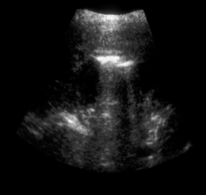
9 Micro- Gallbladder Rule out Gall stones Dogra,VS
10 Microcystic Gall Bladder Duodenum Gall bladder Rule out Gall stones Dogra,VS
11 Micro GallBladder Micro gallbladder is defined as less than 2-3 cm long and cm wide. Micro gallbladder can be seen most commonly in cystic fibrosis but is also seen in idiopathic neonatal hepatitis and alpha-1antitrypsin deficiency. 11

12 Acute Cholecystitis 12


13 Gangrenous Cholecystitis Right Upper Quadrant pain

14 Gangrenous Cholecystitis
15 Acute Cholecystitis RUQ pain+ GallStones or Impacted gall stone in the neck and Ultrasound Murphy sign Murphy Ultrasound sign is the Key in Gallbladder imaging Gallbladder wall thickening and peri cholecystic fluid are secondary signs. Sloughed Mucous membrane or free floating membrane is Key Air in the gallbladder wall is another important feature. 15

16 Gangrenous Cholecystitis

17 Emphysematous Cholecystitis
18 Emphysematous Cholecystitis 18

19 False Emphysematous Cholecystitis 19

20 GB Perforation


21 GB Perforation


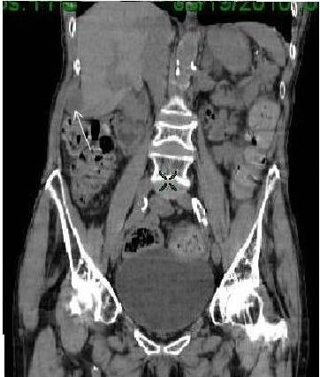
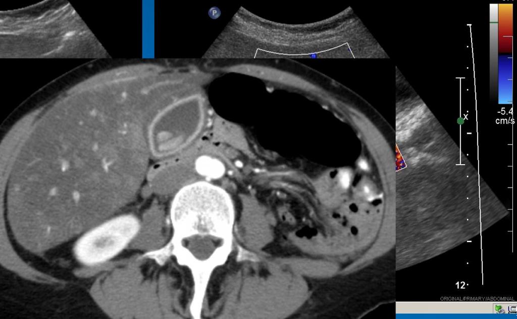
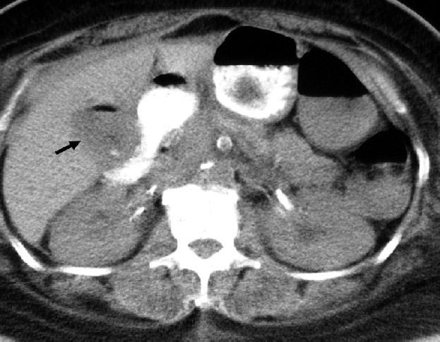
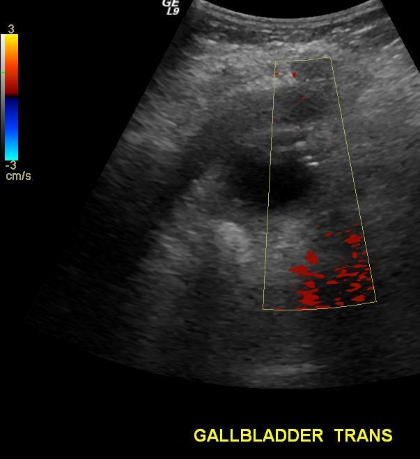
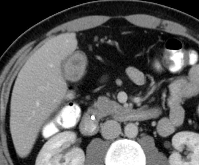
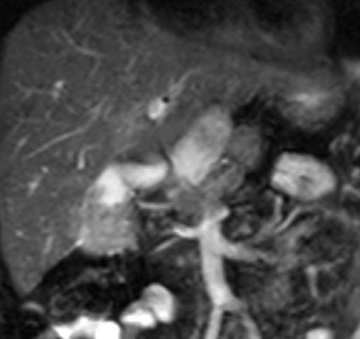
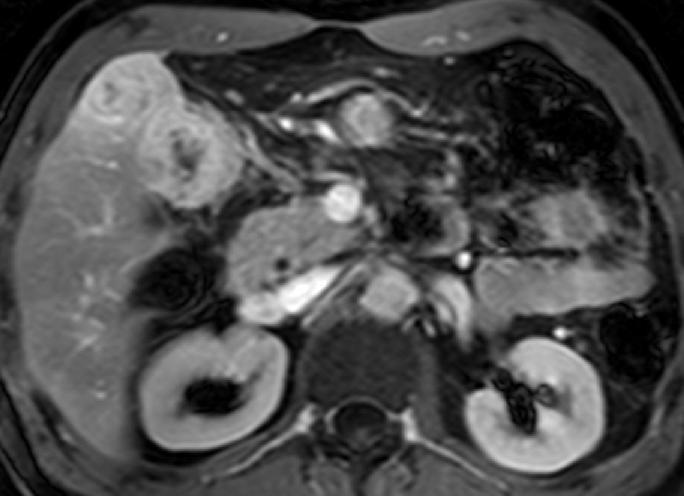
22 50 Year Old Female with RUQ pain Xanthogranulomatous Cholecystitis


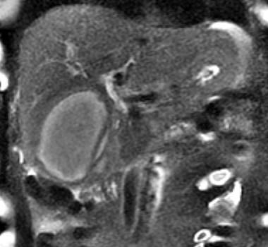

23 Xanthogranulomatous Cholecystitis CT T1 T2 T2
24 Xanthogranulomatous Cholecystitis Clinical presentation is usually one of chronic Cholecystitis Age group is sixth and seventh decades of life Incidence is about 1% and 2% of all cases of cholecystitis The pathologic features parallel xanthogranulomatous pyelonephritis. Sonographic,CT and MRI appearance are non-specific Pathology is confirmatory.

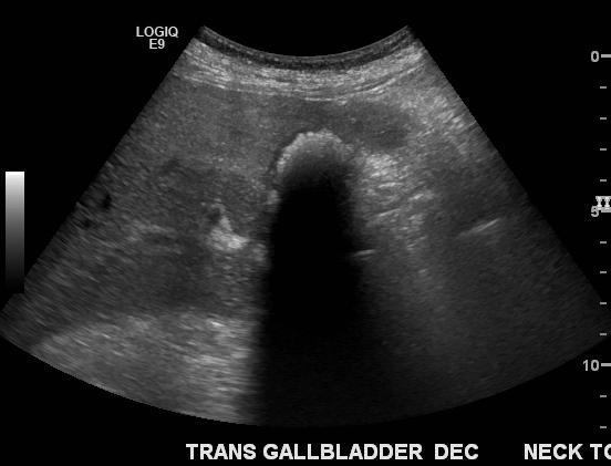
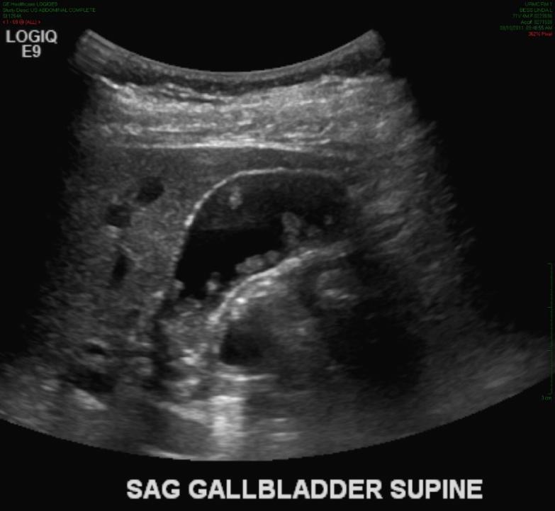
25 The WES Triad
26 Significance: Most of the time the WES sign is indicative of an chronic cholecystitis since the WES sign represents a contracted gallbladder with stones. WES triad helps differentiate Bowel from gallbladder. Pitfall: Calcification of gallbladder wall or poor axial resolution may demonstrate only 1 curvilinear echogenic line.

27 Aids Cholangiopathy 27


28 Right Upper Quadrant pain


29 Right Upper Quadrant pain


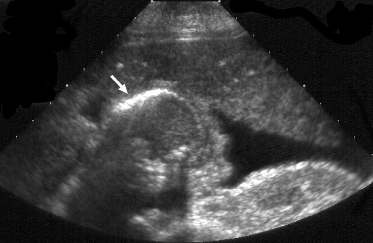
30 55-year-old patient with recurrent abdominal pain


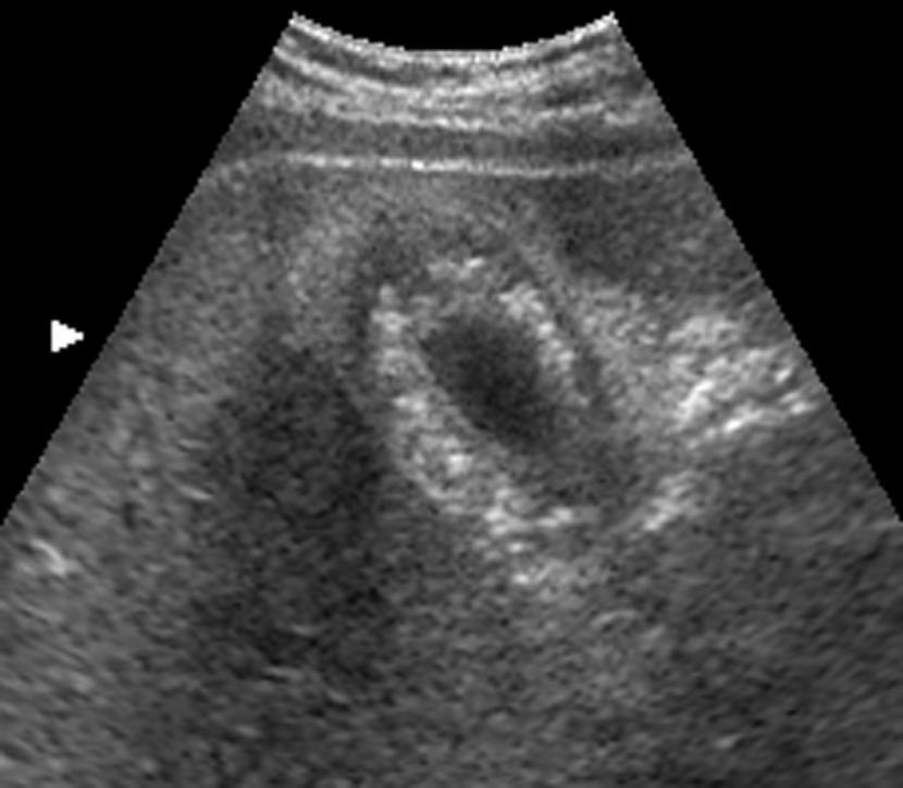
31 55-year-old patient with recurrent abdominal pain Adenomyomatosis- Fundal Type

32 Adenomyomatosis Smooth Muscle Liver Adenomyo Benign glands Cholesterolosis
33 Adenomyomatosis Reverberation artifact from cholesterol crystals is V-shaped and shorter in length than artifact from air. Sometimes, the calcium present within the sinuses may give rise to twinkle artifact. To Differentiate adenomyomatosis from other causes of gallbladder wall thickening, such as inflammation or carcinoma, is difficult..

34 Adenomyomatosis Hour glass type

35 Fundal Type of adenomyomatosis


36 Diffuse Type of Adenomyomatosis

37 Right Upper Quadrant pain


38 Myth of Adherent Gall Stones Adenomyomatosis


39 Myth of Gall Stones


40
41 Adenomyomatosis

42 Adenomyomatosis
43 Hyperplastic Cholecystosis. Better term is Hyperplastic Cholecystosis. Adenomyomatosis Cholesterolosis Overgrowth of the mucosa, thickening of the muscular wall, and formation of intramural diverticula or sinus tracts termed Rokitansky-Aschoff sinuses. Adenomyomatosis may involve the gallbladder in a focal, segmental, or diffuse form.

44 GallBladder Polyposis 44
45 GallBladder Polyps Benign Polyps are: Pseudotumors (cholesterol polyps, inflammatory polyps; cholesterolosis and hyperplasia), Epithelial tumors (adenomas) and mesenchymatous tumors (fibroma, lipoma, and hemangioma). Malignant GPs are gallbladder carcinomas. 45
46 GallBladder Polyps Cholesterol polyp is the most common type. Adenomas or adenomyomatous polyps are very rare (0.4% patients who underwent cholecystectomy) Gallbladder polyps larger than 1.5 cm, especially in solitary sessile hypoechogenic polyps, are associated with a risk of malignancy. Polyps that are smaller than 1 cm are monitored for 6 12 months. 46

47 GallBladder Carcinoma 47

48 60 Year Old Female with RUQ Pain

49
50 GallBladder Carcinoma Adenocarcinoma is most frequent Papillary, Mucinous and Signet cell Sqamous cell, Lymphoma and Mets are least common types. 50
51 GallBladder Carcinoma Gallbladder fossa mass replacing GB and invading Liver is the most common appearance. 25% Polypoid mass, Broad based Mural wall thickening, focal or diffuse, most difficult to diagnose. 51
52 Conclusion Ultrasound Murphy Sign is most important to diagnose Acute Cholecystitis. Free floating membrane is important clue to Gangrenous cholecystitis. Xanthogranulomatous Cholecystitis can not be differentiated from GB carcinoma Aids Cholangiopathy can have marked gallbladder wall thickening. Hyperplastic Cholecystosis can have variable appearance There are no adherent gall stones. 52
53 Gracias por su tiempo 53
Biliary Tree Ultrasound - In a nutshell. Pamela Parker Lead Sonographer
 Biliary Tree Ultrasound - In a nutshell Pamela Parker Lead Sonographer Aims Review what we know about the biliary system Common pathologies Pitfalls Reporting tips The Nutshell Background Biliary examinations
4/9/2018 OBJECTIVES PANCREAOTO BILIARY ULTRASOUND: BEYOND CHOLECYSTITIS
 PANCREAOTO BILIARY ULTRASOUND: BEYOND CHOLECYSTITIS Jean Yves Sewah Kaiser Permanente West Los Angeles 1 OBJECTIVES Discuss the role of ultrasound in the evaluation of the gallbladder, biliary tree and
Abdominal ultrasound:
 Abdominal ultrasound: Non-traumatic acute abdomen Wittanee Na-ChiangMai, MD Department of Radiology ChiangMai University 26/04/2017 Contents Technique of examination Normal anatomy Emergency conditions
Biliary Tree Ultrasound - In a nutshell. Pamela Parker Lead Sonographer
 Biliary Tree Ultrasound - In a nutshell Pamela Parker Lead Sonographer Aims Review what we know about the biliary system Common pathologies Pitfalls Reporting tips The Nutshell Background Biliary examinations
GASTROINTESTINAL IMAGING STUDY GUIDE
 GASTROINTESTINAL IMAGING STUDY GUIDE Pharynx Diverticula Foreign bodies Trauma o Motility Disorders Esophagus Diverticula Trauma Esophagitis Barrett esophagus Rings, webs, and strictures Varices Benign
Anatomy of the biliary tract
 Harvard-MIT Division of Health Sciences and Technology HST.121: Gastroenterology, Fall 2005 Instructors: Dr. Jonathan Glickman Anatomy of the biliary tract Figure removed due to copyright reasons. Biliary
Imaging of Gallbladder Disease
 Acta Radiológica Portuguesa, Vol.XXIII, nº 90, pág. 115-121, Abr.-Jun., 2011 Imaging of Gallbladder Disease Jade Wong Professor of Radiology University of Maryland School of Medicine Visiting lecturer
The Radiologic Features of Xanthogranulomatous Cholecystitis: An Important Mimic of Gallbladder Carcinoma
 The Radiologic Features of Xanthogranulomatous Cholecystitis: An Important Mimic of Gallbladder Carcinoma Poster No.: C-0691 Congress: ECR 2014 Type: Authors: Keywords: DOI: Educational Exhibit H. L. khosa
Case Study: #3: Gallbladder Carcinoma?
 Case Study: #3: Gallbladder Carcinoma? By: Megan Wyatt K. SON Wyatt 225 2B1 RDMS, RVT Patient: Male 85 YOA Caucasian Indication: Elevated Alkaline Phosphatase History Annual physical showed elevated alkaline
Hepatobiliary Ultrasound Rimon Bengiamin, MD, RDMS Assistant Clinical Professor Director of Emergency Ultrasound UCSF Fresno. Objectives. Why?
 Hepatobiliary Ultrasound Rimon Bengiamin, MD, RDMS Assistant Clinical Professor Director of Emergency Ultrasound UCSF Fresno Objectives Discuss the goals of point-of-care biliary ultrasound Review the
Gallbladder Wall Thickening
 Residents Section Pattern of the Month Runner et al. Gallbladder Wall Thickening Residents Section Pattern of the Month Downloaded from www.ajronline.org by 46.3.204.146 on 01/28/18 from IP address 46.3.204.146.
Bedside RUQ Ultrasound. Replace Formal ULS? Why Bedside ULS RUQ? RUQ Ultrasound. Bedside ULS is Limited, Goal-Directed
 Bedside RUQ Ultrasound RUQ Ultrasound Why do it How to do it Elizabeth Kwan UCSF Emergency Ultrasound Fellow Why Bedside ULS RUQ? Dx or Rule Out Acute Cholecystitis Cholelithiasis, Choledocolithiasis Earlier
Intramural Hypoattenuated Nodules in Thickened Wall of the Gallbladder: CT Features According to Their Primary Causes 1
 Intramural Hypoattenuated Nodules in Thickened Wall of the Gallbladder: CT Features ccording to Their Primary Causes 1 Jun Hyung Lee, M.D., Hyun Kwon Ha, M.D., Jeong Hyun Lee, M.D., Jean Hwa Lee, M.D.,
Radiology of hepatobiliary diseases
 GI cycle - Lecture 14 436 Teams Radiology of hepatobiliary diseases Objectives 1. To Interpret plan x-ray radiograph of abdomen with common pathologies. 2. To know the common pathologies presentation.
IT 의료융합 1 차임상세미나 복부질환초음파 이재영
 IT 의료융합 1 차임상세미나 2013-4-3 복부질환초음파 이재영 나는오늘누구를위하여 종을울리나? 전통적의료 의사 공학설계자 의사 최첨단진단장비들 USG, CT, MRI 환자 환자 현대의료 사용자중심의사고 US in the Abdomen Detection DDx Look Behavior Response by external stimuli Guiding Tool
Adenocarcinoma Arising in Segmental Adenomyomatosis of the Gallbladder: A Case Report 1
 Adenocarcinoma Arising in Segmental Adenomyomatosis of the Gallbladder: A Case Report 1 Jin-Sun Yeon, M.D., June-Sik Cho, M.D., Kyung-Sook Shin, M.D., Byung-Seok Lee, M.D. 2, Heon-Young Lee, M.D. 2, In-Sang
Gallbladder & Pancreas Ultrasonography
 복부초음파 : 담낭과췌장 Gallbladder & Pancreas Ultrasonography 김정훈 Department of Radiology 1 Interaction of sound with matter (1) 반사 (Reflection) (2) 굴절 (Refraction) (3) 흡수 (Absorption) (4) 산란 (Scattering) 음향저항
Body MRI from the Liver to the Bladder
 Body MRI from the Liver to the Bladder I Want You! Audience Participation Methodist Hospital Continuing Education Seminar Jordan Swensson, MD November 7, 2015 Objectives Observe the uses of MRI for organs
Contrast enhanced ultrasound (CEUS) in gallbladder and bile duct pathology: technique, interpretation and clinical applications
 in gallbladder and bile duct pathology: technique, interpretation and clinical applications") Contrast enhanced ultrasound (CEUS) in gallbladder and bile duct pathology: technique, interpretation and clinical applications Poster No.: C-2099 Congress: ECR 2011 Type: Scientific Exhibit Authors: E.
Contrast enhanced ultrasound (CEUS) in gallbladder and bile duct pathology: technique, interpretation and clinical applications Poster No.: C-2099 Congress: ECR 2011 Type: Scientific Exhibit Authors: E.
Imaging of common diseases of hepatobiliary and GI system
 Imaging of common diseases of hepatobiliary and GI system Natthaporn Tanpowpong, M.D. Diagnostic radiology Faculty of Medicine, Chulalongkorn University Normal plain radiograph A = Common bile duct
Objectives. Hepatobiliary Ultrasound: Anatomy, Technique, Pathology. RUQ: Normal Anatomy. Emergency Ultrasound: Gallbladder Location
 Hepatobiliary Ultrasound: Anatomy, Technique, Pathology Laleh Gharahbaghian, MD FAAEM Associate Director, EM Ultrasound Co-Director, EM Ultrasound Fellowship Stanford University Medical Center Seric Cusick,
Biliary Ultrasonography Kathleen O Brien MD MPH RDMS Kaiser Permanente South Sacramento
 Biliary Ultrasonography Kathleen O Brien MD MPH RDMS Kaiser Permanente South Sacramento https://www.google.com/search?sa=g&hl=en&q=public+disclosure&tbm=isch&tbs=simg:caqsigeahwelekju2aqaaawlelcmpwgaygpgcamskpib_1qnza7ai
Diffuse Gallbladder Wall Thickening: Differential Diagnosis
 van reda Vriesman et al. Diffuse Gallbladder Wall Thickening Hepatobiliary Imaging Pictorial Essay driaan C. van reda Vriesman 1 Marc R. Engelbrecht 2 Robin H. M. Smithuis 1 Julien. C. M. Puylaert 3 van
Case 1. Intro to Gallbladder & Pancreas Pathology. Case 1 DIAGNOSIS??? Acute Cholecystitis. Acute Cholecystitis. Helen Remotti M.D.
 Cholecystitis acute chronic Gallbladder tumors Adenomyoma (benign) Adenocarcinoma Pancreatitis acute chronic Pancreatic tumors Intro to Gallbladder & Pancreas Pathology Helen Remotti M.D. Case 1 70 year
Emergent Right Upper Quadrant Sonography
 Image Presentation Emergent Right Upper Quadrant Sonography Susanna C. Spence, MD, Davis Teichgraeber, MD, Chitra Chandrasekhar, MD Objective. The purpose of this presentation is to review the sonographic
My Patient Has Abdominal Pain PoCUS of the Biliary Tract and the Urinary Tract
 My Patient Has Abdominal Pain PoCUS of the Biliary Tract and the Urinary Tract Objectives PoCUS for Biliary Disease PoCUS for Renal Colic PoCUS for Urinary Retention Biliary Disease A patient presents
US in non-traumatic acute abdomen. Lalita, M.D. Radiologist Department of radiology Faculty of Medicine ChiangMai university
 US in non-traumatic acute abdomen Lalita, M.D. Radiologist Department of radiology Faculty of Medicine ChiangMai university Sagittal Orientation Transverse (Axial) Orientation Coronal Orientation Intercostal
US Applications. Case Based Wrap-Up 1. Case 1 E-FAST. Case presentations E-FAST Abdominal. Pearls for each indication
 Case Based Wrap-Up 1 Stephanie J. Doniger MD RDMS FAAP FACEP Associate Director, Pediatric Emergency Ultrasound Stanford University Medical Center US Applications Case presentations E-FAST Abdominal Aorta
Symptomatic Adenomyomatosis of the Gallbladder Report of a Case
 Case reports Acta chir belg, 2003, 103, 225-229 Symptomatic Adenomyomatosis of the Gallbladder Report of a Case A. Sermon, J. Himpens, G. Leman Department of Surgery, St. Blasius Hospital, Dendermonde,
REFERRAL GUIDELINES: GALLSTONES
 REFERRAL GUIDELINES: GALLSTONES Document Purpose To ensure patients with gallstones disease are managed appropriately in primary/ secondary care Oxford Radcliffe Hospital Surgical Department Surgical Registrar
Hepatobiliary and Pancreatic Malignancies
 Hepatobiliary and Pancreatic Malignancies Gareth Eeson MD MSc FRCSC Surgical Oncologist and General Surgeon Kelowna General Hospital Interior Health Consultant, Surgical Oncology BC Cancer Agency Centre
Certificate in Clinician Performed Ultrasound (CCPU) Syllabus. Biliary
 Syllabus. Biliary") Certificate in Clinician Performed Ultrasound (CCPU) Syllabus Biliary Page 1 of 6 12/18 Biliary Syllabus Purpose: This unit is designed to cover the theoretical and practical curriculum for basic ultrasound
Certificate in Clinician Performed Ultrasound (CCPU) Syllabus Biliary Page 1 of 6 12/18 Biliary Syllabus Purpose: This unit is designed to cover the theoretical and practical curriculum for basic ultrasound
objectives Pitfalls and Pearls in PET/CT imaging Kevin Robinson, DO Assistant Professor Department of Radiology Michigan State University
 objectives Pitfalls and Pearls in PET/CT imaging Kevin Robinson, DO Assistant Professor Department of Radiology Michigan State University To determine the regions of physiologic activity To understand
Normal Sonographic Anatomy
 hapter 2:The Liver DUNSTAN ABRAHAM Normal Sonographic Anatomy Homogeneous, echogenic texture (Figure 2-1) Measures approximately 15 cm in length and 10 12.5 cm anterior to posterior; measurement taken
Alison Douglass Gillian Lieberman, MD. November. Colon Cancer. Alison Douglass, Harvard Medical School Year III Gillian Lieberman, MD
 November Colon Cancer Alison Douglass, Harvard Medical School Year III Our Patient Mr. K. is a 67 year old man with no prior medical problems other than hemorrhoids which have caused occasional rectal
A Clinicopathological Study of Gallbladder Lesions
 IOSR Journal of Dental and Medical Sciences (IOSR-JDMS) e-issn: 2279-0853, p-issn: 2279-0861.Volume 14, Issue 2 Ver. III (Feb. 2015), PP 15-20 www.iosrjournals.org A Clinicopathological Study of Gallbladder
Imaging of Biliary Tract Emergencies in Jorge A. Soto, MD Professor of Radiology Boston University Medical Center.
 Imaging of Biliary Tract Emergencies in 2011 Jorge A. Soto, MD Professor of Radiology Boston University Medical Center Introduction Biliary emergencies are: Common Come in many flavors Deceiving: frequent
CT Differentiation of Adenomyomatosis and Gallbladder Cancer
 CT of Gallbladder Tumors Abdominal Imaging Original Research Brian H. Ching 1 Benjamin M. Yeh Antonio C. Westphalen Bonnie N. Joe Aliya Qayyum Fergus V. Coakley Ching BH, Yeh BM, Westphalen AC, Joe BN,
CT Findings of Acute Cholecystitis and Its Complications
 Gastrointestinal Imaging Pictorial Essay Shakespear et al. CT of cute Cholecystitis Gastrointestinal Imaging Pictorial Essay Downloaded from www.ajronline.org by 46.3.194.29 on 01/20/18 from IP address
Disclosure. Acknowledgement. What is the Best Workup for Rectal Cancer Staging: US/MRI/PET? Rectal cancer imaging. None
 What is the Best Workup for Rectal Cancer Staging: US/MRI/PET? Zhen Jane Wang, MD Assistant Professor in Residence UC SF Department of Radiology Disclosure None Acknowledgement Hueylan Chern, MD, Department
CT 101 :Pancreas and Spleen
 CT 101 :Pancreas and Spleen Shikha Khullar,, MD, MPH Division of Radiology University of South Alabama The Pancreas Normal Pancreas 3 Phase Pancreatic CT Non contrast Arterial phase : 30-35 35 second
Specialespecifikt kursus i Patologisk Anatomi 2009: Fordøjelseskanalens patologi APPENDIX
 Specialespecifikt kursus i Patologisk Anatomi 2009: Fordøjelseskanalens patologi APPENDIX Appendix Occurrence of lesions (%) Acute appendicitis 72 Normal 16 Fibrosis 3 (Cyst-)Adenoma 3 Diverticulitis
CTA/MRA of Pediatric Hepatic Masses Radiology-Pathology Correlation
 Acta Radiológica Portuguesa, Vol.XVIII, nº70, pág. 41-50, Abr.-Jun., 2006 CTA/MRA of Pediatric Hepatic Masses Radiology-Pathology Correlation Marilyn J. Siegel Mallinckrodt Institute of Radiology, Washington
Contents. Basic Ultrasound Principles and Terminology. Ultrasound Nodule Characteristics
 Contents Basic Ultrasound Principles and Terminology Basic Ultrasound Principles... 1 Ultrasound System... 2 Linear Transducer for Superficial Images and Ultrasound-Guided FNA... 3 Scanning Planes... 4
Acute flank pain in children: Imaging considerations
 Acute flank pain in children: Imaging considerations Carlos J. Sivit MD Rainbow Babies and Children s Hospital Case Western Reserve School of Medicine Flank pain Results from distention of ureter or renal
Abdominal Ultrasound. Diane Hallinen, MD. Bloodroot
 Abdominal Ultrasound Diane Hallinen, MD Bloodroot Abdominal Ultrasound Vasculature Hepatobiliary Spleen Kidney Bladder Bowel Where to put the probe? Vasculature We are going to talk about Celiac Trunk
HEPATO-BILIARY IMAGING
 HEPATO-BILIARY IMAGING BY MAMDOUH MAHFOUZ MD PROF.OF RADIOLOGY CAIRO UNIVERSITY mamdouh.m5@gmail.com www.ssregypt.com CT ABDOMEN Indications Patient preparation Patient position Scanogram Fasting 4-6 hours
Evaluation of Liver Mass Lesions. American College of Gastroenterology 2013 Regional Postgraduate Course
 Evaluation of Liver Mass Lesions American College of Gastroenterology 2013 Regional Postgraduate Course Lewis R. Roberts, MB ChB, PhD Division of Gastroenterology and Hepatology Mayo Clinic College of
Analysis of gallbladder polypoid lesion size as an indication of the risk of gallbladder cancer
 Korean J Hepatobiliary Pancreat Surg 24;8:9-3 http://dx.doi.org/.47/kjhbps.24.8..9 Original Article Analysis of gallbladder polypoid lesion size as an indication of the risk of gallbladder cancer Ji Eun
LAPAROSCOPIC PARTIAL CHOLECYSTECTOMY FOR THE TREATMENT OF GOURD- LIKE GALLBLADDER: CASE REPORT
 LAPAROSCOPIC PARTIAL CHOLECYSTECTOMY FOR THE TREATMENT OF GOURD- LIKE GALLBLADDER: CASE REPORT Bo Wang *, Hai Hu 1, Zhen Zhu 2 and Qing Li 2 * Department of Gallbladder Diseases Center Affiliated Shanghai
Hilar cholangiocarcinoma. Frank Wessels, Maarten van Leeuwen, UMCU utrecht
 Hilar cholangiocarcinoma Frank Wessels, Maarten van Leeuwen, UMCU utrecht Content Anatomy Biliary strictures (Hilar) Cholangiocarcinoom Staging Biliary tract 1 st order Ductus hepatica dextra Ductus hepaticus
Imaging iconography of gallbladder cancer. Assessment by CT.
 1 REVISTA DE IMAGENOLOGIA- EII / Vol. XVI / Num. 2 Imaging iconography of gallbladder cancer. Assessment by CT. Doctors Crisci, Alejandro (1); Landó, Fernando.(2). CASMU CT Department Hospital of Tacuarembó
Case Cholecystoduodenal fistula with migrated gallstone leading to gastric outlet obstruction: Bouveret's syndrome
 Case 14613 Cholecystoduodenal fistula with migrated gallstone leading to gastric outlet obstruction: Bouveret's syndrome Eva De Backer 1, Filip Vanhoenacker 2, 3, 4, Adelard De Backer5 1: Ghent University,
gallbladder' described. It is suggested that, even in the absence of gallstones, cholecystectomy should be
 Gut, 1970, 11, 1029-1034 Acalculous adenomyomatosis of the gallbladder' G. BEVAN2 From the Department of Medicine, UCLA School of Medicine, Center for the Health Sciences, Los Angeles, California, USA
Pathology of the Liver and Biliary Tract 5 Diseases of the Biliary Tract. Shannon Martinson, March 2017
 Pathology of the Liver and Biliary Tract 5 Diseases of the Biliary Tract Shannon Martinson, March 2017 http://people.upei.ca/smartinson/ OUTLINE Normal anatomy & function Hepatobiliary injury and responses
Imaging Ejaculatory Disorders and Hematospermia
 ATHENS 4-6 October 2018 European Society of Urogenital Radiology Imaging Ejaculatory Disorders and Hematospermia Parvati Ramchandani, MD Professor, Radiology and Surgery University of Pennsylvania Medical
Chapter 18 LIVER BILIARY TRACT
 Chapter 18 LIVER & BILIARY TRACT DUCT SYSTEM N O FIBROUS TISSUE PORTAL TRIAD CENTRAL VEIN PATTERNS OF HEPATIC INJURY Degeneration: Balooning, feathery degeneration, fat, pigment Inflammation:
Imaging of Cholecystitis
 Residents Section Structured Review rticle O onnor and Maher Imaging of holecystitis Residents Section Structured Review rticle Downloaded from www.ajronline.org by 148.251.232.83 on 04/21/18 from IP address
Guidelines, Policies and Statements D5 Statement on Abdominal Scanning
 Guidelines, Policies and Statements D5 Statement on Abdominal Scanning Disclaimer and Copyright The ASUM Standards of Practice Board have made every effort to ensure that this Guideline/Policy/Statement
Gallbladder perforation - radiological aspects, types and causes, ultrasound and CT findings
 Gallbladder perforation - radiological aspects, types and causes, ultrasound and CT findings Poster No.: C-1905 Congress: ECR 2013 Type: Educational Exhibit Authors: V. Urban, M. Djosev, T. Nastasic, B.
Reporting Initiative: Motivations and Approach
 The RSNA Structured Reporting Initiative: Motivations and Approach Curtis P. Langlotz, MD, PhD Chair, RSNA Structured Reporting Committee Vice Chair for Informatics, Department of Radiology Professor of
Biliary Papillomatosis: case report
 Chin J Radiol 2003; 28: 407-412 407 Biliary Papillomatosis: case report CHUN-LIN HUANG WEN-PIN CHEN YU-BUN NG JOSEPH HANG LEUNG Department of Medical Imaging, Chiayi Christian Hospital Biliary papillomatosis
Imaging in gastric cancer
 Imaging in gastric cancer Gastric cancer remains a deadly disease because of late diagnosis. Adenocarcinoma represents 90% of malignant tumors. Diagnosis is based on endoscopic examination with biopsies.
Frequency of gall bladder diseases in 200 cholecystectomies lesions.
 International Journal of Current Research in Medical Sciences ISSN: 2454-5716 P-ISJN: A4372-3064, E -ISJN: A4372-3061 www.ijcrims.com Original Research Article Volume 3, Issue 12-2017 Frequency of gall
CASE 01 LA Path Slide Seminar 13 March, 08. Deepti Dhall, MD Department of Pathology and Laboratory Medicine Cedars-Sinai Medical Center
 CASE 01 LA Path Slide Seminar 13 March, 08 Deepti Dhall, MD Department of Pathology and Laboratory Medicine Cedars-Sinai Medical Center Clinical History 60 year old male presented with obstructive jaundice
Carcinoma of the gall bladder (GB) is the fifth most
 is the fifth most") Original Article Computed Tomographic Findings in 50 Cases of Gall Bladder Carcinoma Lt Col RA George *, Col SC Godara +, Lt Col P Dhagat #, Maj PP Som ** Abstract Background : A retrospective assessment
Original Article Computed Tomographic Findings in 50 Cases of Gall Bladder Carcinoma Lt Col RA George *, Col SC Godara +, Lt Col P Dhagat #, Maj PP Som ** Abstract Background : A retrospective assessment
Gastrointestinal pathology 2018 lecture 4. Dr Heyam Awad FRCPath
 Gastrointestinal pathology 2018 lecture 4 Dr Heyam Awad FRCPath Topics to be covered Peptic ulcer disease Hiatal hernia Gastric neoplasms Peptic ulcer disease (PUD)= chronic gastric ulcer Causes H pylori
GALLBLADDER CANCER. Lidie M. Lajoie MD Downstate Surgery M&M July 21, 2011
 GALLBLADDER CANCER Lidie M. Lajoie MD Downstate Surgery M&M July 21, 2011 Agenda Case Presentation Epidemiology Pathogenesis & Pathology Staging Presentation & Diagnosis Stage-wise Management Outcomes/Prognosis
Cholelithiasis & cholecystitis
 1 Cholelithiasis & cholecystitis Dr. Muhammad Shamim FCPS (Pak), FACS (USA), FICS (USA) Assistant Professor, Dept. of Surgery College of Medicine, Prince Sattam bin Abdulaziz University Email: surgeon.shamim@gmail.com
Pediatric Retroperitoneal Masses Radiologic-Pathologic Correlation
 Acta Radiológica Portuguesa, Vol.XVIII, nº 70, pág. 61-70, Abr.-Jun., 2006 Pediatric Retroperitoneal Masses Radiologic-Pathologic Correlation Marilyn J. Siegel Mallinckrodt Institute of Radiology, Washington
Biliary Tract Disease. Emmet Andrews Cork University Hospital 6 th September 2010
 Biliary Tract Disease Emmet Andrews Cork University Hospital 6 th September 2010 Overview Gallstones Biliary tract tumours Other conditions Acute acalculous cholecystitis Mirizzi s syndrome Primary Biliary
Pathology of the Liver and Biliary Tract 5 Diseases of the Biliary Tract. Shannon Martinson, April 2016
 Pathology of the Liver and Biliary Tract 5 Diseases of the Biliary Tract Shannon Martinson, April 2016 http://people.upei.ca/smartinson/ OUTLINE Normal anatomy & function Hepatobiliary Injury and responses
CT Urography. Bladder. Stuart G. Silverman, M.D.
 CT Urography Stuart G. Silverman, M.D. Professor of Radiology Harvard Medical School Director, Abdominal Imaging and Intervention Brigham and Women s Hospital Bladder Boston, MA CT Urography Stuart G.
USMLE and COMLEX II. CE / CK Review. General Surgery. 1. Northwestern Medical Review
 USMLE and COMLEX II CE / CK Review General Surgery 1. Northwestern Medical Review Northwestern Medical Review www.northwesternmedicalreview.com Lansing, Michigan 2014-2015 Acute Abdomen 1. Your patient
Pediatric Hepatobiliary, Pancreatic & Splenic US
 Pediatric Hepatobiliary, Pancreatic & Splenic US Susan J. Back, MD Department of Radiology, The Children s Hospital of Philadelphia No Disclosures Objectives Normal Abnormal: cases and US advances Objectives
Chief Complain. Liver lesion found in routine health check 41 days ago
 Chief Complain Liver lesion found in routine health check 41 days ago Present Illness On 2005-7-26 at 台北署立醫院 he underwent a health check for the first time. Abdominal US showed suspicious of a 6*5 cm hepatoma,
Ultrasonographic Triangular Cord Sign and Gallbladder Abnormality in Diagnosis of Biliary Atresia
 Iranian Journal of Neonatology 14 Ultrasonographic Triangular Cord Sign and Gallbladder Abnormality in Diagnosis of Biliary Atresia Seyed Ali Jafari*, MD,1 Mehrzad Mehdizadeh, MD,2 Fatemeh Farahmand, MD,
Role of multidetector computed tomography (MDCT) in diagnosis and staging of gall bladder carcinoma
 in diagnosis and staging of gall bladder carcinoma") The Egyptian Journal of Radiology and Nuclear Medicine (2013) 44, 1 7 Egyptian Society of Radiology and Nuclear Medicine The Egyptian Journal of Radiology and Nuclear Medicine www.elsevier.com/locate/ejrnm
The Egyptian Journal of Radiology and Nuclear Medicine (2013) 44, 1 7 Egyptian Society of Radiology and Nuclear Medicine The Egyptian Journal of Radiology and Nuclear Medicine www.elsevier.com/locate/ejrnm
INFECTIOUS SCROTAL CONDITIONS
 ATHENS 4-6 October 2018 European Society of Urogenital Radiology INFECTIOUS SCROTAL CONDITIONS Mustafa SECIL, MD Professor of Radiology, Dokuz Eylul University Faculty of Medicine Department of Radiology
Abdomen Sonography Examination Content Outline
 Abdomen Sonography Examination Content Outline (Outline Summary) # Domain Subdomain Percentage 1 2 3 Anatomy, Perfusion, and Function Pathology, Vascular Abnormalities, Trauma, and Postoperative Anatomy
Newcastle HPB MDM updated radiology imaging protocol recommendations. Author Dr John Scott. Consultant Radiologist Freeman Hospital
 Newcastle HPB MDM updated radiology imaging protocol recommendations Author Dr John Scott. Consultant Radiologist Freeman Hospital This document is intended as a guide to aid radiologists and clinicians
Comet tail artifact on ultrasonography: is it a reliable finding of benign gallbladder diseases?
 Comet tail artifact on ultrasonography: is it a reliable finding of benign gallbladder diseases? Sung Hoon Oh, Hyun Young Han, Hee Jin Kim Department of Radiology, Eulji University Hospital, Daejeon, Korea
Biliary tree dilation - and now what?
 Biliary tree dilation - and now what? Poster No.: C-1767 Congress: ECR 2012 Type: Educational Exhibit Authors: I. Ferreira, A. B. Ramos, S. Magalhães, M. Certo; Porto/PT Keywords: Pathology, Diagnostic
IS ROUTINE HISTOPATHOLOGY OF GALL BLADDER AFTER LAPAROSCOPIC CHOLECYSTECTOMY NEEDED? : A LOCAL PERSPECTIVE
 ORIGINAL ARTICLE IS ROUTINE HISTOPATHOLOGY OF GALL BLADDER AFTER LAPAROSCOPIC CHOLECYSTECTOMY NEEDED? : A LOCAL PERSPECTIVE Ali Mohammad Khatri 1 *, Ghani Haider 1, Aliya Hasnain 1, Muhammad Umer Ahmed
Pre-operative prediction of difficult laparoscopic cholecystectomy
 International Surgery Journal http://www.ijsurgery.com pissn 2349-3305 eissn 2349-2902 Research Article DOI: http://dx.doi.org/10.18203/2349-2902.isj20151083 Pre-operative prediction of difficult laparoscopic
Joseph Misdraji, M.D. GI pathology Unit Massachusetts General Hospital
 Joseph Misdraji, M.D. GI pathology Unit Massachusetts General Hospital jmisdraji@partners.org Low-grade appendiceal mucinous neoplasm (LAMN) High-grade appendiceal mucinous neoplasm (HAMN) Adenocarcinoma
Imaging of liver and pancreas
 Imaging of liver and pancreas.. Disease of the liver Focal liver disease Diffusion liver disease Focal liver disease Benign Cyst Abscess Hemangioma FNH Hepatic adenoma HCC Malignant Fibrolamellar carcinoma
A patient with an unusual congenital anomaly of the pancreaticobiliary tree
 A patient with an unusual congenital anomaly of the pancreaticobiliary tree Thomas Hocker, HMS IV BIDMC Core Radiology Case Presentation September 17, 2007 Review of Normal Pancreaticobiliary Tract Anatomy
Malignant Focal Liver Lesions
 Malignant Focal Liver Lesions Other Than HCC Pablo R. Ros, MD, MPH, PhD Departments of Radiology and Pathology University Hospitals Cleveland Medical Center Case Western Reserve University Pablo.Ros@UHhospitals.org
Introduction of GB polyp
 Management of Gallbladder Polyp as Physician's View Sang Hyub Lee, MD, PhD Seoul National University College of Medicine Seoul National University Bundang Hospital Department of Internal Medicine Division
Pitfalls in the CT diagnosis of appendicitis
 The British Journal of Radiology, 77 (2004), 792 799 DOI: 10.1259/bjr/95663370 E 2004 The British Institute of Radiology Pictorial review Pitfalls in the CT diagnosis of appendicitis 1 C D LEVINE, 2 O
Study of validity of ultrasonographic diagnosis in relation to Fine Needle Aspiration Cytology (FNAC) diagnosis
 diagnosis") Original article: Study of validity of ultrasonographic diagnosis in relation to Fine Needle Aspiration Cytology (FNAC) diagnosis *Dr Rajvi Matalia, ** Dr Y.P.Sachdev, ***Dr D.S.Kulkarni *Junior Resident,
Original article: Study of validity of ultrasonographic diagnosis in relation to Fine Needle Aspiration Cytology (FNAC) diagnosis *Dr Rajvi Matalia, ** Dr Y.P.Sachdev, ***Dr D.S.Kulkarni *Junior Resident,
Fig. 59 Malignant phaeochromocytoma, hepatic metastasis.
 Fig. 59 Malignant phaeochromocytoma, hepatic metastasis. X 120 Hyperte nsion Fig. 60 Malignant sympathetic paraganglioma, lymph node metastasis Primary in bladder. x 1 20 Hypertension Fig. 61 Malignant
Ovarian Lesion Benign vs Malignant?
 Ovarian Lesion Benign vs Malignant? Michele Keenan 1,2 Bernice Dunne 2 Mary Moran 1 Therese Herlihy 1 1. Radiography and Diagnostic Imaging, School of Medicine, University College Dublin, Ireland 2. Midland
Cholelithiasis (Gallstones)
") GALL BLADDER Cholelithiasis (Gallstones) Gallstones afflict 10-20% of adult populations in northern hemisphere Western countries. Adult prevalence rates are higher in Latin American countries (20-40%)
GALL BLADDER Cholelithiasis (Gallstones) Gallstones afflict 10-20% of adult populations in northern hemisphere Western countries. Adult prevalence rates are higher in Latin American countries (20-40%)
 Seite 1 von 29 Official reprint from UpToDate www.uptodate.com 2017 UpToDate, Inc. and/or its affiliates. All Rights Reserved. Gallbladder polyps and cholesterolosis Authors: Wisam F Zakko, MD, Salam F
Role of imaging in RCC. Ultrasonography. Solid lesion. Cystic RCC. Solid RCC 31/08/60. From Diagnosis to Treatment: the Radiologist Perspective
 Role of imaging in RCC From Diagnosis to Treatment: the Radiologist Perspective Diagnosis Staging Follow up Imaging modalities Limitations and pitfalls Duangkamon Prapruttam, MD Department of Therapeutic
Abdominal Imaging. Gallbladder perforation: color Doppler findings
 Abdom Imaging 27:47 50 (2002) DOI: 10.1007/s00261-001-0048-1 Abdominal Imaging Springer-Verlag New York Inc. 2002 Gallbladder perforation: color Doppler findings K. Konno, 1 H. Ishida, 1 M. Sato, 1 H.
PATHOLOGY OF LIVER & BILIARY TRACT. Lecture 5. Idiopathic & proliferative conditions; diseases of the biliary tract
 PATHOLOGY OF LIVER & BILIARY TRACT Lecture 5 Idiopathic & proliferative conditions; diseases of the biliary tract Enrique Aburto Winter 2015 IX. Diseases of uncertain origin Equine serum hepatitis Idiopathic
Imaging abdominal vascular emergencies. V.Stoynova
 Imaging abdominal vascular emergencies V.Stoynova Abdominal vessels V. Stoynova 2 Acute liver bleeding trauma anticoagulant therapy liver disease : HCC, adenoma, meta, FNH, Hemangioma Diagnosis :CT angiography