Arastoo Vossough, M.D., Ph.D. Associate Professor of Radiology
|
|
|
- Frank Gordon
- 6 years ago
- Views:
Transcription
1 Disorders of the Temporal Bone Arastoo Vossough, M.D., Ph.D. Associate Professor of Radiology


2 1st Branchial Cleft Cyst Remnant of 1 st branchial apparatus Rare Cystic lesions: I- around pinna II- connecting from angle of mandible/parotid to EAC May present as mass or drainage through sinus tract May get infected (sometimes 1 st clinical presentation) May be confused with an abscess no enhancement Facial nerve at risk during surgery MRI with heavily T2 weighted sequences may delineate tracts


3 History: Lots of cerumen can t see TM External Canal Osteoma


4 Exostosis


5


6
 Look for associated ear anomalies: Middle ear atresia/hypoplasia Ossicular fusion to middle ear wall Oval window atresia Aberrant possition of facial nerve")
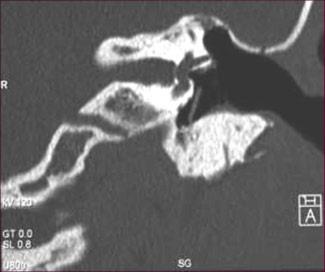
7 External Auditory Canal Atresia More common in males May be bilateral Deformed pinna Bony and/or membranous Isolated or syndromic (e.g. hemifacial microsomia) Look for associated ear anomalies: Middle ear atresia/hypoplasia Ossicular fusion to middle ear wall Oval window atresia Aberrant possition of facial nerve (surgical risk) May later develop cholesteatomas over time


8

9

10

11 Look for the facial nerve


12 EAC Atresia with Dysplastic Ossicles


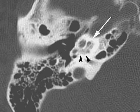
13 Bony Fixation of the Malleus A B


14 Bony Fixation of the Incus A B C


15 Small Dysplastic Ossicles


16 Hemifacial Microsomia?

17 Hemifacial Microsomia

18 Acute Mastoiditis in Children


19 Coalescent Mastoiditis


20 Coalescent Mastoiditis


21 Mastoid Antrum


22

23


24 3 year old s/p mastoidectomy


25


26


27

28


29


30

31


32


33


34


35 Mastoiditis -> Lemierre s syndrome

36 Petrous Apicitis


37


38


39 4 y/o girl transferred with mastoiditis and change in mental status


40


41


42 Typanostomy (myringotomy) tube
43 Chronic Complications of Middle Ear Disease Chronic Otitis Media with Perforation (COM) Chronic Otitis Media with Cholesteatoma (COM)
44
45 Epitympanum EAC TM Mesotympanum
46 Prussak space Scutum Pars flaccida of TM

47

48 Cholesteatoma


49


50 PORP: Partial Ossicular Reconstruction Prosthesis TORP

51 Congenital Middle Ear Cholesteatoma

52
53 Congenital Middle Ear Cholesteatoma Congenitally trapped keratinized squamous epithelium, accumulation of cells and debris Rare (2%-4% of all cholesteatomas) No history or recurrent infections, trauma, or surgery May be found anywhere in middle ear (most commonly in anterior mesotympanum or perieustachian) Slowly grows over time May destroy ossicles and bone if grows


54 5 y/o girl


55 25 y/o External canal cholesteatoma
56 Sensorineural Hearing Loss Congenital and Early SNHL Osseous labyrinth malformations (Michel's aplasia, Mondini malformation,etc.) Membranous labyrinth malformations (complete, partial) Congenital infections (rubella, cytomegalovirus, etc.) Anoxia, birth trauma Ototoxic poisoning (streptomycin, thalidomide, quinine, etc.) Erythroblastosis fetalis (Rh incompatibility) Hyperbilirubnemia Cretinism Prematurity Syndromes (Usher, Pendred, Waardenburg, albinism, onychodystrophy, etc.)
57 Sensorineural Hearing Loss Delayed Sensorineural Hearing Loss Neoplasms (schwannoma, meningioma, lipoma, lymphoma, endolymphatic sac tumor, metastases, etc.) Enlarged endolymphatic sac and duct Infectious meningitis (bacterial, viral) Infectious labyrinthitis (viral, bacterial, spirochetal) Trauma (temporal bone fractures, acoustic trauma) Presbyacusis Endolymphatic hydrops (Meniere's disease) Autoimmune ear disease Ototoxic poisoning (aminoglycosides, diuretics, etc.) Vascular (stroke, vascular loop, AVM) Demyelinating diseases Cochlear otosclerosis/otospongiosis Labyrinthine obliteration (chronic labyrinthitis, Cogan's syndrome) Hemorrhage Perilymphatic fistula Sarcoid Surperficial siderosis Familial progressive SHNL Syndromes (Alport, Hurler, Hunter, Fabry, Klippel-Feil, etc.)
58 Congenital Anomalies Development of Otocyst Aplasia of labyrinth (Michel s aplasia) Division of Otocyst Common cavity malformation or cystic cochleovestibular anomaly Budding of Cochlea Cochlear aplasia or hypolasia Partitioning of Cochlea into 2¾ turns Incomplete partition (Mondini type malformation)

59 Congenital Anomalies Aplasia of Labyrinth (Michel s anomaly) Extremely rare Total absence of osseous and membranous labyrinth


60 Congenital Anomalies Common Cavity Malformation

61 Cystic cochleovestibular anomaly

62 Cochlear hypoplasia (bud)
 IP-II Most")

63 Congenital Anomalies Incomplete partition of cochlea (Mondini-type Malformation) IP-II Most common (55%) of cochlear dysplasias Only 1½ turns in cochlea Middle and apical turns are fused No interscalal septa
64 Mondini Triad Common Error: Mondini Malformation is often applied to anything along spectrum of congenital labyrinthine anomalies Solution: Be descriptive and use hi-res CT and MRI to document all abnormalities


65 Mondini Triad
 Progressive stepwise decline in hearing in childhood and young adulthood Most")

66 Congenital Anomalies Enlarged endolymphatic duct and sac syndrome (Enlarged vestibular aqueduct) Progressive stepwise decline in hearing in childhood and young adulthood Most common radiologically identifiable inner ear anomaly Vestibular aqueduct >1mm in diameter in middle of aqueduct Good rule of thumb: Compare it to the diameter of the posterior semicircular canals

67 Congenital Anomalies Enlarged endolymphatic duct and sac syndrome (Enlarged vestibular aqueduct)

68

69


70 Cochlear Nerve Abnormalities Canal hypoplasia possibly secondary to cochlear nerve hypoplasia or absent cochlear nerve Follow-up with hi-res MR evaluation of cochlear nerve Does not benefit from cochlear implant From: Arastoo Vossough, PhD, MD, Imaging Evaluation of Sensorineural Hearing Loss Applied Radiology, January 2003


71


72 Cochlear Nerve Hypoplasia


73 Narrow IAC


74 Narrow IAC


75 Cochlear Aperture Stenosis or Hypoplasia 9 year old with sensorineural hearing loss


76 46 year old male with hx of SNHLfor follow-up evaluation after cochlear implant. No benefit after implant.


77 Cochlear Aperture Stenosis or Hypoplasia Pre-Implant 46 year old male with Hx of SNHLfor follow-up evaluation after cochlear implant. Post Implant Glastonbury CM, et al, Imaging findings of Cochleaer Nerve Deficiency. AJNR 23: , April 2002
78 Temporal Bone Fractures Longitudinal fractures % - involve tympanic cavity - usually conductive hearing loss from ossicular disruption Transverse Fractures % - involve labyrinth - usually sensorineural hearing loss from inner ear or eight nerve damage


79 Temporal Bone Fractures Otic-capsule Involving
80 Otospongiosis Only affects humans Autosomal dominant inheritance w/variable expressivity Female predominance Peak 2 nd -3 rd decade Bilateral 80% Tinnitus may be presenting symptom Hearing Loss will eventually develop Always conductive component sometimes mixed
81 Otospongiosis Normal persistence of primary endochondral bone in middle layer of otic capsule Otospongiosiotic Phase replacement with spongy vascular new bone CT hypodense to otic capsule Otosclerotic Phase Osteoblastic proliferation Dense sclerotic bone deposition Emedicine Otosclerosis Author: Peter S. Roland, MD
82 Otospongiosis Fenestral (oval window) Cochlear (retrofenestral)
 Curtin and Som, Head & Neck Imaging, 4 th")
83 Otospongiosis Fenestral (oval window) 85% of otospongiosis 85% bilateral Early Decalcification along anterior margins of OW Embryologic fissula ante fenestrum Later: new bone effectively closes OW Complete obliteration in only 2% Fixates stapes footplate CHL Tx: Stapedectomy Cochlear (retrofenestral) Curtin and Som, Head & Neck Imaging, 4 th ed.
84 Otospongiosis Fenestral (oval window) 85% of otospongiosis 85% bilateral Early Decalcification along anterior margins of OW Embryologic fissula ante fenestrum Later: new bone effectively closes OW Compelte obliteration in only 2% Curtin and Som, Head & Neck Imaging, 4 th ed.
 Becomes")

85 Otospongiosis Fenestral (Oval Window) Cochlear (retrofenestral) Conductive +/- SNHL Basilar turn first ( fourth turn ) Becomes sclerotic later than fenestral OS Only relative contraindication to cochlear implant evaluation Curtin and Som, Head & Neck Imaging, 4 th ed.

86 Otospongiosis Fenenstral Otospongiosis limited Ddx Cochlear Otospongiosis Paget Disease Osteogenesis Imperfecta Syphilis OI

87 Postmastoidectomy

88
89 Labyrinthitis Viral, bacterial, spirochetal, autoimmune, traumatic SNHL with vertigo Complication: postinflammatory fibroosseous obliteration: Labyrinthitis ossificans Decreased MRI signal 2 to fibrous change Late: Ossification and obliteration of labyrinth


90 61 year old male with 15 month history of HA s, dizziness, and bilateral hearing losss
91 Labyrinthine Enhancement Schwannoma Autoimmune labyrinthitis Steroid responsive Viral labyrinthitis Perilymphatic fistula Syphylitic labyrinthitis Bacterial Labyrinthitis Middle ear or meningitis spread


92 Postoperative Labyrinthine Enhancement 58 year old woman status-post right acoustic neuroma resection

93 Labyrinthine Enhancement Enhancing inner ear structure: Inflammation or tumor?? High T1 signal on postcontrast images: Enhancement or intrinsic high T1 signal?
94 Labyrinthitis Ossificans

95 Paget s Disease

96 Fibrous Dysplasia
 Acquired Trauma, neoplasm,")
97 Perilymphatic fistula Abnormal communication between middle and inner ear Important cause of SNHL Characteristically fluctuating SNHL (possibly with vertigo/tinnitus) Increased risk for meningitis Congenital A/w congenital deformity of inner ear structures Otitis Media often occurs -> meningitis, labyrinthitis Worsen with trauma (including barotrauma) Acquired Trauma, neoplasm, infection

98 Demyelinating Disease

99 Vestibular Schwannoma

100 Intracochlear Schwannoma


101 Intracochlear Schwannoma

102 Intracochlear Schwannoma Contrast T1-weighted Noncontrast 3D FSE


103 CPA/Intracanalicular/Cochlear Meningioma


104 Endolymphatic sac tumor Papillary adenocarcinomas of the endolymphatic sac Sporadic or associated associated with von Hippel- Lindau syndrome From: Arastoo Vossough, PhD, MD, Imaging Evaluation of Sensorineural Hearing Loss Applied Radiology, January 2003
105 Cochlear Implant Childhood deafness Meningitis Idiopathic and iatrogenic (drug-induced) Congenital deformities Microphone and transducer worn externally Electrodes passed through round window into scala tympani Direct electrical stimulation of residual spiral ganglion cells of cochlear n. Bypassing destroyed hair cells

106 Cochlear Implant
107 Pre-Cochlear Implantation Imaging Evaluation Which type of device to use Which side to implant When surgery should be performed Cochlear patency Round window niche access Degree of mastoid aeration Contraindications
108 Pre-Cochlear Implantation Imaging Evaluation CT and MRI complementary Hypoplasia or acquired atrophy of cochlear nerve High-resolution T2-weighted MRI (FSE or CISS) Normal cochlear nerve = Facial Nerve CT Narrow internal auditory canal (< 2mm) Mastoiditis (Active Infection) Otospongiosis Cochlear Fibrosis/Ossification Choice of cochlear implant model Alter method of insertion Surgical Anatomy Facial N., Carotid A., Jugular V.


109 Cochlear Implantation
The Temporal Bone Anatomy & Pathology
 Department of Radiology University of California San Diego The Temporal Bone Anatomy & Pathology John R. Hesselink, M.D. Temporal Bone Axial View Temporal Bone Coronal View Longitudinal Fracture The Temporal
Pediatric Temporal Bone
 Pediatric Temporal Bone Suresh K. Mukherji, MD, FACR Professor and Chief of Neuroradiology Professor of Radiology, Otolaryngology Head Neck Surgery, Radiation Oncology and Periodontics & Oral Medicine
COCHLEAR IMPLANTS Aetiology of Deafness. Bruce Black MD
 COCHLEAR IMPLANTS Aetiology of Deafness Heterochromia iridis. Cases may be healthy or associated with a variety of conditions, e.g. Waardenburg syndrome. Waardenburg syndrome. Note the snowy lock of hair
Rory Attwood MBChB,FRCS
 Hearing loss Overview Rory Attwood MBChB,FRCS Division of Otorhinolaryngology Faculty of Health Sciences Tygerberg Campus, University of Stellenbosch Not deafness Deaf is a total lack of hearing Deafness
Chronic Ear Disease. Daekeun Joo Resident Lecture Series 11/18/09
 Chronic Ear Disease Daekeun Joo Resident Lecture Series 11/18/09 ETD URIs Viral-induced damage to ET lining resulting in decreased mucociliary clearance Viral invasion of ME mucosa results in inflamm Reflux
Temporal bone anatomy and imaging features of common conditions causing hearing loss: A pictorial review
 Temporal bone anatomy and imaging features of common conditions causing hearing loss: A pictorial review Poster No.: C-1892 Congress: ECR 2012 Type: Educational Exhibit Authors: A. Masukawa, H. Takeuchi,
Cholesteatoma-Pathogenesis and Surgical Management. Grand Rounds Presentation February 24, 1999 Kyle Kennedy, M.D. Jeffrey Vrabec,, M.D.
 Cholesteatoma-Pathogenesis and Surgical Management Grand Rounds Presentation February 24, 1999 Kyle Kennedy, M.D. Jeffrey Vrabec,, M.D. Introduction Cholesteatoma (keratoma)-essentially an accumulation
Middle ear CT imaging: Review of anatomy and common pathology
 Middle ear CT imaging: Review of anatomy and common pathology Poster No.: C-0665 Congress: ECR 2017 Type: Educational Exhibit Authors: M. R. Campos Arenas, M. C. Sánchez-Porro, J. Garrido Rull ; 1 1 2
Cholesteatoma and Non-cholesteatomatous Inflammatory Disease. Cholesteatoma. Disclosures. Overview EAC. Cholesteatoma. None
 Disclosures Cholesteatoma and Non-cholesteatomatous Inflammatory Disease None Amy F Juliano, MD Staff Radiologist, Massachusetts Eye and Ear Infirmary Assistant Professor of Radiology, Harvard Medical
Imaging of Hearing Loss
 Contemporary Imaging of Sensorineural Hearing Loss Imaging of Hearing Loss Discussion Outline (SNHL) Imaging Approaches Anatomic Relationships Lesions: SNHL KL Salzman, MD University of Utah School of
Anatomy of the ear: Lymphatics
 Anatomy of the ear: 1. External ear which consist of auricle and external auditory canal. The auricle has a framework of cartilage except the lobule, the skin is closely adherent to perichonderium at the
Pediatric Ear Diseases
 Pediatric Ear Diseases Yasushi Naito Pediatric Ear Diseases Diagnostic Imaging Atlas and Case Reports 242 figures, 7 in color and 5 tables, 2013 Basel Freiburg Paris London New York New Delhi Bangkok
Petrous Bone Normal anatomy
 Petrous Bone Normal anatomy By Mamdouh Mahfouz MD Prof. of Radiology Cairo University ssregypt.com Axial Coronal Petrous bone External ear Middle ear Inner ear External ear Cartilaginous part Bony part
Acquired Deafness Loss of hearing that occurs or develops sometime in the course of a lifetime, but is not present at birth.
 Page 1 of 5 URMC» Audiology Glossary of Terms A Acoustic Neuroma A tumor, usually benign, which develops on the hearing and balance nerves and can cause gradual hearing loss, tinnitus, and dizziness. Acquired
Chapter 143: Otosclerosis (OS) Sameer Ahmed 2/23/2011
 Sameer Ahmed 2/23/2011") Chapter 143: Otosclerosis (OS) Sameer Ahmed 2/23/2011 Intro Disorder of fibrous osteodystrophy of the human otic capsule Abnormal resorption and deposition of bone Primarily causes CHL SNHL and MHL are
Chapter 143: Otosclerosis (OS) Sameer Ahmed 2/23/2011 Intro Disorder of fibrous osteodystrophy of the human otic capsule Abnormal resorption and deposition of bone Primarily causes CHL SNHL and MHL are
AUDITORY APPARATUS. Mr. P Mazengenya. Tel 72204
 AUDITORY APPARATUS Mr. P Mazengenya Tel 72204 Describe the anatomical features of the external ear Describe the tympanic membrane (ear drum) Describe the walls of the middle ear Outline the structures
The ear: some applied basic science
 Chapter 1 The ear: some applied basic science The pinna The external ear or pinna is composed of cartilage with closely adherent perichondrium and skin. It is developed from six tubercles of the first
Modern Imaging & Current Controversies
 Temporal Bone: Modern Imaging & Current Controversies Suresh K. Mukherji, MD, FACR Professor and Chief of Neuroradiology Professor of Radiology, Otolaryngology Head Neck Surgery, Radiation i Oncology,
Educational Exhibit Authors: P. Digge, R. K. N. Solanki, S. M. Paruthikunnan, D. S. Shah ; 1
 High-field MRI versus high-resolution CT of temporal bone in inner ear pathologies of children with bilateral profound sensorineural hearing loss: A pictorial essay. Poster No.: C-0867 Congress: ECR 2015
RADIOLOGY TEACHING CONFERENCE
 RADIOLOGY TEACHING CONFERENCE John Athas, MD Monica Tadros, MD Columbia University, College of Physicians & Surgeons Department of Otolaryngology- Head & Neck Surgery September 27, 2007 CT SCAN IMAGING
Anatomy of the Ear Region. External ear Middle ear Internal ear
 Ear Lecture Objectives Make a list of structures making the external, middle, and internal ear. Discuss the features of the external auditory meatus and tympanic membrane. Describe the shape, position,
ICD10 CODES CODE DESCRIPTION R Abnormal auditory function study H Abnormal auditory perception, bilateral H Abnormal auditory
 ICD10 CODES CODE DESCRIPTION R94.120 Abnormal auditory function study H93.293 Abnormal auditory perception, bilateral H93.292 Abnormal auditory perception, left ear H93.291 Abnormal auditory perception,
Anatomy and Physiology of Hearing
 Anatomy and Physiology of Hearing The Human Ear Temporal Bone Found on each side of the skull and contains the organs for hearing and balance Divided into four major portions: - squamous - mastoid - tympanic
Section 1 EAR. Section Outlines
 Section 1 EAR Section Outlines Development of the Ear Anatomy of the Ear Physiology of the Ear History Taking with Symptomatology of Ear Diseases Examination of the Ear Congenital Diseases of the External
Assisting in Otolaryngology
 Assisting in Otolaryngology Learning Objectives Identify the structures and explain the functions of the external, middle, and internal ear. Describe the conditions that can lead to hearing loss, including
1. Axial view, left temporal bone. Plane through the upper antrum (A), superior semicircular canal (SSC) and IAC.
, superior semicircular canal (SSC) and IAC.") PA IAC SSC A 1. Axial view, left temporal bone. Plane through the upper antrum (A), superior semicircular canal (SSC) and IAC. IAC VII M I LSC Plane through the IAC, malleus head and incus and the lateral
PA IAC SSC A 1. Axial view, left temporal bone. Plane through the upper antrum (A), superior semicircular canal (SSC) and IAC. IAC VII M I LSC Plane through the IAC, malleus head and incus and the lateral
CE Directed Reading. Temporal Bone CT: Anatomy, Technique, and Associated Pathophysiology
 Temporal Bone CT: Anatomy, Technique, and Associated Pathophysiology Chris Young, MS, R.R.A., R.T.(R) Computed tomography (CT) of the temporal bone is performed to evaluate trauma, tumors, sinuses, the
Congenital Absence of the Oval Window: Radiologic Diagnosis and Associated Anomalies
 AJNR Am J Neuroradiol 21:322 327, February 2000 Congenital Absence of the Oval Window: Radiologic Diagnosis and Associated Anomalies Barbara Zeifer, Paul Sabini, and Jonathan Sonne BACKGROUND AND PURPOSE:
Scrub In. What is the function of cerumen? Which part of the ear collects sound waves and directs them into the auditory canal?
 Scrub In What is the function of cerumen? a. Keeps the ear canal from collapsing b. Helps transmit sound waves c. Protection d. Lubrication Which part of the ear collects sound waves and directs them into
DIAGNOSIS Causes/Etiology of Hearing Loss
 DIAGNOSIS Causes/Etiology of Hearing Loss DIAGNOSIS Causes/Etiology of Hearing Loss VI. How Do We Hear? Sound waves enter our ears and are amplified by the ear drum and middle ear bones (ossicles), allowing
Eye and Ocular Adnexa, Auditory Systems
 Eye and Ocular Adnexa, Auditory Systems CPT copyright 2011 American Medical Association. All rights reserved. Fee schedules, relative value units, conversion factors and/or related components are not assigned
Radiologic Evaluation of Petrous Apex Masses. Pavan Kavali, MS-IV Morehouse School of Medicine November 16, 2009
 Radiologic Evaluation of Petrous Apex Masses Pavan Kavali, MS-IV Morehouse School of Medicine November 16, 2009 Roadmap Petrous Apex Anatomy Patient D.S.: Clinical Presentation Differential diagnosis of
Correlation of HRCT mastoid with clinical presentation and operative findings in ear diseases
 International Journal of Otorhinolaryngology and Head and Neck Surgery Chintale SG et al. Int J Otorhinolaryngol Head Neck Surg. 2017 Jul;3(3):656-660 http://www.ijorl.com pissn 2454-5929 eissn 2454-5937
CT and MR Imaging In Patients Undergoing Evaluation for Cochlear Implantation
 CT and MR Imaging In Patients Undergoing Evaluation for Cochlear Implantation Poster No.: C-1219 Congress: ECR 2015 Type: Educational Exhibit Authors: S. S. Ghuman, T. Buxi, S. Sud, A. Sud, A. Yadav, K.
ENT 318 Artificial Organs Physiology of Ear
 ENT 318 Artificial Organs Physiology of Ear Lecturer: Ahmad Nasrul Norali The Ear The Ear Components of hearing mechanism - Outer Ear - Middle Ear - Inner Ear - Central Auditory Nervous System Major Divisions
Kingdom of Bahrain Arabian Gulf University College of Medicine and Medical Sciences Year 6 ENT SMC Otitis Media (Dr.
 Kingdom of Bahrain Arabian Gulf University College of Medicine and Medical Sciences Year 6 ENT SMC Otitis Media (Dr. Jalal Almarzooq) - Anatomy of the ear: The ear is divided into 3 parts: External ear.
The Ear The ear consists of : 1-THE EXTERNAL EAR 2-THE MIDDLE EAR, OR TYMPANIC CAVITY 3-THE INTERNAL EAR, OR LABYRINTH 1-THE EXTERNAL EAR.
 The Ear The ear consists of : 1-THE EXTERNAL EAR 2-THE MIDDLE EAR, OR TYMPANIC CAVITY 3-THE INTERNAL EAR, OR LABYRINTH 1-THE EXTERNAL EAR Made of A-AURICLE B-EXTERNAL AUDITORY MEATUS A-AURICLE It consists
J.P.S. Bakshi Manual of Ear, Nose and Throat
 J.P.S. Bakshi Manual of Ear, Nose and Throat Reading excerpt Manual of Ear, Nose and Throat of J.P.S. Bakshi Publisher: B. Jain http://www.narayana-publishers.com/b5603 Copying excerpts is not permitted.
Temporal bone imaging in osteogenesis imperfecta patients with hearing loss
 Temporal bone imaging in osteogenesis imperfecta patients with hearing loss F. Swinnen 1, J. Casselman 2, P. Coucke 3, C. Cremers 4, E. De Leenheer 1, I. Dhooge 1 (1) Departement of Otorhinolaryngology,
Structure, Energy Transmission and Function. Gross Anatomy. Structure, Function & Process. External Auditory Meatus or Canal (EAM, EAC) Outer Ear
 Outer Ear") Gross Anatomy Structure, Energy Transmission and Function IE N O ME 1 Structure, Function & Process 4 External Auditory Meatus or Canal (EAM, EAC) Outer third is cartilaginous Inner 2/3 is osseous Junction
Gross Anatomy Structure, Energy Transmission and Function IE N O ME 1 Structure, Function & Process 4 External Auditory Meatus or Canal (EAM, EAC) Outer third is cartilaginous Inner 2/3 is osseous Junction
The Ear. Dr. Heba Kalbouneh Assistant Professor of Anatomy and Histology
 The Ear Dr. Heba Kalbouneh Assistant Professor of Anatomy and Histology The Ear The ear consists of the external ear; the middle ear (tympanic cavity); and the internal ear (labyrinth), which contains
OTOLOGY. 1. BRIEF DESCRIPTION OF OTOLOGIC TRAINING Rotations that include otologic training are a component of each of the four years of training.
 OTOLOGY 1. BRIEF DESCRIPTION OF OTOLOGIC TRAINING Rotations that include otologic training are a component of each of the four years of training. Longwood Rotation PGY-2 through PGY-5 years o Clinic experience
CT of Postmeningitic Deafness: Observations and Predictive Value for Cochlear Implants in Children
 CT of Postmeningitic Deafness: Observations and Predictive Value for Cochlear Implants in Children Michele H. Johnson, M. Suzanne Hasenstab, Michael A. Seicshnaydre, and George H. Williams PURPOSE: To
Congenital Aural Atresia. Miranda S. Dennis, M.D. April 6, 2011
 Congenital Aural Atresia Miranda S. Dennis, M.D. April 6, 2011 Embryology External Auditory Canal first branchial groove starts as a solid core of epithelial cells, which undergoes absorption in a medial
Ear. Utricle & saccule in the vestibule Connected to each other and to the endolymphatic sac by a utriculosaccular duct
 Rahaf Jreisat *You don t have to go back to the slides. Ear Inner Ear Membranous Labyrinth It is a reflection of bony labyrinth but inside. Membranous labyrinth = set of membranous tubes containing sensory
Surgical and Non-Surgical Causes of Progressive Hearing Loss in Children: What can be done about it?
 Surgical and Non-Surgical Causes of Progressive Hearing Loss in Children: What can be done about it? Daniela Carvalho, MD, MMM, FAAP Professor, Surgery Department UCSD Pediatric Otolaryngology Rady Children
Conductive Hearing Loss in Young Children: Options and Opportunities
 Conductive Hearing Loss in Young Children: Options and Opportunities Donna L. Sorkin, M.A., Vice President, Consumer Affairs Jennifer Lake, Clinical Applications Specialist Cochlear Americas Agenda 1.
Almost 9 million people in the UK, 1 in 7 of the population, suffer from deafness or experience significant hearing difficulty i
 Deafness the facts HOW MANY PEOPLE ARE AFFECTED? Almost 9 million people in the UK, 1 in 7 of the population, suffer from deafness or experience significant hearing difficulty i CHILDHOOD DEAFNESS It is
Imaging of the Temporal Bone: A Symptom-Based Approach
 Imaging of the Temporal Bone: A Symptom-Based Approach Tadesse Eshetu, MD, and Nafi Aygun, MD Some of the symptoms associated with the temporal bone diseases are nonspecific, whereas others overlap with
Complications of otitis media
 Chronic otitis media Definition:- Complications of otitis media Otitis media (OM) is broadly defined as inflammation from any cause of the middle ear.this may involve any of the contiguous pneumatized
Cochlear Implant Failure: Imaging Evaluation of the Electrode Course
 Clinical Radiology (2003) 58: 288 293 doi:10.1016/s0009-9260(02)00523-8, available online at www.sciencedirect.com Pictorial Review Cochlear Implant Failure: Imaging Evaluation of the Electrode Course
Assessing the Deaf & the Dizzy. Phil Bird Senior Lecturer University of Otago, Christchurch Consultant Otolaryngologist CPH & Private
 Assessing the Deaf & the Dizzy Phil Bird Senior Lecturer University of Otago, Christchurch Consultant Otolaryngologist CPH & Private Overview Severe & profoundly deaf children & adults Neonatal screening
Case Studies in CPA/IAC
 Outline Case Studies in CPA/IAC Atul K Mallik MD PhD Department of Radiology and Imaging Sciences University of Utah Health Sciences Center Salt Lake City, Utah, USA Case based review of cerebellopontine
Osseous structures in the middle ear cavity(mec): Are they too many or are they too few?
: Are they too many or are they too few?") Osseous structures in the middle ear cavity(mec): Are they too many or are they too few? Poster No.: C-2286 Congress: ECR 2013 Type: Educational Exhibit Authors: P. Mundada, B. S. Purohit, T. Tiong Yong;
Osseous structures in the middle ear cavity(mec): Are they too many or are they too few? Poster No.: C-2286 Congress: ECR 2013 Type: Educational Exhibit Authors: P. Mundada, B. S. Purohit, T. Tiong Yong;
Ear, Nose, and Throat Disorders
 Health Reference Series Second Edition Basic Consumer Health Information about Disorders of the Ears, Hearing Loss, Vestibular Disorders, Nasal and Sinus Problems, Throat and Vocal Cord Disorders, and
Modifying radiology protocols for cochlear implant surgery in a government sponsored scheme: Need of the hour
 Available online at www.ijmrhs.com ISSN No: 2319-5886 International Journal of Medical Research & Health Sciences, 2016, 5, 6:151-157 Modifying radiology protocols for cochlear implant surgery in a government
HEARING IMPAIRMENT LEARNING OBJECTIVES: Divisions of the Ear. Inner Ear. The inner ear consists of: Cochlea Vestibular
 HEARING IMPAIRMENT LEARNING OBJECTIVES: STUDENTS SHOULD BE ABLE TO: Recognize the clinical manifestation and to be able to request appropriate investigations Interpret lab investigations for basic management.
Imaging in patients undergoing cochlear implant: CT and MR technique
 Imaging in patients undergoing cochlear implant: CT and MR technique Poster No.: C-0102 Congress: ECR 2012 Type: Educational Exhibit Authors: G. Posillico Keywords: Ear / Nose / Throat, CT, MR, Comparative
Exposure of facial nerve and endolymphatic sac
 Exposure of facial nerve and endolymphatic sac 1 7 4 2 3 5 6 8 1 Vertical part of the facial nerve exposed 1 Second genu of facial nerve. 2 Vertical part of facial nerve. 3 Horizontal part of facial nerve.
Drug delivery to the inner ear
 Intratympanic Drug Delivery Society of Otorhinolaryngology and Head-Neck Nurses Advantages of intratympanic delivery Higher concentration of drug at site of action Avoid systemic effects May be able to
MICROTIA. The condition is a complex mix of cosmetic, functional, and often psychological difficulties. Microtia: Not only the ear.
 MICROTIA Underdevelopment /deformity of the auricle (pinna) varies from subtle deformities and small pre-auricular rudiments to gross developmental failure, distortion or malpositioned remnants. The external
Implantable Treatments for Different Types of Hearing Loss. Margaret Dillon, AuD Marcia Adunka, AuD
 Implantable Treatments for Different Types of Hearing Loss Margaret Dillon, AuD Marcia Adunka, AuD Implantable Technologies Types of hearing loss Bone-anchored devices Middle ear implantation Cochlear
au/images/conductive-loss-new.jpg
 Biology of the ear http://www.nal.gov. au/images/conductive-loss-new.jpg Agenda Pre-test Lecture Group Gesture Types of hearing losses Audiograms Views Post-test Pretest!! See how much you know Answer
Gross Anatomy of the. TEMPORAL BONE, EXTERNAL EAR, and MIDDLE EAR
 Gross Anatomy of the TEMPORAL BONE, EXTERNAL EAR, and MIDDLE EAR M1 Gross and Developmental Anatomy 9:00 AM, December 11, 2008 Dr. Milton M. Sholley Professor of Anatomy and Neurobiology Assignment: Head
Bruce Black MD EAC TRAUMA
 EAC TRAUMA Bruising in the deep canal due to cotton bud/q-tip selfcleaning attempts. No action required. A granuloma of the deep Lt. EAC. Superficial trauma has become secondarily infected. Clean thoroughly,
ﺎﻨﺘﻤﻠﻋ ﺎﻣ ﻻا ﺎﻨﻟ ﻢﻠﻋ ﻻ ﻚﻧﺎﺤﺒﺳ اﻮﻟﺎﻗ ﻢﻴﻜﺤﻟا ﻢﻴﻠﻌﻟا ﺖﻧأ ﻚﻧا ﻢﻴﻈﻌﻟا ﷲا قﺪﺻ HEARING LOSS
 قالوا سبحانك لا علم لنا الا ما علمتنا انك أنت العليم الحكيم صدق االله العظيم HEARING LOSS 1 Hearing loss: Deviation from normal hearing in one or both ears. Hearing handicap: This term refers to total
Indications and contra-indications of auditory brainstem implants. Systematic review and illustrative cases
 Manuscript: Authors: Journal: Indications and contra-indications of auditory brainstem implants. Systematic review and illustrative cases Merkus P (p.merkus@vumc.nl), Di Lella F, Di Trapani G, Pasanisi
Dr. Vitthalrao Vikhe Patil Foundation s Medical College & Hospital, Ahmednagar, Maharashtra, India
 DOI: 10.21276/sjams.2017.5.3.17 Scholars Journal of Applied Medical Sciences (SJAMS) Sch. J. App. Med. Sci., 2017; 5(3B):770-779 Scholars Academic and Scientific Publisher (An International Publisher for
Narrowest segment of the ear canal. Limited microscopic. Wide endoscopic. field of view. field of view
 Endoscopic Transcanal Management of Cholesteatoma M. Tarabichi American Hospital-Dubai The Endoscope in Otology Mostly for documentation. Mostly diagnostic. Exploration of old mastoid cavities Endoscopic
Middle and Inner Ear: Improved Depiction with Multiplanar Reconstruction of Volumetric CT Data 1
 Note: This copy is for your personal non-commercial use only. To order presentation-ready copies for distribution to your colleagues or clients, contact us at www.rsna.org/rsnarights. EDUCATION EXHIBIT
Dr. Sami Zaqout Faculty of Medicine IUG
 Auricle External Ear External auditory meatus The Ear Middle Ear (Tympanic Cavity) Auditory ossicles Internal Ear (Labyrinth) Bony labyrinth Membranous labyrinth External Ear Auricle External auditory
Hearing. By Jack & Tori
 Hearing By Jack & Tori 3 Main Components of the Human Ear. Outer Ear. Middle Ear. Inner Ear Outer Ear Pinna: >Visible part of ear and ear canal -Acts as a funnel to direct sound Eardrum: >Airtight membrane
Anomalous Facial Nerve Canal with Cochlear Malformations
 AJNR Am J Neuroradiol 22:838 844, May 2001 Anomalous Facial Nerve Canal with Cochlear Malformations Laura Vitale Romo and Hugh D. Curtin BACKGROUND AND PURPOSE: Anteromedial migration of the first segment
Surgery for Conductive Hearing Loss
 THE NEW YORK OTOLARYNGOLOGY GROUP, P.C. The Ear, Nose and Throat Specialists Neil M. Sperling, M.D. Otology/Neuro-Otology Diseases of the Ear Facial Nerve Balance Disorders Surgery for Conductive Hearing
Chapter 1: Applied Anatomy, Physiology and Embryology of the Ear. Anatomy and Physiology. The Outer Ear. The Pinna. The External Ear Canal
 Chapter 1: Applied Anatomy, Physiology and Embryology of the Ear The ear contains two specialized sensory organs, the cochlea and the vestibular apparatus, enclosed in the extremely hard protective casing
Intratympanic therapy of inner ear disease
 Intratympanic therapy of inner ear disease Jack J. Wazen, M.D.FACS. VP & Director of Research Silverstein Institute Ear Research Foundation Sudden Sensorineural Definition Hearing Loss Sudden SNHL is defined
MRI AND HIGH RESOLUTION CT IN CONGENITAL HEARING LOSS
 MRI AND HIGH RESOLUTION CT IN CONGENITAL HEARING LOSS Ahmed A. M. Abd Alla, Ibrahim A. Lebda, Sonia Y. Mohamed, Ezzat A. Merwad Department of Radiology, Faculty of medicine, Zagazig University. Department
New EAONO Cholesteatoma Classification with imaging illustration. Milan Profant, Katarina Sláviková
 New EAONO Cholesteatoma Classification with imaging illustration Milan Profant, Katarina Sláviková EAONO/JOS Joint Consensus Statements on the Definitions, Classification and Staging of Middle Ear Cholesteatoma
Cholesteatoma in children
 Cholesteatoma in children British Association of Paediatricians in Audiology London Conference, Jan.2012 Matthew Clark FRCS (ORL-HNS) Consultant Otologist Gloucestershire Royal Hospital Overview: Cholesteatoma
Gross Anatomy of the. TEMPORAL BONE, EXTERNAL EAR, and MIDDLE EAR. Assignment: Head to Toe Temporomandibular Joint (TMJ)
") Gross Anatomy the TEMPORAL BONE, EXTERNAL EAR, and MIDDLE EAR M1 Gross and Developmental Anatomy 9:00 AM, December 11, 2008 Dr. Milton M. Sholley Pressor Anatomy and Neurobiology Assignment: Head to Toe
Gross Anatomy the TEMPORAL BONE, EXTERNAL EAR, and MIDDLE EAR M1 Gross and Developmental Anatomy 9:00 AM, December 11, 2008 Dr. Milton M. Sholley Pressor Anatomy and Neurobiology Assignment: Head to Toe
Unit VIII Problem 9 Anatomy of The Ear
 Unit VIII Problem 9 Anatomy of The Ear - The ear is an organ with 2 functions: Hearing. Maintenance of equilibrium/balance. - The ear is divided into 3 parts: External ear. Middle ear (which is also known
CITY & HACKNEY PATHFINDER CLINICAL COMMISSIONING GROUP. Vertigo. (1) Vertigo. (4) Provisional Diagnosis. (5) Investigations. lasting days or weeks
 Vertigo. (4) Provisional Diagnosis. (5) Investigations. lasting days or weeks") Authors: Dr Lucy O'Rouke and Mr N Eynon-Lewis Review date: January 2017 Vertigo (1) Vertigo (2) History (3) Examination (4) Provisional Diagnosis (5) Investigations (6) Medical Cause (7) Psychiatric Cause
Authors: Dr Lucy O'Rouke and Mr N Eynon-Lewis Review date: January 2017 Vertigo (1) Vertigo (2) History (3) Examination (4) Provisional Diagnosis (5) Investigations (6) Medical Cause (7) Psychiatric Cause
Abnormal direction of internal auditory canal and vestibulocochlear nerve
 Medicine Otorhinolaryngology fields Okayama University Year 2004 Abnormal direction of internal auditory canal and vestibulocochlear nerve Shin Kariya kazunori Nishizaki Hirofumi Akagi Michael M. Paparella
CONFLICTS OF INTEREST
 COCHLEAR IMPLANTATION: A SURGEON S PERSPECTIVE Ravi N. Samy, M.D., F.A.C.S. Ravi.Samy@UC.edu Director, Adult Cochlear Implant Program Program Director, Neurotology Fellowship CONFLICTS OF INTEREST RESEARCH
Chapter 17, Part 2! The Special Senses! Hearing and Equilibrium!
 Chapter 17, Part 2! The Special Senses! Hearing and Equilibrium! SECTION 17-5! Equilibrium sensations originate within the inner ear, while hearing involves the detection and interpretation of sound waves!
Chapter 17, Part 2! Chapter 17 Part 2 Special Senses! The Special Senses! Hearing and Equilibrium!
 Chapter 17, Part 2! The Special Senses! Hearing and Equilibrium! SECTION 17-5! Equilibrium sensations originate within the inner ear, while hearing involves the detection and interpretation of sound waves!
An optical coherence tomography study for imaging the round window niche and the promontorium tympani
 An optical coherence tomography study for imaging the round window niche and the promontorium tympani T. Just *a, E. Lankenau b, G. Hüttmann b, H.W. Pau a a Department of Otorhinolaryngology, University
Sasan Dabiri, MD, Assistant Professor
 Sasan Dabiri, MD, Assistant Professor Department of Otorhinolaryngology Head & Neck Surgery Amir A lam hospital Tehran University of Medical Sciences October 2015 Outlines Anatomy of Vestibular System
High-resolution Computed Tomography Study of Temporal Bone Pathologies
 Original Article Print ISSN: 2321-6379 Online ISSN: 2321-595X DOI: 10.17354/ijss/2016/375 High-resolution Computed Tomography Study of Temporal Bone Pathologies Manjit Bagul Senior Resident, Department
Management of Ear, Hearing and Balance Disorders: Fact, Fiction, and Future
 Management of Ear, Hearing and Balance Disorders: Fact, Fiction, and Future George W. Hicks, M,D. 7440 N. Shadeland Avenue, Suite 150 Indianapolis, IN 46250 904 N. Samuel Moore Parkway Mooresville, IN
Congenital Neck Masses C. Stefan Kénel-Pierre, MD
 Congenital Neck Masses C. Stefan Kénel-Pierre, MD SUNY-LICH Medical Center Department of Surgery Case Presentation xx year old male presents with sudden onset left lower neck swelling x 1 week Denies pain,
The close anatomic relationship between the cochlea and
 ORIGINAL RESEARCH R.J. Young D.R. Shatzkes J.S. Babb A.K. Lalwani The Cochlear-Carotid Interval: Anatomic Variation and Potential Clinical Implications BACKGROUND AND PURPOSE: A temporal bone CT study
Anatomy and Pathophysiology for ICD-10 Module 11. Ear and Mastoid
 Anatomy and Pathophysiology for ICD-10 Module 11 Ear and Mastoid Ear Anatomy Outer Ear Ear Flap (Pinna) Ear Canal (Meatus) Middle Ear Tympanic Membrane (Eardrum) Hammer (Malleus) Anatomy and Physiology
8 External Ear Canal Surgery
 30 Chapter 8 8 External Ear Canal Surgery Henning Hildmann, Holger Sudhoff Surgery in the external auditory canal without surgery in the middle ear may be necessary: 1. After surgery 2. After trauma 3.
UC SF. Safe Surgery Rule #1. Cholesteatoma. It s hard to have a surgical complication when you are not operating
 UC SF Cholesteatoma Chronic Ear Surgery: Staying Out of Trouble! Lawrence R. Lustig, MD Department of Oto-HNS University of California San Francisco Ligaments and folds Spaces NU Epitympanic Cholesteatoma
CLASSIFICATION OF CONGENITAL MIDDLE AND EXTERNAL EAR MALFORMATIONS: CT STUDY
 VolumeS Medicalloumal of the Islamic Republic of Iran Number 3,4 Payiz & Zemestan 1370 Fall & Winter 1991 CLASSIFICATION OF CONGENITAL MIDDLE AND EXTERNAL EAR MALFORMATIONS: CT STUDY D. SAYIC, MD, DMS:
High resolution computed tomography of temporal bone in the evaluation of otologic diseases
 International Journal of Otorhinolaryngology and Head and Neck Surgery Handi PS et al. Int J Otorhinolaryngol Head Neck Surg. 2018 Jan;4(1):87-92 http://www.ijorl.com pissn 2454-5929 eissn 2454-5937 Original
HEARING AND COCHLEAR IMPLANTS
 HEARING AND COCHLEAR IMPLANTS FRANCIS CREIGHTON, MD NEUROTOLOGY & SKULL BASE SURGERY FELLOW JOHNS HOPKINS SCHOOL OF MEDICINE NOV 9 TH, 2017 THANKS TO: CHARLIE DELLA SANTINA, HEIDI NAKAJIMA AND DOUG MATTOX