Cholesteatoma and Non-cholesteatomatous Inflammatory Disease. Cholesteatoma. Disclosures. Overview EAC. Cholesteatoma. None
|
|
|
- Gordon Hamilton
- 5 years ago
- Views:
Transcription

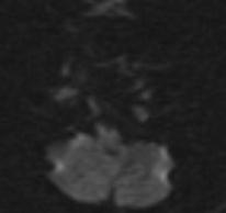
 surrounded by two layers Inner layer (matrix): keratinizing squamous epithelium")
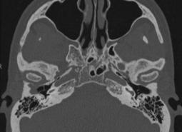
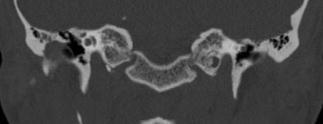
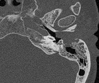
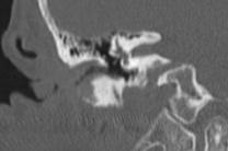
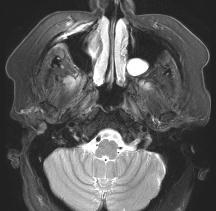
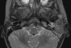
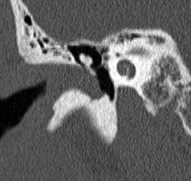
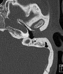
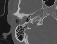
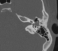
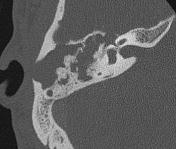
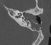
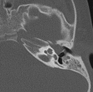
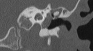
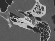
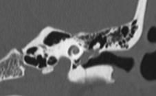
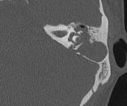
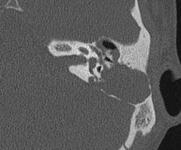
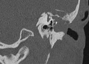
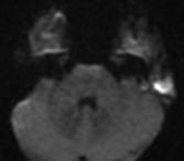
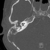
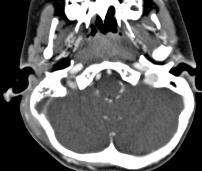
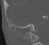
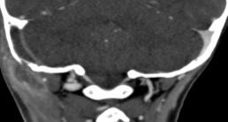
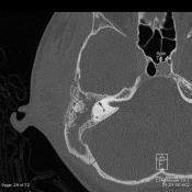
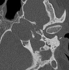
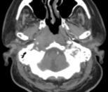
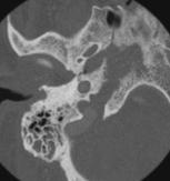
1 Disclosures Cholesteatoma and Non-cholesteatomatous Inflammatory Disease None Amy F Juliano, MD Staff Radiologist, Massachusetts Eye and Ear Infirmary Assistant Professor of Radiology, Harvard Medical School Overview Cholesteatoma What is it? By location: cholesteatoma and ddx EAC Middle ear Mastoid Petrous Apex Non-cholesteatomatous inflammatory processes Necrotizing external otitis Facial nerve Inner ear Ossicular complications Cholesteatoma Accumulation of desquamated Perimatrix keratin epithelium (connective Acellular keratin debris tissue) surrounded by two layers Inner layer (matrix): keratinizing squamous epithelium produces keratin Outer layer (perimatrix): subepithelial connective tissue produces proteolytic enzymes that can resorb bone Middle ear > other pneumatized areas (e.g. EAC, mastoid, petrous air cells) Matrix (epithelium) Cholesteatoma EAC CT: expansile opacified air cell T1: hypointense T2: hyperintense DWI: reduced diffusivity Need to use coronal, nonecho planar (non EPI) DWI EAC cholesteatoma Mimickers Malignancies Granulomatous diseases Keratosis obturans Osteoradionecrosis
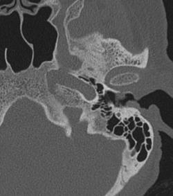
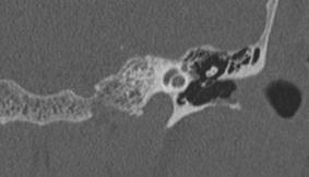
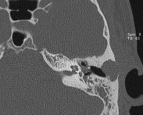
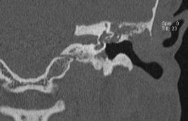
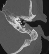
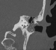
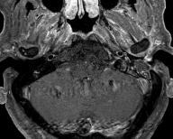
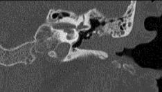
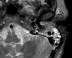
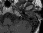

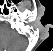
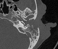
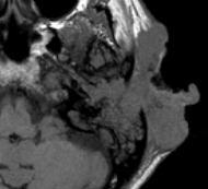
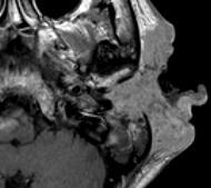
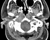
2 EAC cholesteatoma EAC cholesteatoma EAC cholesteatoma EAC scc, looks like a cholesteatoma EAC scc EAC basal cell carcinoma Much less frequent than SCC Uniformly associated with actinic damage to epidermis Almost always seen in men Rarely fatal

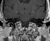
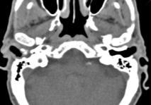
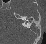
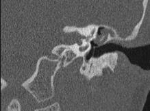
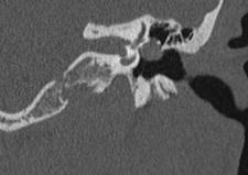
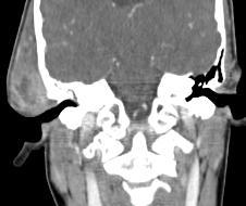
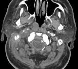
3 EAC sarcoid Langerhans cell histiocytosis EAC KO EAC ORN Middle ear Middle ear cholesteatoma Middle ear cholesteatoma Mimickers AOM with effusion COM and its sequelae: effusion, granulation tissue, cholesterol granuloma ETD with effusion


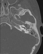
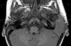
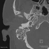
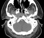
4 Middle ear cholesteatoma Middle ear congenital cholesteatoma Middle ear carcinoid read out as cholesteatoma Mastoid Mastoid cholesteatoma Mimickers Malignancies Coalescent mastoiditis Mastoid cholesteatoma R coalescent mastoiditis, Bezold, sigmoid sinus thrombosis (vs compressed sinus vs epidural abscess)
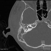
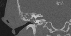
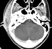


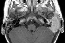
5 Coalescent mastoiditis from actinomycosis, Bezold Petrous Apex Petrous apex cholesteatoma Mimickers Mucocele Cholesterol granuloma Meningocele Not really: effusion, asymmetric pneumatization Petrous apicitis Malignancies: metastasis, chondrosarcoma, etc Cholesteatoma Petrous apex mucocele Cholesterol granuloma

 Petrous apex air cell (with obstructed drainage) Mastoid,")


, Dorello s canal (VI) Otomastoiditis Deep")


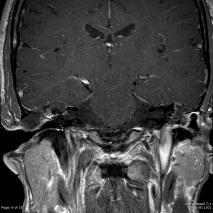
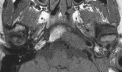
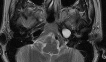
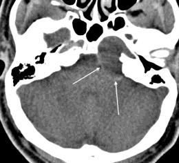
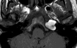
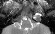
6 Cholesterol granuloma An air space with negative pressure rupture of blood vessels Breakdown of RBCs Release of cholesterol Foreign body giant cell reaction Granuloma is formed, with cholesterol elements and blood products Locations Middle ear (with Eustachian tube dysfunction) Petrous apex air cell (with obstructed drainage) Mastoid, mastoid bowl Ddx: expansile opacified PA air cell Mucocele Cholesterol granuloma Cholesteatoma Meningocele, arachnoid cyst Petrous apicitis Infection in the mastoid spreading medially to the petrous apex Osteitis, disruption of cortex/septations, meningitis Occurs in the setting of pneumatized petrous apex Gradenigo s syndrome triad of symptoms, bacterial otitis media leading to petrous apicitis and spread of infection to meninges, Gasserian ganglion (V), Dorello s canal (VI) Otomastoiditis Deep retroorbital pain in distribution of V Diplopia due to VI palsy CT: fluid in air cells, bone erosion MR: abnormal enhancement of adjacent meninges Look for complications: arterial complications, cavernous sinus/sigmoid/ij thrombosis, epidural abscess, subdural empyema, meningitis, cerebritis Petrous apicitis Petrous apicitis? Petrous apex effusion Petrous apicitis? Metastatic neuroblastoma Opacified petrous apex air cell, no bone erosion, no expansile margins effusion Beware of mimics: if bone erosion is very extensive and IAC is involved, get MR. Look for abnormal soft tissue mass.


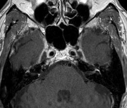
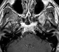
7 Petrous apicitis Petrous apicitis Petrous apex effusion Opacified petrous apex air cell, no bone erosion, no expansile margins effusion Petrous apex osteomyelitis Others Occurs in setting of non-pneumatized petrous apex Petrous marrow hyperintense on T2, enhances Inflammation of pneumatized petrous apex petrous apicitis If not pneumatized petrous apex osteomyelitis NEO Facial nerve enhancement - Bell

 autoimmune,")


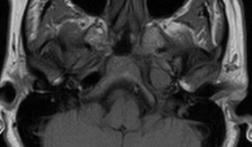
8 Facial nerve enhancement Ramsay Hunt Facial nerve enhancement Lyme Labyrinthitis Etiology: infectious (viral, bacterial, luetic) autoimmune, toxins, post-traumatic Particularly associated with acute bacterial meningitis due to H. influenza or S. pneumoniae Children present with SNHL, vertigo; can progress rapidly to profound deafness Labyrinthitis Acute CT normal MR faint enhancement, preserved fluid signal Fibrous stage CT normal MR enhancement, loss of fluid signal on heavily T2-weighted sequence Ossific stage (labyrinthitis ossificans) CT abnormal MR loss of fluid signal on heavily T2-weighted sequence MR is more sensitive than CT for detection of early acute labyrinthitis Acute labyrinthitis Fibrous/late stage of labyrinthitis on MR


 and by")


 Thank")


9 Evolution of labyrinthitis on CT Transverse / OCV fracture Labyrinthine fractures heal slowly (paucity of vascularity) and by fibrous union Complications Labyrinthitis ossificans Transverse / OCV fracture Labyrinthine fractures heal slowly (paucity of vascularity) and by fibrous union Complications Labyrinthitis ossificans Perilymphatic fistula (e.g. round window fluid, pneumolabyrinth) Thank you amy_juliano@meei.harvard.edu Pneumolabyrinth = perilymphatic fistula
The Temporal Bone Anatomy & Pathology
 Department of Radiology University of California San Diego The Temporal Bone Anatomy & Pathology John R. Hesselink, M.D. Temporal Bone Axial View Temporal Bone Coronal View Longitudinal Fracture The Temporal
Imaging of Petrous Apex: Anatomy and Pathology
 University of Utah Head and Neck Conference 2018 Petrous apex Imaging of Petrous Apex: Anatomy and Pathology Philip Chapman MD University of Alabama, Birmingham Good News PAs tend to be symmetric A quick
Cholesteatoma in children
 Cholesteatoma in children British Association of Paediatricians in Audiology London Conference, Jan.2012 Matthew Clark FRCS (ORL-HNS) Consultant Otologist Gloucestershire Royal Hospital Overview: Cholesteatoma
Chronic Ear Disease. Daekeun Joo Resident Lecture Series 11/18/09
 Chronic Ear Disease Daekeun Joo Resident Lecture Series 11/18/09 ETD URIs Viral-induced damage to ET lining resulting in decreased mucociliary clearance Viral invasion of ME mucosa results in inflamm Reflux
Complications of otitis media
 Chronic otitis media Definition:- Complications of otitis media Otitis media (OM) is broadly defined as inflammation from any cause of the middle ear.this may involve any of the contiguous pneumatized
ACUTE PAEDIATRIC EAR PRESENTATIONS PROF IAIN BRUCE PAEDIATRIC OTOLARYNGOLOGIST & ADULT OTOLOGIST
 www.manchesterchildrensent.com ACUTE PAEDIATRIC EAR PRESENTATIONS PROF IAIN BRUCE PAEDIATRIC OTOLARYNGOLOGIST & ADULT OTOLOGIST A CHILD WITH EARACHE UNCOMPLICATED AOM ACUTE OTITIS MEDIA Acute otitis media
THE MANAGEMENT of COMPLICATED OTITIS MEDIA. IFOS, Lima, 2018
 THE MANAGEMENT of COMPLICATED OTITIS MEDIA IFOS, Lima, 2018 VINCENT C COUSINS ENT-Otoneurology Unit, The Alfred Hospital & Department of Surgery. Monash University MELBOURNE, AUSTRALIA Otologic Complications
Middle ear CT imaging: Review of anatomy and common pathology
 Middle ear CT imaging: Review of anatomy and common pathology Poster No.: C-0665 Congress: ECR 2017 Type: Educational Exhibit Authors: M. R. Campos Arenas, M. C. Sánchez-Porro, J. Garrido Rull ; 1 1 2
Cholesteatoma-Pathogenesis and Surgical Management. Grand Rounds Presentation February 24, 1999 Kyle Kennedy, M.D. Jeffrey Vrabec,, M.D.
 Cholesteatoma-Pathogenesis and Surgical Management Grand Rounds Presentation February 24, 1999 Kyle Kennedy, M.D. Jeffrey Vrabec,, M.D. Introduction Cholesteatoma (keratoma)-essentially an accumulation
RADIOLOGY TEACHING CONFERENCE
 RADIOLOGY TEACHING CONFERENCE John Athas, MD Monica Tadros, MD Columbia University, College of Physicians & Surgeons Department of Otolaryngology- Head & Neck Surgery September 27, 2007 CT SCAN IMAGING
Petrositis, the big unknown: a review of recent work
 Petrositis, the big unknown: a review of recent work Poster No.: C-1165 Congress: ECR 2017 Type: Authors: Keywords: DOI: Educational Exhibit B. Rodriguez Chikri, E. R. Amador González, A. B. Marin Quiles,
SKULL BASE LESIONS THAT MAY MIMICK DISEASE
 SKULL BASE LESIONS THAT MAY MIMICK DISEASE AUTHORS: MYERS, TANDBERG, LORENZO UNIVERSITY OF NEW MEXICO DIAGNOSTIC RADIOLOGY Learning Objectives The participant will identify normal anatomic variants that
Imaging of Hearing Loss
 Contemporary Imaging of Sensorineural Hearing Loss Imaging of Hearing Loss Discussion Outline (SNHL) Imaging Approaches Anatomic Relationships Lesions: SNHL KL Salzman, MD University of Utah School of
Case Studies in the Skull Base
 Case Studies in the Skull Base Amy C Tsai, MD Neuroradiology Fellow Department of Radiology and Imaging Sciences University of Utah Health Sciences Center Salt Lake City, Utah, USA No disclosures related
Injury retrotympanic white, blue and red. Clinicalradiological
 Injury retrotympanic white, blue and red. Clinicalradiological correlation Poster No.: C-0211 Congress: ECR 2013 Type: Educational Exhibit Authors: R. Esteban Saiz, R. Castañón Martinez, M. Rebolledo Vicente,
Case Studies in CPA/IAC
 Outline Case Studies in CPA/IAC Atul K Mallik MD PhD Department of Radiology and Imaging Sciences University of Utah Health Sciences Center Salt Lake City, Utah, USA Case based review of cerebellopontine
Nasopharyngeal Carcinoma Presenting as Gradenigo s Syndrome
 1 Nasopharyngeal Carcinoma Presenting as Gradenigo s Syndrome Jay C. BRADLEY MD Arshad M. KHANANI MD Guy HIRSCH III MD Kenn A. FREEDMAN MD From the: Dept of Ophthalmology and Visual Sciences (Dr. Bradley,
New EAONO Cholesteatoma Classification with imaging illustration. Milan Profant, Katarina Sláviková
 New EAONO Cholesteatoma Classification with imaging illustration Milan Profant, Katarina Sláviková EAONO/JOS Joint Consensus Statements on the Definitions, Classification and Staging of Middle Ear Cholesteatoma
Complications of At t i c o a n t ral Cholesteato ma:
 Complications of t t i c o a n t ral Cholesteato ma: MR Manifestations 1 Jeong Hyun Lee, M.D., Ho Kyu Lee, M.D., Soo Mi Lim, M.D., Ji Hoon Shin, M.D., Choong Gon Choi, M.D., Dae Chul Suh, M.D., Kwang Sun
Modern Imaging & Current Controversies
 Temporal Bone: Modern Imaging & Current Controversies Suresh K. Mukherji, MD, FACR Professor and Chief of Neuroradiology Professor of Radiology, Otolaryngology Head Neck Surgery, Radiation i Oncology,
1. Axial view, left temporal bone. Plane through the upper antrum (A), superior semicircular canal (SSC) and IAC.
, superior semicircular canal (SSC) and IAC.") PA IAC SSC A 1. Axial view, left temporal bone. Plane through the upper antrum (A), superior semicircular canal (SSC) and IAC. IAC VII M I LSC Plane through the IAC, malleus head and incus and the lateral
PA IAC SSC A 1. Axial view, left temporal bone. Plane through the upper antrum (A), superior semicircular canal (SSC) and IAC. IAC VII M I LSC Plane through the IAC, malleus head and incus and the lateral
PITUITARY PARASELLAR LESIONS. Kim Learned, MD
 PITUITARY PARASELLAR LESIONS Kim Learned, MD DIFFERENTIALS Pituitary Sella Clivus, Sphenoid Sinus Suprasellar Optic chiasm, Hypothalamus, Circle of Willis Parasellar Cavernous Sinus Case 1 17 YEAR-OLD
Dr Melanie Souter. Consultant Otolaryngologist/Otologist Christchurch Public Hospital Christchurch. 12:00-12:15 Ears Made Easy
 Dr Melanie Souter Consultant Otolaryngologist/Otologist Christchurch Public Hospital Specialists @nine Christchurch 12:00-12:15 Ears Made Easy Ears made Easy Dr Melanie Souter Otology / Otolaryngology
Radiologic Evaluation of Petrous Apex Masses. Pavan Kavali, MS-IV Morehouse School of Medicine November 16, 2009
 Radiologic Evaluation of Petrous Apex Masses Pavan Kavali, MS-IV Morehouse School of Medicine November 16, 2009 Roadmap Petrous Apex Anatomy Patient D.S.: Clinical Presentation Differential diagnosis of
Pediatric Ear Diseases
 Pediatric Ear Diseases Yasushi Naito Pediatric Ear Diseases Diagnostic Imaging Atlas and Case Reports 242 figures, 7 in color and 5 tables, 2013 Basel Freiburg Paris London New York New Delhi Bangkok
Outline. Neuroradiology. Diffusion Imaging in. Clinical Applications of. Basics of Diffusion Imaging. Basics of Diffusion Imaging
 Clinical Applications of Diffusion Imaging in Neuroradiology No disclosures Stephen F. Kralik Assistant Professor of Radiology Indiana University School of Medicine Department of Radiology and Imaging
The ear: some applied basic science
 Chapter 1 The ear: some applied basic science The pinna The external ear or pinna is composed of cartilage with closely adherent perichondrium and skin. It is developed from six tubercles of the first
Magnetic Resonance Imaging (MRI) and High Resolution Computed Tomography (HRCT): Can they improve the evaluation of Middle ear cholesteatoma?
 and High Resolution Computed Tomography (HRCT): Can they improve the evaluation of Middle ear cholesteatoma?") Magnetic Resonance Imaging (MRI) and High Resolution Computed Tomography (HRCT): Can they improve the evaluation of Middle ear cholesteatoma? Poster No.: C-1249 Congress: ECR 2013 Type: Educational Exhibit
Magnetic Resonance Imaging (MRI) and High Resolution Computed Tomography (HRCT): Can they improve the evaluation of Middle ear cholesteatoma? Poster No.: C-1249 Congress: ECR 2013 Type: Educational Exhibit
Petrous Bone Normal anatomy
 Petrous Bone Normal anatomy By Mamdouh Mahfouz MD Prof. of Radiology Cairo University ssregypt.com Axial Coronal Petrous bone External ear Middle ear Inner ear External ear Cartilaginous part Bony part
DISEASES OF THE JAWS I
 DISEASES OF THE JAWS I ODONTOGENIC AND PERIODONTAL INFECTIONS ODONTOGENIC INFECTIONS PERIAPICAL GRANULOMA PERIAPICAL ABSCESS APICAL PERIODONTAL CYST PHOENIX ABSCESS FISTULA, DRAINING SINUS SPACE INFECTIONS
The Ear The ear consists of : 1-THE EXTERNAL EAR 2-THE MIDDLE EAR, OR TYMPANIC CAVITY 3-THE INTERNAL EAR, OR LABYRINTH 1-THE EXTERNAL EAR.
 The Ear The ear consists of : 1-THE EXTERNAL EAR 2-THE MIDDLE EAR, OR TYMPANIC CAVITY 3-THE INTERNAL EAR, OR LABYRINTH 1-THE EXTERNAL EAR Made of A-AURICLE B-EXTERNAL AUDITORY MEATUS A-AURICLE It consists
Pediatric Temporal Bone
 Pediatric Temporal Bone Suresh K. Mukherji, MD, FACR Professor and Chief of Neuroradiology Professor of Radiology, Otolaryngology Head Neck Surgery, Radiation Oncology and Periodontics & Oral Medicine
Refresher Course EAR TUMOR. Sasikarn Chamchod, MD Chulabhorn Hospital
 Refresher Course EAR TUMOR Sasikarn Chamchod, MD Chulabhorn Hospital Reference: Perez and Brady s Principles and Practice of radiation oncology sixth edition Outlines Anatomy Epidemiology Clinical presentations
DISCLOSURES LEARNING OBJECTIVES WE WILL NOT DISCUSS. CSB: Birdseye View MESSAGE NAVIGATING THE SELLA AND CENTRAL SKULL BASE
 NAVIGATING THE SELLA AND CENTRAL SKULL BASE Christopher P. Hess, M.D., Ph.D. DISCLOSURES Research Support, General Electric SLIDES: http://www.radiology.ucsf.edu/research/meetings/rsna LEARNING OBJECTIVES
Combined TBH / GSH meeting. 11 September 2007 Eric F Post
 Combined TBH / GSH meeting 11 September 2007 Eric F Post Case 32 yo male Presenting: Vertigo x since a.m. Tinnitus 5/7 Hearing loss worsened 4/7 Otorhea (L) x 2/ 52 Case History Ear surgery 1990 @ Umtata
Arastoo Vossough, M.D., Ph.D. Associate Professor of Radiology
 Disorders of the Temporal Bone Arastoo Vossough, M.D., Ph.D. Associate Professor of Radiology 1st Branchial Cleft Cyst Remnant of 1 st branchial apparatus Rare Cystic lesions: I- around pinna II- connecting
Bruce Black MD EAC TRAUMA
 EAC TRAUMA Bruising in the deep canal due to cotton bud/q-tip selfcleaning attempts. No action required. A granuloma of the deep Lt. EAC. Superficial trauma has become secondarily infected. Clean thoroughly,
Kathleen R. Fink, MD Virginia Mason Medical Center. 6 th Nordic Emergency Radiology Course 2017
 Kathleen R. Fink, MD Virginia Mason Medical Center 6 th Nordic Emergency Radiology Course 2017 Disclosure My spouse has a financial relationship with a commercial organization that may have a direct or
1. GOAL 2. OBJECTIVES a) KNOWLEDGE b) SKILLS c) INTEGRATION
 KNOWLEDGE b) SKILLS c) INTEGRATION") 1. GOAL The broad goal of the teaching of undergraduate students in Otorhinolaryngology is that the undergraduate student have acquired adequate knowledge and skills for optimally dealing with common disorders
1. GOAL The broad goal of the teaching of undergraduate students in Otorhinolaryngology is that the undergraduate student have acquired adequate knowledge and skills for optimally dealing with common disorders
UC SF. Safe Surgery Rule #1. Cholesteatoma. It s hard to have a surgical complication when you are not operating
 UC SF Cholesteatoma Chronic Ear Surgery: Staying Out of Trouble! Lawrence R. Lustig, MD Department of Oto-HNS University of California San Francisco Ligaments and folds Spaces NU Epitympanic Cholesteatoma
WSC , Conference 9. Case 1. Tissue from a rhesus macaque.
 Case 1. Tissue from a rhesus macaque. MICROSCOPIC DESCRIPTION: Esophagus: There is multifocal loss of the mucosal lining (1 pt). In these areas, the denuded subepithelial fibrous connective tissue is infiltrated
Correlation of HRCT mastoid with clinical presentation and operative findings in ear diseases
 International Journal of Otorhinolaryngology and Head and Neck Surgery Chintale SG et al. Int J Otorhinolaryngol Head Neck Surg. 2017 Jul;3(3):656-660 http://www.ijorl.com pissn 2454-5929 eissn 2454-5937
CHAPTER 13. FACIAL NERVE PARALYSIS
 CHAPTER 13. FACIAL NERVE PARALYSIS Introduction Facial nerve paralysis, whilst not a disease of the ear itself, commonly arises within the ear due to its anatomical course, and often as a result of ear
COM DEFINITION : TYPES
 COM DEFINITION : The diagnosis of chronic otitis media (COM) implies a permanent abnormality of the pars tensa or flaccida, most likely a result of earlier acute otitis media, negative middle ear pressure
Superior View of the Skull (Norma Verticalis) Anteriorly the frontal bone articulates with the two parietal bones AT THE CORONAL SUTURE
 Anteriorly the frontal bone articulates with the two parietal bones AT THE CORONAL SUTURE") Superior View of the Skull (Norma Verticalis) Anteriorly the frontal bone articulates with the two parietal bones AT THE CORONAL SUTURE 1 The two parietal bones articulate in the midline AT THE SAGITTAL
Superior View of the Skull (Norma Verticalis) Anteriorly the frontal bone articulates with the two parietal bones AT THE CORONAL SUTURE 1 The two parietal bones articulate in the midline AT THE SAGITTAL
Principles of Chronic Ear Surgery. Principles of Chronic Ear Surgery. Preoperative Considerations
 Principles of Chronic Ear Surgery Principles of Chronic Ear Surgery David S. Haynes, MD, FACS The Otology Group of Vanderbilt Professor, Dept. of Otolaryngology, Professor Dept. of Neurosurgery Professor,
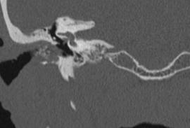
Cochlear Implant Failure: Imaging Evaluation of the Electrode Course
 Clinical Radiology (2003) 58: 288 293 doi:10.1016/s0009-9260(02)00523-8, available online at www.sciencedirect.com Pictorial Review Cochlear Implant Failure: Imaging Evaluation of the Electrode Course
Petrous Apex Cephalocele: Report of Two Cases and
 Petrous Apex Cephalocele: Report of Two Cases and Review of the Literature 1 Bo Seong Jeong, M.D., Ghi Jai Lee, M.D., Jae Chan Shim, M.D., Jae Myeong Lee, M.D., Mee Young Nam, M.D., Ho Kyun Kim, M.D. A
CSF Leaks. Abnormal communication between the subarachnoid space and the tympanomastoid space or nasal cavity. Presenting symptoms:
 CSF Leaks Steven Wright, M.D. Faculty Advisor: Matthew Ryan, M.D. The University of Texas Medical Branch Department of Otolaryngology Grand Rounds Presentation January 5, 2005 CSF Leaks Abnormal communication
AMSER Case of the Month July 2018 Complicated Headache with Fever
 AMSER Case of the Month July 2018 Complicated Headache with Fever Benjamin Park, MS IV Dr. Karen Xie Department of Radiology University of Illinois College of Medicine at Chicago Patient Presentation CC:
Dr. Vitthalrao Vikhe Patil Foundation s Medical College & Hospital, Ahmednagar, Maharashtra, India
 DOI: 10.21276/sjams.2017.5.3.17 Scholars Journal of Applied Medical Sciences (SJAMS) Sch. J. App. Med. Sci., 2017; 5(3B):770-779 Scholars Academic and Scientific Publisher (An International Publisher for
Vascular and Parameningeal Infections of the Head and Neck
 Vascular and Parameningeal Infections of the Head and Neck Kevin B. Laupland, MD, MSc, FRCPC Associate Professor Departments of Medicine, Critical Care Medicine, Pathology and Laboratory Medicine, and
Subspecialty Rotation: Otolaryngology
 Subspecialty Rotation: Otolaryngology Faculty: Evelyn Kluka, M.D. GOAL: Hearing Loss. Understand the morbidity of hearing loss, intervention strategies, and the pediatrician's and other specialists' roles
Gerard J. Gianoli, MD, FACS The Ear and Balance Institute Baton Rouge, Louisiana
 Gerard J. Gianoli, MD, FACS The Ear and Balance Institute Baton Rouge, Louisiana SSCD is defined anatomically as the absence of bone between the SSC and the middle fossa dura PSCD is a defect of the PSC
Facial and Temporal Bone Trauma Diagnostic imaging and therapeutic challenges in emergency
 Facial and Temporal Bone Trauma Diagnostic imaging and therapeutic challenges in emergency ATTYE A, KRAINIK A Department of Neuroradiology and MRI University Hospital Grenoble / University Grenoble Alpes
High resolution computed tomography of temporal bone in the evaluation of otologic diseases
 International Journal of Otorhinolaryngology and Head and Neck Surgery Handi PS et al. Int J Otorhinolaryngol Head Neck Surg. 2018 Jan;4(1):87-92 http://www.ijorl.com pissn 2454-5929 eissn 2454-5937 Original
Neoplasms that present as a swelling in the neck may be either
 Problems in otolaryngology Inflammatory swellings Viral and bacterial infection are frequent causes of swellings in the neck. Enlargement of the cervical lymph nodes is most likely but a dormant branchial
The Ear. Dr. Heba Kalbouneh Assistant Professor of Anatomy and Histology
 The Ear Dr. Heba Kalbouneh Assistant Professor of Anatomy and Histology The Ear The ear consists of the external ear; the middle ear (tympanic cavity); and the internal ear (labyrinth), which contains
RADIOGRAPHIC INTERPRETATION Differential Diagnosis
 RADIOGRAPHIC INTERPRETATION Differential Diagnosis MODULE 1: The Introduction. Chief complaint Demographics Age Sex Race Historical findings Physical findings Clinical Radiographic Location Maxilla/mandible
OTITIS MEDIA DR AKPALABA I.O.
 OTITIS MEDIA DR AKPALABA I.O. OTITIS MEDIA OUTLINE Introduction and Classification Brief Anatomy of the middle ear Acute Suppurative Otitis Media (ASOM) Chronic Suppurative Otitis Media (CSOM) Nonsuppurative
Orbital facia. Periororbital facia Orbital septum Bulbar facia Muscular facia
 Anatomy Orbital facia Periororbital facia Orbital septum Bulbar facia Muscular facia Physiology of symptoms 1) Proptosis ( exophthalmos) Pseudoproptosis Axial Non axial Pulsating Positional Intermittent
Advanced ENT Imaging. Objectives. Sinus Disease 3/18/2014. Tanya J. Rath, MD. Review what studies to order for common clinical scenarios
 Advanced ENT Imaging Tanya J. Rath, MD Director of head and Neck Imaging Assistant Professor University of Pittsburgh Medical Center University of Pittsburgh School of Medicine Fourth Annual ENT ENT for
DIZZINESS Varieties. : Fainting, hypotension : Rotatory, spinning. : Muscular incoordination : Collapse without LOC: ELH : Disturbed awareness
 DIZZINESS Varieties head Syncope Vertigo Dysequilibrium Ataxia Drop attacks Confusion Panic Attacks Non-organic : Fainting, hypotension : Rotatory, spinning : Unsteadiness on moving : Muscular incoordination
7. Anatomy and physiology of the vestibular system. Harmonic and disharmonic vestibular syndrome.
 7. Anatomy and physiology of the vestibular system. Harmonic and disharmonic vestibular syndrome. 8. Fundamental examination tools of otoneurology. 20. Ménière s syndrome and Ménière s disease. Therapeutic
Diseases of the Inner Ear
 Diseases of the Inner Ear A Clinical, Radiologic, and Pathologic Atlas Bearbeitet von Masoud Motasaddi Zarandy, John Rutka 1st Edition. 2010. Buch. vii, 93 S. Hardcover ISBN 978 3 642 05057 2 Format (B
Definition. Otitis Media with effusion (OME)
") Otitis Media. 1 Dr,wegdan saeed ALFHAL 2 Definition Acute Otitis Media (AOM) acute onset of symptoms, evidence of a middle ear effusion, and signs or symptoms of middle ear inflammation. Otitis Media with
Otitis Media. 1 Dr,wegdan saeed ALFHAL 2 Definition Acute Otitis Media (AOM) acute onset of symptoms, evidence of a middle ear effusion, and signs or symptoms of middle ear inflammation. Otitis Media with
C. Douglas Phillips MD FACR Director of Head and Neck Imaging Weill Cornell Medical College/NewYork-Presbyterian Hospital
 C. Douglas Phillips MD FACR Director of Head and Neck Imaging Weill Cornell Medical College/NewYork-Presbyterian Hospital Disclosures Neither I nor any family members have any pertinent financial relations
! Women greater than men (4:1)» Typical of other autoimmune diseases
» Typical of other autoimmune diseases") 1 2 3 4 : Overview and Diagnosis Suzanne K. Freitag, M.D. Director, Ophthalmic Plastic Surgery Massachusetts Eye and Ear Infirmary Harvard Medical School! I have no financial disclosures. Learning Objectives!
1 2 3 4 : Overview and Diagnosis Suzanne K. Freitag, M.D. Director, Ophthalmic Plastic Surgery Massachusetts Eye and Ear Infirmary Harvard Medical School! I have no financial disclosures. Learning Objectives!
INFECTION OF EXTERNAL EAR. Miguel G. Wagner R1 HUSE 2017
 INFECTION OF EXTERNAL EAR Miguel G. Wagner R1 HUSE 2017 ANATOMY AURICLE + EXTERNAL AUDITORY CANAL (EAC) + EPITELIAL SURFACE TYMPANIC MB Auricle Fibroelastic cartilage (except lobule) + perichondrium +
Skullbase Lesions. Skullbase Surgery Open vs endoscopic. Choice Of Surgical Approaches 12/28/2015. Skullbase Surgery: Evolution
 Skullbase Lesions Skullbase Surgery Open vs endoscopic Prof Asim Mahmood,FRCS,FACS,FICS,FAANS, Professor of Neurosurgery Henry Ford Hospital Detroit, MI, USA Anterior Cranial Fossa Subfrontal meningioma
The Child s Ear. Normal? Abnormal? And what do we do next?
 The Child s Ear Normal? Abnormal? And what do we do next? Anatomy of the Ear: Outer (External) Ear External Ear: Middle Ear: Inner Ear: Inner Ear: Cochlea Inner Ear: Semicircular Canals Why do we care?
TUMOURS OF THE TEMPORAL BONE
 TUMOURS OF THE TEMPORAL BONE RISK FACTORS : NF2 gene Chr 22 VHL (Von Hippel Lindau) disease Chr 3p Ionizing radiation Criteria for radiation induced temporal bone tumours by LUSTIG is as follows : 1. The
High-resolution Computed Tomography Study of Temporal Bone Pathologies
 Original Article Print ISSN: 2321-6379 Online ISSN: 2321-595X DOI: 10.17354/ijss/2016/375 High-resolution Computed Tomography Study of Temporal Bone Pathologies Manjit Bagul Senior Resident, Department
Metastasis. 57 year old with progressive Headache and Right Sided Visual Loss
 Metastasis 1% of sellar/parasellar masses Usually occurs with known primary Can involve third ventricle, hypothalamus, infundibular stalk May be both supra-, intrasellar 57 year old with progressive Headache
CT and MRI imaging of chronic otitis media complications.
 CT and MRI imaging of chronic otitis media complications. Poster No.: C-2131 Congress: ECR 2014 Type: Scientific Exhibit Authors: V. Bizimi, M. Tsitskari, K. Spyrou, V. Papalouka, A. Economou; Athens/GR
Kingdom of Bahrain Arabian Gulf University College of Medicine and Medical Sciences Year 6 ENT SMC Otitis Media (Dr.
 Kingdom of Bahrain Arabian Gulf University College of Medicine and Medical Sciences Year 6 ENT SMC Otitis Media (Dr. Jalal Almarzooq) - Anatomy of the ear: The ear is divided into 3 parts: External ear.
Efficacy of pre-operative computed tomography scans on clinical management and temporal bone surgery in cases of chronic otitis media
 Boston University OpenBU Theses & Dissertations http://open.bu.edu Boston University Theses & Dissertations 2014 Efficacy of pre-operative computed tomography scans on clinical management and temporal
Multidisciplinary care of a paediatric patient with Gradenigo s syndrome
 CASE REPORT Rare disease Multidisciplinary care of a paediatric patient with Gradenigo s syndrome Noor Janjua, 1 Mohammed Bajalan, 2 Samantha Potter, 3 Andrea Whitney, 2 Fabian Sipaul 2 1 Department of
Difficult Cases: Controversies in Cochlear Implantation
 Difficult Cases: Controversies in Cochlear Implantation David S Haynes, MD FACS Fred F Telischi, MD MEE FACS Lawrence R. Lustig, MD Robert F Labadie, PhD MD Nikolas H Blevins, MD Matthew L. Carlson, MD
Course Description 343 DDS- Clinical Oral and Maxillofacial Radiology II ( )
") King Saud University College of Dentistry Dept. of Oral Medicine & Diagnostic Sciences Division of Oral & Maxillofacial Radiology Course Description 343 DDS- Clinical Oral and Maxillofacial Radiology II
King Saud University College of Dentistry Dept. of Oral Medicine & Diagnostic Sciences Division of Oral & Maxillofacial Radiology Course Description 343 DDS- Clinical Oral and Maxillofacial Radiology II
Small lesions involving scalp and skull in pediatric age.
 Small lesions involving scalp and skull in pediatric age. Poster No.: C-1149 Congress: ECR 2013 Type: Educational Exhibit Authors: M. J. Yi, J. H. Yoo; Seoul/KR Keywords: Education and training, Education,
Benign brain lesions
 Benign brain lesions Diagnostic and Interventional Radiology Hung-Wen Kao Department of Radiology, Tri-Service General Hospital, National Defense Medical Center Computed tomography Hounsfield unit (HU)
A Pediatrician s Guide to Healthy Ears
 A Pediatrician s Guide to Healthy Ears Matthew L. Bush, M.D. Assistant Professor - Otology, Neurotology and Cranial Base Surgery Otolaryngology Head and Neck Surgery Disclosures No financial relationships
Sensorineural Hearing Loss in Complicated Cholesteatomatous Ear Disease
 Research in Otolaryngology 2014, 3(2): 29-35 DOI: 10.5923/j.otolaryn.20140302.04 Sensorineural Hearing Loss in Complicated Cholesteatomatous Ear Disease Borlingegowda Viswanatha 1,*, Khaja Naseeruddin
Small lesions involving scalp and skull in pediatric age.
 Small lesions involving scalp and skull in pediatric age. Poster No.: C-1149 Congress: ECR 2013 Type: Educational Exhibit Authors: M. J. Yi, J. H. Yoo; Seoul/ Keywords: Education and training, Education,
CT as first diagnostic approach in non-traumatic conditions of temporal bone
 CT as first diagnostic approach in non-traumatic conditions of temporal bone Award: Certificate of Merit Poster No.: C-1408 Congress: ECR 2016 Type: Educational Exhibit Authors: M. Bernabéu Rodríguez,
Vasculitis local: systemic
 Vasculitis Inflammation of the vessel wall. Signs and symptoms: 1- local: according to the involved tissue 2- systemic:(fever, myalgia, arthralgias, and malaise) Pathogenesis 1- immune-mediated 2- infectious
Major Anatomic Components of the Orbit
 Major Anatomic Components of the Orbit 1. Osseous Framework 2. Globe 3. Optic nerve and sheath 4. Extraocular muscles Bony Orbit Seven Bones Frontal bone Zygomatic bone Maxillary bone Ethmoid bone Sphenoid
CELL AND TISSUE INJURY COURSE-II PATHOLOGY LABORATORY
 CELL AND TISSUE INJURY COURSE-II PATHOLOGY LABORATORY PATHOLOGY of INFECTIOUS DISEASES MICROSCOPY Rengin Ahıskalı Macroscopy samples are shown in the macroscopy presentations of the first two courses.
Spectrum of lesions involving the petrous apex
 Spectrum of lesions involving the petrous apex Poster No.: C-1796 Congress: ECR 2010 Type: Educational Exhibit Topic: Head and Neck Authors: I. Alba, A. Paniagua Bravo, J. A. Blanco, L. Ibañez, J. C. Albillos,
Facial Paralysis: Objectives: Discuss the anatomy of the facial nerve. Look at common patterns of facial nerve palsy
 Facial Paralysis: Objectives: Discuss the anatomy of the facial nerve Look at common patterns of facial nerve palsy Discuss imaging appearance of lesions that lead to facial paralysis. Lindell R. Gentry,
70480 CT Orbit, et al without contrast CAT 9023
 70480 CT Orbit, et al without contrast CAT 9023 190.0 MALIG NEO EYEBALL 190.1 MALIG NEO ORBIT 190.2 MALIG NEO LACRIMALIG GLAND 190.9 MALIG NEO EYE UNSPEC 224.1 BENIGN NEO ORBIT 360.51 FB MAGNET ANT CHAMB
Course Description 343 DDS- Clinical Oral and Maxillofacial Radiology II ( )
") King Saud University College of Dentistry Dept. of Oral Medicine & Diagnostic Sciences Division of Oral & Maxillofacial Radiology Course Description 343 DDS- Clinical Oral and Maxillofacial Radiology II
King Saud University College of Dentistry Dept. of Oral Medicine & Diagnostic Sciences Division of Oral & Maxillofacial Radiology Course Description 343 DDS- Clinical Oral and Maxillofacial Radiology II
Parapharyngeal And Retropharyngeal Space Abscess: An Unusual Complication Of Chronic Suppurative Otitis Media
 ISPUB.COM The Internet Journal of Head and Neck Surgery Volume 1 Number 2 Parapharyngeal And Retropharyngeal Space Abscess: An Unusual Complication Of Chronic Suppurative Rijuneeta, P Kumar Parida, S Bhagat
Vertebral and Paravertebral Diseases
 Department of Radiology University of California San Diego Vertebral and Paravertebral Diseases John R. Hesselink, M.D. Vertebral / Paravertebral Disease (Extradural) Metastatic disease Primary bone tumors
objectives Pitfalls and Pearls in PET/CT imaging Kevin Robinson, DO Assistant Professor Department of Radiology Michigan State University
 objectives Pitfalls and Pearls in PET/CT imaging Kevin Robinson, DO Assistant Professor Department of Radiology Michigan State University To determine the regions of physiologic activity To understand
Pott s Puffy Tumor. Shahad Almohanna 15/1/2018
 Pott s Puffy Tumor Shahad Almohanna R2 15/1/2018 Definition First described in 1760 by Sir Percival Pott. s he originally suggested that trauma of the frontal bone was causative for this lesion, but later,
Narrowest segment of the ear canal. Limited microscopic. Wide endoscopic. field of view. field of view
 Endoscopic Transcanal Management of Cholesteatoma M. Tarabichi American Hospital-Dubai The Endoscope in Otology Mostly for documentation. Mostly diagnostic. Exploration of old mastoid cavities Endoscopic
The University of Arizona Pediatric Residency Program. Primary Goals for Rotation. Otolaryngology
 The University of Arizona Pediatric Residency Program Primary Goals for Rotation Otolaryngology 1. GOAL: Hearing Loss. Understand the morbidity of hearing loss, intervention strategies, and the pediatrician's
Dermatopathology: The tumor is composed of keratinocytes which show atypia, increase mitoses and abnormal mitoses.
 Squamous cell carcinoma (SCC): A common malignant tumor of keratinocytes arising in the epidermis, usually from a precancerous condition: 1- UV induced actinic keratosis, usually of low grade malignancy.