Petrous Bone Normal anatomy
|
|
|
- Mariah Hardy
- 5 years ago
- Views:
Transcription
1 Petrous Bone Normal anatomy By Mamdouh Mahfouz MD Prof. of Radiology Cairo University ssregypt.com

2


3 Axial


4 Coronal

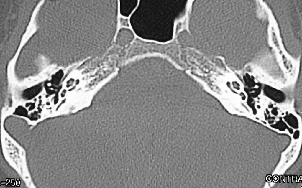
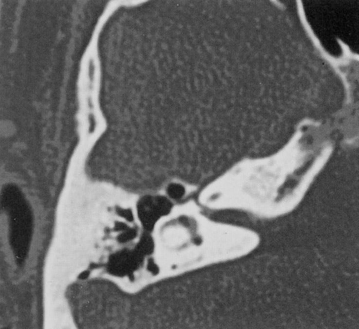
5 Petrous bone External ear Middle ear Inner ear
 The drum is")

6 External ear Cartilaginous part Bony part End by the tympanic membrane (drum) The drum is attached to the tympanic annulus The anterior and superior part of tympanic annulus= scutum

7 External ear
8 Middle ear Medial to the drum Contents Ossicles (Incus, malleous, stapes) Muscles and nerves
9 Middle ear Connections To the nasopharynx via Eustachian tube To the mastoid antrum via aditus ad antrum To the vestibule via oval window To the cochlea via the round window


10 ANATOMIC LANDMARK Ice cream cone

11 Ice cream cone

12 Ice cream Incus Malleus Aditus ad antrum Mastoid antrum Incudo- malleal joint Mesotympanum


13 Incus Malleus Eustachian tube


14 Incus Malleus Stapes Mastoid


15 Sinus tympani Facial recess Tympanic P. Eustachian tube Mastoid Hypo tympanum
16


17 Tegmen tympani Mastoid Epitympanum


18 Middle ear anatomy
19 Inner ear Vestibule Semicircular canals Cochlea Internal auditory canal Cochlear aqueduct Vestibular aqueduct

20 ANATOMIC LANDMARK Lateral SCC

21


22 Lateral SCC Posterior SCC Vestibule


23

24 Middle ear cleft Ice cream cone Mastoid Lateral SCC Posterior SCC Dome of cochlea IAC


25 Middle ear cleft Ice cream cone Mastoid Lateral SCC Posterior SCC Dome cochlea IAC of


26 Superior SCC


27 Superior SCC
28 Cochlea


29 Middle ear cleft Ice cream cone Mastoid Lateral SCC Posterior SCC Dome of cochlea IAC Vestibule


30 Cochlea


31


32


33 Sinus tympani Facial recess Tympanic pyramid Basal turn of cochlea Eustachian tube Niche of round window


34 Cochlear aqueduct

35 Vestibular aqueduct
36 Facial nerve Geniculate ganglion
37 Facial nerve Middle ear cleft Mastoid Lateral SCC Posterior SCC Dome of cochlea IAC Facial nerve Geniculate ganglion
38


39


40 MRI CT


41 Coronal images Tegmen tympani Oval window Facial nerve


42 Facial nerve


43 Vessels at the CP angle Loop of the anterior inferior cerebellar artery

44 Petrosal vein of Dandy Vessels at the CP angle

45 Q

46 Q

47 Q

48 Q

49 Q

50 Q

51 Q
52 Q

53 Coronal images Q
54 Petrous Bone Pathology


55 External ear Artesia and hypoplasia Abnormal embryogenesis of the 1 st & 2 nd branchial arches The canal is obstructed by bone or soft tissue CT to assess associated middle ear anomalies Soft tissue filling the external canal with fused malleus and incus


56 Bony Atresia Thick bony Artesia plate with fused ossicular mass


57 Soft tissue atresia of the left external auditory canal

58 Bony atresia of the left external auditory canal


59 Malignant ostitis externa Aggressive infection of the external ear Typically seen in diabetic, elderly, immune compromised patients Organism: pseudomonas aeruginosa Soft tissue mass at the external ear with bone destruction Extensions to middle ear, mastoid, intracranially


60 Malignant ostitis externa


61 Malignant ostitis externa


62 Neoplastic lesions Surfer s ear: Exostosis as a result of prolonged exposure to cold sea water, bilateral stenosis but not occlusion of the ear Osteoma: Unilateral solitary, found more laterally in the external ear Cutaneous malignancies: Squamous cell, basal cell, adenoid cystic carcinomas, melanoma


63 Cutaneous malignancies: Squamous cell, basal cell, adenoid cystic carcinomas, melanoma

![Ossicular anomalies [ Fusion, absence] Oval window normally](/docs-images/85/92782028/images/64-2.jpg "2mm [ coronal image] Anomalies of the facial nerve, carotid")
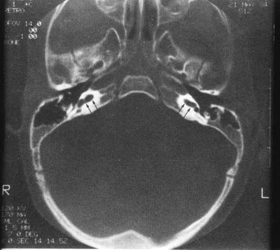
64 Middle ear anomalies Contracted middle ear cavity = small cavity < 3mm may be inadequate for ossicular reconstruction Ossicular anomalies [ Fusion, absence] Oval window normally 2mm [ coronal image] Anomalies of the facial nerve, carotid artery


65


66

67 Labyrinthine ossification


68 Congenital cholesteatoma Ectopic rest of epithelial tissue When occur intracranially it is called epidermoid Cholesteatoma without history of middle ear infection Evaluation of external & middle ear anomalies should include assessment of the presence of congenital cholesteatoma


69 Congenital cholesteatoma May occur any where in the middle ear Near the Eustachian tube / Near the stapes [are common sites] Non enhancing mass with bone destruction Enhancement of the labyrinth on MR images when fistula occurs


70 Otitis media versus cholesteatoma


71 Acquired cholesteatoma
![Acquired cholesteatoma Pars flaccida [Prussak s space]](/docs-images/85/92782028/images/72-0.jpg "[attic] Pars Tensa [ sinus cholesteatoma] A complication of")

72 Acquired cholesteatoma Pars flaccida [Prussak s space] [attic] Pars Tensa [ sinus cholesteatoma] A complication of chronic otitis media Non dependant, non enhancing + bone erosion Ossicles intact in 30% attic cholesteatoma 10% sinus cholesteatoma


73 Cholesteatoma affecting the facial recess Cholesteatoma eroding the tegmen tympani

74 Labyrinthine fistula


75 Cholesteatoma?!


76 Acquired cholesteatoma complications Erosion of the facial canal facial palsy Erosion of the SCC labyrinthine fistula Erosion of the tegmen tympani intracranial extension Dural sinus thrombosis Brain abscess


77 Acquired cholesteatoma complications Erosion of the tegmen tympani intracranial extension


78 Mastoid cholesteatoma Metastatic lung carcinoma, Axial section demonstrates infiltrative lesion of right mastoid (*)


79 Cholesterol granuloma

 MRI intense")
80 Glomus tymanicum Male: Female = 1:3 Pulsatile tinnitus, CHL Vascular retro tympanic mass CT a nodule of soft tissue density in the middle ear Large lesions do not cause bone erosion (typical finding) MRI intense enhancement


81 Glomus tymanicum


82 Glomus tymanicum
 in a patient")

83 A round soft tissue mass on the cochlear promontory(white arrowhead) in a patient with pulsatile tinnitus. This lesion enhanced avidly with contrast on MRI. A glomus tympanicum.


84 Trauma Clinical Otorrhea, hemo tympanum Conductive or SN HL Vertigo, facial nerve palsy CSF leakage, meningitis Longitudinal fractures 79%- 90% involve the middle ear CHL Transverse fracture 10%- 30% involve the inner ear SNHL Facial nerve injury 50%


85 Longitudinal and transverse fractures


86 Long fracture with incus dislocation


87 Transverse fracture


88 Pneumolabyrinth [ air in the vestibule ], blood in the middle ear Subluxation of the mallouncudal joint


89 Complete incus dislocation The head of the malleus is intact. The incus is in the mastoid antrum Longitudinal fracture with dislocation of the mallouncudal joint

90 Longitudinal fracture Hemo-tympanum Subluxation of incudo-malleal joint Labyrinthine fistula Pneumo-labyrinth

![system Enlargement of the vestibular aqueduct [VAS] Normal](/docs-images/85/92782028/images/91-1.jpg "diameter of the vestibular aqueduct = 1.")
91 Inner ear Congenital anomalies Large endolymphatic duct and sac [LEDS] Congenital enlargement of the inner ear endolymphatic system Enlargement of the vestibular aqueduct [VAS] Normal diameter of the vestibular aqueduct = 1.5 mm One of the most common causes of SNHL


92 Vestibular aqueduct syndrome


93 Inner ear Congenital anomalies Inner ear dysplasia [ 3 rd -8 th gestation week ] Michel anomaly total aplasia of the inner ear Mondini anomaly A range of morphologic anomalies of cochlea

94 Mondini anomaly with ear infection


95 Mondini anomaly

96 Normal Short, broad LCC with vestibular aqueduct syndrome


97 Normal canal Mega canal
![Facial nerve Schwannoma Uncommon May involve a segment [ geniculate ganglion] or multiple segments The motor axons of the facial nerve are](/docs-images/85/92782028/images/98-0.jpg "less sensitive to compression facial neuroma inside the IAC SNHL facial neuroma inside the IAC cann t be differentiated from acoustic")
98 Facial nerve Schwannoma Uncommon May involve a segment [ geniculate ganglion] or multiple segments The motor axons of the facial nerve are less sensitive to compression facial neuroma inside the IAC SNHL facial neuroma inside the IAC cann t be differentiated from acoustic neuroma


99 Normal canal Nodular or tubular lesion at the region of GG Facial nerve schwannoma Homogenous enhancement CT expansion of the facial nerve canal


100 Facial neuritis Non specific inflammation (Bell s palsy) Idiopathic, herpes simplex infection Ramsay- Hunt syndrome: herpes Zoster infection painful vesicles around the external ear Classical clinical presentation no need for imaging Atypical clinical course [unresolving or progressive]? tumor Enhanced MRI uniform enhancement of the intratemporal facial nerve with little or no enlargement.no Focal nodularity Comparison with the other ear is important
101 A 42-year-old man with right-sided Bell s palsy. Axial pre- and post contrast T1-weighted MR images demonstrate focal enhancement of the right facial nerve at the fundus of the internal auditory canal (green arrow). Note the symmetric, intense enhancement of the facial nerves around the geniculate fossa on both sides [orange arrows], attributable to the prominent normal circumneural arteriovenous plexus located in this area.


102 T1+ C T2 Viral labyrinthitis


103 Cochlear nerve atrophy Vascular Inflammatory Traumatic Nerve degeneration SNHL
104 سبحانك اللهم و بحمدك نشهد ان ال اله اال انت! نستغفرك و نتوب اليك! THANK YOU MAMDOUH MAHFOUZ MD
105 Cerebello-pontine angle lesions Acoustic neuroma 90% Meningioma 2-4% Epidermoid 1%
106 Acoustic neuroma 5-10 % of all intracranial tumors 85% of all intracranial neuromas 90% of cerebellopontine angle tumors Age adults y M:F = 1:2 Type II neurofibromatosis CT iso orhypodense SOL Homogenous enhancement Air cisternography MR iso or hypointense in T1 WIs Hyper intense in T2 WIs Strong homogenous enhancement.

107


108 CT MR Neurofibromatosis type II

109 Intra canalicular acoustic neuroma


110 CT AIR CISTERNOGRAPHY N Intra canalicular acoustic neuroma

111


112 Axial T1WI without contrast Axial T1 WI With contrast


113 Acoustic neuroma


114


115
116 Inner ear Internal auditory canal and CPA Meningioma 5-10% of all CPA masses Cerebellar dysfunction Calcification is common 75% Hyperostosis (CT) Dural tail (MRI)
117 Posterior fossa meningioma 9-15% Cerebellar convexity 5% Tentorium 2-4% Cerebello pontine 2-4%

118 2-4%


119 CPA meningioma


120 CPA meningioma


121 CPA meningioma
122 Epidermoid cyst 0.2 % - 1.8% of brain masses Cerebellopontine angle, supra and parasellar region middle cranial fossa and cisterna magna CT: hypodense lesions may simulate arachnoid cyst Surface is lobulated May show calcifications MRI: hypointense T1 between CSF and brain parenchyma Marked hyperintense T2 > CSF

123


124


125 Glomus jugulare: Contrast enhanced CT scan showing homogenously enhancing soft tissue mass lesion seen eroding the jugular fossa as well as the adjacent part of the petrous bone


126 Glomus jugulare


127 Glomus tumor


128 Glomus tumors

129

130

131


132 Normal anatomy


133 Normal anatomy


134 CT Anatomy Axial anatomy Arrowheads indicate the isthmus. The mastoid segment of facial nerve canal indicated with an arrowhead.

 RW: Round window CAQ:.")
135 Axial anatomy AP: apical turn of cochlea MD: middle turn of cochlea PROM: promonotory (basilar turn of cochlea) RW: Round window CAQ:. Cochlear aquduct
: internal audiotory canal, LSC: lateral")

136 Axial anatomy.f:entrance of facial nerve canal GF: geniculate fossa IAC (SUP): internal audiotory canal, LSC: lateral semicircular canal PSC: posterior semicircular canal SVN: canal for superior vestibular nerve. Arrow indicates posterior SC CN: canal. For cochlear nerve IAC: (INF): internal auditory canal- IVN: canal for inferior vestibular nerve MOD, OSL modiolus of cochlea with osseous spiral lamina; OW: oval window; V: vestibule
 is shown by long outlined arrow.")
 is indicated by arrow.")
137 Axial anatomy F: facial recess (arrowhead); S: Sinus tympani (arrowhead); pyramidal eminence (with second genu of facial nerve) is shown by long outlined arrow. incudostapedial joint is shown by large arrowhead A arrow: anterior epitympanic cell. First genu of facial nerve canal (geniculate ganglion) is indicated by arrow. V, small arrow: vestibular aqueduct
.")
, mastoid")
138 Axial anatomy C: cochlea; v: vestibule. SPI: short process of incus. Malleoincudal articulation (unlabeled arrow). Small arrow, tympanic segment of facial nerve canal. Ant.: anterior malleolar ligament; mia: malleoincudal articulation; antrum (arrow), mastoid antrum; single arrowhead: aditus ad antrum. Triple arrowhead: tympanic segment of facial nerve canal.


139 Coronal anatomy Large number of tegmental air cell (T). AP: apical turn of cochlea; C: carotid canal; DLS: distal labyrinthine segment; MID: middle turn of cochlea; PTS: proximal tympanic segment (Facial nerve canal).
.")
.")
140 Coronal anatomy mh: malleus head; mn: malleus neck; c: carotid canal. Tegmental air cells above external auditory (arrow). LO: long process of incus; LE: lenticular process of incus; J: jugular fossa; TEG: tegmen tympani (arrow). Tympanic segment of facial nerve canal (arrowhead).


141 Coronal anatomy Arrow indicates vestibule. BAS: basilar turn of cochlea. IVN: canal for inferior vestibular nerve; LSC: lateral semicircular canal; OW: oval widow ; CF; crista falciformis; SSC: superior semicircular canal; SVN: canal for superior vestibular nerve. C: Most posterior section round window (RW[DU], SSC: superior semicircular canal; T. SEG: tympanic segment; lateral semicircular canal: OW: oval widow.
142 Inner ear Congenital anomalies Cochlear nerve aplasia Failure of normal otic development (3-7 th weeks of gestation) aplasia or hypoplasia of cochlear nerve The presence of 8 th & 7 th nerves is necessary for normal formation of the internal auditory canal Usually presents by SNHL MRI T2 FSE
143 Middle ear Granulation tissue and cholesterol granuloma Chronic otomastoiditis accumulation of granulation tissue in the middle ear with possible bone erosions How to differentiate?! Granulation tissue Dependant opacity Enhancement [ marginal, heterogenous] Hemorrhagic nature high signal in T1 and T2 MR images Little bone erosions?! Little symptoms?!


144 Granulation tissue


145


146 Viral labyrinthitis

147 Facial neuritis


148 Non dehescant JB Glomus tympanicum

149 Mondini anomaly

150

151


152 Cholesterol cyst
153 Neoplastic lesions Primary tumors (rare) Osteo cartilagenous Giant cell tumour Aneurysmal bone cyst Osteoblastoma Osteosarcoma Paget s diseases Hematopoetic Lymphoma Myeloma Secondary Histeocytosis Rhabdomyosarcoma

154

155 Ice cream Malleus head Incus Incus Malleus Aditus ad antrum Mastoid antrum Incudo- malleal joint Mesotympanum


156 Malleus head Incus


157 T1 CT


158 MRI T2 T1 T1

159 ?

160 ?

161 ?

162 ?

163 ?


164 Coronal images


165 Non dehiscent JB Dehiscent J B


166


167


168


169 Recurrent cholesteatoma: Destructive mass lesion eroding the ossicles as well as the lateral semi circular canal.
The Temporal Bone Anatomy & Pathology
 Department of Radiology University of California San Diego The Temporal Bone Anatomy & Pathology John R. Hesselink, M.D. Temporal Bone Axial View Temporal Bone Coronal View Longitudinal Fracture The Temporal
1. Axial view, left temporal bone. Plane through the upper antrum (A), superior semicircular canal (SSC) and IAC.
, superior semicircular canal (SSC) and IAC.") PA IAC SSC A 1. Axial view, left temporal bone. Plane through the upper antrum (A), superior semicircular canal (SSC) and IAC. IAC VII M I LSC Plane through the IAC, malleus head and incus and the lateral
PA IAC SSC A 1. Axial view, left temporal bone. Plane through the upper antrum (A), superior semicircular canal (SSC) and IAC. IAC VII M I LSC Plane through the IAC, malleus head and incus and the lateral
Gross Anatomy of the. TEMPORAL BONE, EXTERNAL EAR, and MIDDLE EAR
 Gross Anatomy of the TEMPORAL BONE, EXTERNAL EAR, and MIDDLE EAR M1 Gross and Developmental Anatomy 9:00 AM, December 11, 2008 Dr. Milton M. Sholley Professor of Anatomy and Neurobiology Assignment: Head
Gross Anatomy of the. TEMPORAL BONE, EXTERNAL EAR, and MIDDLE EAR. Assignment: Head to Toe Temporomandibular Joint (TMJ)
") Gross Anatomy the TEMPORAL BONE, EXTERNAL EAR, and MIDDLE EAR M1 Gross and Developmental Anatomy 9:00 AM, December 11, 2008 Dr. Milton M. Sholley Pressor Anatomy and Neurobiology Assignment: Head to Toe
Gross Anatomy the TEMPORAL BONE, EXTERNAL EAR, and MIDDLE EAR M1 Gross and Developmental Anatomy 9:00 AM, December 11, 2008 Dr. Milton M. Sholley Pressor Anatomy and Neurobiology Assignment: Head to Toe
AUDITORY APPARATUS. Mr. P Mazengenya. Tel 72204
 AUDITORY APPARATUS Mr. P Mazengenya Tel 72204 Describe the anatomical features of the external ear Describe the tympanic membrane (ear drum) Describe the walls of the middle ear Outline the structures
Imaging of Hearing Loss
 Contemporary Imaging of Sensorineural Hearing Loss Imaging of Hearing Loss Discussion Outline (SNHL) Imaging Approaches Anatomic Relationships Lesions: SNHL KL Salzman, MD University of Utah School of
Middle ear CT imaging: Review of anatomy and common pathology
 Middle ear CT imaging: Review of anatomy and common pathology Poster No.: C-0665 Congress: ECR 2017 Type: Educational Exhibit Authors: M. R. Campos Arenas, M. C. Sánchez-Porro, J. Garrido Rull ; 1 1 2
The Ear The ear consists of : 1-THE EXTERNAL EAR 2-THE MIDDLE EAR, OR TYMPANIC CAVITY 3-THE INTERNAL EAR, OR LABYRINTH 1-THE EXTERNAL EAR.
 The Ear The ear consists of : 1-THE EXTERNAL EAR 2-THE MIDDLE EAR, OR TYMPANIC CAVITY 3-THE INTERNAL EAR, OR LABYRINTH 1-THE EXTERNAL EAR Made of A-AURICLE B-EXTERNAL AUDITORY MEATUS A-AURICLE It consists
The Ear. Dr. Heba Kalbouneh Assistant Professor of Anatomy and Histology
 The Ear Dr. Heba Kalbouneh Assistant Professor of Anatomy and Histology The Ear The ear consists of the external ear; the middle ear (tympanic cavity); and the internal ear (labyrinth), which contains
Pediatric Temporal Bone
 Pediatric Temporal Bone Suresh K. Mukherji, MD, FACR Professor and Chief of Neuroradiology Professor of Radiology, Otolaryngology Head Neck Surgery, Radiation Oncology and Periodontics & Oral Medicine
Cholesteatoma and Non-cholesteatomatous Inflammatory Disease. Cholesteatoma. Disclosures. Overview EAC. Cholesteatoma. None
 Disclosures Cholesteatoma and Non-cholesteatomatous Inflammatory Disease None Amy F Juliano, MD Staff Radiologist, Massachusetts Eye and Ear Infirmary Assistant Professor of Radiology, Harvard Medical
Anatomy of the ear: Lymphatics
 Anatomy of the ear: 1. External ear which consist of auricle and external auditory canal. The auricle has a framework of cartilage except the lobule, the skin is closely adherent to perichonderium at the
Arastoo Vossough, M.D., Ph.D. Associate Professor of Radiology
 Disorders of the Temporal Bone Arastoo Vossough, M.D., Ph.D. Associate Professor of Radiology 1st Branchial Cleft Cyst Remnant of 1 st branchial apparatus Rare Cystic lesions: I- around pinna II- connecting
Dr. T. Venkat Kishan Asst. Prof Department of Radiodiagnosis
 Dr. T. Venkat Kishan Asst. Prof Department of Radiodiagnosis Schwannomas (also called neurinomas or neurilemmomas) constitute the most common primary cranial nerve tumors. They are benign slow-growing
Radiologic Evaluation of Petrous Apex Masses. Pavan Kavali, MS-IV Morehouse School of Medicine November 16, 2009
 Radiologic Evaluation of Petrous Apex Masses Pavan Kavali, MS-IV Morehouse School of Medicine November 16, 2009 Roadmap Petrous Apex Anatomy Patient D.S.: Clinical Presentation Differential diagnosis of
Major Anatomic Components of the Orbit
 Major Anatomic Components of the Orbit 1. Osseous Framework 2. Globe 3. Optic nerve and sheath 4. Extraocular muscles Bony Orbit Seven Bones Frontal bone Zygomatic bone Maxillary bone Ethmoid bone Sphenoid
Case Studies in CPA/IAC
 Outline Case Studies in CPA/IAC Atul K Mallik MD PhD Department of Radiology and Imaging Sciences University of Utah Health Sciences Center Salt Lake City, Utah, USA Case based review of cerebellopontine
Imaging of Petrous Apex: Anatomy and Pathology
 University of Utah Head and Neck Conference 2018 Petrous apex Imaging of Petrous Apex: Anatomy and Pathology Philip Chapman MD University of Alabama, Birmingham Good News PAs tend to be symmetric A quick
Temporal bone anatomy and imaging features of common conditions causing hearing loss: A pictorial review
 Temporal bone anatomy and imaging features of common conditions causing hearing loss: A pictorial review Poster No.: C-1892 Congress: ECR 2012 Type: Educational Exhibit Authors: A. Masukawa, H. Takeuchi,
Chronic Ear Disease. Daekeun Joo Resident Lecture Series 11/18/09
 Chronic Ear Disease Daekeun Joo Resident Lecture Series 11/18/09 ETD URIs Viral-induced damage to ET lining resulting in decreased mucociliary clearance Viral invasion of ME mucosa results in inflamm Reflux
Anatomy of the Ear Region. External ear Middle ear Internal ear
 Ear Lecture Objectives Make a list of structures making the external, middle, and internal ear. Discuss the features of the external auditory meatus and tympanic membrane. Describe the shape, position,
Cholesteatoma-Pathogenesis and Surgical Management. Grand Rounds Presentation February 24, 1999 Kyle Kennedy, M.D. Jeffrey Vrabec,, M.D.
 Cholesteatoma-Pathogenesis and Surgical Management Grand Rounds Presentation February 24, 1999 Kyle Kennedy, M.D. Jeffrey Vrabec,, M.D. Introduction Cholesteatoma (keratoma)-essentially an accumulation
Interpretation of Computed Tomography of the Petrous Temporal Bone
 Systematic Review Article & Research Interpretation of Computed Tomography of the Petrous Temporal Bone Abstract This review will familiarise the reader with the normal radiological anatomy of the temporal
Spinal Imaging. ssregypt.com. Mamdouh Mahfouz MD
 Spinal Imaging Degenerative diseases ssregypt.com Mamdouh Mahfouz MD mamdouh.m5@gmail.com MRI Open MRI Closed Extremity MRI Dynamic MRI Dynamic MRI The bed rotates from Upright to Recumbent, stopping at
Skull Base Course. Dissection with fresh temporal bones and half heads
 Skull Base Course Dissection with fresh temporal bones and half heads 711 November 2016 Gruppo Otologico Via Emmanueli 42 Piacenza 29122 t +39 0523 754 362 fax +39 0523 453 708 www.gruppootologico.com
Head&Neck Imaging. ssregypt.com. Parapharyngeal Spaces. Mamdouh mahfouz MD
 Head&Neck Imaging Parapharyngeal Spaces ssregypt.com Mamdouh mahfouz MD mamdouh.m5@gmail.com Definitio n Fat filled triangular space lateral the pharynx Extends from the skull base to the oropharynx Parapharyngeal
High resolution computed tomography of temporal bone in the evaluation of otologic diseases
 International Journal of Otorhinolaryngology and Head and Neck Surgery Handi PS et al. Int J Otorhinolaryngol Head Neck Surg. 2018 Jan;4(1):87-92 http://www.ijorl.com pissn 2454-5929 eissn 2454-5937 Original
Kingdom of Bahrain Arabian Gulf University College of Medicine and Medical Sciences Year 6 ENT SMC Otitis Media (Dr.
 Kingdom of Bahrain Arabian Gulf University College of Medicine and Medical Sciences Year 6 ENT SMC Otitis Media (Dr. Jalal Almarzooq) - Anatomy of the ear: The ear is divided into 3 parts: External ear.
Dr. Vitthalrao Vikhe Patil Foundation s Medical College & Hospital, Ahmednagar, Maharashtra, India
 DOI: 10.21276/sjams.2017.5.3.17 Scholars Journal of Applied Medical Sciences (SJAMS) Sch. J. App. Med. Sci., 2017; 5(3B):770-779 Scholars Academic and Scientific Publisher (An International Publisher for
Unit VIII Problem 9 Anatomy of The Ear
 Unit VIII Problem 9 Anatomy of The Ear - The ear is an organ with 2 functions: Hearing. Maintenance of equilibrium/balance. - The ear is divided into 3 parts: External ear. Middle ear (which is also known
RADIOLOGY TEACHING CONFERENCE
 RADIOLOGY TEACHING CONFERENCE John Athas, MD Monica Tadros, MD Columbia University, College of Physicians & Surgeons Department of Otolaryngology- Head & Neck Surgery September 27, 2007 CT SCAN IMAGING
The ear: some applied basic science
 Chapter 1 The ear: some applied basic science The pinna The external ear or pinna is composed of cartilage with closely adherent perichondrium and skin. It is developed from six tubercles of the first
Dr. Sami Zaqout Faculty of Medicine IUG
 Auricle External Ear External auditory meatus The Ear Middle Ear (Tympanic Cavity) Auditory ossicles Internal Ear (Labyrinth) Bony labyrinth Membranous labyrinth External Ear Auricle External auditory
Anatomy and Physiology of Hearing
 Anatomy and Physiology of Hearing The Human Ear Temporal Bone Found on each side of the skull and contains the organs for hearing and balance Divided into four major portions: - squamous - mastoid - tympanic
CE Directed Reading. Temporal Bone CT: Anatomy, Technique, and Associated Pathophysiology
 Temporal Bone CT: Anatomy, Technique, and Associated Pathophysiology Chris Young, MS, R.R.A., R.T.(R) Computed tomography (CT) of the temporal bone is performed to evaluate trauma, tumors, sinuses, the
UC SF. Safe Surgery Rule #1. Cholesteatoma. It s hard to have a surgical complication when you are not operating
 UC SF Cholesteatoma Chronic Ear Surgery: Staying Out of Trouble! Lawrence R. Lustig, MD Department of Oto-HNS University of California San Francisco Ligaments and folds Spaces NU Epitympanic Cholesteatoma
Pediatric Ear Diseases
 Pediatric Ear Diseases Yasushi Naito Pediatric Ear Diseases Diagnostic Imaging Atlas and Case Reports 242 figures, 7 in color and 5 tables, 2013 Basel Freiburg Paris London New York New Delhi Bangkok
Correlation of HRCT mastoid with clinical presentation and operative findings in ear diseases
 International Journal of Otorhinolaryngology and Head and Neck Surgery Chintale SG et al. Int J Otorhinolaryngol Head Neck Surg. 2017 Jul;3(3):656-660 http://www.ijorl.com pissn 2454-5929 eissn 2454-5937
Cone Beam CT Atlas of the Normal Suspensory Apparatus of the Middle Ear Ossicles
 Cone Beam CT Atlas of the Normal Suspensory Apparatus of the Middle Ear Ossicles Poster No.: C-2036 Congress: ECR 2013 Type: Authors: Educational Exhibit B. Smet 1, I. De Kock 2, P. Gillardin 2, M. Lemmerling
Osseous structures in the middle ear cavity(mec): Are they too many or are they too few?
: Are they too many or are they too few?") Osseous structures in the middle ear cavity(mec): Are they too many or are they too few? Poster No.: C-2286 Congress: ECR 2013 Type: Educational Exhibit Authors: P. Mundada, B. S. Purohit, T. Tiong Yong;
Osseous structures in the middle ear cavity(mec): Are they too many or are they too few? Poster No.: C-2286 Congress: ECR 2013 Type: Educational Exhibit Authors: P. Mundada, B. S. Purohit, T. Tiong Yong;
CT as first diagnostic approach in non-traumatic conditions of temporal bone
 CT as first diagnostic approach in non-traumatic conditions of temporal bone Award: Certificate of Merit Poster No.: C-1408 Congress: ECR 2016 Type: Educational Exhibit Authors: M. Bernabéu Rodríguez,
High-resolution Computed Tomography Study of Temporal Bone Pathologies
 Original Article Print ISSN: 2321-6379 Online ISSN: 2321-595X DOI: 10.17354/ijss/2016/375 High-resolution Computed Tomography Study of Temporal Bone Pathologies Manjit Bagul Senior Resident, Department
Facial Paralysis: Objectives: Discuss the anatomy of the facial nerve. Look at common patterns of facial nerve palsy
 Facial Paralysis: Objectives: Discuss the anatomy of the facial nerve Look at common patterns of facial nerve palsy Discuss imaging appearance of lesions that lead to facial paralysis. Lindell R. Gentry,
Anatomy of External and Middle ear. Dr Sai Manohar
 Anatomy of External and Middle ear. Dr Sai Manohar 1 Human Ear For Anatomical description, Ear is divided into Auricle (or pinna) The external auditory canal The Middle Ear and its derivatives The Inner
HEPATO-BILIARY IMAGING
 HEPATO-BILIARY IMAGING BY MAMDOUH MAHFOUZ MD PROF.OF RADIOLOGY CAIRO UNIVERSITY mamdouh.m5@gmail.com www.ssregypt.com CT ABDOMEN Indications Patient preparation Patient position Scanogram Fasting 4-6 hours
Injury retrotympanic white, blue and red. Clinicalradiological
 Injury retrotympanic white, blue and red. Clinicalradiological correlation Poster No.: C-0211 Congress: ECR 2013 Type: Educational Exhibit Authors: R. Esteban Saiz, R. Castañón Martinez, M. Rebolledo Vicente,
Exposure of facial nerve and endolymphatic sac
 Exposure of facial nerve and endolymphatic sac 1 7 4 2 3 5 6 8 1 Vertical part of the facial nerve exposed 1 Second genu of facial nerve. 2 Vertical part of facial nerve. 3 Horizontal part of facial nerve.
Brain Imaging. IC calcifications. Mamdouh mahfouz MD
 Brain Imaging IC calcifications www.ssregypt.com Mamdouh mahfouz MD mamdouh.m5@gmail.com CT Hyper dense [ more than100 HU ] MRI Low signal in T1 and T2 WIs [non mobile protons] Exceptions Minute calcifications
New EAONO Cholesteatoma Classification with imaging illustration. Milan Profant, Katarina Sláviková
 New EAONO Cholesteatoma Classification with imaging illustration Milan Profant, Katarina Sláviková EAONO/JOS Joint Consensus Statements on the Definitions, Classification and Staging of Middle Ear Cholesteatoma
Bruce Black MD EAC TRAUMA
 EAC TRAUMA Bruising in the deep canal due to cotton bud/q-tip selfcleaning attempts. No action required. A granuloma of the deep Lt. EAC. Superficial trauma has become secondarily infected. Clean thoroughly,
For the following questions, indicate the letter that corresponds to the SINGLE MOST APPROPRIATE ANSWER
 GROSS ANATOMY EXAMINATION May 15, 2000 For the following questions, indicate the letter that corresponds to the SINGLE MOST APPROPRIATE ANSWER 1. Pain associated with an infection limited to the middle
ORIGINAL ARTICLE. A New Staging System for Tympano-mastoid Cholesteatoma. Aziz Belal, Mahmoud Reda, Ahmed Mehana, Yousef Belal
 Int. Adv. Otol. 2012; 8:(1) 63-68 ORIGINAL ARTICLE A New Staging System for Tympano-mastoid Cholesteatoma Aziz Belal, Mahmoud Reda, Ahmed Mehana, Yousef Belal Alexandria Ear Hospital Alexandria Egypt (AB,
Middle and Inner Ear: Improved Depiction with Multiplanar Reconstruction of Volumetric CT Data 1
 Note: This copy is for your personal non-commercial use only. To order presentation-ready copies for distribution to your colleagues or clients, contact us at www.rsna.org/rsnarights. EDUCATION EXHIBIT
Structure, Energy Transmission and Function. Gross Anatomy. Structure, Function & Process. External Auditory Meatus or Canal (EAM, EAC) Outer Ear
 Outer Ear") Gross Anatomy Structure, Energy Transmission and Function IE N O ME 1 Structure, Function & Process 4 External Auditory Meatus or Canal (EAM, EAC) Outer third is cartilaginous Inner 2/3 is osseous Junction
Gross Anatomy Structure, Energy Transmission and Function IE N O ME 1 Structure, Function & Process 4 External Auditory Meatus or Canal (EAM, EAC) Outer third is cartilaginous Inner 2/3 is osseous Junction
Introduction. Types of Cholesteatoma
 TITLE: Cholesteatoma SOURCE: Grand Rounds Presentation, UTMB, Dept. of Otolaryngology DATE: January 25, 2006 RESIDENT PHYSICIAN: Garrett Hauptman, MD FACULTY PHYSICIAN: Tomoko Makishima, MD, PhD SERIES
Anatomy and Pathology of the
 Residents Section Pattern of the Month Ho et al. natomy and Pathology of the Facial Nerve Residents Section Pattern of the Month Downloaded from www.ajronline.org by 37.44.200.121 on 01/30/18 from IP address
Case Studies in the Skull Base
 Case Studies in the Skull Base Amy C Tsai, MD Neuroradiology Fellow Department of Radiology and Imaging Sciences University of Utah Health Sciences Center Salt Lake City, Utah, USA No disclosures related
Year 2003 Paper two: Questions supplied by Tricia
 question 43 A 42-year-old man presents with a two-year history of increasing right facial numbness. He has a history of intermittent unsteadiness, mild hearing loss and vertigo but has otherwise been well.
Complications of otitis media
 Chronic otitis media Definition:- Complications of otitis media Otitis media (OM) is broadly defined as inflammation from any cause of the middle ear.this may involve any of the contiguous pneumatized
Refresher Course EAR TUMOR. Sasikarn Chamchod, MD Chulabhorn Hospital
 Refresher Course EAR TUMOR Sasikarn Chamchod, MD Chulabhorn Hospital Reference: Perez and Brady s Principles and Practice of radiation oncology sixth edition Outlines Anatomy Epidemiology Clinical presentations
Activity 1: Anatomy of the Eye and Ear Lab
 Activity 1: Anatomy of the Eye and Ear Lab 1. Launch the view! Launch Human Anatomy Atlas. Navigate to Quizzes/Lab Activities, find the Eye and Ear Lab section. Launch Augmented Reality mode and scan the
Gerard J. Gianoli, MD, FACS The Ear and Balance Institute Baton Rouge, Louisiana
 Gerard J. Gianoli, MD, FACS The Ear and Balance Institute Baton Rouge, Louisiana SSCD is defined anatomically as the absence of bone between the SSC and the middle fossa dura PSCD is a defect of the PSC
Ear. Utricle & saccule in the vestibule Connected to each other and to the endolymphatic sac by a utriculosaccular duct
 Rahaf Jreisat *You don t have to go back to the slides. Ear Inner Ear Membranous Labyrinth It is a reflection of bony labyrinth but inside. Membranous labyrinth = set of membranous tubes containing sensory
ISSN: Volume 5 Issue CASE REPORT. Anju Chauhan, Vikram Wadhwa, Samuel Rajan, P.K. Rathore
 ISSN: 2250-0359 Volume 5 Issue 2 2015 CONGENITAL CHOLESTEATOMA ISOLATED TO MASTOID PROCESS : A CASE REPORT Anju Chauhan, Vikram Wadhwa, Samuel Rajan, P.K. Rathore Maulana Azad Medical College, New Delhi,
OTOLOGY. 1. BRIEF DESCRIPTION OF OTOLOGIC TRAINING Rotations that include otologic training are a component of each of the four years of training.
 OTOLOGY 1. BRIEF DESCRIPTION OF OTOLOGIC TRAINING Rotations that include otologic training are a component of each of the four years of training. Longwood Rotation PGY-2 through PGY-5 years o Clinic experience
Congenital Absence of the Oval Window: Radiologic Diagnosis and Associated Anomalies
 AJNR Am J Neuroradiol 21:322 327, February 2000 Congenital Absence of the Oval Window: Radiologic Diagnosis and Associated Anomalies Barbara Zeifer, Paul Sabini, and Jonathan Sonne BACKGROUND AND PURPOSE:
Modern Imaging & Current Controversies
 Temporal Bone: Modern Imaging & Current Controversies Suresh K. Mukherji, MD, FACR Professor and Chief of Neuroradiology Professor of Radiology, Otolaryngology Head Neck Surgery, Radiation i Oncology,
Chapter 17, Part 2! The Special Senses! Hearing and Equilibrium!
 Chapter 17, Part 2! The Special Senses! Hearing and Equilibrium! SECTION 17-5! Equilibrium sensations originate within the inner ear, while hearing involves the detection and interpretation of sound waves!
Chapter 17, Part 2! Chapter 17 Part 2 Special Senses! The Special Senses! Hearing and Equilibrium!
 Chapter 17, Part 2! The Special Senses! Hearing and Equilibrium! SECTION 17-5! Equilibrium sensations originate within the inner ear, while hearing involves the detection and interpretation of sound waves!
1. GOAL 2. OBJECTIVES a) KNOWLEDGE b) SKILLS c) INTEGRATION
 KNOWLEDGE b) SKILLS c) INTEGRATION") 1. GOAL The broad goal of the teaching of undergraduate students in Otorhinolaryngology is that the undergraduate student have acquired adequate knowledge and skills for optimally dealing with common disorders
1. GOAL The broad goal of the teaching of undergraduate students in Otorhinolaryngology is that the undergraduate student have acquired adequate knowledge and skills for optimally dealing with common disorders
External and middle ear diseases: radiological diagnosis based on clinical signs and symptoms
 Insights Imaging (2012) 3:33 48 DOI 10.1007/s13244-011-0126-z PICTORIAL REVIEW External and middle ear diseases: radiological diagnosis based on clinical signs and symptoms Agnieszka Trojanowska & Andrzej
Cochlear Schwannoma Removed Through the External Auditory Canal by a Transcanal Exclusive Endoscopic Technique
 The Laryngoscope VC 2013 The American Laryngological, Rhinological and Otological Society, Inc. Case Report Cochlear Schwannoma Removed Through the External Auditory Canal by a Transcanal Exclusive Endoscopic
Magnetic Resonance Imaging (MRI) and High Resolution Computed Tomography (HRCT): Can they improve the evaluation of Middle ear cholesteatoma?
 and High Resolution Computed Tomography (HRCT): Can they improve the evaluation of Middle ear cholesteatoma?") Magnetic Resonance Imaging (MRI) and High Resolution Computed Tomography (HRCT): Can they improve the evaluation of Middle ear cholesteatoma? Poster No.: C-1249 Congress: ECR 2013 Type: Educational Exhibit
Magnetic Resonance Imaging (MRI) and High Resolution Computed Tomography (HRCT): Can they improve the evaluation of Middle ear cholesteatoma? Poster No.: C-1249 Congress: ECR 2013 Type: Educational Exhibit
Complications of At t i c o a n t ral Cholesteato ma:
 Complications of t t i c o a n t ral Cholesteato ma: MR Manifestations 1 Jeong Hyun Lee, M.D., Ho Kyu Lee, M.D., Soo Mi Lim, M.D., Ji Hoon Shin, M.D., Choong Gon Choi, M.D., Dae Chul Suh, M.D., Kwang Sun
COCHLEAR IMPLANTS Aetiology of Deafness. Bruce Black MD
 COCHLEAR IMPLANTS Aetiology of Deafness Heterochromia iridis. Cases may be healthy or associated with a variety of conditions, e.g. Waardenburg syndrome. Waardenburg syndrome. Note the snowy lock of hair
Fracture mimics on temporal bone CT - a guide for the radiologist
 Fracture mimics on temporal bone CT - a guide for the radiologist Award: Certificate of Merit Poster No.: C-0158 Congress: ECR 2012 Type: Educational Exhibit Authors: Y. Kwong, D. Yu, J. Shah; Nottingham/UK
Imaging of the Temporal Bone: A Symptom-Based Approach
 Imaging of the Temporal Bone: A Symptom-Based Approach Tadesse Eshetu, MD, and Nafi Aygun, MD Some of the symptoms associated with the temporal bone diseases are nonspecific, whereas others overlap with
Expand The Scope of Temporal Bone Reporting: Why, What and Where to Look For.
 Expand The Scope of Temporal Bone Reporting: Why, What and Where to Look For. Award: Certificate of Merit Poster No.: C-1521 Congress: ECR 2014 Type: Educational Exhibit Authors: P. Mundada, B. S. Purohit,
Dr Melanie Souter. Consultant Otolaryngologist/Otologist Christchurch Public Hospital Christchurch. 12:00-12:15 Ears Made Easy
 Dr Melanie Souter Consultant Otolaryngologist/Otologist Christchurch Public Hospital Specialists @nine Christchurch 12:00-12:15 Ears Made Easy Ears made Easy Dr Melanie Souter Otology / Otolaryngology
Congenital Aural Atresia. Miranda S. Dennis, M.D. April 6, 2011
 Congenital Aural Atresia Miranda S. Dennis, M.D. April 6, 2011 Embryology External Auditory Canal first branchial groove starts as a solid core of epithelial cells, which undergoes absorption in a medial
Sinonasal Imaging. Mamdouh Mahfouz MD Professor of Radiology Cairo University. ssregypt.com
 Sinonasal Imaging Mamdouh Mahfouz MD Professor of Radiology Cairo University ssregypt.com Scanning Techniques Routine Study CORONAL Coronal 3-5mm sections from the posterior wall of the sphenoid sinus
REVIEW/PREVIEW OF HEAD AND NECK ANATOMY FOR ENT EXAM
 REVIEW/PREVIEW OF HEAD AND NECK ANATOMY FOR ENT EXAM - 2017 PALPATE CAROTID ARTERY: AT LEVEL OF CAROTID BIFURCATION VERTEBRAL LEVEL C4 Sternocleidomastoid Muscle INTERNAL CAROTID EXTERNAL CAROTID COMMON
Role of high resolution computed tomography in the evaluation of temporal bone lesions: our experience
 International Journal of Otorhinolaryngology and Head and Neck Surgery Jyothi AC et al. Int J Otorhinolaryngol Head Neck Surg. 2016 Jul;2(3):135-139 http://www.ijorl.com pissn 2454-5929 eissn 2454-5937
Imaging of facial paralysis
 Imaging of facial paralysis Poster No.: C-2151 Congress: ECR 2013 Type: Educational Exhibit Authors: N. Martinez Molina, L. Aleman Romero, L. A. Sanchez Alonso, A. Puerta Sales, V. Garcia Medina; Murcia/ES
Cholesteatoma in children
 Cholesteatoma in children British Association of Paediatricians in Audiology London Conference, Jan.2012 Matthew Clark FRCS (ORL-HNS) Consultant Otologist Gloucestershire Royal Hospital Overview: Cholesteatoma
Neuro - imaging. Sella. ssregypt.com
 Neuro - imaging Sella ssregypt.com Bony Sella AP diameter Depth Contents 16mm 14mm Pituitary gland, part of infundibular stalk, CSF CT Technique 5 mm slices Axial and coronal Contrast injection Bone and
MECHANISM OF HEARING
 MECHANISM OF HEARING Sound: Sound is a vibration that propagates as an audible wave of pressure, through a transmission medium such as gas, liquid or solid. Sound is produced from alternate compression
Title. Author(s) Takahashi, Haruo. Issue Date Right.
 Takahashi, Haruo. Issue Date Right.") NAOSITE: Nagasaki University's Ac Title Author(s) Citation A case with posterior fossa epiderm symptoms caused by insufficiency of usefulness of free DICOM image view Takasaki, Kenji; Kumagami, Hidetaka
NAOSITE: Nagasaki University's Ac Title Author(s) Citation A case with posterior fossa epiderm symptoms caused by insufficiency of usefulness of free DICOM image view Takasaki, Kenji; Kumagami, Hidetaka
Neurovascular elements of the PCF
 Level I: Neurovascular elements of the PCF Trigeminal and Abducent Superior cerebellar artery and vein Dandy s vein Level II: Facial and Cochleovestibular AICA and internal auditory artery, veins Level
Rebecca J. Clark-Bash, R. EEG\EP T., CNIMeKnowledgePlus.net Page 1
 Navigating the Auditory Pathway: Technical & Physiological Impact on IOM Rebecca Clark-Bash, R. EEG\EP T, CLTM, CNIM, F.ASET, FASNM Faculty Rebecca Clark-Bash R. EEG\EP T., CLTM, CNIM, F.ASNM, F.ASET ASNM
External carotid blood supply to acoustic neurinomas
 External carotid blood supply to acoustic neurinomas Report of two cases HARVEY L. LEVINE, M.D., ERNEST J. FERmS, M.D., AND EDWARD L. SPATZ, M.D. Departments of Radiology, Neurology, and Neurosurgery,
CHAPTER 13. FACIAL NERVE PARALYSIS
 CHAPTER 13. FACIAL NERVE PARALYSIS Introduction Facial nerve paralysis, whilst not a disease of the ear itself, commonly arises within the ear due to its anatomical course, and often as a result of ear
Key words: Chronic suppurative otitis media, High resolution Computed tomography, cholesteatoma,
 ORIGINAL ARTICLE Study of Radiological findings in High resolution computed tomography (HRCT) temporal bone in Chronic suppurative otitis Media (CSOM): A hospital Based cross sectional study. Vijay vaidya
Cochlear Implant Failure: Imaging Evaluation of the Electrode Course
 Clinical Radiology (2003) 58: 288 293 doi:10.1016/s0009-9260(02)00523-8, available online at www.sciencedirect.com Pictorial Review Cochlear Implant Failure: Imaging Evaluation of the Electrode Course
Chapter 1: Applied Anatomy, Physiology and Embryology of the Ear. Anatomy and Physiology. The Outer Ear. The Pinna. The External Ear Canal
 Chapter 1: Applied Anatomy, Physiology and Embryology of the Ear The ear contains two specialized sensory organs, the cochlea and the vestibular apparatus, enclosed in the extremely hard protective casing
CSF Leaks. Abnormal communication between the subarachnoid space and the tympanomastoid space or nasal cavity. Presenting symptoms:
 CSF Leaks Steven Wright, M.D. Faculty Advisor: Matthew Ryan, M.D. The University of Texas Medical Branch Department of Otolaryngology Grand Rounds Presentation January 5, 2005 CSF Leaks Abnormal communication
THE COCHLEA AND AUDITORY PATHWAY
 Dental Neuroanatomy Suzanne S. Stensaas, PhD February 23, 2012 Reading: Waxman, Chapter 16, Review pictures in a Histology book Computer Resources: http://www.cochlea.org/ - Promenade around the Cochlea
Eye and Ocular Adnexa, Auditory Systems
 Eye and Ocular Adnexa, Auditory Systems CPT copyright 2011 American Medical Association. All rights reserved. Fee schedules, relative value units, conversion factors and/or related components are not assigned
Real role of MRI in ORL diagnosis protocol in hearing loss and vertiginous syndrome: A daily challenge for the clinician and the radiologist
 Real role of MRI in ORL diagnosis protocol in hearing loss and vertiginous syndrome: A daily challenge for the clinician and the radiologist Poster No.: C-0848 Congress: ECR 2012 Type: Scientific Exhibit
PITUITARY PARASELLAR LESIONS. Kim Learned, MD
 PITUITARY PARASELLAR LESIONS Kim Learned, MD DIFFERENTIALS Pituitary Sella Clivus, Sphenoid Sinus Suprasellar Optic chiasm, Hypothalamus, Circle of Willis Parasellar Cavernous Sinus Case 1 17 YEAR-OLD