SLLF FOR TMJ CASES IN ADULT DENTITION SEVERE BRACHIFA BRACHIF FACIAL
|
|
|
- Rudolf Carr
- 5 years ago
- Views:
Transcription
1 SLLF FOR TMJ CASES IN ADULT DENTITION SEVERE BRACHIFAFACIAL


2


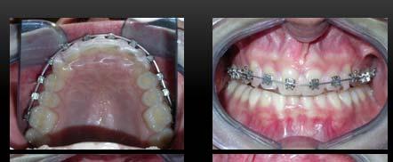
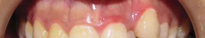
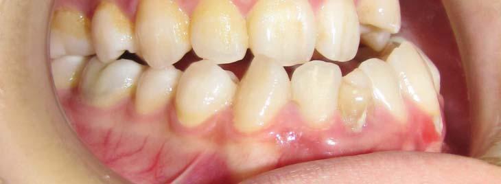
3 TMJ: Severe Postural Imbalance+Severe Myofascial Pain Syndrome, severe soreness Temporalis Tendon RL, Sternocleidomastoideus RL Age:39 years 2 months Passive Aligner + Corticision First + Incognito 6 Months therapy


4 VTO : Incognito set-up models Virtual Corticision


5 VTO : Incognito set-up modes Virtual Corticision


6


7


8


9


10 1 Month after Piezosurgery+Incognito


11 1 Month after Corticision+Incognito


12 1 Month after Corticision First+Incognito


13 1 Month after Corticision First+Incognito Bone reconstruction starts inside lateral cuts
14


15 Asiatic Homo Erectus Caucasic Homo Erectus IN ASIA Hominids had a flatter and larger maxilla related to Caucasians Sangiran 17, "Pithecanthropus VIII", Homo erectus Discovered by Sastrohamidjojo Sartono in 1969 at Sangiran on Java. This consists of a fairly complete cranium, with a brain size of about 1000 cc. It is the most complete erectus fossil from Java. This skull is very robust, with a slightly projecting face and huge flaring cheekbones. It has been thought to be about 800, years old.


16 ASIAN HOMINIDS HAD A FLATTER AND LARGER MAXILLA ANCHE SE GLI OMINIDI DIFFERISCONO O TRA LE VARIE AREE Sangiran 17, "Pithecanthropus VIII", Homo erectus Discovered by Sastrohamidjojo Sartono in 1969 at Sangiran on Java. This consists of a fairly complete cranium, with a brain size of about 1000 cc. It is the most complete erectus fossil from Java. This skull is very robust, with a slightly projecting face and huge flaring cheekbones. It has been thought to be about 800, years old.
17 Chirurgia eseguita dal Prof. G. Iannetti e Dr. Mario Pagnoni Age:26 years presurgical ortho Months straight-wire 12 Months retention P i Aligners+ Passive Ali T Tongue / i exercises /spine i TMJ: Severe occipital pain, Retrofaringeal Muscles, Lower Trapezius


18 Chirurgia eseguita dal Prof. G. Iannetti e Dr. Mario Pagnoni
19 Chirurgia eseguita dal Prof. G. Iannetti e Dr. Mario Pagnoni


20 Chirurgia eseguita dal Prof. G. Iannetti e Dr. Mario Pagnoni


21 Chirurgia eseguita dal Prof. G. Iannetti e Dr. Mario Pagnoni


22 MAXILLA BODILY 1 MM. UP anterior DOWN 7 MM. BSSO 10 MM.. Chirurgia eseguita dal Prof. G. Iannetti e Dr. Mario Pagnoni


23


24

25 Apert syndrome Crouzon syndrome Mutation of the FGFR2 gene (10q25-q26) Craniosynostosis of the coronal suture. Birth prevalence of about 1 per 65,000 live births Turricephaly Exophthalmos Skin or bone Syndactyly, may be partial or total Maxillary hypoplasia Possible mental retardation Mutation of the FGFR2 gene (10q25- q26) impairment of the bone with endochondral ossification. Turricephaly or oxycephaly with abnormal lbulging of the bregma maxillary hypoplasia exophthalmos
26


27 UPPER THIRD FACE SURGICAL ADVANCEMENT LE FORT III Subperiosteal undermining allows exposure of the fronto-nasal and fronto-malar sutures The osteotomy line is then performed between these sutures, along the lateral wall of the orbit, reaching the inferior orbital fissure. The osteotomy line continues along the medial orbital wall behind the naso-lacrymal canal. The zygomatic body and arch are also interrupted medially or laterally, depending upon the preoperative planning. The osteotomy is then completed with the pterigo-maxillary disjunction. The mobilization of the maxillo-facial skeleton is achieved with the use of the Rowe disimpaction forceps. Thanks to prof. G. Iannetti for the surgical part of the study


28 Crouzon and Apert cases Surgery performed from Prof. G. Iannetti, Director Department of Maxillofacial Surgery La Sapienza University Rome ITALY The original technique was characterized by a one-stage acute midface advancement, but it presented a limiting factor determined by the muscular and soft-tissue resistance. In order to overcome these limits, recently, a midface advancement with distraction ti osteogenesis has been proposed. Thanks to prof. G. Iannetti for the surgical part of the study


29 The Rigid External Distractor (RED) is applied. The halo-type external fixation device of the RED is secured to the calvaria and connected, through anchored-bars, with plates at the inferior orbital rim and at the pyramidal apophysis of the upper maxilla, bilaterally. Traction is initiated iti t at a rate of 0.5 mm twice a day to achieve the desired advancement in the sagittal and vertical plane. After the distraction process is completed, a 2-3 months consolidation phase is required. After advancing the midface for at least 20 mm the occlusion was corrected from class III in class II with overcorrection in all patients Thanks to prof. G. Iannetti for the surgical part of the study
30 INCLUSION CRITERIA 12 subjects suffering from Apert and Crouzon syndrome were evaluated in the sample, 6 subjects suffering from Crouzon Syndrome and 6 from Apert Syndrome. Age was in a range from 5-9 y. old. In the sample there were 5 females and 7 males Thus, to include the patients t in this study we utilized some cephalometrical and clinical impair index, as gravity index; after this selection only 4 patients are eligible for the study. Thanks to prof. G. Iannetti for the surgical part of the study
31 ALL SUBJECTS INCLUDED SHOWED: Frequent episodes of obstruptive sleep apnea (OSAS): Characterized by recurrent complete or partial obstruction of the upper airways, during sleep, deterioration of artery blood gas and increasing inspiratory effort to provide airway permanence. Class III malocclusion due to midface retrusion (ANB angl: 0, A to N perpendicular mm, Pog to N perpendicular mm) Thanks to prof. G. Iannetti for the surgical part of the study
.")

32 EXPERIMENTAL PROTOCOL The subjects were limited to those treated only with Le Fort III midface advancement, and all operations were performed by the same operator (Prof. G. Iannetti). The pre-operative (T0) and post-operative (T1: 6 months after surgery) 3D craniofacial CT scans of the subjects were collected and retrospectively analyzed. The airway space volume and orbital volume before and after treatment were analyzed and compared; also the airway surfaces and orbital surfaces on the axial, coronal, and sagittal CT scans were calculated and compared. Informed consent had been obtained from all subjects. Thanks to prof. G. Iannetti for the surgical part of the study


33 Patient affected by Crouzon syndrome pre-treatment photo Patient affected by Crouzon syndrome post-treatment photo Thanks to prof. G. Iannetti for the surgical part of the study


34 Patient affected by Apert syndrome pre-treatment photo Patient affected by Apert syndrome post-treatment photo Thanks to prof. G. Iannetti for the surgical part of the study


35 Th k t f G Thanks to prof. G. Iannetti for the surgical part of the study
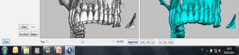
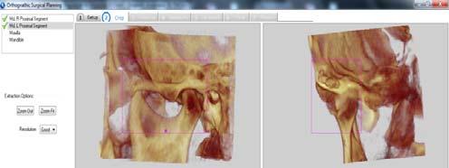
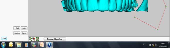
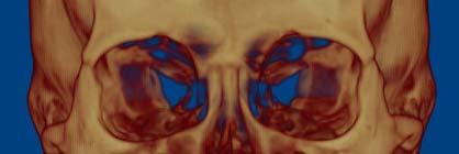
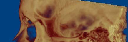
36 NEW INDIVIDUALIZED EUROPEAN TECHNOLOGIES (NIET) FOR TMJ 3D diagnostic tools 3D CBCT Craniofacial Volume Analysis UPPER AIRWAYS 3D Alfa Segmentation Analysis A= EXTERNAL B=INTERNAL CONDYLE A ORBIT A B
37 Thanks to prof. G. Iannetti for the surgical part of the study


38
39 BETA SEGMENTATION ALFA SEGMENTATION


40 BETA SEGMENTATION ALFA SEGMENTATION

 was fixed on the lateral")
.")
41 ALFA SEGMENTATION On O the sagittal plane, the 2D reference plane was located at the entry of On the axial cut, the 2D reference plane the optic nerve, most medial section (Figure 1c) was fixed on the lateral frontozygomatic On the frontal plane, the 2D suture (Figure 1a). reference area corresponded to the section of the area of the lacrimal sack.

, as well as a")


42 The increase of the orbital volume after Le Fort III advancement was statistically significant for both right and left eyes (P <0,001), as well as a statistically significant variation of the surface of the right and left eyes was observed (P <0,001). Right orbit segmentation PRE POST Axial plane Sagittal plane Coronal plane
By JOHN MARQUIS CONVERSE, M.D., and DAUBERT TELSEY, D.D.S.
 THE TRIPARTITE OSTEOTOMY OF THE MID-FACE FOR ORBITAL EXPANSION AND CORRECTION OF THE DEFORMITY IN CRANIOSTENOSIS By JOHN MARQUIS CONVERSE, M.D., and DAUBERT TELSEY, D.D.S. Center for Craniofacial Anomalies
Correction of Dentofacial Deformities (Orthognathic Surgery)
") Correction of Dentofacial Deformities (Orthognathic Surgery) BDS, MSc, German board of Oral and Maxillofacial Surgery ( Berlin-Germany), Doctoral degree by LBMS Definition Orthognathic surgery is a combination
Correction of Dentofacial Deformities (Orthognathic Surgery) BDS, MSc, German board of Oral and Maxillofacial Surgery ( Berlin-Germany), Doctoral degree by LBMS Definition Orthognathic surgery is a combination
Bones of the skull & face
 Bones of the skull & face Cranium= brain case or helmet Copyright The McGraw-Hill Companies, Inc. Permission required for reproduction or display. The cranium is composed of eight bones : frontal Occipital
Maxillofacial Injuries Practical Tips
 Saturday, October 29, 2016 Maxillofacial Injuries Practical Tips Suyash Mohan MD, PDCC THE ROOTS OF PENN RADIOLOGY RADIOLOGICAL Assistant Professor of Radiology Assistant Professor of Neurosurgery Neuroradiology
TRAUMA TO THE FACE AND MOUTH
 Dr.Yahya A. Ali 3/10/2012 F.I.C.M.S TRAUMA TO THE FACE AND MOUTH Bailey & Love s 25 th edition Injuries to the orofacial region are common, but the majority are relatively minor in nature. A few are major
AXIAL SKELETON SKULL
 AXIAL SKELETON SKULL CRANIAL BONES (8 total flat bones w/ 2 paired) 1. Frontal forms forehead & upper portion of eyesocket (orbital) 2. Parietal paired bones; form superior & lateral walls of cranium 3.
CT of Maxillofacial Injuries
 CT of Maxillofacial Injuries Stuart E. Mirvis, M.D., FACR Department of Radiology University of Maryland School of Medicine Viking 1 1976 MGS 2001 Technology changes the diagnosis Technologic Evolution
Core Curriculum Syllabus Emergencies in Otolaryngology-Head and Neck Surgery FACIAL FRACTURES
 Core Curriculum Syllabus Emergencies in Otolaryngology-Head and Neck Surgery A. General Considerations FACIAL FRACTURES Look for other fractures like skull and/or cervical spine fractures Test function
What is Craniosynostosis?
 What is Craniosynostosis? Craniosynostosis is defined as the premature closure of the cranial sutures (what some people refer to as soft spots). This results in restricted and abnormal growth of the head.
Chapter 7 Part A The Skeleton
 Chapter 7 Part A The Skeleton Why This Matters Understanding the anatomy of the skeleton enables you to anticipate problems such as pelvic dimensions that may affect labor and delivery The Skeleton The
Bones of the Skull Lateral View
 Bones of the Skull Lateral View Frontal Bone Parietal Bone Occipital Bone Temporal Bone Sphenoid Bone Pterion Sutures of the Skull Lateral View Coronal Suture Lambdoid Suture Squamous Suture Sutures of
MAXILLOFACIAL TRAUMA. The on-call maxillofacial surgeons can be contacted through the switchboard at the Southern General Hospital
 MAXILLOFACIAL TRAUMA The on-call maxillofacial surgeons can be contacted through the switchboard at the Southern General Hospital Mandibular Injuries Mechanism of injury Assault, falls, RTA-Direct trauma
Maxillary Osteotomies and Bone Grafts for. Correction of Contoural and Occlusal
 Maxillary Osteotomies and Bone Grafts for Correction of Contoural and Occlusal Deformities* M. L. LEWIN, M.D. RAVELO V. ARGAMASO, M.D. ABRAHAM I. FINGEROTH, D.D.S. Bronx, New York One of the major aims
OPERATIVE CORRECTION BY OSTEOTOMY OF RECESSED MALAR MAXILLARY COMPOUND IN A CASE OF OXYCEPHALY
 OPERATIVE CORRECTION BY OSTEOTOMY OF RECESSED MALAR MAXILLARY COMPOUND IN A CASE OF OXYCEPHALY By Sir HAROLD GILLIES, C.B.E., F.R.C.S., and STEWART H. HARRISON, F.R.C.S., L.D.S., R.C.S. From the Plastic
Structure Location Function
 Frontal Bone Cranium forms the forehead and roof of the orbits Occipital Bone Cranium forms posterior and inferior portions of the cranium Temporal Bone Cranium inferior to the parietal bone forms the
Chapter 7: Head & Neck
 Chapter 7: Head & Neck Osteology I. Overview A. Skull The cranium is composed of irregularly shaped bones that are fused together at unique joints called sutures The skull provides durable protection from
Case Report. Orthognathic Correction of Class II Open Bite. Using the Piezoelectric System and MatrixORTHOGNATHIC Plating System.
 Case Report Orthognathic Correction of Class II Open Bite. Using the Piezoelectric System and MatrixORTHOGNATHIC Plating System. Orthognathic Correction of Class II Open Bite. Using the Piezoelectric System
Thickened and thinner parts of the skull = important base for understanding of the functional structure of the skull - the transmission of masticatory
 Functional structure of the skull and Fractures of the skull Thickened and thinner parts of the skull = important base for understanding of the functional structure of the skull - the transmission of masticatory
Measuring Upper Airway Volume: Accuracy and Reliability of Dolphin 3D Software Compared to Manual Segmentation in Craniosynostosis Patients
 CRANIOMAXILLOFACIAL DEFORMITIES/COSMETIC SURGERY Measuring Upper Airway Volume: Accuracy and Reliability of Dolphin 3D Software Compared to Manual Segmentation in Craniosynostosis Patients Valerie R. de
The America Association of Oral and Maxillofacial Surgeons classify occlusion/malocclusion in to the following three categories:
 Subject: Orthognathic Surgery Policy Effective Date: 04/2016 Revision Date: 07/2018 DESCRIPTION Orthognathic surgery is an open surgical procedure that corrects anomalies or malformations of the lower
Interactions of the endocrine system, bone and oral health
 Interactions of the endocrine system, bone and oral health All bones are not equal! Dense high proportion of cortical bone High proportion of trabecular bone Mandible Functions: mastication, respiration,
APPENDICULAR SKELETON 126 AXIAL SKELETON SKELETAL SYSTEM. Cranium. Skull. Face. Skull and associated bones. Auditory ossicles. Associated bones.
 SKELETAL SYSTEM 206 AXIAL SKELETON 80 APPENDICULAR SKELETON 26 Skull Skull and associated s 29 Cranium Face Auditory ossicles 8 4 6 Associated s Hyoid Thoracic cage 25 Sternum Ribs 24 Vertebrae 24 column
CT of Maxillofacial Fracture Patterns. CT of Maxillofacial Fracture Patterns
 CT of Maxillofacial Fracture Patterns CT of Maxillofacial Fracture Patterns Stuart E. Mirvis, M.D., FACR Department of Radiology University of Maryland School of Medicine Viking 1 1976 MGS 2001 Technology
Anatomy and Physiology. Bones, Sutures, Teeth, Processes and Foramina of the Human Skull
 Anatomy and Physiology Chapter 6 DRO Bones, Sutures, Teeth, Processes and Foramina of the Human Skull Name: Period: Bones of the Human Skull Bones of the Cranium: Frontal bone: forms the forehead and the
Pre- and Postsurgical Facial Growth in Patients. with Crouzon's and Apert's Syndromes
 Pre- and Postsurgical Facial Growth in Patients with Crouzon's and Apert's Syndromes Sven KrRreiBora, D.D.S., Dr. Obpont. HowArRDp Apuss, D.D.S., M.S. Our report deals with 8 patients with Crouzon's and
Computed-Tomography of maxillofacial fractures: What do surgeons want to know?
 Computed-Tomography of maxillofacial fractures: What do surgeons want to know? Poster No.: C-0968 Congress: ECR 2016 Type: Educational Exhibit Authors: A. Ammar, M. Jrad, I. KASRAOUI, A. Zoubli, H. Mizouni
Skeletal System -Axial System. Chapter 7 Part A
 Skeletal System -Axial System Chapter 7 Part A Skeleton Learn: Names of the s. Identify specific landmarks that allow: Bones to fit into each other, Organs to fit into the cavities, Muscles to attach,
Musculoskeletal System (Part A-1) Module 7 -Chapter 10 Overview. Functions
 Module 7 -Chapter 10 Overview. Functions") Musculoskeletal System (Part A-1) Module 7 -Chapter 10 Overview Susie Turner, M.D. 1/8/13 Muscles Attachments Bones Bone types Surface features of bones Divisions of the skeletal system Joints or Articulations
Musculoskeletal System (Part A-1) Module 7 -Chapter 10 Overview Susie Turner, M.D. 1/8/13 Muscles Attachments Bones Bone types Surface features of bones Divisions of the skeletal system Joints or Articulations
Chapter 7. Skeletal System
 Chapter 7 Skeletal System 1 Skull A. The skull is made up of 22 bones: 8 cranial bones, 13 facial bones, and the mandible. B. The Cranium encloses and protects the brain, provides attachments for muscles,
 Evaluation of the maxillary morphological changes following distraction in CLP patients decrease in the Ul to NF except for Case 6. me [35] instance,血e small maxillary advancement of 2.4 mm and maxillary
ORTHOGNATHIC SURGERY
 Status Active Medical and Behavioral Health Policy Section: Surgery Policy Number: IV-16 Effective Date: 10/22/2014 Blue Cross and Blue Shield of Minnesota medical policies do not imply that members should
North Oaks Trauma Symposium Friday, November 3, 2017
 + Evaluation and Management of Facial Trauma D Antoni Dennis, MD North Oaks ENT an Allergy November 3, 2017 + Financial Disclosure I do not have any conflicts of interest or financial interest to disclose
Medical Policy Oral and Maxillofacial Surgery and Procedures
 Medical Policy Oral and Maxillofacial Surgery and Procedures Document Number: 003 Commercial and Qualified Health Plans MassHealth Authorization required X X No Prior Authorization *Cleft lip and palate
Mesial Step Class I or Class III Dependent upon extent of step seen clinically and patient s growth pattern Refer for early evaluation (by 8 years)
") Orthodontics and Dentofacial Development Overview Development of Dentition Treatment Retention and Relapse Growth of Naso-Maxillary Complex Develops postnatally entirely by intramenbranous ossification
Orthodontics and Dentofacial Development Overview Development of Dentition Treatment Retention and Relapse Growth of Naso-Maxillary Complex Develops postnatally entirely by intramenbranous ossification
Temporal region. temporal & infratemporal fossae. Zhou Hong Ying Dept. of Anatomy
 Temporal region temporal & infratemporal fossae Zhou Hong Ying Dept. of Anatomy Temporal region is divided by zygomatic arch into temporal & infratemporal fossae. Temporal Fossa Infratemporal fossa Temporal
ACTIVITY 3: AXIAL SKELETON AND LONG BONE DISSECTION COW BONE DISSECTION
 ACTIVITY 3: AXIAL SKELETON AND LONG BONE DISSECTION Objectives: 1) How to get ready: Read Chapter 7, McKinley et al., Human Anatomy, 4e. All text references are for this textbook. Learning the meanings
MAHP Orthognathic Surgery Guidelines. Medical Policy Statement. Criteria
 Introduction The word orthognathic comes from the Greek words for straighten and jaw. Orthognathic surgery is the surgical correction of abnormalities of the mandible and/or maxilla. 1 It involves the
Diagnosis of Midface Fractures with CT: What the Surgeon Needs to Know 1
 Note: This copy is for your personal non-commercial use only. To order presentation-ready copies for distribution to your colleagues or clients, contact us at www.rsna.org/rsnarights. EDUCATION EXHIBIT
ANATOMY & PHYSIOLOGY I Laboratory Version B Name Section. REVIEW SHEET Exercise 10 Axial Skeleton
 ANATOMY & PHYSIOLOGY I Laboratory Version B Name Section REVIEW SHEET Exercise 10 Axial Skeleton 1 POINT EACH. THE SKULL MULTIPLE CHOICE 1. The major components of the axial skeleton include the 7. The
Skeletal System: Skull.
 Skeletal System: Skull www.fisiokinesiterapia.biz Bones of the Skull SPLANCHNOCRANIUM Nasal (2) Maxilla (2) Lacrimal (2) Zygomatic (2) Palatine (2) Inferior concha (2) Vomer Mandible NEUROCRANIUM Frontal
Skeletal system. Prof. Abdulameer Al-Nuaimi. E. mail:
 Skeletal system Prof. Abdulameer Al-Nuaimi E-mail: a.al-nuaimi@sheffield.ac.uk E. mail: abdulameerh@yahoo.com Functions of Bone and The Skeletal System Support: The skeleton serves as the structural framework
Monoblock Craniofacial Internal Distraction in a Child with Pfeiffer Syndrome: A Case Report
 J Korean Med Sci 2008; 23: 342-6 ISSN 1011-8934 DOI: 10.3346/jkms.2008.23.2.342 Copyright The Korean Academy of Medical Sciences Monoblock Craniofacial Internal Distraction in a Child with Pfeiffer Syndrome:
Osteodistraction. Rigid External Distraction RED II System
 Osteodistraction Rigid External Distraction RED II System Rigid External Distraction RED II System Clinical example: LeFort I region Clinical example: LeFort III region 2 Introduction Distraction Osteogenesis
MAXILLOFACIAL TRAUMATOLOGY Department of Maxillofacial Surgery Semmelweis University, Budapest. Dr. Huszár Tamás
 MAXILLOFACIAL TRAUMATOLOGY Department of Maxillofacial Surgery Semmelweis University, Budapest Dr. Huszár Tamás Maxillofacial injuries isolated maxillofacial injury multiple injuries polytrauma (injury
NEUROCRANIUM VISCEROCRANIUM VISCEROCRANIUM VISCEROCRANIUM
 LECTURE 4 SKULL NEUROCRANIUM VISCEROCRANIUM VISCEROCRANIUM VISCEROCRANIUM CRANIUM NEUROCRANIUM (protective case around brain) VISCEROCRANIUM (skeleton of face) NASOMAXILLARY COMPLEX MANDIBLE (DESMOCRANIUM)
06/12/18. [Note: When orthognathic surgery is not a covered benefit, it is non-covered for any diagnosis, including sleep apnea.]
![06/12/18. [Note: When orthognathic surgery is not a covered benefit, it is non-covered for any diagnosis, including sleep apnea.]](/thumbs/84/91182117.jpg "06/12/18. [Note: When orthognathic surgery is not a covered benefit, it is non-covered for any diagnosis, including sleep apnea.]") Reference #: MC/B002 Page: 1 of 5 PRODUCT APPLICATION: PreferredOne Community Health Plan (PCHP) PreferredOne Administrative Services, Inc. (PAS) ERISA PreferredOne Administrative Services, Inc. (PAS)
Reference #: MC/B002 Page: 1 of 5 PRODUCT APPLICATION: PreferredOne Community Health Plan (PCHP) PreferredOne Administrative Services, Inc. (PAS) ERISA PreferredOne Administrative Services, Inc. (PAS)
Biology 218 Human Anatomy. Adapted from Martini Human Anatomy 7th ed. Chapter 6 The Skeletal System: Axial Division
 Adapted from Martini Human Anatomy 7th ed. Chapter 6 The Skeletal System: Axial Division Introduction The axial skeleton: Composed of bones along the central axis of the body Divided into three regions:
Case Study. Micrognathia Secondary to Pierre Robin Sequence. Treated with distraction osteogenesis using an internal mandible distractor.
 Case Study Micrognathia Secondary to Pierre Robin Sequence. Treated with distraction osteogenesis using an internal mandible distractor. Micrognathia Secondary to Pierre Robin Sequence Patient profile
 www.oralradiologists.com CONE BEAM CT REPORT CASE ---- Case Information Referring Doctor: - Patient Name: - Scan Date: December 1, 2015 Patient DOB: - Reason for Exam: - Study Details: icat Flex, 160x160x112
Extraoral radiography Introduction: Extraoral radiographs (outside the mouth) are taken when large areas of the skull or jaw must be examined or when
 are taken when large areas of the skull or jaw must be examined or when") Extraoral radiography Introduction: Extraoral radiographs (outside the mouth) are taken when large areas of the skull or jaw must be examined or when patients are unable to open their mouths for film placement.
Extraoral radiography Introduction: Extraoral radiographs (outside the mouth) are taken when large areas of the skull or jaw must be examined or when patients are unable to open their mouths for film placement.
Technique Guide. Midface Distractor System. For the temporary stabilization and gradual lengthening of the cranial or midfacial bones.
 Technique Guide Midface Distractor System. For the temporary stabilization and gradual lengthening of the cranial or midfacial bones. Table of Contents Introduction Midface Distractor System 2 Indications
The future of health is digital
 Dated: XX/XX/XXXX Name: XXXXXXXX XXXXXXXXXXX Birth Date: XX/XX/XXXX Date of scan: XX/XX/XXXX Examination of the anatomical volume: The following structures are reviewed and evaluated for bilateral symmetry,
Exercise 10. The Axial Skeleton
 Exercise 10 The Axial Skeleton The Axial Skeleton Consists of the skeletal structures found along the midline of the body. Includes the skull, hyoid, vertebrae, ribs, sternum, and sacrum. The cartilages
SKULL AS A WHOLE + ANTERIOR CRANIAL FOSSA
 SKULL AS A WHOLE + ANTERIOR CRANIAL FOSSA LEARNING OBJECTIVES At the end of this lecture, the student should be able to know: Parts of skeleton (axial and appendicular) Parts of skull Sutures of skull
ORTHOGNATHIC SURGERY
 ORTHOGNATHIC SURGERY MEDICAL POLICY Effective Date: February 1, 2017 Review Dates: 1/93, 7/95, 10/97, 4/99, 10/00, 8/01, 12/01, 4/02, 2/03, 1/04, 1/05, 12/05, 12/06, 12/07, 12/08, 12/09, 12/10, 12/11,
The Aetiology of Malocclusion
 The Aetiology of Malocclusion Dr. John Flutter For thirty-three years I have been practicing orthodontics and dentofacial orthopaedics. I have observed abnormal muscle habits and breathing patterns that
Oral and Maxillofacial Surgeons and the seriously injured patient. Barts and The London NHS Trust
 Oral and Maxillofacial Surgeons and the seriously injured patient Barts and The London NHS Trust How do you assess this? Primary Survey A B C D E Airway & Cervical Spine Breathing & Ventilation Circulation
Post-operative stability of the maxilla treated with Le Fort I and horseshoe osteotomies in bimaxillary surgery
 European Journal of Orthodontics 24 (2002) 471 476 2002 European Orthodontic Society Post-operative stability of the maxilla treated with Le Fort I and horseshoe osteotomies in bimaxillary surgery Kiyoshi
Midface fractures; what the radiologist should know.
 Midface fractures; what the radiologist should know. Poster No.: C-1056 Congress: ECR 2013 Type: Educational Exhibit Authors: J. Garcia Villanego, E.-M. Heursen, A. Rodriguez Piñero; Cadiz/ES Keywords:
Imaging Orbit/Periorbital Injury
 Imaging Orbit/Periorbital Injury 9 th Nordic Trauma Radiology Course 2016 Stuart E. Mirvis, M.D., FACR Department of Radiology University of Maryland School of Medicine Fireworks Topics to Cover Struts
Intraoperative measurement of maxillary repositioning in a series of 30 patients with maxillomandibular vertical asymmetries
 Giancarlo Renzi, MD Andrea Carboni, MD Maurizio Perugini, MD Fellow Maxilllofacial Surgery Department University La Sapienza Rome, Italy Roberto Becelli, PhD, MDS Professor Maxillofacial Surgery Teaching
Orthognathic surgery. Dr. Mohamed Rahil. ((Maxillofacial surgeon))
)") Orthognathic surgery Dr. Mohamed Rahil ((Maxillofacial surgeon)) Tikrit dentistry college 2015 2016 Orthognathic surgery is the art and science of diagnosis, and treatment of facial disproportion. These
Orthognathic surgery Dr. Mohamed Rahil ((Maxillofacial surgeon)) Tikrit dentistry college 2015 2016 Orthognathic surgery is the art and science of diagnosis, and treatment of facial disproportion. These
SURGICAL - ORTHODONTIC TREATMENT OF CLASS II DIVISION 1 MALOCCLUSION IN AN ADULT PATIENT: A CASE REPORT
 Case Report International Journal of Dental and Health Sciences Volume 02, Issue 02 SURGICAL - ORTHODONTIC TREATMENT OF CLASS II DIVISION 1 MALOCCLUSION IN AN ADULT PATIENT: A CASE REPORT Amit Dahiya 1,Minakshi
Dr. Sami Zaqout, IUG Medical School
 The skull The skull is composed of several separate bones united at immobile joints called sutures. Exceptions? Frontal bone Occipital bone Vault Cranium Sphenoid bone Zygomatic bones Base Ethmoid bone
What is Hemifacial Microsomia? By Pravin K. Patel, MD and Bruce S. Bauer, MD Children s Memorial Hospital, Chicago, IL
 What is Hemifacial Microsomia? By Pravin K. Patel, MD and Bruce S. Bauer, MD Children s Memorial Hospital, Chicago, IL 773-880-4094 Early in the child s embryonic development the structures destined to
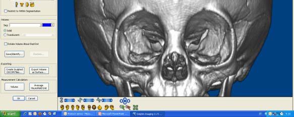
Dolphin 3D Imaging 11.7 beta
 Dolphin 3D Imaging 11.7 beta Dolphin 3D Surgery Dolphin s 3D Surgery has expanded to include even more features! This revolutionary treatment planning and presentation software module now includes a fully
Sample Case #1. Disclaimer
 ABO Sample Cases Disclaimer Sample Case #1 The following sample questions and answers were composed and vetted by a panel of experts in orthodontics and are intended to provide an example of the types
CT in the Evaluation of the Orbit and the Bony Interorbital Distance
 265 CT in the Evaluation of the Orbit and the Bony Interorbital Distance Mahmood F. Mafee' Samuel Pruzansky 2 Manuel M. Corrales' Mohan G. Phatak' Galdino E. Valvassori' Glen D Dobben' Vlastimil Capek'
Cranium Facial bones. Sternum Rib
 Figure 7.1 The human skeleton. Skull Thoracic cage (ribs and sternum) Cranium Facial bones Sternum Rib Bones of pectoral girdle Vertebral column Sacrum Vertebra Bones of pelvic girdle (a) Anterior view
Correlation between Gonial Angle and Different Variables after Bilateral Sagittal Split Ramus Osteotomy
 Original Article Correlation between Gonial Angle and Different Variables after Bilateral Sagittal Split Ramus Osteotomy M. Bayat 1,2, M. Ja'farian 3, O. Ghassemi Habashi 4 1 Assistant Professor, Department
LATERAL CEPHALOMETRIC EVALUATION IN CLEFT PALATE PATIENTS
 POLSKI PRZEGLĄD CHIRURGICZNY 2009, 81, 1, 23 27 10.2478/v10035-009-0004-2 LATERAL CEPHALOMETRIC EVALUATION IN CLEFT PALATE PATIENTS PRADEEP JAIN, ANAND AGARWAL, ARVIND SRIVASTAVA Department of Plastic
DISTRACTION PRODUCT OVERVIEW. For a wide variety of facial applications
 DISTRACTION PRODUCT OVERVIEW For a wide variety of facial applications DISTRACTION PRODUCT OVERVIEW. STRONG, MODULAR, VERSATILE CRANIOFACIAL DISTRACTION External Midface Distractor Distraction of the maxilla,
Advancement Case. Surgical-Orthodontic Treatment A Dual-Jaw. clinical orthodontics case. by Drs. Wm. Randol Womack and Reed Day
 Surgical-Orthodontic Treatment A Dual-Jaw Advancement Case by Drs. Wm. Randol Womack and Reed Day Orthodontic malocclusions involving jaw discrepancies and airway problems are best managed with a combination
Coding For Craniosynostosis. Peggy Feeley RHIA, CCS, CCS-P, COC AHIMA Approved ICD-10-CM/PCS Trainer
 Coding For Craniosynostosis Peggy Feeley RHIA, CCS, CCS-P, COC AHIMA Approved ICD-10-CM/PCS Trainer Cranial sagittal Synostosis Cranium job is to protect the brain The top portion of the skull, which protects
TRANSVERSE SECTION PLANE Scalp 2. Cranium. 13. Superior sagittal sinus
 TRANSVERSE SECTION PLANE 1 1. Scalp 2. Cranium 3. Superior sagittal sinus 4. Dura mater 5. Falx cerebri 6. Frontal lobes of the cerebrum 7. Middle meningeal artery 8. Cortex, grey matter 9. Cerebral vessels
2018 Dental Code Set For dates of service from 1/1/ /31/2018
 D0120 PERIODIC ORAL EVALUATION - ESTABLISHED PATIENT D0140 LIMITED ORAL EVALUATION - PROBLEM FOCUSED D0150 COMPREHENSIVE ORAL EVALUATION - NEW OR ESTABLISHED PATIENT D0160 DETAILED AND EXTENSIVE ORAL EVALUATION
2018 Dental Code Set
 D0120 D0140 D0150 D0160 D0180 D0210 D0220 D0230 D0240 D0250 D0251 D0270 D0272 D0273 D0274 D0277 D0290 D0310 D0330 D0340 D0350 D0393 D0470 D0502 PERIODIC ORAL EVALUATION ESTABLISHED PATIENT LIMITED ORAL
Anatomy images for MSS practical exam- 2019
 Anatomy images for MSS practical exam- 2019 Ilium Ischium Pubis Acetabulaum Iliac crest Iliac tubercle ASIS (muscle and ligament attached) AIIS (muscle attached) PSIS PIIS Ischial spine Ischial tuberosity
YOU MUST BRING YOUR OWN GLOVES FOR THIS ACTIVITY.
 ACTIVITY 3: AXIAL SKELETON AND LONG BONE DISSECTION Objectives: 1) How to get ready: Read Chapter 7, McKinley et al., Human Anatomy, 5e. All text references are for this textbook. Learning the meanings
Major Anatomic Components of the Orbit
 Major Anatomic Components of the Orbit 1. Osseous Framework 2. Globe 3. Optic nerve and sheath 4. Extraocular muscles Bony Orbit Seven Bones Frontal bone Zygomatic bone Maxillary bone Ethmoid bone Sphenoid
Craniosynostosis. Diagnosis and Treatment
 Craniosynostosis Diagnosis and Treatment 2015 For more information about the Weill Cornell Craniofacial Program ABOUT The Weill Cornell Craniofacial Program takes a multidisciplinary approach to treating
Airway and Airflow Characteristics In OSAS
 Airway and Airflow Characteristics In OSAS 16 th Annual Advances in Diagnostics and Treatment of Sleep Apnea and Snoring February 12-13, 2010 San Francisco, CA Nelson B. Powell M.D., D.D.S. Adjunct Clinical
BIO 137 AXIAL SKELETON BONE STUDY THE HUMAN SKELETON
 BIO 137 THE AXIAL SKELETON MARY CATHERINE FLATH, Ph.D. THE HUMAN SKELETON AXIAL SKULL HYOID THORACIC CAGE VERTEBRAL COLUMN APPENDICULAR PECTORAL GIRDLE UPPER LIMBS PELVIC GIRDLE LOWER LIMBS AXIAL SKELETON
Non-Discrimination Statement and Multi-Language Interpreter Services information are located at the end of this document.
 ORTHOGNATHIC SURGERY Non-Discrimination Statement and Multi-Language Interpreter Services information are located at the end of this document. Coverage for services, procedures, medical devices and drugs
DLT 111 DENTAL ANATOMY/PHYSIOLOGY
 DLT 111 DENTAL ANATOMY/PHYSIOLOGY COURSE DESCRIPTION: Prerequisites: Corequisites: Enrollment in the Dental Laboratory Technology program None This course introduces the anatomy of the individual tooth
Jaw relation (Maxillomandibular relationship): any one of the infinite
: any one of the infinite") Maxillo-mandibular Relationship Jaw relation (Maxillomandibular relationship): any one of the infinite spatial relationships of the mandible to the maxilla. Jaw relation record: It is a registration of
Maxillo-mandibular Relationship Jaw relation (Maxillomandibular relationship): any one of the infinite spatial relationships of the mandible to the maxilla. Jaw relation record: It is a registration of
Introduction to Local Anesthesia and Review of Anatomy
 5-Sep Introduction and Anatomy Review 12-Sep Neurophysiology and Pain 19-Sep Physiology and Pharmacology part 1 26-Sep Physiology and Pharmacology part 2 Introduction to Local Anesthesia and Review of
Stability and Relapse in Orthognathic Surgery
 Stability and Relapse in Orthognathic Surgery Neeraj Panchal, DDS, MD, MA Christine Ellis, DDS, MSD Paul Tiwana, DDS, MD, MS, FACS INTRODUCTION The long-term success of orthognathic reconstructive surgery
McHenry Western Lake County EMS System Paramedic, EMT-B and PHRN Optional Continuing Education 2019 #1 Facial Trauma
 McHenry Western Lake County EMS System Paramedic, EMT-B and PHRN Optional Continuing Education 2019 #1 Facial Trauma The face is vital to human appearance and function. Facial injuries can impair a patient
Dr. Esam Ahmad Z. Omar BDS, MSc-OMFS, FFDRCSI. Monitor the vital signs. Monitor the vital signs. Complications of Facial Traumas.
 Complications of Facial Traumas 1) Immediate Complications 2) Late Complications Dr. Esam Ahmad Z. Omar BDS, MSc-OMFS, FFDRCSI Assistant Professor Oral & Maxillofacial Surgeon Taibah University Monitor
Current concepts in midface fracture management
 REVIEW C URRENT OPINION Current concepts in midface fracture management AQ1 Alf L. Nastri and Ben Gurney AQ4 Purpose of review Management of midface trauma is complex and challenging and requires a clear
Postnatal Growth. The study of growth in growing children is for two reasons : -For health and nutrition assessment
 Growth of The Soft Tissues Postnatal Growth Postnatal growth is defined as the first 20 years of growth after birth krogman 1972 The study of growth in growing children is for two reasons : -For health
Anatomy and physiology of Temporomandibular Joint
 Anatomy and physiology of Temporomandibular Joint Temporomandibular joint (TMJ): It is the articulation of the condyle of the mandible, and the inter-articular disc; with the mandibular fossa (glenoid
Skull basic structures. Neurocranium
 Assoc. Prof. Květuše Lovásová, M.V.D., PhD. Skull basic structures Skull consists of two groups of bones: neurocranium (bones forming the brain box) splanchnocranium (bones forming the facial skeleton)
2016 Dental Code Set For dates of service from 1/1/16-12/31/16
 HCPCS DESCRIPTIONS D0120 D0140 D0150 D0160 D0180 D0210 D0220 D0230 D0240 D0250 D0260 D0270 D0272 D0273 D0274 D0277 D0290 D0310 D0330 D0340 D0350 D0470 D0502 D1110 D1206 D1208 D1352 D2140 D2150 D2160 D2161
Anatomy Made Easy MSS
 Anatomy Made Easy MSS part #1 هذا الملف يشمل تفريغ المحاضرة الثانية لعون بدءا من الصفحة 11 وحتى األخير Done By :MohamedA. Diabat Edited by Awn Academic team The Axial Skeleton The axial skeleton consist
Lab Exercise #04 The Skeletal System Student Performance Objectives
 Lab Exercise #04 The Skeletal System Student Performance Objectives The material that you are required to learn in this exercise can be found in either the lecture text or the supplemental materials provided
Facial and Temporal Bone Trauma Diagnostic imaging and therapeutic challenges in emergency
 Facial and Temporal Bone Trauma Diagnostic imaging and therapeutic challenges in emergency ATTYE A, KRAINIK A Department of Neuroradiology and MRI University Hospital Grenoble / University Grenoble Alpes
Case Report: Long-Term Outcome of Class II Division 1 Malocclusion Treated with Rapid Palatal Expansion and Cervical Traction
 Case Report Case Report: Long-Term Outcome of Class II Division 1 Malocclusion Treated with Rapid Palatal Expansion and Cervical Traction Roberto M. A. Lima, DDS a ; Anna Leticia Lima, DDS b Abstract: