Interactions of the endocrine system, bone and oral health
|
|
|
- Violet Rose
- 5 years ago
- Views:
Transcription
1 Interactions of the endocrine system, bone and oral health

2 All bones are not equal! Dense high proportion of cortical bone High proportion of trabecular bone

3 Mandible Functions: mastication, respiration, speech
4 Formation and Development of the Mandible
5 Bones are formed through two distinct processes Intramembranous Ossification Endochondral Ossification
6 Endochondral Bone Formation (Endochondral Ossification) Mouse embryo, E14.5 Mouse embryo, E18.0 First, cartilage primordia are formed (blue). Then, osteoblasts replace cartilage (red).
 Intramembraneous ossification")
7 Meckel s cartilage: 1 st pharyngeal arch derivative Close positional relationship to mandible Does not become the mandible Malleus Incus Sphenomandibular ligament Formation of Mandible: Dependent on Meckel s cartilage Does not derive from Meckel s cartilage Derives from mesenchymal condensations which form along the lateral aspects of Meckel s cartilage (week 6) Intramembraneous ossification (week 7) spreads rapidly along the lateral aspect of Meckel s cartilage both anteriorly and posteriorly


8 Meckel s Cartilage & the Mandible: Mandibular branch of trigeminal nerve develops together with Meckel s cartilage Ramus of the mandible develops by ossification posteriorly into the 1st arch, turning away from Meckel s cartilage Points of divergence marked by the lingula

9 Mandibular Development: Subsequent mandibular growth until birth is influenced strongly by appearance of three secondary cartilages (coronoid, condylar and symphyseal) and the development of muscular attachments. Secondary cartilages undergo endochondral ossification Condylar cartilage is most significant contributor to mandibular growth coronoid process condylar process symphysis
10 Mandibular Formation Forms by what type of ossification process???
11 Mandibular formation occurs by both endochondral and intramembraneous ossification. Intramembraneous: body, ramus Endochondral: condyle, coronoid, symphysis
12 Directions of Mandibular Growth Predominant growth vectors are posterior and superior Grows directly toward its articular contact in glenoid fossa (also a good example of displacement )
13 What happens if the mandible does not grow adequately?
 Cleft palate (lack of fusion of palatal shelves) Upper airway obstruction Etiology unknown Jamshidi N et al.")
14 Infant with evident micrognathia due to Pierre Robin Sequence Micrognathia (small mandible) = primary defect Glossoptosis (posterior displacement or retraction of the tongue) Cleft palate (lack of fusion of palatal shelves) Upper airway obstruction Etiology unknown Jamshidi N et al. J Med Genet 2004;41:e1-e1


15 Bones are formed through two distinct processes Intramembranous Ossification Endochondral Ossification
16 Endochondral Bone Formation (Endochondral Ossification) Mouse embryo, E14.5 Mouse embryo, E18.0 First, cartilage primordia are formed (blue). Then, osteoblasts replace cartilage (red).

 (Mishina,")
17 Sizes and shapes of each bone in skull / face determine the shape. (Sadler, 2012) (Mishina, 2014) Human Mouse

18
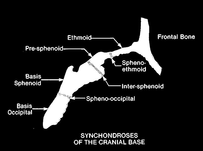
19 Cranial Base Synchondroses
 Mouse embryo, E14.")

20 Endochondral Bone Formation (Endochondral Ossification) Mouse embryo, E14.5 Mouse embryo, E18.0 First, cartilage primordia are formed (blue). Then, osteoblasts replace cartilage (red).

21 Sutural Bone Growth Bone deposition along osteogenic front of growing facial or cranial bone Suture contains precursor cells that differentiate to form osteoblasts along the edge of the growing cranial bone Some sutures normally fuse and some remain patent osteogenic front suture bone Fundamentals of Craniofacial Growth. Dixon, Hoyte and Ronning (1997)


22 Craniofacial Suture Function 1. Bone growth 2. Provide firm union between bony segments 3. Allow for mechanical movement between bony segments for response to mechanical stress
 and bone remodeling Facial growth requires freedom of movement of facial")
23 Facial Bone Sutures zygomatico-frontal suture zygomatico-maxillary suture midpalatal suture Midfacial growth occurs by bone deposition at bony sutures and by periosteal apposition (bone deposition) and bone remodeling Facial growth requires freedom of movement of facial sutures


24 Disorders of the skull vault in human Craniosynostosis caused by premature fusion of sutures Parietal foramina Wilkie AO & Morriss-Kay GM, Nature reviews, Genetics, 2001
 osteoprogenitor (proliferation & differentiation)")
25 Sizes and shapes of each bone in skull / face determine the shape. Intramembranous Bone Formation time mesenchymal condensation (proliferation) osteoprogenitor (proliferation & differentiation) osteoblast (osteogenesis) osteocyte (osteogenic maturation) apoptosis

 Abnormal ossification: Suture")
26 Craniosynostosis? Suture Mutations in critical genes (FGF etc.) Abnormal ossification: Suture fused Facial deformity Delays of brain growth
27 Craniosynostosis Clinical condition of premature cranial suture fusion Results in abnormal craniofacial shape, increased intracranial pressure Team treatment commonly includes surgery, genetic counseling, dentistry, orthodontics, medical and social support. High incidence (1/2500 live births) Occurs sporadically or as part of a genetic syndrome Severity depends upon timing and number of fused sutures Etiology: genetic, environmental, multifactorial


 Lambdoid suture")
28 Clinical symptoms of Craniosynostosis are viable. Sagittal synostosis/ scaphocephaly Metopic synostosis/ trigonocephaly Coronal synostosis/ lambdoid synostosis (Plagiocephaly) Lambdoid suture Sagittal suture Coronal sutures Metopic suture
 Sagittal suture Axin1 and Fgfr1 Compound LOF Sagittal suture BMP2 enhancer SNP (GOF?")
29 Causative genes for Craniosynostosis Gene mutation suture FGF receptors GOF Coronal suture MSX2 GOF Coronal suture TWIST1 Het Coronal suture TCF12 LOF Coronal suture BBS9 SNP (GOF?) Sagittal suture Axin1 and Fgfr1 Compound LOF Sagittal suture BMP2 enhancer SNP (GOF?) Sagittal suture ERF LOF All sutures Bmpr1a GOF Metopic Lambdoid suture Sagittal suture Coronal sutures Metopic suture
30 Syndromic Craniosynostosis: Human malformation syndromes Abnormal development of the craniofacial skeleton Craniosynostosis Facial skeleton abnormalities Digit abnormalities Associated with genetic mutations
 Msx2 (a transcription factor) Twist (a transcription factor) Autosomal Dominant Full Penetrance Variable")
31 Genetic Basis of Syndromic Craniosynostosis: Apert, Crouzon, Pfeiffer, Jackson-Weiss: FGFR s Boston-Type: Msx2 Saethre-Chotzen: Twist FGFR s (Fibroblast Growth Factor Receptor) Msx2 (a transcription factor) Twist (a transcription factor) Autosomal Dominant Full Penetrance Variable Expression
32 Craniosynostosis Associated FGFR Mutations

33 FGFR Structure extracellular intracellular Ig1 Ig2 Ig3 TM TK1 TK2 FGF binding Ig = immunoglobulin like domain TM = transmembrane domain TK = tyrosine kinase domain, allows for receptor signaling
34 Molecular Genetics of Apert Syndrome Molecular Genetics of Crouzon Syndrome Mutation: Activating mutation in FGFR2 gene Mutation is in area of FGF binding site Two mutations only Molecular phenotype: Enhanced FGFR to ligand binding FGFR2 binding to wrong ligands Mutations can arise spontaneously Risk of mutation increases with paternal age Mutation: Activating mutation in FGFR2 gene Mutation is in area of FGF binding site Two common mutations with numerous additional less common mutations. Molecular phenotype: Ligand independent FGFR signaling Mutations can arise spontaneously Risk of mutation increases with paternal age
35 FGFR2 Mutations extracellular intracellular Ig1 Ig2 Ig3 SP TM TK1 TK2 FGF binding Apert mutations Crouzon common mutations

")

36 Apert Syndrome: Full penetrance, variable expression Prenatal Coronal Synostosis Increased intracranial pressure NL to diminished IQ Brain abnormalities (defects in septum pellucidum) Proptosis, hypertelorism Midface hypoplasia (facial sutures) Class III malocclusion Progressive cervical spine fusions Tracheal abnormalities Digit abnormalities: syndactyly Higher risk with higher paternal age
 NL intelligence")

37 Crouzon Syndrome: Full penetrance, variable expression Coronal Synostosis, rare pansynostosis Increased intracranial pressure Brain abnormalities (defects in corpus callosum) NL intelligence Proptosis, hypertelorism Midface hypoplasia (facial sutures) Class III malocclusion Vertebral fusions Stylohyoid ligament calcification Tracheal abnormalities Digit abnormalities: None Higher risk with higher paternal age
38 FGF signals have distinct roles in the developing skull vault Missense mutations of FGF receptors (Gain-of-function mutations) lead to craniosynostosis. (Approx. 30 % of cases) Enhancement of endogenous FGF signaling pathway extracellular signal-regulated kinases 1 and 2 (ERK1/2) is observed. Morriss-Kay GM & Wilkie AO, J. Anat. Review 2005
39 FGFR Function: Cellular competence for responsiveness to FGF s Cell to cell signaling tissue boundaries, induction of cell behavior changes


 are fused in the mutant.")
40 Craniosynostosis can be treated by inhibition of FGF signaling. Gain-of-function mutation for FGFR2 U0126 is a chemical inhibitor for ERK1/2 (signaling molecules). Coronal suture (arrows) are fused in the mutant. Inhibition of FGF signaling by U0126 rescues premature fusion of the suture.


41 How can we target growth factor signaling for therapeutic use? Cell A Ligands Receptors Cell B Multiple ligands interact with multiple receptors. They share common functions, but they also have unique functions. Growth factor signaling interact with each other. Tissues interact with each other. Specificity! Signaling molecules Gene Expression
42
43
44 FGFR splice variants FGFR2IIIb (yellow) Expressed in epithelial lineages (dural cells) Specific for ligands that are expressed by mesenchymal lineage cells FGFR2IIIc (green) Expressed in mesenchymal lineages (osteoblastic cells) Specific for ligands that are expressed by epithelial lineage cells

45 Prenatal Maxillary Bone Growth Lingual surface: bone deposition Nasal surface: initially bone deposition, then bone resorption nasal sinus Infraorbital surface: bone deposition and remodeling growing eyeball Exterior anterior surface: bone deposition increase in arch length Exterior posterior surface: bone deposition increase in arch length Increase in arch length allows for development and enlargement of tooth buds Zygomatic (malar) secondary cartilage in developing zygomatic process also contributes to maxillary growth
Lecture 3: Skeletogenesis and diseases
 Jilin University School of Stomatology Skeletogenesis Lecture 3: Skeletogenesis and diseases Aug. 21, 2015 Yuji Mishina, Ph.D. mishina@umich.edu Bone Development Mouse embryo, E14.5 Mouse embryo, E18.0
Lecture 2: Skeletogenesis
 Jilin University School of Stomatology Skeletogenesis Lecture 2: Skeletogenesis Aug. 18, 2015 Yuji Mishina, Ph.D. mishina@umich.edu Student will describe Development of Bone - the general anatomy of bone
What is Craniosynostosis?
 What is Craniosynostosis? Craniosynostosis is defined as the premature closure of the cranial sutures (what some people refer to as soft spots). This results in restricted and abnormal growth of the head.
Essentials in Head and Neck Embryology. Part 3 Development of the head, face, and oral cavity
 Essentials in Head and Neck Embryology Part 3 Development of the head, face, and oral cavity Outline General overview of prenatal development Embryonic period phase 1 Formation of bilaminar disk Formation
DEFINATION Growth was concieved by an anatomist, born to a biologist, delivered by a physician, left on a chemist doorstep, and adopted by a physiolog
 DEFINATION Growth was concieved by an anatomist, born to a biologist, delivered by a physician, left on a chemist doorstep, and adopted by a physiologist.at an early age- she eloped with a statistician,
NEUROCRANIUM VISCEROCRANIUM VISCEROCRANIUM VISCEROCRANIUM
 LECTURE 4 SKULL NEUROCRANIUM VISCEROCRANIUM VISCEROCRANIUM VISCEROCRANIUM CRANIUM NEUROCRANIUM (protective case around brain) VISCEROCRANIUM (skeleton of face) NASOMAXILLARY COMPLEX MANDIBLE (DESMOCRANIUM)
Skeletal System: Skull.
 Skeletal System: Skull www.fisiokinesiterapia.biz Bones of the Skull SPLANCHNOCRANIUM Nasal (2) Maxilla (2) Lacrimal (2) Zygomatic (2) Palatine (2) Inferior concha (2) Vomer Mandible NEUROCRANIUM Frontal
Skeletal System. Prof. Dr. Malak A. Al-yawer Department of Anatomy/Embryology Section
 Skeletal System Prof. Dr. Malak A. Al-yawer Department of Anatomy/Embryology Section Learning objectives At the end of this lecture, the medical student will be able to: State the embryonic origin of skeletal
04 Development of the Face and Neck. Development of the Face Development of the neck
 04 Development of the Face and Neck Development of the Face Development of the neck Development of the face Overview of facial development The fourth week ~ the twelfth week of prenatal development Between
Anatomy and Physiology. Bones, Sutures, Teeth, Processes and Foramina of the Human Skull
 Anatomy and Physiology Chapter 6 DRO Bones, Sutures, Teeth, Processes and Foramina of the Human Skull Name: Period: Bones of the Human Skull Bones of the Cranium: Frontal bone: forms the forehead and the
Cranium Facial bones. Sternum Rib
 Figure 7.1 The human skeleton. Skull Thoracic cage (ribs and sternum) Cranium Facial bones Sternum Rib Bones of pectoral girdle Vertebral column Sacrum Vertebra Bones of pelvic girdle (a) Anterior view
Chapter 7: Head & Neck
 Chapter 7: Head & Neck Osteology I. Overview A. Skull The cranium is composed of irregularly shaped bones that are fused together at unique joints called sutures The skull provides durable protection from
Pharyngeal Apparatus. Pouches Endoderm Grooves Ectoderm Arch Neural Crest Somitomeres Aortic Arch - Vessel
 Pharyngeal Apparatus Pouches Endoderm Grooves Ectoderm Arch Neural Crest Somitomeres Aortic Arch - Vessel Segmental Organization Humans: Arch 1-4 prominent Arch 5 absent Arch 6 - transient First Arch Face
Chapter 7. Skeletal System
 Chapter 7 Skeletal System 1 Skull A. The skull is made up of 22 bones: 8 cranial bones, 13 facial bones, and the mandible. B. The Cranium encloses and protects the brain, provides attachments for muscles,
Infratemporal fossa: Tikrit University college of Dentistry Dr.Ban I.S. head & neck Anatomy 2 nd y.
 Infratemporal fossa: This is a space lying beneath the base of the skull between the lateral wall of the pharynx and the ramus of the mandible. It is also referred to as the parapharyngeal or lateral pharyngeal
Chapter 7 Part A The Skeleton
 Chapter 7 Part A The Skeleton Why This Matters Understanding the anatomy of the skeleton enables you to anticipate problems such as pelvic dimensions that may affect labor and delivery The Skeleton The
Skeletal System -Axial System. Chapter 7 Part A
 Skeletal System -Axial System Chapter 7 Part A Skeleton Learn: Names of the s. Identify specific landmarks that allow: Bones to fit into each other, Organs to fit into the cavities, Muscles to attach,
Skeletal Development Multiple Cellular Origins. Intramembranous Bone. Endochondrial Bone. Cartilage template of the limb in the Chick wing
 Skeletal Development Multiple Cellular Origins 1 - Paraxial Mesoderm Somite, Sclerotome Axial Skeleton (e.g. vertebra) 2 - Lateral Plate Mesoderm Appendicular Skeleton (e.g. limb) 3 - Neural Crest Head
Craniosynostosis - making the head fit the hat
 Craniosynostosis - making the head fit the hat Poster No.: R-0173 Congress: 2014 CSM Type: Scientific Exhibit Authors: S. Constantine, B. Clark; NORTH ADELAIDE/AU Keywords: Head and neck, Bones, Pediatric,
Bones of the skull & face
 Bones of the skull & face Cranium= brain case or helmet Copyright The McGraw-Hill Companies, Inc. Permission required for reproduction or display. The cranium is composed of eight bones : frontal Occipital
Biology 218 Human Anatomy. Adapted from Martini Human Anatomy 7th ed. Chapter 6 The Skeletal System: Axial Division
 Adapted from Martini Human Anatomy 7th ed. Chapter 6 The Skeletal System: Axial Division Introduction The axial skeleton: Composed of bones along the central axis of the body Divided into three regions:
The Skull and Temporomandibular joint II Prof. Abdulameer Al-Nuaimi. E. mail:
 The Skull and Temporomandibular joint II Prof. Abdulameer Al-Nuaimi E-mail: a.al-nuaimi@sheffield.ac.uk E. mail: abdulameerh@yahoo.com Temporal fossa The temporal fossa is a depression on the temporal
DEVELOPMENTAL ANATOMY OF THE FACE, JAW AND NECK. O.M. Oluwatosin Department of Surgery
 DEVELOPMENTAL ANATOMY OF THE FACE, JAW AND NECK O.M. Oluwatosin Department of Surgery 1 2 By the end of this lecture, you should be able to: Discuss the embryology of the face Relate congenital anomalies
THE SKELETAL SYSTEM. Focus on the Skull
 THE SKELETAL SYSTEM Focus on the Skull Review Anatomical Terms Anterior/Posterior Dorsal/Ventral Medial/Lateral Superior/Inferior Bone Markings - Review Projections for attachment of muscles, ligaments
International Journal of Current Research and Academic Review ISSN: Volume 3 Number 1 (January-2015) pp
 pp") International Journal of Current Research and Academic Review ISSN: 47 Volume Number (January) pp. 66 www.ijcrar.com Clinical Profile of Patients with Craniosynostosis: A Descriptive Study Nagaraj V. Gadwal*
International Journal of Current Research and Academic Review ISSN: 47 Volume Number (January) pp. 66 www.ijcrar.com Clinical Profile of Patients with Craniosynostosis: A Descriptive Study Nagaraj V. Gadwal*
Structure Location Function
 Frontal Bone Cranium forms the forehead and roof of the orbits Occipital Bone Cranium forms posterior and inferior portions of the cranium Temporal Bone Cranium inferior to the parietal bone forms the
Pharyngeal apparatus. - At the third week, it is a 3 layered structure: ectoderm, mesoderm and endoderm. This is called trilaminar disc
 Pharyngeal apparatus Remember from the first year embryology - The embryo was disc shaped in the second week of development (this is called embryonic disc) and it is a 2 layered disc (composed of two layers)---bilaminar
4.1 Classification of Craniosynostosis: Therapeutical implications.
 ISPN course 23 rd Nov, 2015 Cranial & Craniofacial disorders 4.1 Classification of Craniosynostosis: Therapeutical implications. Kazuaki Shimoji, Masakazu Miyajima and Hajime Arai Department of Neurosurgery,
AXIAL SKELETON SKULL
 AXIAL SKELETON SKULL CRANIAL BONES (8 total flat bones w/ 2 paired) 1. Frontal forms forehead & upper portion of eyesocket (orbital) 2. Parietal paired bones; form superior & lateral walls of cranium 3.
Development of the skull
 Development of the skull Ágnes Nemeskéri 2017 Semmelweis University Budapest Department of Anatomy, Histology and Embryology Clinical Anatomy Research Laboratory nemeskeri.agnes@med.semmelweis-univ.hu
Remember from the first year embryology Trilaminar disc has 3 layers: ectoderm, mesoderm, and endoderm
 Development of face Remember from the first year embryology Trilaminar disc has 3 layers: ectoderm, mesoderm, and endoderm The ectoderm forms the neural groove, then tube The neural tube lies in the mesoderm
Cleidocranial Dysplasia
 BRIEF REPORTS Cleidocranial Dysplasia Anita Sharma Rohtash Yadav Kuldip Ahlawat Cleidocranial dysplasia (CCD), is characterized by short stature, typical facial features and variable degree of pan-skeletal
APPENDICULAR SKELETON 126 AXIAL SKELETON SKELETAL SYSTEM. Cranium. Skull. Face. Skull and associated bones. Auditory ossicles. Associated bones.
 SKELETAL SYSTEM 206 AXIAL SKELETON 80 APPENDICULAR SKELETON 26 Skull Skull and associated s 29 Cranium Face Auditory ossicles 8 4 6 Associated s Hyoid Thoracic cage 25 Sternum Ribs 24 Vertebrae 24 column
FORMATION OF BONE. Intramembranous Ossification. Bone-Lec-10-Prof.Dr.Adnan Albideri
 FORMATION OF BONE All bones are of mesodermal origin. The process of bone formation is called ossification. We have seen that formation of most bones is preceded by the formation of a cartilaginous model,
An Introduction to the Axial Skeleton. Copyright 2009 Pearson Education, Inc., publishing as Pearson Benjamin Cummings
 An Introduction to the Axial Skeleton Copyright 2009 Pearson Education, Inc., publishing as Pearson Benjamin Cummings Terms: Structures of Bones Articulations: Contacts with other bones Landmarks (Bone
SLLF FOR TMJ CASES IN ADULT DENTITION SEVERE BRACHIFA BRACHIF FACIAL
 SLLF FOR TMJ CASES IN ADULT DENTITION SEVERE BRACHIFAFACIAL TMJ: Severe Postural Imbalance+Severe Myofascial Pain Syndrome, severe soreness Temporalis Tendon RL, Sternocleidomastoideus RL Age:39 years
Chapter 7: Skeletal System: Gross Anatomy
 Chapter 7: Skeletal System: Gross Anatomy I. General Considerations A. How many bones in an average adult skeleton? B. Anatomic features of bones are based on II. Axial Skeleton A. Skull 1. Functionally
pressure 6-7 and cranial growth restriction.
 Josephine Jung graduated from Medical School in Berlin in 2014. After completing her M.D. Thesis in Berlin she moved to London and is working as a Clinical Research Fellow in Neurosurgery at King s College
Headlines Craniofacial Support
 Other leaflets available from Headlines Craniofacial Support Please contact Group Administrator Justine Tweedie - info@headlines.org.uk for details of how to obtain copies 1 What causes Craniosynostosis?
ANATOMY & PHYSIOLOGY I Laboratory Version B Name Section. REVIEW SHEET Exercise 10 Axial Skeleton
 ANATOMY & PHYSIOLOGY I Laboratory Version B Name Section REVIEW SHEET Exercise 10 Axial Skeleton 1 POINT EACH. THE SKULL MULTIPLE CHOICE 1. The major components of the axial skeleton include the 7. The
ACTIVITY 3: AXIAL SKELETON AND LONG BONE DISSECTION COW BONE DISSECTION
 ACTIVITY 3: AXIAL SKELETON AND LONG BONE DISSECTION Objectives: 1) How to get ready: Read Chapter 7, McKinley et al., Human Anatomy, 4e. All text references are for this textbook. Learning the meanings
Temporal region. temporal & infratemporal fossae. Zhou Hong Ying Dept. of Anatomy
 Temporal region temporal & infratemporal fossae Zhou Hong Ying Dept. of Anatomy Temporal region is divided by zygomatic arch into temporal & infratemporal fossae. Temporal Fossa Infratemporal fossa Temporal
Parotid Gland, Temporomandibular Joint and Infratemporal Fossa
 M1 - Anatomy Parotid Gland, Temporomandibular Joint and Infratemporal Fossa Jeff Dupree Sanger 9-057 jldupree@vcu.edu Parotid gland: wraps around the mandible positioned between the mandible and the sphenoid
BONE CHALLENGE DANIL HAMMOUDI.MD
 BONE CHALLENGE DANIL HAMMOUDI.MD Bone Basic functions? A. support B. protection C. movement assistance in D. RBC formation-hemopoiesis E. mineral homeostasis +importance of calcium F. energy supply -yellow
Subject Index. AXIN2, cleft defects 24, 26
 Subject Index ADAMTS, mouse mutants and palate development 37, 38 Africa, cleft lip and palate prevalence 6, 7 Alcohol dependence, pregnancy risks for cleft 25, 61 Altitude, pregnancy risks for cleft 25,
GENETIC ALTERATIONS IN CRANIOSYNOSTOSIS, GENOTYPE- PHENOTYPE CORRELATIONS. Beáta Bessenyei. Supervisor: Prof. Dr. Éva Oláh
 SHORT THESIS FOR THE DEGREE OF DOCTOR OF PHILOSOPHY (PhD) GENETIC ALTERATIONS IN CRANIOSYNOSTOSIS, GENOTYPE- PHENOTYPE CORRELATIONS by Beáta Bessenyei Supervisor: Prof. Dr. Éva Oláh UNIVERSITY OF DEBRECEN
Sheets 16&17. Dr. Heba Kalbouneh. Dr. Heba Kalbouneh. Dr. Heba Kalbouneh
 Sheets 16&17 Dr. Heba Kalbouneh Dr. Heba Kalbouneh Dr. Heba Kalbouneh Ossification (formation of bone) - Osteoblasts are responsible for producing the extracellular matrix of the bone and these osteoblasts
Osteology. Dr. Carmen E. Rexach Anatomy 35 Mt San Antonio College
 Osteology Dr. Carmen E. Rexach Anatomy 35 Mt San Antonio College Functions of the Skeletal System: Support Movement Protection Hemopoiesis Electrolyte balance (Ca ++ /PO -3 4 ) Acid-base balance Storage
IJCMR 433. CASE REPORT Crouzons Syndrome With Multiple Impacted Permanent Teeth - A Case Report. Saraswathi Gopal.K 1, Madhubala.
 IJCMR 433 CASE REPORT With Multiple Impacted Permanent Teeth - A Case Report Saraswathi Gopal.K 1, Madhubala.E 2 Source of Support: Nil ABSTRACT Introduction: Crouzon syndrome (CS) is a rare genetic disorder
Skull basic structures. Neurocranium
 Assoc. Prof. Květuše Lovásová, M.V.D., PhD. Skull basic structures Skull consists of two groups of bones: neurocranium (bones forming the brain box) splanchnocranium (bones forming the facial skeleton)
TRAUMA TO THE FACE AND MOUTH
 Dr.Yahya A. Ali 3/10/2012 F.I.C.M.S TRAUMA TO THE FACE AND MOUTH Bailey & Love s 25 th edition Injuries to the orofacial region are common, but the majority are relatively minor in nature. A few are major
Human Anatomy and Physiology - Problem Drill 07: The Skeletal System Axial Skeleton
 Human Anatomy and Physiology - Problem Drill 07: The Skeletal System Axial Skeleton Question No. 1 of 10 Which of the following statements about the axial skeleton is correct? Question #01 A. The axial
CT of Maxillofacial Fracture Patterns. CT of Maxillofacial Fracture Patterns
 CT of Maxillofacial Fracture Patterns CT of Maxillofacial Fracture Patterns Stuart E. Mirvis, M.D., FACR Department of Radiology University of Maryland School of Medicine Viking 1 1976 MGS 2001 Technology
In Vitro Assessment of Osteoblast Behavior in. Craniosynostosis
 In Vitro Assessment of Osteoblast Behavior in Craniosynostosis by Tatiana Karine Simon Cypel A thesis submitted in conformity with the requirements for the degree of Master of Science The Institute of
 Drawings illustrating the human pharyngeal apparatus. Drawings illustrating the human pharyngeal apparatus. Drawings illustrating the human pharyngeal apparatus. Drawings illustrating the human pharyngeal
Pre- and Postsurgical Facial Growth in Patients. with Crouzon's and Apert's Syndromes
 Pre- and Postsurgical Facial Growth in Patients with Crouzon's and Apert's Syndromes Sven KrRreiBora, D.D.S., Dr. Obpont. HowArRDp Apuss, D.D.S., M.S. Our report deals with 8 patients with Crouzon's and
YOU MUST BRING YOUR OWN GLOVES FOR THIS ACTIVITY.
 ACTIVITY 3: AXIAL SKELETON AND LONG BONE DISSECTION Objectives: 1) How to get ready: Read Chapter 7, McKinley et al., Human Anatomy, 5e. All text references are for this textbook. Learning the meanings
Bone. Development. Tim Arnett. University College London. Department of Anatomy and Developmental Biology
 Bone Development Tim Arnett Department of Anatomy and Developmental Biology University College London Bone development Outline Bone composition matrix + mineral Bone formation - intramembranous & endochondral
Anatomy and Physiology II. Review Spine and Neck
 Anatomy and Physiology II Review Spine and Neck Spine regions How many cervical vertibrae are there? 7 The curvature is the cervical region posterior? Concave posterior How many thoracic? And curvature?
Maxillofacial Injuries Practical Tips
 Saturday, October 29, 2016 Maxillofacial Injuries Practical Tips Suyash Mohan MD, PDCC THE ROOTS OF PENN RADIOLOGY RADIOLOGICAL Assistant Professor of Radiology Assistant Professor of Neurosurgery Neuroradiology
Lab Exercise #04 The Skeletal System Student Performance Objectives
 Lab Exercise #04 The Skeletal System Student Performance Objectives The material that you are required to learn in this exercise can be found in either the lecture text or the supplemental materials provided
Temporomandibular Joint. Dr Noman ullah wazir
 Temporomandibular Joint Dr Noman ullah wazir Type of Joint TMJ is a Synovial joint between : The condylar head of the mandible. The mandibular fossa of squamous part of temporal bone. The joint cavity
Craniosynostosis. Diagnosis and Treatment
 Craniosynostosis Diagnosis and Treatment 2015 For more information about the Weill Cornell Craniofacial Program ABOUT The Weill Cornell Craniofacial Program takes a multidisciplinary approach to treating
By JOHN MARQUIS CONVERSE, M.D., and DAUBERT TELSEY, D.D.S.
 THE TRIPARTITE OSTEOTOMY OF THE MID-FACE FOR ORBITAL EXPANSION AND CORRECTION OF THE DEFORMITY IN CRANIOSTENOSIS By JOHN MARQUIS CONVERSE, M.D., and DAUBERT TELSEY, D.D.S. Center for Craniofacial Anomalies
The Skull DANIL HAMMOUDI.MD
 The Skull DANIL HAMMOUDI.MD summary of bones/structures in Chapter 15 of the manual need tp be print as soon as possible http://www.mnsu.edu/emuseum/biology/humananatomy/skeletal/skul l/frontal/frontal.html
The SKELETAL System. The framework of bones and cartilage which protect organs, and provides a lever system that allows locomotion.
 The SKELETAL System The framework of bones and cartilage which protect organs, and provides a lever system that allows locomotion. Functions of the Skeletal System Support Protection Movement Facilitation
Bones Ethmoid bone Inferior nasal concha Lacrimal bone Maxilla Nasal bone Palatine bone Vomer Zygomatic bone Mandible
 splanchnocranium - Consists of part of skull that is derived from branchial arches - The facial bones are the bones of the anterior and lower human skull Bones Ethmoid bone Inferior nasal concha Lacrimal
Core Curriculum Syllabus Emergencies in Otolaryngology-Head and Neck Surgery FACIAL FRACTURES
 Core Curriculum Syllabus Emergencies in Otolaryngology-Head and Neck Surgery A. General Considerations FACIAL FRACTURES Look for other fractures like skull and/or cervical spine fractures Test function
Temporal fossa Infratemporal fossa Pterygopalatine fossa Terminal branches of external carotid artery Pterygoid venous plexus
 Outline of content Temporal fossa Infratemporal fossa Pterygopalatine fossa Terminal branches of external carotid artery Pterygoid venous plexus Boundary Content Communication Mandibular division of trigeminal
Nervous & Skeletal Systems. Virtual Science University
 Nervous & Skeletal Systems Virtual Science University 1 Nervous & Skeletal Systems Texas TEK B.10(A) The student will interpret the function of systems in organisms (humans) including the nervous and skeletal
Crouzon s syndrome with adenotonsillitis: conventional surgery in altered anatomy.
 ISSN:2250-0359 Volume 3 Issue 3 2013 Crouzon s syndrome with adenotonsillitis: conventional surgery in altered anatomy. Sudhir M Naik 1, Mohan Appaji 2, Ravishankar S 3, Goutham MK 4, Annapurna SM 5, N
Skeletal system. Prof. Abdulameer Al-Nuaimi. E. mail:
 Skeletal system Prof. Abdulameer Al-Nuaimi E-mail: a.al-nuaimi@sheffield.ac.uk E. mail: abdulameerh@yahoo.com Functions of Bone and The Skeletal System Support: The skeleton serves as the structural framework
University of Palestine. Midterm Exam 2013/2014 Total Grade:
 Course No: DNTS2208 Course Title: Head and Neck Anatomy Date: 09/11/2013 No. of Questions: (50) Time: 1hour Using Calculator (No) University of Palestine Midterm Exam 2013/2014 Total Grade: Instructor
Functions of the Skeletal System
 SKELETAL SYSTEM Functions of the Skeletal System Support: Internal framework that supports and anchors all soft organs. Protection: Bones protect soft body organs Body movement skeletal muscle attached
Human Anatomy & Physiology
 PowerPoint Lecture Slides prepared by Barbara Heard, Atlantic Cape Community College Ninth Edition Human Anatomy & Physiology C H A P T E R 6 Annie Leibovitz/Contact Press Images 2013 Pearson Education,
Coding For Craniosynostosis. Peggy Feeley RHIA, CCS, CCS-P, COC AHIMA Approved ICD-10-CM/PCS Trainer
 Coding For Craniosynostosis Peggy Feeley RHIA, CCS, CCS-P, COC AHIMA Approved ICD-10-CM/PCS Trainer Cranial sagittal Synostosis Cranium job is to protect the brain The top portion of the skull, which protects
Anatomy and Physiology 1 Chapter 7 self quiz Pro, Dima Darwish,MD.
 Anatomy and Physiology 1 Chapter 7 self quiz Pro, Dima Darwish,MD. 1) How many bones make up the axial skeleton? A) 50 B) 60 C) 70 D) 80 E) 90 2) Which of the following is a function of the axial skeleton?
Department of Neurosurgery. Differentiating Craniosynostosis from Positional Plagiocephaly
 Department of Neurosurgery Differentiating Craniosynostosis from Positional Plagiocephaly The number of infants with head shape deformities has risen over the past several years, likely due to increased
in compact bone, large vertical canals carrying blood vessels and nerves. in compact bone, large horizontal canals carrying blood vessels and nerves.
 Carl Christensen, PhD Skeletal System (Bones`) Bio. 2304 Human Anatomy 1. Identify a term for each of the following: shaft of a long bone ends of a long bone ossified remnant of the "growth plate" connective
Musculoskeletal System (Part A-1) Module 7 -Chapter 10 Overview. Functions
 Module 7 -Chapter 10 Overview. Functions") Musculoskeletal System (Part A-1) Module 7 -Chapter 10 Overview Susie Turner, M.D. 1/8/13 Muscles Attachments Bones Bone types Surface features of bones Divisions of the skeletal system Joints or Articulations
Musculoskeletal System (Part A-1) Module 7 -Chapter 10 Overview Susie Turner, M.D. 1/8/13 Muscles Attachments Bones Bone types Surface features of bones Divisions of the skeletal system Joints or Articulations
Dr. Heba Kalbouneh. Saba Alfayoumi. Heba Kalbouneh
 11 Dr. Heba Kalbouneh Saba Alfayoumi Heba Kalbouneh 2- Bone Bone tissue is also classified into primary bone and secondary bone. In the beginning, the first bone that is deposited by the osteoblasts is
Postnatal Growth. The study of growth in growing children is for two reasons : -For health and nutrition assessment
 Growth of The Soft Tissues Postnatal Growth Postnatal growth is defined as the first 20 years of growth after birth krogman 1972 The study of growth in growing children is for two reasons : -For health
Skeletal System. It s all about the bones!!!
 Skeletal System It s all about the bones!!! The Skeletal System in Action!! The Skeletal System in Action! https://www.youtube.com/watch?v=icwllrqkv cg&list=plzile25upgebvru0jneppcabh0fhktgt Q 1. FYI 5
CT of Maxillofacial Injuries
 CT of Maxillofacial Injuries Stuart E. Mirvis, M.D., FACR Department of Radiology University of Maryland School of Medicine Viking 1 1976 MGS 2001 Technology changes the diagnosis Technologic Evolution
REVIEW OF CLINICAL EMBRYOLOGY OF HEAD AND NECK
 REVIEW OF CLINICAL EMBRYOLOGY OF HEAD AND NECK OUTLINE - EMBRYOLOGY UNDERLYING CLINICAL CONDITIONS I. EARLY DEVELOPMENT OF FACE: CLEFT LIP, CLEFT PALATE, OBSTRUCTED NASOLACRIMAL DUCT II. BRANCHIAL ARCHES
Chapter 66 Craniofacial Defects
 Página 1 de 20 Copyright 1999 Lippincott Williams & Wilkins McMillan, Julia A., DeAngelis, Catherine D., Feigin, Ralph D., Warshaw, Joseph B. Oski's Pediatrics: Principles and Practice, 3rd Edition Chapter
Anesthetic consideration in Clefts & Craniofacial surgery
 Anesthetic consideration in Clefts & Craniofacial surgery พญ.เด อนเพ ญ ห อร ตนาเร อง ภาคว ชาว ส ญญ ว ทยา คณะแพทย แพทยศาสตร มหาว ทยาล ยขอนแก น Preoperative evaluation Cleft lip & Cleft palate reconstruction
Upper arch. 1Prosthodontics. Dr.Bassam Ali Al-Turaihi. Basic anatomy & & landmark of denture & mouth
 1Prosthodontics Lecture 2 Dr.Bassam Ali Al-Turaihi Basic anatomy & & landmark of denture & mouth Upper arch Palatine process of maxilla: it form the anterior three quarter of the hard palate. Horizontal
Muscles of mastication [part 1]
![Muscles of mastication [part 1]](/thumbs/76/73586850.jpg "Muscles of mastication [part 1]") Muscles of mastication [part 1] In this lecture well have the muscles of mastication, neuromuscular function, and its relationship to the occlusion morphology. The fourth determinant of occlusion is the
Muscles of mastication [part 1] In this lecture well have the muscles of mastication, neuromuscular function, and its relationship to the occlusion morphology. The fourth determinant of occlusion is the
ORTHOdontics - CRANIOFACIAL GROWTH AND DEVELOPMENT
 ORTHOdontics - PG I CRANIOFACIAL GROWTH AND DEVELOPMENT FOCUS ON BASIC ROLE OF GROWTH AND DEVELOPMENT IN DIAGNOSIS, TIMING AND TYPE OF TREATMENT IN GROWING INDIVIDUALS Series includes lectures,seminars,
Chapter 8A. The Skeletal System: The Axial Skeleton. The Skeletal System: The Axial Skeleton. Types of Bones. Types of Bones
 Chapter 8A The Skeletal System: The Axial Skeleton The Skeletal System: The Axial Skeleton 206 named bones Axial Skeleton 80 bones lie along longitudinal axis skull, hyoid, vertebrae, ribs, sternum, ear
Omran Saeed. Luma Taweel. Mohammad Almohtaseb. 1 P a g e
 2 Omran Saeed Luma Taweel Mohammad Almohtaseb 1 P a g e I didn t include all the photos in this sheet in order to keep it as small as possible so if you need more clarification please refer to slides In
EVALUATION AND MANAGEMENT OF PATIENTS WITH CLEFT LIP AND PALATE
 EVALUATION AND MANAGEMENT OF PATIENTS WITH CLEFT LIP AND PALATE DEFINING TERMS PRIMARY PALATE- Structures anterior to the incisive foramen Includes the nose, lip alveolus, and hard palate back to the incisive
Development of the Pharyngeal Arches
 Development of the Pharyngeal Arches Thomas A. Marino, Ph.D. Temple University School of Medicine Competencies: Upon completion of this section of the course, the student must be able to: 1. Recall the
Pitfalls in counselling: the craniosynostoses
 J7 Med Genet 1991; 28: 117-121 Pitfalls in counselling: the craniosynostoses Romana Marini, Karen Temple, Lyn Chitty, Sally Genet, Michael Baraitser Abstract We describe three families to highlight the
Skeletal Tissues Dr. Ali Ebneshahidi
 Skeletal Tissues Dr. Ali Ebneshahidi Functions of Bones 1. Support and protection: Bones give shape to body structure. Bones provide support to body weight. Certain bones protect vital internal organs
External Acoustic Meatus. Mastoid Process. Zygomatic Process. Temporal Bone
 Bone lab review 1. Frontal Bone 2. Supra-Orbital Foramen 3. Orbit (Orbital Cavity) 4. Superior Orbital Fissure 5. Inferior Orbital Fissure 6. Zygomatic Bone 7. Infra-Orbital Foramen 8. Maxilla 9. Mandible
University of Palestine. Midterm Exam 2013/2014 Total Grade:
 [ Course No: DNTS2208 Course Title: Head and Neck Anatomy Date: 17/11/1024 No. of Questions: (52) Time: 2hours Using Calculator (No) University of Palestine Midterm Exam 2013/2014 Total Grade: Instructor