Jolanta Marszalek CN 1,9-12
|
|
|
- Elinor Maxwell
- 5 years ago
- Views:
Transcription
1 Jolanta Marszalek CN 1,9-12
2 Olfactory SVA, special visceral afferent, which carries the modality of smell Shortest of the CNs Does not emanate from brainstem (CN II) The afferent nerve fibers of the olfactory receptor neurons, transmit nerve impulses about odors to the central nervous system, where they are perceived by the sense of smell (olfaction). Capable of some regeneration if damaged

3
4 I The specialized olfactory receptor neurons of the olfactory nerve are located in the olfactory mucosa of the upper parts of the nasal cavity The olfactory nerves consist of a collection of many sensory nerve fibers that extend from the olfactory epithelium to the olfactory bulb, passing through the many openings of the cribriform plate of the ethmoid bone. The sense of smell arises from the stimulation of receptors by small molecules in inspired air of varying spatial, chemical, and electrical properties that reach the nasal epithelium in the nasal cavity during inhalation. These stimulants are transduced into electrical activity in the olfactory neurons, which then transmit these impulses to the olfactory bulb and from there to the rest of the central nervous system via the olfactory tract.
5 The olfactory tract is a bundle of afferent nerve fibers from the mitral and tufted cells of the olfactory bulb that connects to several target regions in the brain, including the piriform cortex, amygdala, and entorhinal cortex.
6 I Lesions to the olfactory nerve can occur because of "blunt trauma", such as coup-contra-coup damage, meningitis, and tumors of the frontal lobe of the brain. These injuries often lead to a reduced ability to taste and smell. Destruction to the olfactory tract results in ipsilateral anosmia. Lesions of the olfactory nerve do not lead to a reduced ability to sense pain from the nasal epithelium.
7 This is because pain from the nasal epithelium is not carried to the central nervous system by the trigeminal nerve.
8 I Anterior commissure - connects two halves of olfactory system Olfactory cortex - portions of cortex receiving direct projections from olfactory bulb, the one area of cortex that receives direct sensory input without interposed thalamic connection
9 When testing for olfactory impairment it is necessary to keep two things in mind: 1) The nasal cavities and olfactory pathways up to the level of the anterior commissure are completely separate so each nostril can be tested separately in order to detect a unilateral anosmia 2) There are endings of the trigeminal nerve (free nerve endings) within the nasal cavity which respond to irritating or pungent odors. Odors of this type must therefore be avoided in testing for anosmia
10 Glossopharyngeal Mixed nerve carrying motor efferents and sensory afferents. Mediates 5 distinct functions: 1. Branchial motor function providing voluntary control of the stylopharyngeal muscle (dilates and elevates pharynx during swallowing and speech); 2. Visceral motor functions to innervate the parotid gland via preganglionic fibers to the otic ganglion and postganglionic fibers to the parotid gland 3. Visceral sensory functions providing visceral sensory input from the carotid sinus and carotid bodies 4. General somatic afferent functions providing sensory information for the skin of the external ear, the internal surface of the tympanic membrane, the upper pharynx, the soft palate, the tonsils, and the posterior third of the tongue 5. Special sensory function providing sensation from the posterior third of the tongue.
11 IX Branches of the nerve include the tympanic pharyngeal lingual the nerve to carotid sinus tonsillar stylopharyngeal branch. communicating branch to the vagal nerve
12 IX The branchial motor branch originates from the nucleus ambiguus in the reticular formation of the medulla, emerges from the lateral aspect of the medulla, exits the skull via the jugular foramen, and descends to the styloid process to innervate the target muscles. The visceral (parasympathetic) motor branch of the nerve innervates the ipsilateral parotid gland. Preganglionic fibers originate from the inferior salivatory nucleus (receives afferents from the hypothalamus and the olfactory system) to exit the medulla between olive and inferior cerebellar peduncle. Together with other components of the nerve, parasympathetic fibers cross the two glossopharyngeal ganglia in the jugular foramen to form the tympanic nerve.
13

14
15 IX The tympanic nerve ascends into the tympanic cavity to form a plexus, which provides the general sensation of the middle ear. Parasympathetic fibers leave the plexus as the lesser petrosal nerve, re-enter the middle cranial fossa to exit the skull via the foramen ovale along with the mandibular nerve and synapses in the otic ganglion. Postganglionic fibers travel with the auriculo-temporal branch of the mandibular nerve to enter the parotid gland.
16 IX The visceral sensory component carries impulses from the baroreceptors of the carotid sinus and the chemoreceptors of the carotid body via the sinus nerve to join the CN IX at the inferior glossopharyngeal ganglion. From there these projections reach the tractus solitarius to synapse in the caudal nucleus solitarius. Connections are made with the reticular formation and the hypothalamus to mediate cardiovascular and respiratory reflex responses to changes of blood pressure or CO2/O2.

17
18 IX The general sensory component originates from the skin of the external ear, the upper pharynx, and the posterior third of the tongue to travel via the pharyngeal branch and the tympanic nerve to the spinal trigeminal tract. Ascending neurons project to the thalamus. These sensory fibers mediate the afferent limb of the gag reflex, while the efferent impulse is carried by the branchial motor fibers of the vagal nerve. The 9th cranial nerve is also involved in the afferent side of the cough reflex. Special sensory fibers carry taste sensation from the posterior third of the tongue to the inferior ganglion, and then pass the jugular foramen to ascend to the tractus solitarius and synapse with the nucleus solitarius. Hiccups are due to a brief contraction of the inspiratory muscles plus a glottis adduction of the stylopharyngeal muscles
19 Vagus Mixed nerve carrying motor efferents and sensory afferents The nerve conducts five distinct qualities, which are carried along General visceral efferent fibers - parasympathetic innervation of pharyngeal, laryngeal, bronchial, and gastrointestinal mucosa) General visceral afferent fibers - sensory information from the thoracic and abdominal viscera, the aortic body, aortic arch Special visceral afferent fibers - carry taste of the epiglottal region General somatic afferent fibers - carry sensation from the external auditory meatus, outer tympanic membrane, back of the ear, part of meninges, pharynx) Special visceral efferent fibers - innervate skeletal muscles of the pharynx and larynx.
20 X Skeletal muscles: cricothyroid - external branch of the superior laryngeal nerve levator veli palatini salpingopharyngeus palatoglossal palatopharyngeus pharyngeal constrictor laryngeal muscles - RLN (except cricothyroid)
21 Innervation of these muscles is involved during speech or opening of the larynx during breathing.
22 X Efferent parasympathetic fibers control heart rate, peristalsis, and sweating. Stimulation of the efferent parasympathetic fibers lowers heart rate or blood pressure % of the vagal fibers are afferent, only 10 20% are efferent fibers
23 Vagal nerve fibers originate from or converge to four medullar nuclei, which include: Dorsal nucleus of the vagal nerve (responsible for parasympathetic output to viscera), Nucleus ambiguus (origin of branchial efferent motor fibers and of preganglionic parasympathetic to the heart), Solitary nucleus (receives taste information and afferents from viscera) Spinal trigeminal nucleus (receives sensory input from the outer ear, dura, posterior cranial fossa, and mucosa).

24
25 The vagal nerve leaves the medulla between the pyramid and inferior cerebellar peduncle, crosses the jugular foramen, and passes into the carotid sheath down to the neck, chest and abdomen. It splits into the : auricular pharyngeal (innervates palate and pharynx) superior laryngeal (innervates constrictor and cricothyroid) recurrent laryngeal nerves anterior and posterior vagal trunk and provides superior and inferior cervical cardiac branches and thoracic cardiac branches to the heart and the esophageal and pulmonary plexus (vagus nerve branches)
26 X The superior laryngeal nerve divides into the internal and external laryngeal nerves. The external laryngeal branch supplies the inferior constrictors. The vocal cord is also innervated by the superior laryngeal nerve and the external and internal rami of the inferior laryngeal nerve
27 The right vagus crosses anteriorly of the subclavian artery, runs posterior of the superior vena cava, descends posterior to the right main bronchus to form the cardiac, pulmonary and esophageal plexus. Distally, the right vagal nerve passes over into the posterior vagal trunk, which crosses the diaphragm through the esophageal hiatus. The right recurrent laryngeal nerve deviates from the right vagal nerve and hooks around the right subclavian artery and ascends to the neck.

28

29
30 From the left vagal nerve deviates the left recurrent laryngeal nerve, which hooks around the aortic arch to ascend back to the neck. After having contributed to the esophageal, cardiac, and pulmonary plexus, the left vagal nerve forms the anterior vagal trunk, which enters the abdomen via the esophageal hiatus.

31
32 X The right vagal nerve innervates the sinus node, whereas the left vagal nerve innervates the atrioventricular-node. One of the most important reflexes mediated by the vagal nerve is the gag reflex. Also, the cough reflex is partially mediated via general visceral afferent parasympathetic fibers.
33 The cough reflex is initiated by cough receptors. Vagal sensory fibers together with trigeminal and glossopharyngeal fibers carry information from the cough receptors to the cerebral cough center (nucleus tractus solitarius). From there efferent fibers of the phrenic nerve, spinal nerves, and laryngeus recurrens nerve project to the diaphragm, abdominal, intercostal, and laryngeal muscles. Parasympathetic compensation of sympathetic overstimulation in response to pain, carotid sinus massage, the Valsalva manoeuvre, or gastrointestinal illness, may cause a vasovagal syncope.
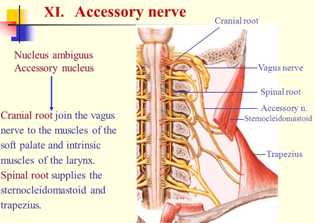
34 Accessory The accessory nerve is anatomically split into a spinal and a cranial portion. Spinal portion originate from the upper spinal cord (spinal accessory nucleus located in the postero-lateral aspect of the anterior horn). Cranial portion originates from the nucleus ambiguus, which is continuous with the lateral horn of the cervical segments. The spinal roots form the spinal accessory component, which enters the skull through the foramen magnum. The cranial part descends from the medulla oblongata to connect with the spinal accessory component.
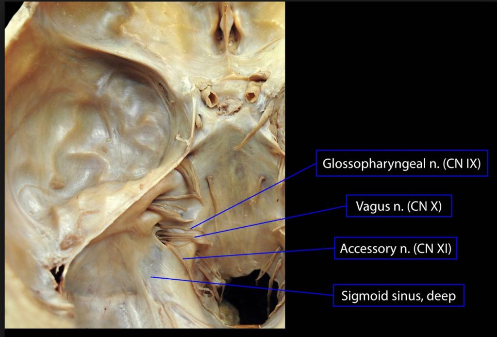
35 XI The nerve then courses along the skull to the jugular foramen through which it exits together with IX and X. CN X lies most medial in the foramen. CN IX exits together with the inferior petrosal sinus through the anterior compartment of the foramen. Vagus and accessory nerves exit via the middle compartment of the foramen. The sigmoid sinus exits via the posterior compartment of the foramen. Shortly before crossing the foramen, the cranial component branches off the accessory nerve to join the vagal nerve (the 11th cranial nerve is a transitional nerve).

36
37
38
39
40 XI In the neck, the nerve crosses the internal jugular vein anteriorly (80% of cases) or posteriorly (20% of cases) to enter the target muscles shortly afterwards. The only cranial nerve which enters and exits the skull and is thus sometimes not regarded as a true cranial nerve.
41 XI mixed or transitional nerve conducting general somatic qualities (innervation of the sternocleidomastoid and trapezius muscles) originating from the cranial portion and a distinct modality originating from the spinal portion. Anastomoses between the spinal component of the accessory nerve and the dorsal roots of the cervical nerves exist below level C1
42 Hypoglossal Pure motor nerve The hypoglossal nerve innervates the muscles of the tongue and is involved in tongue movements during speech, food manipulation, and swallowing. The nerve directs not only voluntary activities of the tongue, but also involuntary functions, such as: clearing the mouth of saliva by swallowing adjusting the tongue to new movements to create desired sounds when learning a new language
43 XII originates from the hypoglossal nucleus. It leaves the medulla oblongata between the olive and the pyramid to pass through the hypoglossal canal. Immediately after leaving the skull it gives off a meningeal branch and unites with a branch of the anterior root C1, which innervates the geniothyroid and thyrohyoid muscles. The nerve then winds behind the CN X and passes between internal carotid artery and internal jugular vein to the posterior belly of the digastric muscle to continue to the submandibular region lateral to the hypoglossal muscle and inferior of the lingual nerve to finally reach the tongue.
44 XII All muscles of the tongue are supplied except for the palatoglossal muscle (IX).
45 Thus, symptoms of LCN lesions include: dysphagia, dysarthria, hoarse voice, chocking during ingestion of fluids, taste disturbances (posterior third of tongue), sensory disturbances (posterior third of the tongue, soft palate, throat, external auditory meatus), palpitations, tachycardia, bradycardia, tachypnea, bradypnea, dyspnea, arterial hypotension, arterial hypertension, fainting, syncope, dizziness, pain in the posterior oral cavity, the face, the neck, the head, obstipation, or diarrhoea. Affection of the 11th cranial nerve may lead to weakness of head rotation, head anteflexion, or shoulder elevation, to scapular winging or to shoulder pain. Affection of the 12th cranial nerve may lead to slurring of speech and the feeling of a thick, heavy, or clumsy tongue. If additional neural or nonneural structures are involved, symptomatology may be even broader.
46 Affection of the LCN may go along with pain, such as neuralgias. Neuralgias of the LCN are the rare glossopharyngeal neuralgia and the even more rare neuralgia of the laryngeal nerve. Glossopharyngeal neuralgia is characterized by excruciating pain attacks of the short duration of the ear, pharynx, neck, tonsil, or base of the tongue. Pain may be triggered by contact of the tongue with the teeth or with the back of the tongue. Glossopharyngeal neuralgia may be associated with syncope (fainting) in 10% of the cases due to reflex bradycardia (efferent limb of reflex via vagal nerve) or occasionally induced by swallowing. Glossopharyngeal neuralgia is most frequently primary (compression of the nerve by adjacent vessel loop or secondary to an intrinsic brain lesion such as in multiple sclerosis Neuralgia of the laryngeal nerve is a rare condition and may be due to a deviated hyoid bone, due to previous acute laryngitis, a pharyngeal diverticulum, or idiopathic. Patients present with paroxysmal, lancing pain located to the area of the thyrohyoid membrane, which can be induced or worsened by swallowing.
47 Examination IX - includes asking the patient to swallow or cough. In addition, taste and a general sensation of the posterior third of the tongue and the throat can be evaluated. The bitter taste (posterior 1/3 of the tongue) is used to test for this nerve. There may also be hypoesthesia of the soft palate or reduced palatal or pharyngeal reflexes. X - eliciting the gag reflex, to look if the uvula is deviated from the side of the lesion, if there is failure of palate elevation upon phonation, and evaluation of speech impairment. In case of bilateral vagal nerve lesions, heart rate and breath rate may be increased. The somatic sensory function can be assessed by testing the sensibility of the external auditory meatus.
48 Examination XI - assessing strength, mass, and spontaneous activity of the trapezius and sternocleidomastoid muscle. The patient is asked to shrug the shoulders with and without resistance. The sternocleidomastoid is tested by asking the patient to turn the head to the left or right or to anteflect the head against resistance. Inspection of the muscles may reveal wasting or fasciculations. XII - tested by asking the patient to stick the tongue out straight. If there is a lesion of the nerve, the tongue will deviate toward the affected side or there may be wasting and fasciculations if the lesion is not acute. The strength of the tongue can be tested by pressing it toward the cheek against the finger of the examiner outside. Weakness of the tongue will also manifest as slurred speech.
49 Stroke Ischemic stroke is a frequent cause of LCN affection, particularly if nuclei or fascicles of any of the four LCN are affected (nuclear or fascicular cranial nerve lesions). According to the location of the ischemic lesion in the brain stem, a dorsal, lateral, paramedian, or median location of the vascular lesion is delineated. Various syndromes are differentiated of which the Wallenbergsyndrome (lateral medullary syndrome) is the most common. More rare ischemic medullary stroke syndromes include the Avellissyndrome, the Jackson-syndrome, and the Opalski-syndrome
50
51

52

53
54
55 Thanks! RITE questions to come!
Cranial Nerves IX-X (Glossopharyngeal & Vagus Nerves)
") Cranial Nerves IX-X (Glossopharyngeal & Vagus Nerves) Please view our Editing File before studying this lecture to check for any changes. Color Code Important Doctors Notes Notes/Extra explanation Objectives
Cranial Nerves IX-X (Glossopharyngeal & Vagus Nerves) Please view our Editing File before studying this lecture to check for any changes. Color Code Important Doctors Notes Notes/Extra explanation Objectives
Cranial nerves.
 Cranial nerves eaglezhyxzy@163.com Key Points of Learning Name Components Passing through Peripheral distribution Central connection Function Cranial nerves Ⅰ olfactory Ⅱ optic Ⅲ occulomotor Ⅳ trochlear
Anatomy #9. Rashed AL-Jomared. The Cranial Nerves IX. Amneh Hazaimeh & Alanood Bostanji
 Anatomy #9 The Cranial Nerves IX Rashed AL-Jomared Amneh Hazaimeh & Alanood Bostanji السالم عليكم This lecture talks about the cranial nerves IX & X:: *Glossopharyngeal nerve : The nerve gets out of the
Prevertebral Region, Pharynx and Soft Palate
 Unit 20: Prevertebral Region, Pharynx and Soft Palate Dissection Instructions: Step1 Step 2 Step 1: Insert your fingers posterior to the sternocleidomastoid muscle, vagus nerve, internal jugular vein,
Cranial Nerves and Spinal Cord Flashcards
 1. Name the cranial nerves and their Roman numeral. 2. What is Cranial Nerve I called, and what does it 3. Scientists who are trying to find a way to make neurons divide to heal nerve injuries often study
Cranial Nerves. Steven McLoon Department of Neuroscience University of Minnesota
 Cranial Nerves Steven McLoon Department of Neuroscience University of Minnesota 1 Course News Change in Lab Sequence Week of Oct 2 Lab 5 Week of Oct 9 Lab 4 2 Sensory and Motor Systems Sensory Systems:
Brain and spinal nerve. By: shirin Kashfi
 Brain and spinal nerve By: shirin Kashfi Nervous system: central nervous system (CNS) peripheral nervous system (PNS) Brain (cranial) nerves Spinal nerves Ganglions (dorsal root ganglions, sympathetic
C h a p t e r PowerPoint Lecture Slides prepared by Jason LaPres North Harris College Houston, Texas
 C h a p t e r 15 The Nervous System: The Brain and Cranial Nerves PowerPoint Lecture Slides prepared by Jason LaPres North Harris College Houston, Texas Copyright 2009 Pearson Education, Inc., publishing
ParasymPathetic Nervous system. Done by : Zaid Al-Ghnaneem
 ParasymPathetic Nervous system Done by : Zaid Al-Ghnaneem In this lecture we are going to discuss Parasympathetic, in the last lecture we took sympathetic and one of the objectives of last lecture was
Functional components
 Facial Nerve VII cranial nerve Emerges from Pons Two roots Functional components: 1. GSA (general somatic afferent) 2. SA (Somatic afferent) 3. GVE (general visceral efferent) 4. BE (Special visceral/branchial
Lec#10 Part 2. Dawood Alatefi. Mariam Hassouneh. Dr.Mohammed Al-Salem
 Lec#10 Part 2 Dawood Alatefi Mariam Hassouneh Dr.Mohammed Al-Salem This part s record is found on the Batch s channel as LabA.3, starting from 0.00min until 26.20min. Hope this ll be an easy part on you;
Cranial Nerve VII - Facial Nerve. The facial nerve has 3 main components with distinct functions
 Cranial Nerve VII - Facial Nerve The facial nerve has 3 main components with distinct functions Somatic motor efferent Supplies the muscles of facial expression; posterior belly of digastric muscle; stylohyoid,
Cranial Nerves VII to XII
 Cranial Nerves VII to XII MSTN121 - Neurophysiology Session 13 Department of Myotherapy Cranial Nerve VIII: Vestibulocochlear Sensory nerve with two distinct branches. Vestibular branch transmits information
PERIPHERAL NERVOUS SYSTEM
 CHAPTER 13 PERIPHERAL NERVOUS SYSTEM Functional division of nervous system = afferent info to the CNS ascending spinal cord = efferent info from CNS descending spinal cord somatic skin, muscles visceral
cardiac plexus is continuous with the coronary and no named branches pain from the heart and lungs
 Nerves of the Thoracic Region Nerve Source Branches Motor Sensory Notes cardiac plexus cardiac brs. of the vagus n. and cervical ; thoracic l nn. the heart and lungs cardiac, cervical cardiac, vagal vagus
Tympanic Bulla Temporal Bone. Digastric Muscle. Masseter Muscle
 Superior view Hyoid Bone The hyoid bone does not articulate with any other bones. It is held in place by ligaments to the styloid process of the temporal bone and the thyroid cartilage of the larynx. It
By : Prof Saeed Abuel Makarem & Dr.Sanaa Alshaarawi
 By : Prof Saeed Abuel Makarem & Dr.Sanaa Alshaarawi OBJECTIVES By the end of the lecture, students shouldbe able to: List the nuclei of the deep origin of the trigeminal and facial nerves in the brain
Introduction to Head and Neck Anatomy
 Introduction to Head and Neck Anatomy Nervous Tissue Controls and integrates all body activities within limits that maintain life Three basic functions 1. sensing changes with sensory receptors 2. interpreting
General Sensory Pathways of the Face Area, Taste Pathways and Hearing Pathways
 General Sensory Pathways of the Face Area, Taste Pathways and Hearing Pathways Lecture Objectives Describe pathways for general sensations (pain, temperature, touch and proprioception) from the face area.
Autonomic Nervous System, Visceral Sensation and Visceral Reflexes Jeff Dupree, Ph.D.
 Autonomic Nervous System, Visceral Sensation and Visceral Reflexes Jeff Dupree, Ph.D. OBJECTIVES After studying the material of this lecture, the student should know the: 1. basic divisions of the autonomic
lecture #2 Done by : Tyma'a Al-zaben
 lecture #2 Done by : Tyma'a Al-zaben ** Hello SERTONIN! note:: the slide included within the sheet but make sure back to slide for pictures in the previous lecture we talk about ascending tract and its
Lab 16: PNS: Nerves and Autonomic NS Hamilton Answers to Pre- Lab Assignments
 Lab 16: PNS: Nerves and Autonomic NS Hamilton Answers to Pre- Lab Assignments Pre-Lab Activity 1: 1. a. olfactory nerve b. optic nerve c. oculomotor nerve d. abducens nerve e. trochlear nerve f. trigeminal
For the following questions, indicate the letter that corresponds to the SINGLE MOST APPROPRIATE ANSWER
 GROSS ANATOMY EXAMINATION May 15, 2000 For the following questions, indicate the letter that corresponds to the SINGLE MOST APPROPRIATE ANSWER 1. Pain associated with an infection limited to the middle
Laith Sorour. Facial nerve (vii):
:") Laith Sorour Cranial nerves 7 & 8 Hello, there are edited slides please go back to them to see pictures, they are not that much important in this lecture but still, and yes slides are included :p Let s
Laith Sorour Cranial nerves 7 & 8 Hello, there are edited slides please go back to them to see pictures, they are not that much important in this lecture but still, and yes slides are included :p Let s
Brainstem and Cranial Nerves II. Nerves covered in other lectures. A reminder about embryology. Prof. Stuart Bunt
 Brainstem and Cranial Nerves II Prof. Stuart Bunt Nerves covered in other lectures 1 Olfactory 2 Optic 3,4,6 Extraocular eye muscles 8 Vestibulo-cochlear 5 Motor and Sensory to the face and muscles of
SCHOOL OF ANATOMICAL SCIENCES Mock Run Questions. 4 May 2012
 SCHOOL OF ANATOMICAL SCIENCES Mock Run Questions 4 May 2012 1. With regard to the muscles of the neck: a. the platysma muscle is supplied by the accessory nerve. b. the stylohyoid muscle is supplied by
Veins of the Face and the Neck
 Veins of the Face and the Neck Facial Vein The facial vein is formed at the medial angle of the eye by the union of the supraorbital and supratrochlear veins. connected through the ophthalmic veins with
Brainstem. Telencephalon Diencephalon Cerebellum Brain stem
 Brainstem Brainstem 脑 脊髓 Brainstem Telencephalon Diencephalon Cerebellum Brain stem Ventral view Lateral view 10 pairs of the cranial nerves are attached to the brain stem The brainstem Midbrain Pons Medulla
Synapse Homework. Back page last question not counted. 4 pts total, each question worth 0.18pts. 26/34 students answered correctly!
 Synapse Homework Back page last question not counted 26/34 students answered correctly! 4 pts total, each question worth 0.18pts Business TASS hours extended! MWF 1-2pm, Willamette 204 T and Th 9:30-10:30am,
Unit VIII Problem 3 Neuroanatomy: Brain Stem, Cranial Nerves and Scalp
 Unit VIII Problem 3 Neuroanatomy: Brain Stem, Cranial Nerves and Scalp - Brain stem: It is connected to the cerebellum and cerebral hemispheres. Rostral end of brain stem: diencephalon is the area which
b. The groove between the two crests is called 2. The neural folds move toward each other & the fuse to create a
 Chapter 13: Brain and Cranial Nerves I. Development of the CNS A. The CNS begins as a flat plate called the B. The process proceeds as: 1. The lateral sides of the become elevated as waves called a. The
Lec [8]: Mandibular nerve:
![Lec [8]: Mandibular nerve:](/thumbs/94/121295776.jpg "Lec [8]: Mandibular nerve:") Lec [8]: Mandibular nerve: The mandibular branch from the trigeminal ganglion lies in the middle cranial fossa lateral to the cavernous sinus. With the motor root of the trigeminal nerve [motor roots lies
Lec [8]: Mandibular nerve: The mandibular branch from the trigeminal ganglion lies in the middle cranial fossa lateral to the cavernous sinus. With the motor root of the trigeminal nerve [motor roots lies
CRANIAL NERVES. Dr. Amani A. Elfaki Associate Professor Department of Anatomy
 CRANIAL NERVES Dr. Amani A. Elfaki Associate Professor Department of Anatomy LEARNING OBJECTIVES Named the cranial nerves Identify the funcunal component of each cranial nerve Identify the effect of each
Cranial Nerve VII & VIII
 Cranial Nerve VII & VIII Lecture Objectives Follow up the course of facial nerve from its point of central connections, exit and down to its target areas. Follow up the central connections of the facial
Biology 323 Human Anatomy for Biology Majors Week 10; Lecture 1; Tuesday Dr. Stuart S. Sumida. Cranial Nerves and Soft Tissues of the Skull
 Biology 323 Human Anatomy for Biology Majors Week 10; Lecture 1; Tuesday Dr. Stuart S. Sumida Cranial Nerves and Soft Tissues of the Skull FOREBRAIN MIDBRAIN HINDBRAIN Forebrain: Cerebrum Perception,
Chapter 16. APR Enhanced Lecture Slides
 Chapter 16 APR Enhanced Lecture Slides See separate PowerPoint slides for all figures and tables pre-inserted into PowerPoint without notes and animations. Copyright The McGraw-Hill Companies, Inc. Permission
CN I Olfactory. CN II Optic. CN III Oculomotor. Special Sensory Efferent fibers to Olfactory Bulb. Cribiform Plate of Ethmoid
 CN I Olfactory Efferent fibers to Olfactory Bulb Olfactory Tract Olfactory Bulb Cribiform Plate of Ethmoid Anosmia Loss of sense of smell Uncinate Fits olfactory hallucinations To Olfactory Epithelium
ACTIVITY 7: NERVOUS SYSTEM HISTOLOGY, BRAIN, CRANIAL NERVES
 ACTIVITY 7: NERVOUS SYSTEM HISTOLOGY, BRAIN, CRANIAL NERVES LABORATORY OBJECTIVES: 1. Histology: Identify structures indicated on three different slides or images of nervous system tissue. These images
human anatomy 2016 lecture fifteen Dr meethak ali ahmed neurosurgeon
 Cranial Nerves Organization of the Cranial Nerves The cranial nerves are named as follows: I. Olfactory II. Optic III. Oculomotor IV. Trochlear V. Trigeminal VI. Abducent VII. Facial VIII. Vestibulocochlear
Human Anatomy. Autonomic Nervous System
 Human Anatomy Autonomic Nervous System 1 Autonomic Nervous System ANS complex system of nerves controls involuntary actions. Works with the somatic nervous system (SNS) regulates body organs maintains
Spinal nerves and cervical plexus Prof. Abdulameer Al Nuaimi. E mail: a.al E. mail:
 Spinal nerves and cervical plexus Prof. Abdulameer Al Nuaimi E mail: a.al nuaimi@sheffield.ac.uk E. mail: abdulameerh@yahoo.com Branches of ophthalmic artery Muscles of face A spinal nerve Spinal
Neural Integration I: Sensory Pathways and the Somatic Nervous System
 15 Neural Integration I: Sensory Pathways and the Somatic Nervous System PowerPoint Lecture Presentations prepared by Jason LaPres Lone Star College North Harris An Introduction to Sensory Pathways and
Nervous System. Student Learning Objectives:
 Nervous System Student Learning Objectives: Identify the primary parts of the neuron Identify the major structures of the central nervous system Identify the major structures of the peripheral nervous
PTERYGOPALATINE FOSSA
 PTERYGOPALATINE FOSSA Outline Anatomical Structure and Boundaries Foramina and Communications with other spaces and cavities Contents Pterygopalatine Ganglion Especial emphasis on certain arteries and
The NIHSS score is 4 (considering 2 pts for the ataxia involving upper and lower limbs.
 Neuroscience case 5 1. Speech comprehension, ability to speak, and word use were normal in Mr. Washburn, indicating that aphasia (cortical language problem) was not involved. However, he did have a problem
Dr.Ban I.S. head & neck anatomy 2 nd y. جامعة تكريت كلية طب االسنان املرحلة الثانية أ.م.د. بان امساعيل صديق 6102/6102
 جامعة تكريت كلية طب االسنان التشريح مادة املرحلة الثانية أ.م.د. بان امساعيل صديق 6102/6102 Parotid region The part of the face in front of the ear and below the zygomatic arch is the parotid region. The
Brain Stem. 1. Midbrain 2. Pons 3. Medulla Oblongata
 Brain Stem 1. Midbrain 2. Pons 3. Medulla Oblongata 1 Ext. features Medulla Oblongata *Direct continuation of Spinal Cord *Extend from foramen magnum to lower Pons *More than 2.5 cm in length. *Lower part
Autonomic Nervous System. Ms. DS Pillay Room 2P24
 Autonomic Nervous System Ms. DS Pillay Room 2P24 OVERVIEW OF THE NERVOUS SYSTEM NERVOUS SYSTEM CNS PNS BRAIN SPINAL CORD SOMATIC ANS SYMPATHEIC PARASYMPATHEIC LOCATION OF GANGLIA IN THE ANS Short post-ganglionic
Biology 218 Human Anatomy
 Chapter 20 Adapted form Tortora 10 th ed. LECTURE OUTLINE A. Introduction (p. 632) 1. The autonomic nervous system (ANS) regulates the activity of smooth muscle, cardiac muscle, and certain glands. 2.
NERVOUS SYSTEM ANATOMY
 INTRODUCTION to NERVOUS SYSTEM ANATOMY M1 - Gross and Developmental Anatomy Dr. Milton M. Sholley Professor of Anatomy and Neurobiology and Dr. Michael H. Peters Professor of Chemical and Life Science
Peripheral Nervous System Dr. Gary Mumaugh
 Peripheral Nervous System Dr. Gary Mumaugh Spinal Nerves Overview Thirty-one pairs of spinal nerves are connected to the spinal cord No special names; numbered by level of vertebral column at which they
HBA THE BODY Head & Neck Written Examination October 23, 2014
 HBA 531 - THE BODY Head & Neck Written Examination October 23, 2014 Name: NOTE 2: When asked to trace nerve, artery, or vein pathways, do so by using arrows, e.g., structure a structure b structure c...
Dr. Weyrich G07: Superior and Posterior Mediastina. Reading: 1. Gray s Anatomy for Students, chapter 3
 Dr. Weyrich G07: Superior and Posterior Mediastina Reading: 1. Gray s Anatomy for Students, chapter 3 Objectives: 1. Subdivisions of mediastinum 2. Structures in Superior mediastinum 3. Structures in Posterior
Human Nervous System:
 OLLI Brain: Making Sense of Our World: Lecture 3 Human Nervous System: The Motor & Sensory Divisions Copyright 2004 Pearson Education, Inc., publishing as Benjamin Cummings Organization of the Nervous
Organisation of the nervous system
 Chapter1 Organisation of the nervous system 1. Subdivisions of the nervous system The nervous system is divided: i) Structurally The central nervous system (CNS) composed of the brain and spinal cord.
The neurvous system senses, interprets, and responds to changes in the environment. Two types of cells makes this possible:
 NERVOUS SYSTEM The neurvous system senses, interprets, and responds to changes in the environment. Two types of cells makes this possible: the neuron and the supporting cells ("glial cells"). Neuron Neurons
The Neck the lower margin of the mandible above the suprasternal notch and the upper border of the clavicle
 The Neck is the region of the body that lies between the lower margin of the mandible above and the suprasternal notch and the upper border of the clavicle below Nerves of the neck Cervical Plexus Is formed
Tracing the Cranial Nerves Osteologically
 CN I II III IV V 1 Supra-orbital ethmoidal nn. Ext. nasal V 2 Tracing the Cranial Nerves Osteologically Nucleus of Origin Olfactory tracts of frontal lobe of cerebrum Optic tracts from optic chiasma and
Organization of The Nervous System PROF. SAEED ABUEL MAKAREM
 Organization of The Nervous System PROF. SAEED ABUEL MAKAREM Objectives By the end of the lecture, you should be able to: List the parts of the nervous system. List the function of the nervous system.
The Nervous System PART C. PowerPoint Lecture Slide Presentation by Patty Bostwick-Taylor, Florence-Darlington Technical College
 PowerPoint Lecture Slide Presentation by Patty Bostwick-Taylor, Florence-Darlington Technical College The Nervous System 7 PART C Protection of the Central Nervous System Scalp and skin Skull and vertebral
Unit 18: Cranial Cavity and Contents
 Unit 18: Cranial Cavity and Contents Dissection Instructions: The calvaria is to be removed without damage to the dura mater which is attached to the inner surface of the calvaria. Cut through the outer
The Pharynx. Dr. Nabil Khouri MD. MSc, Ph.D
 The Pharynx Dr. Nabil Khouri MD. MSc, Ph.D Introduction The pharynx is the Musculo-fascial halfcylinder that links the oral and nasal cavities in the head to the larynx and esophagus in the neck Common
Lecture 4 The BRAINSTEM Medulla Oblongata
 Lecture 4 The BRAINSTEM Medulla Oblongata Introduction to brainstem 1- Medulla oblongata 2- Pons 3- Midbrain - - - occupies the posterior cranial fossa of the skull. connects the narrow spinal cord
Doctor Osama Asa ad Khader. Mohammad Alsalem
 6 Doctor 2015 Osama Asa ad Khader Mohammad Alsalem A quick revision for the spinal cord blood supply: Arterial Blood supply of spinal cord The spinal cord got its arterial supply by two ways: Longitudinal
INTRODUCTION: ANATOMY UNDERLYING CLINICAL TESTS OF CRANIAL NERVES
 INTRODUCTION: ANATOMY UNDERLYING CLINICAL TESTS OF CRANIAL NERVES CRANIAL NERVE I - OLFACTORY I - OLFACTORY NERVE - SMELL TEST: SMELL ODORS (note: not ammonia; pain in nasal cavity CN5 DAMAGE: LOSS OF
Temporal region. temporal & infratemporal fossae. Zhou Hong Ying Dept. of Anatomy
 Temporal region temporal & infratemporal fossae Zhou Hong Ying Dept. of Anatomy Temporal region is divided by zygomatic arch into temporal & infratemporal fossae. Temporal Fossa Infratemporal fossa Temporal
NERVOUS SYSTEM ANATOMY
 NTRODUCTON to NERVOUS SYSTEM ANATOMY M1 - Gross and Developmental Anatomy Dr. Milton M. Sholley Professor of Anatomy and Neurobiology and Dr. Michael H. Peters Professor of Chemical and Life Science Engineering
Peripheral Nervous System
 Peripheral Nervous System Sensory Receptors Motor Endings Cranial Nerves The Four Plexuses Extremities Review of Reflexes Fast, preprogrammed, inborn, automatic responses Occur in the CNS at the spinal
Human Anatomy - Problem Drill 11: The Spinal Cord and Spinal Nerves
 Human Anatomy - Problem Drill 11: The Spinal Cord and Spinal Nerves Question No. 1 of 10 Instructions: (1) Read the problem statement and answer choices carefully, (2) Work the problems on paper as needed,
Parotid Gland, Temporomandibular Joint and Infratemporal Fossa
 M1 - Anatomy Parotid Gland, Temporomandibular Joint and Infratemporal Fossa Jeff Dupree Sanger 9-057 jldupree@vcu.edu Parotid gland: wraps around the mandible positioned between the mandible and the sphenoid
Principles of Anatomy and Physiology
 Principles of Anatomy and Physiology 14 th Edition CHAPTER 14 The Brain and Cranial Nerves Introduction The purpose of the chapter is to: 1. Understand how the brain is organized, protected, and supplied
Dr.Ban I.S. head & neck anatomy 2 nd y. جامعة تكريت كلية طب االسنان املرحلة الثانية
 جامعة تكريت كلية طب االسنان التشريح مادة املرحلة الثانية أ.م.د. بان امساعيل صديق 6102-6102 1 The Palate The palate forms the roof of the mouth and the floor of the nasal cavity. It is divided into two
I: To describe the pyramidal and extrapyramidal tracts. II: To discuss the functions of the descending tracts.
 Descending Tracts I: To describe the pyramidal and extrapyramidal tracts. II: To discuss the functions of the descending tracts. III: To define the upper and the lower motor neurons. 1. The corticonuclear
Sympathetic Nervous System
 Sympathetic Nervous System Lecture Objectives Review the subdivisions of the nervous system. Review the general arrangement and compare the sympathetic and parasympathetic parts. Describe the following
Cranial nerve Dept. of Anatomy Zhou Hong Ying
 Cranial nerve Dept. of Anatomy Zhou Hong Ying Key Points of Learning Name Components Passing through Peripheral distribution Central connection Function Cranial nerve Ⅰ olfactory Ⅱ optic Ⅲ occulomotor
Oral cavity : consist of two parts: the oral vestibule and the oral cavity proper. Oral vestibule : is slit like space between.
 Oral cavity Oral cavity : consist of two parts: the oral vestibule and the oral cavity proper Oral vestibule : is slit like space between the teeth, buccal gingiva, lips, and cheeks 1 Oral cavity Oral
The Nervous System: Sensory and Motor Tracts of the Spinal Cord
 15 The Nervous System: Sensory and Motor Tracts of the Spinal Cord PowerPoint Lecture Presentations prepared by Steven Bassett Southeast Community College Lincoln, Nebraska Introduction Millions of sensory
[ANATOMY #12] April 28, 2013
![[ANATOMY #12] April 28, 2013](/thumbs/86/93473883.jpg "[ANATOMY #12] April 28, 2013") Sympathetic chain : Sympathetic chain is each of the pair of ganglionated longitudinal cords of the sympathetic nervous system; extend from level of atlas (base of skull) till coccyx. It is paravertebral
Sympathetic chain : Sympathetic chain is each of the pair of ganglionated longitudinal cords of the sympathetic nervous system; extend from level of atlas (base of skull) till coccyx. It is paravertebral
Tikrit University collage of dentistry Dr.Ban I.S. head & neck anatomy 2 nd y. Lec [5] / Temporal fossa :
![Tikrit University collage of dentistry Dr.Ban I.S. head & neck anatomy 2 nd y. Lec [5] / Temporal fossa :](/thumbs/88/115294566.jpg "Tikrit University collage of dentistry Dr.Ban I.S. head & neck anatomy 2 nd y. Lec [5] / Temporal fossa :") Lec [5] / Temporal fossa : Borders of the Temporal Fossa: Superior: Superior temporal line. Inferior: gap between zygomatic arch and infratemporal crest of sphenoid bone. Anterior: Frontal process of the
Lec [5] / Temporal fossa : Borders of the Temporal Fossa: Superior: Superior temporal line. Inferior: gap between zygomatic arch and infratemporal crest of sphenoid bone. Anterior: Frontal process of the
Brainstem. By Dr. Bhushan R. Kavimandan
 Brainstem By Dr. Bhushan R. Kavimandan Development Ventricles in brainstem Mesencephalon cerebral aqueduct Metencephalon 4 th ventricle Mylencephalon 4 th ventricle Corpus callosum Posterior commissure
Mohammad Hisham Al-Mohtaseb. Lina Mansour. Reyad Jabiri. 0 P a g e
 2 Mohammad Hisham Al-Mohtaseb Lina Mansour Reyad Jabiri 0 P a g e This is only correction for the last year sheet according to our record. If you already studied this sheet just read the yellow notes which
Medical Neuroscience Tutorial Notes
 Medical Neuroscience Tutorial Notes Cranial Nerve Nuclei MAP TO NEUROSCIENCE CORE CONCEPTS 1 NCC1. The brain is the body's most complex organ. LEARNING OBJECTIVES After study of the assigned learning materials,
Nervous System. The Peripheral Nervous System Agenda Review of CNS v. PNS PNS Basics Cranial Nerves Spinal Nerves Reflexes Pathways
 Nervous System Agenda Review of CNS v. PNS PNS Basics Cranial Nerves Spinal Nerves Sensory Motor Review of CNS v. PNS Central nervous system (CNS) Brain Spinal cord Peripheral nervous system (PNS) All
Done by : Areej Al-Hadidi
 Brainstem &diencephalon Done by : Areej Al-Hadidi Brainstem Functions Ascending and descending tracts Reflex centers Cardiovascular and respiratory centers Coughing, sneezing, swallowing Nuclei of the
M555 Medical Neuroscience Lab 1: Gross Anatomy of Brain, Crainal Nerves and Cerebral Blood Vessels
 M555 Medical Neuroscience Lab 1: Gross Anatomy of Brain, Crainal Nerves and Cerebral Blood Vessels Anatomical Directions Terms like dorsal, ventral, and posterior provide a means of locating structures
The Nervous System. Functions of the Nervous System input gathering To monitor occurring inside and outside the body Changes =
 The Nervous System Functions of the Nervous System input gathering To monitor occurring inside and outside the body Changes = To process and sensory input and decide if is needed output A response to integrated
Chp. 16: AUTONOMIC N.S. (In Review: Peripheral N. S.)
") Chp. 16: AUTONOMIC N.S. (In Review: Peripheral N. S.) Peripheral nerves contain both motor and sensory neurons Among the motor neurons, some of these are somatic and innervate skeletal muscles while some
Chp. 16: AUTONOMIC N.S. (In Review: Peripheral N. S.) Peripheral nerves contain both motor and sensory neurons Among the motor neurons, some of these are somatic and innervate skeletal muscles while some
The Nervous System: Autonomic Nervous System
 17 The Nervous System: Autonomic Nervous System PowerPoint Lecture Presentations prepared by Steven Bassett Southeast Community College Lincoln, Nebraska Introduction The autonomic nervous system functions
THE GOOFY ANATOMIST QUIZZES
 THE GOOFY ANATOMIST QUIZZES 5. NERVES Q1. Which of the following classifications of the nervous systems is correct? A. The autonomic nervous system is composed of the brain, cranial nerves and spinal nerves.
Blood supply to the brain Blood brain barrier isolates neural tissue from general circulation
 The Brain and Cranial Nerves Objectives Name the major regions of the brain and describe their functions. Discuss the formation, circulation, and functions of the CSF. List the main components of the medulla
Temporal fossa Infratemporal fossa Pterygopalatine fossa Terminal branches of external carotid artery Pterygoid venous plexus
 Outline of content Temporal fossa Infratemporal fossa Pterygopalatine fossa Terminal branches of external carotid artery Pterygoid venous plexus Boundary Content Communication Mandibular division of trigeminal
The Ear The ear consists of : 1-THE EXTERNAL EAR 2-THE MIDDLE EAR, OR TYMPANIC CAVITY 3-THE INTERNAL EAR, OR LABYRINTH 1-THE EXTERNAL EAR.
 The Ear The ear consists of : 1-THE EXTERNAL EAR 2-THE MIDDLE EAR, OR TYMPANIC CAVITY 3-THE INTERNAL EAR, OR LABYRINTH 1-THE EXTERNAL EAR Made of A-AURICLE B-EXTERNAL AUDITORY MEATUS A-AURICLE It consists
Nose & Mouth OUTLINE. Nose. - Nasal Cavity & Its Walls. - Paranasal Sinuses. - Neurovascular Structures. Mouth. - Oral Cavity & Its Contents
 Dept. of Human Anatomy, Si Chuan University Zhou hongying eaglezhyxzy@163.com Nose & Mouth OUTLINE Nose - Nasal Cavity & Its Walls - Paranasal Sinuses - Neurovascular Structures Mouth - Oral Cavity & Its
ORAL CAVITY, ESOPHAGUS AND STOMACH
 ORAL CAVITY, ESOPHAGUS AND STOMACH 1 OBJECTIVES By the end of the lecture you should be able to: Describe the anatomy the oral cavity, (boundaries, parts, nerve supply). Describe the anatomy of the palate,
Part 1. Copyright 2011 Pearson Education, Inc. Copyright 2011 Pearson Education, Inc. Stimulatory
 PowerPoint Lecture Slides prepared by Leslie Hendon University of Alabama, Birmingham C H A P T E R 15 Part 1 The Autonomic Nervous System and Visceral Sensory Neurons The ANS and Visceral Sensory Neurons
Tymaa Al-zaben & Amin Al-ajalouni
 Done by: Tymaa Al-zaben & Amin Al-ajalouni ** Hello SERTONIN! SLIDE 3 note:: the slide included within the sheet but make sure back to slide for pictures The Autonomic Nervous System Function : Regulate
Spinal nerves. Aygul Shafigullina. Department of Morphology and General Pathology
 Spinal nerves Aygul Shafigullina Department of Morphology and General Pathology Spinal nerve a mixed nerve, formed in the vicinity of an intervertebral foramen, where fuse a dorsal root and a ventral root,
Organization of The Nervous System PROF. MOUSAED ALFAYEZ & DR. SANAA ALSHAARAWY
 Organization of The Nervous System PROF. MOUSAED ALFAYEZ & DR. SANAA ALSHAARAWY Objectives At the end of the lecture, the students should be able to: List the parts of the nervous system. List the function
THE SURGEON S LIBRARY
 THE SURGEON S LIBRARY THE HISTORY AND SURGICAL ANATOMY OF THE VAGUS NERVE Lee J. Skandalakis, M.D., Chicago, Illinois, Stephen W. Gray, PH.D., and John E. Skandalakis, M.D., PH.D., F.A.C.S., Atlanta, Georgia