Cranial nerve Dept. of Anatomy Zhou Hong Ying
|
|
|
- Gloria Goodman
- 5 years ago
- Views:
Transcription

1 Cranial nerve Dept. of Anatomy Zhou Hong Ying
2 Key Points of Learning Name Components Passing through Peripheral distribution Central connection Function
3 Cranial nerve Ⅰ olfactory Ⅱ optic Ⅲ occulomotor Ⅳ trochlear Ⅴ trigeminal Ⅵ abducens Ⅶ facial Ⅷ vestibulocochlear Ⅸ glossophryngeal Ⅹ vagus Ⅺ accessory Ⅻ hypoglossal

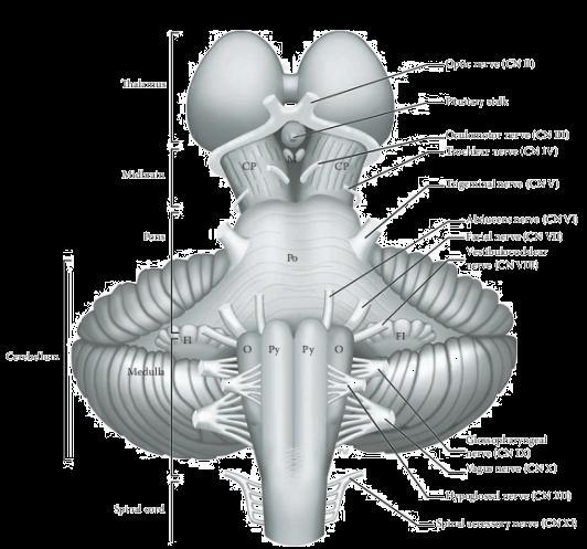
4 Cranial nerve: connect with brain


5 Openings and fissures on base of skull for CN
6 CRANIAL NERVE General somatic motor (GSM) General visceral motor (GVM) Special visceral motor (SVM) General somatic sensory (GSS) Special somatic sensory (SSS) General visceral sensory (GVS) Special visceral sensory (SVS) Every pair of CN has not all of the above components.

7 CRANIAL NERVE
8 Pure sensory CN olfactory n. optic n. vestibulocochlear n.

9 Olfactory nerve CN I Pure sensory (SVS) -- smell Olfactory bulb of telencephalon Leaves cranial cavity through foramina in cribriform plate Distributes to mucosa of superior part of lateral and septal walls of nasal cavity.

10 Olfactory nerve CN I

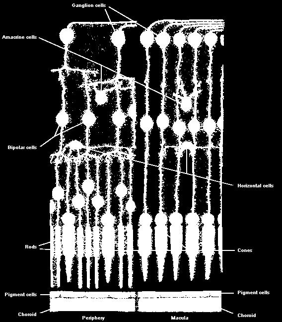
11 pure sensory (SSS)-- sight Connects with optic chiasma of diancephlon. Leaves cranial cavity through optic canal. Distributes to retina of eyeball optic n.(Ⅱ)


12 optic nerve, chiasma, optic tract


13 Ⅷ vestibulocochlear conveys impulses from inner ear. sense of position terminates in vestibular N, sense of hearing in cochlear N


14 Pure sensory (SSS) Internal acoustic meatus internal ear Vestibular nerve is responsible for equilibrium Cochlear nerve is responsible for hearing Vestibulocochlear N.
15 Pure motor CN oculomotor n. trochlear n. abducens n. accessory n. hypoglossal nerve
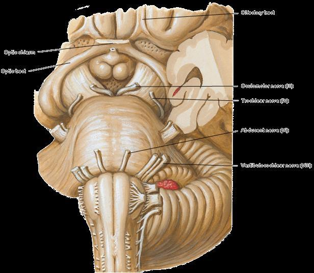
16 oculomotor n. trochlear n. abducens n.

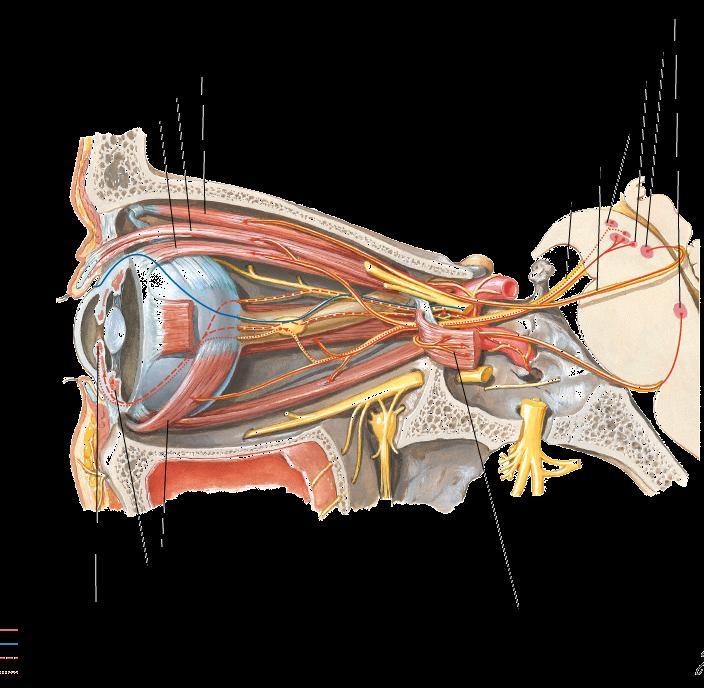
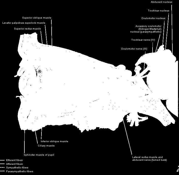
17 Oculomotor n.(gsm and GVM) Midbrain Superior orbital fissure Superior division--superior rectus & levator palpebrae superioris Inferior division supplies inferior & medial rectus, & inferior oblique. GVM fibers parasympathetic, supply ciliary muscle and sphincter pupillae. Trochlear N Pure motor (GSM) ; midbrain ; Superior orbital fissure ; Superior oblique m Abducens N Pure motor (GSM) ; Pons ; Superior orbital fissure Lateral rectus m
18

19 Two roots Pure motor Medulla & spinal cord Jugular foramen Cranial root--joins X Spinal root-- sternocleidomastoid & trapezius accessory n.
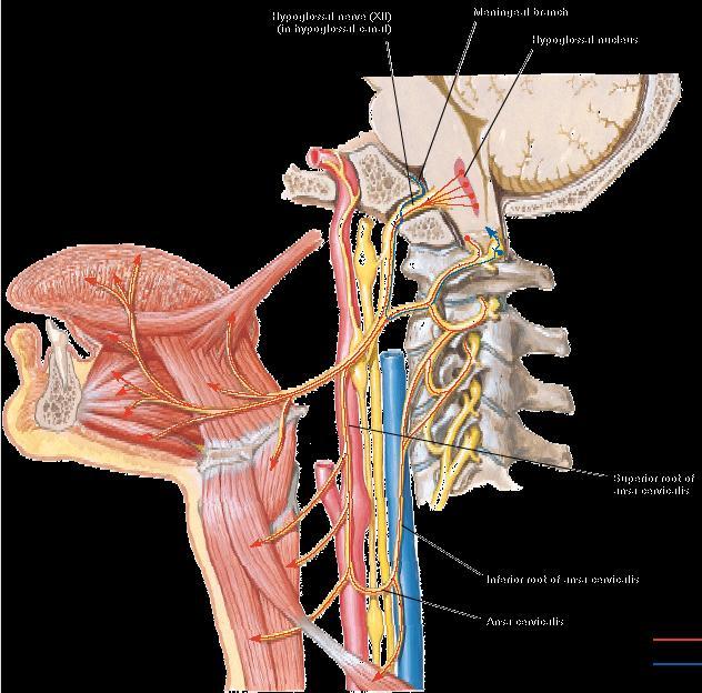
20 (Ⅻ)Hypoglossal n.

21 (Ⅻ)Hypoglossal nerve Pure motor (GSM) Medulla Hypoglossal canal extrinsic and intrinsic muscles of tongue.
22 Mixed CN trigeminal n. facial n. glossopharyngeal n. vagus n.
23 Trigeminal nerve V pons Has three divisions: ophthalmic, maxillary, and mandibular nerve Has both sensory and motor components. It is the main sensory nerve for head and face. Its motor nerve innervates the muscles of mastication, etc.


24 Three divisions: Ophthalmic Superior orbital fissure into orbital cavity Maxillary-foramen rotundum into pteygopalatine Mandibular foramen ovale

25 Trigeminal nerve ophthalmic N


26 Trigeminal nerve ophthalmic N superior orbital fissure Lacrimal nerve to lacrimal gland. Frontal nerve to forehead Nasociliary nerve to eyeball, part of nasal cavity, and skin of dorsum of nose from root to top.


27 Trigeminal nerve maxillary N foramen rotundum pterygopalatine fossa inferior orbital fissure, sulcus, canal and foramen to part of nasal cavity, maxilla, upper teeth, and skin between eye and mouth


28 Lingual nerve to anterior 2/3 of tongue Inferior alveolar nerve to mandible, lower teeth, and chin Buccal nerve to cheek Auriculotemporal nerve to parotid gland and temporal region SVM fibers supply muscles of mastication, tensor veli palatini, and tensor tempani, etc. Mandibular n.

29 tensor tympani M

30 Anterior belly of digastric M

31 Sensory fibers of CN V

32 Ⅶ Facial nerve in the temporal bone and main branches and distribution Mixed (SVM, GVM, SVS) Pons internal acoustic meatus canal of facial nerve stylomastoid foramen parotid gland SVM fibers supply muscles of facial expression & stapedius GVM fibers synapse in pterygo-palatine ganglion to supply lacrimal gland; in submandibular ganglion submandibular & sublingual gland SVS fibers receive taste from anterior 2/3 of tongue


33 Ⅶ Facial nerve facial n in the internal auditory meatus


34 facial n in the internal auditory meatus, canal, through the stylomastioid foramen

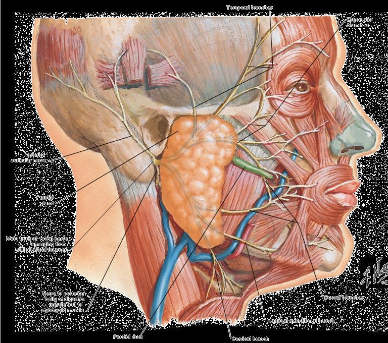
35 Ⅶ Facial nerve distributes to expression muscles

36 Ⅶ Facial nerve distributes to mucous of tongue taste

37 CN Ⅶ distributes to submandibular gland and sublingual gland through the submadibular ganglion
38 (Ⅸ) glossopharyngeal n. Mixed; medulla; jugular foramen GS fibers to carotid sinus and body, oropharynx, and posterior 1/3 of tongue SVS fibers receive taste from posterior 1/3 of tongue SVM fibers supply stylopharyngeus GVM fibers synapse in otic ganglion to supply the parotid gland

39 (Ⅸ)glossopharyngeal n.distributes to pharyngeal muscle, parotid salivary gland
 carotid sinus")
40 (Ⅸ) distributes to carotid body (chemoreceptors) carotid sinus (baroreceptors )

41 (Ⅸ)glossopharyngeal n. distributes to tongue general somatic and special taste sensory(posterior third)
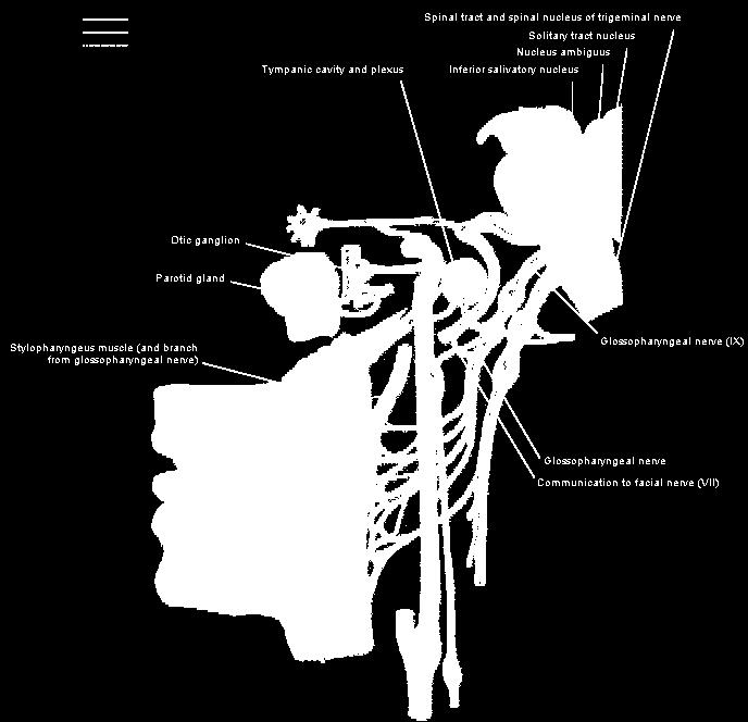
42 (Ⅸ)glossopharyngeal n.

43 The vagus nerve in the neck, thorax, and abdomen Mixed; medulla; jugular foramen; carotid sheath, on the side of mediastinum, esophgeal plexus, esophgeal hiatus anterior and posterior vagal trunks


44 Parasympathetic fibers in the vagus nerve Parasympathetic fibers originate from the dorsal motor nucleus of vagus. They are distributed widely throughout the respiratory,gastrointestinal system and cardiovascular. GVM
45 The afferent fibers of vagus n. 1.receptor for general sensation in tympanic membrane, external autitory meatus, and part of the concha of the external ear. 2. receptor for general viscera sensation are distributed widely throughout the mucous membrane of the respiratory,gastrointestinal system and internal layer of the cardiovascular. 3.chemoreceptors are distributed in aortic bodies and baroreptors in the aortic arch GVS, GSS


46 motor fibers in the vagus nerve innervates to the muscles of larynx and pharynx through the superior laryngeal nerves and the recurrent nerves SVM
47 Ophthalmic division Sensory (GSA) Trigeminal Cornea, skin of forehead, scalp, eyelids, and nose; also mucous membrane of paranasal sinuses and nasal cavity Maxillary division Sensory (GSA) Skin of face over maxilla; teeth of upper jaw; mucous membrane of nose, the maxillary sinus, and palate Mandibular division Motor (SVE) Sensory (GSA) Muscles of mastication, mylohyoid, anterior belly of digastric, tensor veli palatini, and tensor tympani Skin of cheek, skin over mandible and side of head, teeth of lower jaw and temporomandibular joint; mucous membrane of mouth and anterior part of tongue
48 Motor (SVE) Facial Muscles of face and scalp, stapedius muscle, posterior belly of digastric and stylohyoid muscles Sensory (SVA) Secretomotor (GVE) parasympathetic Taste from anterior two-thirds of tongue, from floor of mouth and palate Submandibular and sublingual salivary glands, the lacrimal gland, and glands of nose and palate Motor (SVE) Glossopharyngeal Stylopharyngeus muscle assists swallowing Secretomotor (GVE) parasympathetic Sensory (GVA, SVA, GSA) Parotid salivary gland General sensation and taste from posterior one-third of tongue and pharynx; carotid sinus (baroreceptor); and carotid body (chemoreceptor)

49 SUMMARIZE Nerves of eye Nerves of tongue nerves of pharynx
50 Case A 49-year-old man woke up one morning to find the right side of his face paralyzed. When examined by his local medical practitioner, he was found to have complete paralysis of the entire right side of the face. He was also found to have severe hypertension. The patient talked with slightly slurred speech. The physician told the patient that he had suffered a mild stroke, and he was admitted to the hospital. The patient was later seen by a neurologist who disagreed with the diagnosis.

51 External Features of Brain Stem External structure: superficial markers Internal structure: grey matter, white matter, reticular formation Important section levels
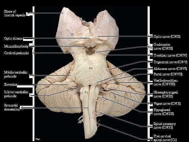
52 External structures Midbrain Pons Medulla oblongata


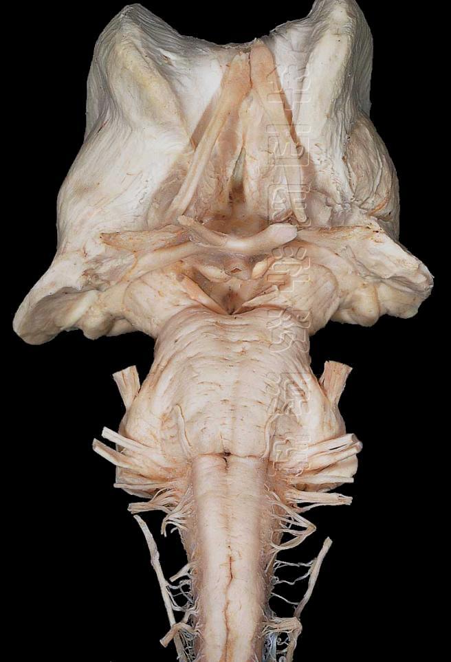
53 Ventral aspect of medulla Anterior median fissure Hypoglossal nerve Pyramid Glossopharyngeal nerve Decussation of pyramid Vagus nerve Olive Accessory nerve Preolivary sulcus & Postolivary sulcus

54 middle cerebellum peduncle basilar sulcus cranial nerves Ⅴ,Ⅵ,Ⅶ,Ⅷ The ventral surface of pons

55 Ventral view of midbrain Massive crural cerebri interpeduncular fossa oculomotor nerve Floor of the fossa is called posterior perforated substance due to the many blood vessels perforate the midbrain.
56


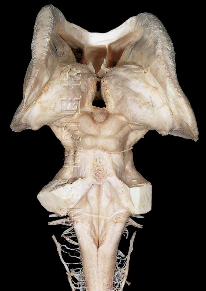
57 Dorsal surfaces of the pons and medulla rhomboid-shaped fossa Boundary Lateral recess/aperture Median sulcus Aqueduct
58 Dorsal surfaces of the pons and medulla stria medullaris sulcus limitans medial eminence vestibular area: vestibular nuclei lie beneath the vestibular area facial colliculus : abducens nucleus

59 Dorsal surfaces of the pons and medulla hypoglossal triangle lie within the medial eminence just inferior to stria medullaris. dorsa vagus motor nucleus lie beneath vagus motor dorsal triangle. gracilis tubercle, cuneate tubercle


60 dorsa surface of mid brain superior and inferior colliculi/quadrigemina corpora tectum of the midbrain Cranial nerve Ⅳ
Cranial nerves.
 Cranial nerves eaglezhyxzy@163.com Key Points of Learning Name Components Passing through Peripheral distribution Central connection Function Cranial nerves Ⅰ olfactory Ⅱ optic Ⅲ occulomotor Ⅳ trochlear
Introduction to Head and Neck Anatomy
 Introduction to Head and Neck Anatomy Nervous Tissue Controls and integrates all body activities within limits that maintain life Three basic functions 1. sensing changes with sensory receptors 2. interpreting
Brainstem. Telencephalon Diencephalon Cerebellum Brain stem
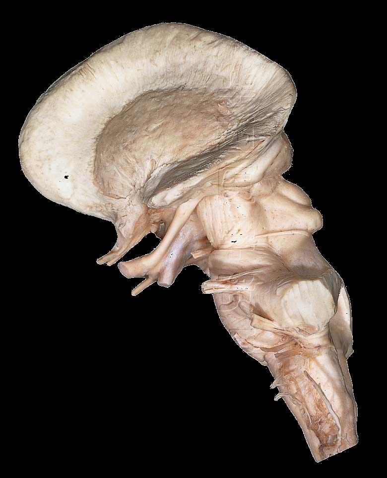
 Brainstem Brainstem 脑 脊髓 Brainstem Telencephalon Diencephalon Cerebellum Brain stem Ventral view Lateral view 10 pairs of the cranial nerves are attached to the brain stem The brainstem Midbrain Pons Medulla
human anatomy 2016 lecture fifteen Dr meethak ali ahmed neurosurgeon
 Cranial Nerves Organization of the Cranial Nerves The cranial nerves are named as follows: I. Olfactory II. Optic III. Oculomotor IV. Trochlear V. Trigeminal VI. Abducent VII. Facial VIII. Vestibulocochlear
C h a p t e r PowerPoint Lecture Slides prepared by Jason LaPres North Harris College Houston, Texas
 C h a p t e r 15 The Nervous System: The Brain and Cranial Nerves PowerPoint Lecture Slides prepared by Jason LaPres North Harris College Houston, Texas Copyright 2009 Pearson Education, Inc., publishing
Biology 323 Human Anatomy for Biology Majors Week 10; Lecture 1; Tuesday Dr. Stuart S. Sumida. Cranial Nerves and Soft Tissues of the Skull
 Biology 323 Human Anatomy for Biology Majors Week 10; Lecture 1; Tuesday Dr. Stuart S. Sumida Cranial Nerves and Soft Tissues of the Skull FOREBRAIN MIDBRAIN HINDBRAIN Forebrain: Cerebrum Perception,
By : Prof Saeed Abuel Makarem & Dr.Sanaa Alshaarawi
 By : Prof Saeed Abuel Makarem & Dr.Sanaa Alshaarawi OBJECTIVES By the end of the lecture, students shouldbe able to: List the nuclei of the deep origin of the trigeminal and facial nerves in the brain
Tracing the Cranial Nerves Osteologically
 CN I II III IV V 1 Supra-orbital ethmoidal nn. Ext. nasal V 2 Tracing the Cranial Nerves Osteologically Nucleus of Origin Olfactory tracts of frontal lobe of cerebrum Optic tracts from optic chiasma and
CRANIAL NERVES. Dr. Amani A. Elfaki Associate Professor Department of Anatomy
 CRANIAL NERVES Dr. Amani A. Elfaki Associate Professor Department of Anatomy LEARNING OBJECTIVES Named the cranial nerves Identify the funcunal component of each cranial nerve Identify the effect of each
Brain and spinal nerve. By: shirin Kashfi
 Brain and spinal nerve By: shirin Kashfi Nervous system: central nervous system (CNS) peripheral nervous system (PNS) Brain (cranial) nerves Spinal nerves Ganglions (dorsal root ganglions, sympathetic
PERIPHERAL NERVOUS SYSTEM
 CHAPTER 13 PERIPHERAL NERVOUS SYSTEM Functional division of nervous system = afferent info to the CNS ascending spinal cord = efferent info from CNS descending spinal cord somatic skin, muscles visceral
Nose & Mouth OUTLINE. Nose. - Nasal Cavity & Its Walls. - Paranasal Sinuses. - Neurovascular Structures. Mouth. - Oral Cavity & Its Contents
 Dept. of Human Anatomy, Si Chuan University Zhou hongying eaglezhyxzy@163.com Nose & Mouth OUTLINE Nose - Nasal Cavity & Its Walls - Paranasal Sinuses - Neurovascular Structures Mouth - Oral Cavity & Its
Temporal region. temporal & infratemporal fossae. Zhou Hong Ying Dept. of Anatomy
 Temporal region temporal & infratemporal fossae Zhou Hong Ying Dept. of Anatomy Temporal region is divided by zygomatic arch into temporal & infratemporal fossae. Temporal Fossa Infratemporal fossa Temporal
Unit VIII Problem 3 Neuroanatomy: Brain Stem, Cranial Nerves and Scalp
 Unit VIII Problem 3 Neuroanatomy: Brain Stem, Cranial Nerves and Scalp - Brain stem: It is connected to the cerebellum and cerebral hemispheres. Rostral end of brain stem: diencephalon is the area which
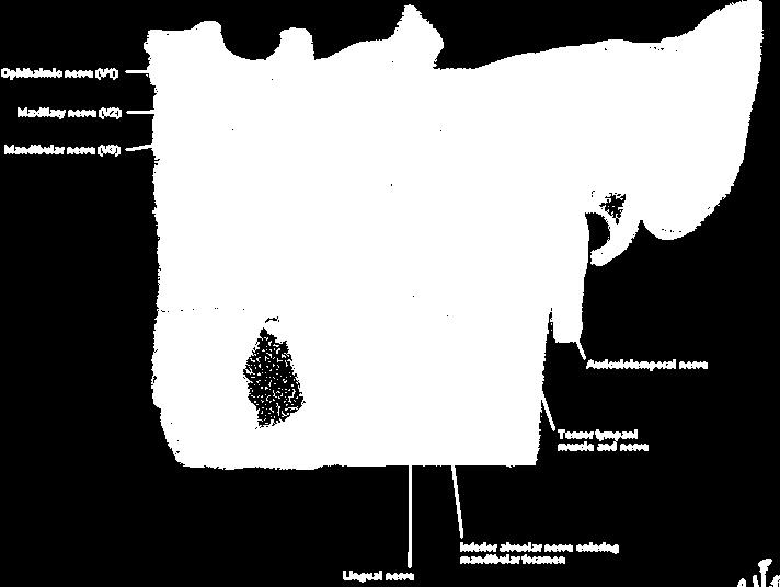
Lec [8]: Mandibular nerve:
![Lec [8]: Mandibular nerve:](/thumbs/94/121295776.jpg "Lec [8]: Mandibular nerve:") Lec [8]: Mandibular nerve: The mandibular branch from the trigeminal ganglion lies in the middle cranial fossa lateral to the cavernous sinus. With the motor root of the trigeminal nerve [motor roots lies
Lec [8]: Mandibular nerve: The mandibular branch from the trigeminal ganglion lies in the middle cranial fossa lateral to the cavernous sinus. With the motor root of the trigeminal nerve [motor roots lies
Temporal fossa Infratemporal fossa Pterygopalatine fossa Terminal branches of external carotid artery Pterygoid venous plexus
 Outline of content Temporal fossa Infratemporal fossa Pterygopalatine fossa Terminal branches of external carotid artery Pterygoid venous plexus Boundary Content Communication Mandibular division of trigeminal
Parotid Gland, Temporomandibular Joint and Infratemporal Fossa
 M1 - Anatomy Parotid Gland, Temporomandibular Joint and Infratemporal Fossa Jeff Dupree Sanger 9-057 jldupree@vcu.edu Parotid gland: wraps around the mandible positioned between the mandible and the sphenoid
function - sensory & postganglionic sympathetic [communication from the internal carotid plexus in the cavernous sinus] innervation of the mucosa of
![function - sensory & postganglionic sympathetic [communication from the internal carotid plexus in the cavernous sinus] innervation of the mucosa of](/thumbs/74/71276096.jpg "function - sensory & postganglionic sympathetic [communication from the internal carotid plexus in the cavernous sinus] innervation of the mucosa of") Nerves I. Cranial nerves A. Olfactory (CN I) 1. Olfactory bulb 2. Olfactory tract B. Optic n. (CNII) function - carries visual sensory information from the neural retina to the diencephalon & midbrain
Nerves I. Cranial nerves A. Olfactory (CN I) 1. Olfactory bulb 2. Olfactory tract B. Optic n. (CNII) function - carries visual sensory information from the neural retina to the diencephalon & midbrain
Dr.Ban I.S. head & neck anatomy 2 nd y. جامعة تكريت كلية طب االسنان املرحلة الثانية أ.م.د. بان امساعيل صديق 6102/6102
 جامعة تكريت كلية طب االسنان التشريح مادة املرحلة الثانية أ.م.د. بان امساعيل صديق 6102/6102 Parotid region The part of the face in front of the ear and below the zygomatic arch is the parotid region. The
Maxilla, ORBIT and infratemporal fossa. Neophytos C Demetriades MD, DDS, MSc Associate professor European University of Cyprus School of Medicine
 Maxilla, ORBIT and infratemporal fossa Neophytos C Demetriades MD, DDS, MSc Associate professor European University of Cyprus School of Medicine MAXILLA Superior, middle, and inferior meatus Frontal sinus
CN I Olfactory. CN II Optic. CN III Oculomotor. Special Sensory Efferent fibers to Olfactory Bulb. Cribiform Plate of Ethmoid
 CN I Olfactory Efferent fibers to Olfactory Bulb Olfactory Tract Olfactory Bulb Cribiform Plate of Ethmoid Anosmia Loss of sense of smell Uncinate Fits olfactory hallucinations To Olfactory Epithelium
Functional components
 Facial Nerve VII cranial nerve Emerges from Pons Two roots Functional components: 1. GSA (general somatic afferent) 2. SA (Somatic afferent) 3. GVE (general visceral efferent) 4. BE (Special visceral/branchial
Brainstem and Cranial Nerves II. Nerves covered in other lectures. A reminder about embryology. Prof. Stuart Bunt
 Brainstem and Cranial Nerves II Prof. Stuart Bunt Nerves covered in other lectures 1 Olfactory 2 Optic 3,4,6 Extraocular eye muscles 8 Vestibulo-cochlear 5 Motor and Sensory to the face and muscles of
Cranial Nerve VII - Facial Nerve. The facial nerve has 3 main components with distinct functions
 Cranial Nerve VII - Facial Nerve The facial nerve has 3 main components with distinct functions Somatic motor efferent Supplies the muscles of facial expression; posterior belly of digastric muscle; stylohyoid,
General Sensory Pathways of the Face Area, Taste Pathways and Hearing Pathways
 General Sensory Pathways of the Face Area, Taste Pathways and Hearing Pathways Lecture Objectives Describe pathways for general sensations (pain, temperature, touch and proprioception) from the face area.
Laith Sorour. Facial nerve (vii):
:") Laith Sorour Cranial nerves 7 & 8 Hello, there are edited slides please go back to them to see pictures, they are not that much important in this lecture but still, and yes slides are included :p Let s
Laith Sorour Cranial nerves 7 & 8 Hello, there are edited slides please go back to them to see pictures, they are not that much important in this lecture but still, and yes slides are included :p Let s
SCHOOL OF ANATOMICAL SCIENCES Mock Run Questions. 4 May 2012
 SCHOOL OF ANATOMICAL SCIENCES Mock Run Questions 4 May 2012 1. With regard to the muscles of the neck: a. the platysma muscle is supplied by the accessory nerve. b. the stylohyoid muscle is supplied by
Faculty of Dental Medicine and Surgery. Sem 4 Cranial Nerves Dr. Abbas Garib Alla
 Faculty of Dental Medicine and Surgery Sem 4 Cranial Nerves Dr. Abbas Garib Alla Cranial Nerves I through XII FUNCTIPONAL CLSSIFICATION OF THE CN parasympathetic nerves 1973 PHARYNGEAL ARCHES nerves 1975
Doctor Osama Asa ad Khader. Mohammad Alsalem
 6 Doctor 2015 Osama Asa ad Khader Mohammad Alsalem A quick revision for the spinal cord blood supply: Arterial Blood supply of spinal cord The spinal cord got its arterial supply by two ways: Longitudinal
DEVELOPMENT OF BRAIN
 Ahmed Fathalla OBJECTIVES At the end of the lecture, students should: List the components of brain stem. Describe the site of brain stem. Describe the relations between components of brain stem & their
INTRODUCTION: ANATOMY UNDERLYING CLINICAL TESTS OF CRANIAL NERVES
 INTRODUCTION: ANATOMY UNDERLYING CLINICAL TESTS OF CRANIAL NERVES CRANIAL NERVE I - OLFACTORY I - OLFACTORY NERVE - SMELL TEST: SMELL ODORS (note: not ammonia; pain in nasal cavity CN5 DAMAGE: LOSS OF
Dr. Sami Zaqout, IUG Medical School
 The skull The skull is composed of several separate bones united at immobile joints called sutures. Exceptions? Frontal bone Occipital bone Vault Cranium Sphenoid bone Zygomatic bones Base Ethmoid bone
MAXILLA, ORBIT & PTERYGOPALATINE FOSSA. Neophytos C Demetriades MD, DDS, MSc Associate professor European University of Cyprus School of Medicine
 MAXILLA, ORBIT & PTERYGOPALATINE FOSSA Neophytos C Demetriades MD, DDS, MSc Associate professor European University of Cyprus School of Medicine Maxilla MAXILLA Superior, middle, and inferior meatus Frontal
Trigeminal Nerve Anatomy. Dr. Mohamed Rahil Ali
 Trigeminal Nerve Anatomy Dr. Mohamed Rahil Ali Trigeminal nerve Largest cranial nerve Mixed nerve Small motor root and large sensory root Motor root Nucleus of motor root present in the pons and medulla
Trigeminal Nerve (V)
") Trigeminal Nerve (V) Lecture Objectives Discuss briefly how the face is developed. Follow up the course of trigeminal nerve from its point of central connections, exit and down to its target areas. Describe
Trigeminal Nerve (V) Lecture Objectives Discuss briefly how the face is developed. Follow up the course of trigeminal nerve from its point of central connections, exit and down to its target areas. Describe
CN modalities Sensory: SSA (Vision) Mixed: GSE, proprioceptive. Mixed: GSE, proprioceptive
 Mixed: GSE, proprioceptive. Mixed: GSE, proprioceptive") CN 2 3 4 6 modalities Sensory: SSA (Vision) course Rods and cones of the retina bipolar neurons gangli on cells Optic nerve optic foramen Optic chiasm Optic tracts Sup colliculi LGN Optic radiation cortex
CN 2 3 4 6 modalities Sensory: SSA (Vision) course Rods and cones of the retina bipolar neurons gangli on cells Optic nerve optic foramen Optic chiasm Optic tracts Sup colliculi LGN Optic radiation cortex
Anatomy of the Trigeminal Nerve
 19 Anatomy of the Trigeminal Nerve.1 Introduction 0. The Central Part of the Trigeminal Nerve 1..1 Origin 1.. Trigeminal Nuclei.3 The Peripheral Part of the Trigeminal Nerve 4.3.1 Ophthalmic Nerve 4.3.
ACTIVITY 7: NERVOUS SYSTEM HISTOLOGY, BRAIN, CRANIAL NERVES
 ACTIVITY 7: NERVOUS SYSTEM HISTOLOGY, BRAIN, CRANIAL NERVES LABORATORY OBJECTIVES: 1. Histology: Identify structures indicated on three different slides or images of nervous system tissue. These images
PHYSIOLOHY OF BRAIN STEM
 PHYSIOLOHY OF BRAIN STEM Learning Objectives The brain stem is the lower part of the brain. It is adjoining and structurally continuous with the spinal cord. 1 Mid Brain 2 Pons 3 Medulla Oblongata The
Done by : Areej Al-Hadidi
 Brainstem &diencephalon Done by : Areej Al-Hadidi Brainstem Functions Ascending and descending tracts Reflex centers Cardiovascular and respiratory centers Coughing, sneezing, swallowing Nuclei of the
PTERYGOPALATINE FOSSA
 PTERYGOPALATINE FOSSA Outline Anatomical Structure and Boundaries Foramina and Communications with other spaces and cavities Contents Pterygopalatine Ganglion Especial emphasis on certain arteries and
Cranial Nerve VII & VIII
 Cranial Nerve VII & VIII Lecture Objectives Follow up the course of facial nerve from its point of central connections, exit and down to its target areas. Follow up the central connections of the facial
For the following questions, indicate the letter that corresponds to the SINGLE MOST APPROPRIATE ANSWER
 GROSS ANATOMY EXAMINATION May 15, 2000 For the following questions, indicate the letter that corresponds to the SINGLE MOST APPROPRIATE ANSWER 1. Pain associated with an infection limited to the middle
Oral cavity : consist of two parts: the oral vestibule and the oral cavity proper. Oral vestibule : is slit like space between.
 Oral cavity Oral cavity : consist of two parts: the oral vestibule and the oral cavity proper Oral vestibule : is slit like space between the teeth, buccal gingiva, lips, and cheeks 1 Oral cavity Oral
Cranial Nerves and Spinal Cord Flashcards
 1. Name the cranial nerves and their Roman numeral. 2. What is Cranial Nerve I called, and what does it 3. Scientists who are trying to find a way to make neurons divide to heal nerve injuries often study
Cranial Nerves IX-X (Glossopharyngeal & Vagus Nerves)
") Cranial Nerves IX-X (Glossopharyngeal & Vagus Nerves) Please view our Editing File before studying this lecture to check for any changes. Color Code Important Doctors Notes Notes/Extra explanation Objectives
Cranial Nerves IX-X (Glossopharyngeal & Vagus Nerves) Please view our Editing File before studying this lecture to check for any changes. Color Code Important Doctors Notes Notes/Extra explanation Objectives
This lab activity is aligned with Visible Body s Human Anatomy Atlas app.
 1 This lab activity is aligned with Visible Body s Human Anatomy Atlas app. Learn more at visiblebody.com/professors We've split our Cranial Nerves lab activity into two parts. Part 1 is pre-lab exercises
Skull-2. Norma Basalis Interna Norma Basalis Externa. Dr. Heba Kalbouneh Associate Professor of Anatomy and Histology
 Skull-2 Norma Basalis Interna Norma Basalis Externa Dr. Heba Kalbouneh Associate Professor of Anatomy and Histology Norma basalis interna Base of the skull- superior view The interior of the base of the
Lec#10 Part 2. Dawood Alatefi. Mariam Hassouneh. Dr.Mohammed Al-Salem
 Lec#10 Part 2 Dawood Alatefi Mariam Hassouneh Dr.Mohammed Al-Salem This part s record is found on the Batch s channel as LabA.3, starting from 0.00min until 26.20min. Hope this ll be an easy part on you;
Lecture 4 The BRAINSTEM Medulla Oblongata
 Lecture 4 The BRAINSTEM Medulla Oblongata Introduction to brainstem 1- Medulla oblongata 2- Pons 3- Midbrain - - - occupies the posterior cranial fossa of the skull. connects the narrow spinal cord
ACTIVITY 7: NERVOUS SYSTEM HISTOLOGY, BRAIN, CRANIAL NERVES NERVOUS SYSTEM TISSUES: HISTOLOGY SLIDES
 ACTIVITY 7: NERVOUS SYSTEM HISTOLOGY, BRAIN, CRANIAL NERVES OBJECTIVES: 1) How to get ready: Read Chapter 14 & 15 McKinley et al., Human Anatomy, 4e. All text references are for this textbook. Read dissection
Blood supply to the brain Blood brain barrier isolates neural tissue from general circulation
 The Brain and Cranial Nerves Objectives Name the major regions of the brain and describe their functions. Discuss the formation, circulation, and functions of the CSF. List the main components of the medulla
Cranial Nerves. Steven McLoon Department of Neuroscience University of Minnesota
 Cranial Nerves Steven McLoon Department of Neuroscience University of Minnesota 1 Course News Change in Lab Sequence Week of Oct 2 Lab 5 Week of Oct 9 Lab 4 2 Sensory and Motor Systems Sensory Systems:
Learning Outcomes. The Carotid 20/02/2013. Scalp, Face, Parotid. Layers of the Scalp. The Parotid Gland. The Scalp. The Carotid The Facial Artery
 Learning Outcomes The Scalp Layers of the Scalp Bleeding from the Scalp The Carotid The Facial Artery Major Muscles of the Face and Jaw(s) Muscles of Mastication Muscles of Facial Expression The Parotid
V. Trigeminal Nerve V 1 Ophthalmic Nerve V 2 Maxillary Nerve V 3 Mandibular Nerve. Clinical Considerations VI. Abducens Nerve
 Cranial Nerves 18 Chapter Outline Cranial Nerves Cranial Nerve Modalities I. Olfactory Nerve II. Optic Nerve III. Oculomotor Nerve IV. Trochlear Nerve V. Trigeminal Nerve V 1 Ophthalmic Nerve V 2 Maxillary
Bones of the skull & face
 Bones of the skull & face Cranium= brain case or helmet Copyright The McGraw-Hill Companies, Inc. Permission required for reproduction or display. The cranium is composed of eight bones : frontal Occipital
Trigeminal Nerve Worksheets, Distributions Page 1
 Trigeminal Nerve Worksheet #1 Distribution by Nerve Dr. Darren Hoffmann Dental Gross Anatomy, Spring 2013 We have drawn out each of the branches of CN V in lecture and you have an idea now for their basic
Infratemporal fossa: Tikrit University college of Dentistry Dr.Ban I.S. head & neck Anatomy 2 nd y.
 Infratemporal fossa: This is a space lying beneath the base of the skull between the lateral wall of the pharynx and the ramus of the mandible. It is also referred to as the parapharyngeal or lateral pharyngeal
Veins of the Face and the Neck
 Veins of the Face and the Neck Facial Vein The facial vein is formed at the medial angle of the eye by the union of the supraorbital and supratrochlear veins. connected through the ophthalmic veins with
Lab 16: PNS: Nerves and Autonomic NS Hamilton Answers to Pre- Lab Assignments
 Lab 16: PNS: Nerves and Autonomic NS Hamilton Answers to Pre- Lab Assignments Pre-Lab Activity 1: 1. a. olfactory nerve b. optic nerve c. oculomotor nerve d. abducens nerve e. trochlear nerve f. trigeminal
The functional Anatomy of the Cranial nerves
 The State Medical and Pharmaceutical University Nicolae Testemitanu Republic of Moldova Department of Human Anatomy The functional Anatomy of the Lecturer Globa Lilian Like spinal nerves, cranial nerves
Introduction to Local Anesthesia and Review of Anatomy
 5-Sep Introduction and Anatomy Review 12-Sep Neurophysiology and Pain 19-Sep Physiology and Pharmacology part 1 26-Sep Physiology and Pharmacology part 2 Introduction to Local Anesthesia and Review of
b. The groove between the two crests is called 2. The neural folds move toward each other & the fuse to create a
 Chapter 13: Brain and Cranial Nerves I. Development of the CNS A. The CNS begins as a flat plate called the B. The process proceeds as: 1. The lateral sides of the become elevated as waves called a. The
mistake ;slides in bold but you still have to go back to our slides to see the figure, tables and some scheme
 Khozama jehad : I am doing my best and I am sorry for any unintended mistake ;slides in bold but you still have to go back to our slides to see the figure, tables and some scheme The Orbit, Orbital Contents
Major Anatomic Components of the Orbit
 Major Anatomic Components of the Orbit 1. Osseous Framework 2. Globe 3. Optic nerve and sheath 4. Extraocular muscles Bony Orbit Seven Bones Frontal bone Zygomatic bone Maxillary bone Ethmoid bone Sphenoid
Parotid Gland. Parotid Gland. Largest of 3 paired salivary glands (submandibular; sublingual) Ramus of Mandible. Medial pterygoid.
 Ramus of Mandible. Medial pterygoid.") Parotid region Parotid Gland Largest of 3 paired salivary glands (submandibular; sublingual) Ramus of Mandible Medial pterygoid Cross section of mandible Masseter D S SCM Parotid Gland Mastoid Process
Parotid region Parotid Gland Largest of 3 paired salivary glands (submandibular; sublingual) Ramus of Mandible Medial pterygoid Cross section of mandible Masseter D S SCM Parotid Gland Mastoid Process
Omran Saeed. Luma Taweel. Mohammad Almohtaseb. 1 P a g e
 2 Omran Saeed Luma Taweel Mohammad Almohtaseb 1 P a g e I didn t include all the photos in this sheet in order to keep it as small as possible so if you need more clarification please refer to slides In
Peripheral Nervous System
 Peripheral Nervous System Sensory Receptors Motor Endings Cranial Nerves The Four Plexuses Extremities Review of Reflexes Fast, preprogrammed, inborn, automatic responses Occur in the CNS at the spinal
Omar Sami. Aseel Abdeen. Muhammad Al-Salem. 1 P a g e
 Omar Sami Aseel Abdeen Muhammad Al-Salem 1 P a g e Using only section 2 record, I wrote this sheet; as the video is not ready yet. Despite pointing the structures, I ve tried to include all the scientific
The Seventh Cranial Nerve The Facial By Prof. Dr. Muhammad Imran Qureshi
 The Seventh Cranial Nerve The Facial By Prof. Dr. Muhammad Imran Qureshi Functional Components: SVE: Fibers originate from nucleus of facial nerve, and supply facial muscles GVE: Fibers derived from superior
THE BRAINSTEM. Raymond S. Price, MD University of Pennsylvania
 THE BRAINSTEM Raymond S. Price, MD University of Pennsylvania Overview of Brainstem Functions The brainstem serves numerous crucial neurologic functions. The most clinically relevant functions include:
lecture #2 Done by : Tyma'a Al-zaben
 lecture #2 Done by : Tyma'a Al-zaben ** Hello SERTONIN! note:: the slide included within the sheet but make sure back to slide for pictures in the previous lecture we talk about ascending tract and its
The orbit-1. Dr. Heba Kalbouneh Assistant Professor of Anatomy and Histology
 The orbit-1 Dr. Heba Kalbouneh Assistant Professor of Anatomy and Histology Orbital plate of frontal bone Orbital plate of ethmoid bone Lesser wing of sphenoid Greater wing of sphenoid Lacrimal bone Orbital
Skull-2. Norma Basalis Interna. Dr. Heba Kalbouneh Assistant Professor of Anatomy and Histology
 Skull-2 Norma Basalis Interna Dr. Heba Kalbouneh Assistant Professor of Anatomy and Histology Norma basalis interna Base of the skull- superior view The interior of the base of the skull is divided into
University of Palestine. Midterm Exam 2013/2014 Total Grade:
 Course No: DNTS2208 Course Title: Head and Neck Anatomy Date: 09/11/2013 No. of Questions: (50) Time: 1hour Using Calculator (No) University of Palestine Midterm Exam 2013/2014 Total Grade: Instructor
Ms. K. GOWRI. M.Pharm., Lecturer.
 Ms. K. GOWRI. M.Pharm., Lecturer. CENTRAL NERVOUS SYSTEM (CNS) central nervous system consists of brain and spinal cord membrane covering the brain and spinal cord are surrounded by three membrane Meninges
Mohammad Hisham Al-Mohtaseb. Lina Mansour. Reyad Jabiri. 0 P a g e
 2 Mohammad Hisham Al-Mohtaseb Lina Mansour Reyad Jabiri 0 P a g e This is only correction for the last year sheet according to our record. If you already studied this sheet just read the yellow notes which
Prevertebral Region, Pharynx and Soft Palate
 Unit 20: Prevertebral Region, Pharynx and Soft Palate Dissection Instructions: Step1 Step 2 Step 1: Insert your fingers posterior to the sternocleidomastoid muscle, vagus nerve, internal jugular vein,
M555 Medical Neuroscience Lab 1: Gross Anatomy of Brain, Crainal Nerves and Cerebral Blood Vessels
 M555 Medical Neuroscience Lab 1: Gross Anatomy of Brain, Crainal Nerves and Cerebral Blood Vessels Anatomical Directions Terms like dorsal, ventral, and posterior provide a means of locating structures
The sebaceous glands (glands of Zeis) open directly into the eyelash follicles, ciliary glands (glands of Moll) are modified sweat glands that open
 open directly into the eyelash follicles, ciliary glands (glands of Moll) are modified sweat glands that open") The Orbital Region The orbits are a pair of bony cavities that contain the eyeballs; their associated muscles, nerves, vessels, and fat; and most of the lacrimal apparatus upper eyelid is larger and more
The Orbital Region The orbits are a pair of bony cavities that contain the eyeballs; their associated muscles, nerves, vessels, and fat; and most of the lacrimal apparatus upper eyelid is larger and more
Chapter 14: The Brain and Cranial Nerves. Copyright 2009, John Wiley & Sons, Inc.
 Chapter 14: The Brain and Cranial Nerves Development of the Brain Three to four-week embryo: prosencephalon, mesencephalon and rhombencephalon. Five-week embryo: telencephalon (cerebrum), diencephalon
REVIEW OF HEAD AND NECK CRANIAL NERVES AND EVERYTHING ELSE
 REVIEW OF HEAD AND NECK CRANIAL NERVES AND EVERYTHING ELSE OLFACTORY NERVE CN I ANTERIOR CRANIAL FOSSA CRISTA GALLI OF ETHMOID OLFACTORY FORAMINA IN CRIBIFORM PLATE OF ETHMOID BONE CN I OLFACTORY NERVE
APRIL
 APRIL - 2003 OCTOBER - 2003 February 2009 [KU 652] Sub. Code : 4131 FIRST B.D.S DEGREE EXAMINATION (Modified Regulations III) Paper I HUMAN ANATOMY, HISTOLOGY AND EMBRYOLOGY Time : Three hours
Biology 218 Human Anatomy. Adapted from Martini Human Anatomy 7th ed. Chapter 6 The Skeletal System: Axial Division
 Adapted from Martini Human Anatomy 7th ed. Chapter 6 The Skeletal System: Axial Division Introduction The axial skeleton: Composed of bones along the central axis of the body Divided into three regions:
Anatomic Relations Summary. Done by: Sohayyla Yasin Dababseh
 Anatomic Relations Summary Done by: Sohayyla Yasin Dababseh Anatomic Relations Lecture 1 Part-1 - The medial wall of the nose is the septum. - The vestibule lies directly inside the nostrils (Nares). -
Dr. Sami Zaqout Faculty of Medicine IUG
 Auricle External Ear External auditory meatus The Ear Middle Ear (Tympanic Cavity) Auditory ossicles Internal Ear (Labyrinth) Bony labyrinth Membranous labyrinth External Ear Auricle External auditory
University of Palestine. Midterm Exam 2013/2014 Total Grade:
 [ Course No: DNTS2208 Course Title: Head and Neck Anatomy Date: 17/11/1024 No. of Questions: (52) Time: 2hours Using Calculator (No) University of Palestine Midterm Exam 2013/2014 Total Grade: Instructor
The Cranial Nerves & Spinal nerves. Departemen Anatomi Fakultas Kedokteran USU
 The Cranial Nerves & Spinal nerves Departemen Anatomi Fakultas Kedokteran USU Names of cranial nerves ⅠOlfactory nerve ⅡOptic nerve ⅢOculomotor nerve Ⅳ Trochlear nerve ⅤTrigeminal nerve ⅥAbducent nerve
Sensory system. Dr. Carmen E. Rexach Anatomy 35 Mt San Antonio College
 Sensory system Dr. Carmen E. Rexach Anatomy 35 Mt San Antonio College Sensory receptors Detect stimuli Classified by structure Origin Distribution Modality Structural Classification naked nerve endings
Bony orbit Roof The orbital plate of the frontal bone Lateral wall: the zygomatic bone and the greater wing of the sphenoid
 Bony orbit Roof: Formed by: The orbital plate of the frontal bone, which separates the orbital cavity from the anterior cranial fossa and the frontal lobe of the cerebral hemisphere Lateral wall: Formed
Review or skim Ch 12 on the vascular supply of the brain. Just look at pictures and legends for the clinical part at the end.
 Dental Neuroanatomy January 20 and 27, 10-12, 2011 Suzanne S. Stensaas, Ph.D. Dear Students: Please print these notes and bring them with you. My style is to use a Tablet PC and I draw on either a Word
HEAD AND NECK ANATOMY PRACTICE QUESTIONS
 HEAD AND NECK ANATOMY PRACTICE QUESTIONS 1. A patient complains that he has lost sensation on his face and that the skin of his face feels numb. The physician tests tactile acuity by touching the forehead
Unit 18: Cranial Cavity and Contents
 Unit 18: Cranial Cavity and Contents Dissection Instructions: The calvaria is to be removed without damage to the dura mater which is attached to the inner surface of the calvaria. Cut through the outer
EXAM NUMBER STRUCTURAL BASIS OF MEDICAL PRACTICE EXAMINATION 7 October 28, PART l. Answer in the space provided. (9 pts)
") STRUCTURAL BASIS OF MEDICAL PRACTICE EXAMINATION 7 October 28, 2005 PART l. Answer in the space provided. (9 pts) 1. Identify the structures. (3 pts) a. _Frontal Sinus b. _Lateral Posterior Inferior Nasal
STRUCTURAL BASIS OF MEDICAL PRACTICE EXAMINATION 7 October 28, 2005 PART l. Answer in the space provided. (9 pts) 1. Identify the structures. (3 pts) a. _Frontal Sinus b. _Lateral Posterior Inferior Nasal
Brain and Cranial Nerves (Ch. 15) Human Anatomy lecture. caudal = toward the spinal cord)
 Human Anatomy lecture. caudal = toward the spinal cord)") Insight: Some cranial nerve disorders Brain and Cranial Nerves (Ch. 15) Human Anatomy lecture I. Overview (Directional terms: rostral = toward the forehead caudal = toward the spinal cord) A. 3 Major parts
Insight: Some cranial nerve disorders Brain and Cranial Nerves (Ch. 15) Human Anatomy lecture I. Overview (Directional terms: rostral = toward the forehead caudal = toward the spinal cord) A. 3 Major parts
Chapter 27: The ear, intracranial region and cranial nerves. Ear. The External Ear. This consists of the auricle and the external acoustic meatus.
 Chapter 27: The ear, intracranial region and cranial nerves Ear The ear is the organ of hearing and balance. It is divided into an external ear directing sound waves to the tympanic membrane, a middle
3-Deep fascia: is absent (except over the parotid gland & buccopharngeal fascia covering the buccinator muscle)
") The Face 1-Skin of the Face The skin of the face is: Elastic Vascular (bleed profusely however heal rapidly) Rich in sweat and sebaceous glands (can cause acne in adults) It is connected to the underlying
The Face 1-Skin of the Face The skin of the face is: Elastic Vascular (bleed profusely however heal rapidly) Rich in sweat and sebaceous glands (can cause acne in adults) It is connected to the underlying
Chapter 13: The Peripheral Nervous System
 Chapter 13: The Peripheral Nervous System Objectives: 1. Define peripheral nervous system and list its components. 2. Classify general sensory receptors by structure, stimulus detected, and body location.