Cardiomyopathies Dilated cardiomyopathy: idiopathic, ischemic and others
|
|
|
- Jeremy Snow
- 5 years ago
- Views:
Transcription
1 EUROECHO 2011 TEACHING COURSE Cardiomyopathies Dilated cardiomyopathy: idiopathic, ischemic and others Regina Ribeiras M.D., FESC Hospital de Santa Cruz Carnaxide, Portugal
2 EHJ (2008) 29, Dilated Cardiomyopathy (DCM) A myocardial disorder in which the heart muscle is structurally and functionally abnormal, in the absence of : coronary artery disease, hypertension, valvular disease and congenital heart disease, sufficient to cause the observed myocardial abnormality.


3 45% 34% DCM phenotype: LV remodeling 27% LVEDd >117% may precede systolic dysfunction EF% <45% /FS<25% LV spherical dilatation Normal or reduced wall thickness a progressive disease along time


4 DCM phenotype : LV remodeling No regional WMAbn Mechanical Dyssynchrony RV involvment 4 chambers dilatation MV Regurgitation (functional)


5 RWMab Regional wall motion abnormalities (RWMab) Ischemic DCM - coronary distribution (can be faint in extreme LV remodelling) idcm some regionality basal posterolateral segments relatively preserved systolic function Friedrich s ataxia - posterior wall motion abnormality Takotsubo - apical hypokinesia/akinesia with preservation of the basal segments Radiation RWMab


6 Functional Mitral Regurgitation incomplete closure of normal MV leaflets Secondary MR to DCM apical tenting, annular dilatation, and ventricular dyssynchrony.
 subendocardial transmural Non-Ischemic patterns (GLE): Mid-wall Epicardial Global")
7 Magnetic Resonance Imaging : DELAYED ENHANCEMENT Ischemic Non-Ischemic Ischemic Patterns (GLE) subendocardial transmural Non-Ischemic patterns (GLE): Mid-wall Epicardial Global endocardial


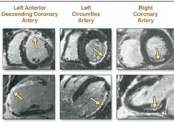
8 ISCHEMIC Non-ISCHEMIC No-LGE Ananthasubramaniam R et al. Heart Fail Rev Dec10 Iles L et al JACC 11, 57:821
 ±12% Mestroni et al. EHJ 1999 ; 20:93 Luk A, et al,j Clin Pathol 2009;62:219")
9 Diagnostic criteria for DCM or using the formula =( 45.3x(BSA) 1/3 (0.03xage)-7.2) ±12% Mestroni et al. EHJ 1999 ; 20:93 Luk A, et al,j Clin Pathol 2009;62:219
10 EHJ (2008) 29, specific morphological and functional phenotypes

 the clinical manifestation of the disease appears to be typically")
11 Familial DCM more > 35% (40-60%) of cases of DCM have a genetic etiology (a monogenic familial disease) Elliott P, et al. Eur Heart J 2008;29: Mode of inheritance : autosomal (33genes); X-Linked (2) variable penetrance = 80% in families with an autosomal dominant pattern of transmission (which is the most frequent mode of inheritance) the clinical manifestation of the disease appears to be typically age-dependent < 20 yrs 10 % yrs 34 % yrs 60 % >40 yrs 90 % Mestroni L, et al. Familial dilated cardiomyopathy. Br Heart J 1994; 72:
12 sarcomeric proteins cytoskeleton proteins phenotype is highly heterogeneous Isolated or Syndromic proteins of the nuclear membrane cardiolaminopathies

13 LMNA gene mutations Need for Early Diagnosis Probands and mutated healthy persons highly penetrant, age-dependent, male gender early indications for ICD implantation so as to prevent SCD and provide a possible bridge to heart transplantation in end stage HF. malignant disease : Risk factors: NSVT LVEF<45% ICD implantation in patients who are candidates for pacemaker implantation due to AVBlock low dynamic noncompetitive sport activity high rates of maligant arrhythmias even with mild LV-DCM phenotype (HF / high risk of SCD before the onset of HF). Fatkin D et al. NEJM 1999;341: Pasotti, et al. JACC 2008;52:1250
14 1 st : TAKE A 3-4 generation FAMILY HISTORY JACC 2011;57: d : If Positive ( >1 familly member afected) ECHO screening of first-degree relatives 3 d : Consider GENETIC TESTING : check for phenotipic red flags for high-risk mutations (Lamin A/C)

15 4 th : Genetic testing Identification probands and family members Combination of DCM and AVBlock Co-existent myopathy (EDMD2 or variable myopathy) and increased serum creatine phosphokinase levels, especially if AVBlock, Familial Dilated Cardiomyopathy Registry Research Group. J Am Coll Cardiol 2003;41: Clinical and Genetic Issues in Familial Dilated Cardiomyopathy : Hershberger and Siegfried. JACC 2011;57:1641 9
16 5 th : ECHO Screening of first degree relatives In Asymptomatic Relatives of a proband (genotype-phenotype patient) isolated LVE 20% mild contractile impairement (dfs)- 6% DCM -histological and immunohistochemical changes similar to those with established disease 3% Baig MK. et al. JACC 1998: 31 Necessity of ECHOCardiographic Serial Assessment over a lengthy FUP 4.6% asymptomatic relatives with DCM have a significant medium-term risk for disease progression For pre-clinical Dx based on the observed incidence and risk for progression Progression to DCM - occurred in 13 (10%) relatives with LVE or dfs versus 3 (1.3%) healthy relatives Mahon N.et al. Ann Intern Med. 2005;143:108
 every 3 to 5 years Because.")
17 Stage A At high risk for HF, no structural disease or symptons of HF Pre-Clinical diagnosis Early Detection of Structural Abnormalities subjects with negative DCM-phenotype in a Familial-DCM kindred warrant rescreening for disease detection (examination, ECG and echocardiography) every 3 to 5 years Because.. Age-dependency and early treatment by ACE-inhibitors or beta-blockers could limit the progression of the disease once detected 2009 ACCF/AHA Heart Failure Guidelines Circulation April 14, 2009 Clinical and Genetic Issues in Familial Dilated Cardiomyopathy : Hershberger and Siegfried. JACC 2011;57:1641 9
 for each")

18 controls <10yrs >10yrs MDE- MDE+ Nr. EF% EF<55% A. progressive reduction in circumferential strain (CS) for each stratum (Groups B to E) B. ALL control subjects have CS higher -16%, and no DMD subjects have CS better than -16%. Hor N. et al. JACC 2009;53:
19 EHJ (2008) 29, Non-Familial DCM ( Disease sub-type) Myocarditis (infective/toxic/immune) Kawasaki disease Eosinophilic (Churg Strauss syndrome) Viral persistence Drugs ( ephedra, anthracyclines, trastuzumab, cyclofosfamide) Pregnancy Endocrine Nutritional thiamine, carnitine, selenium, hypophosphataemia, hypocalcaemia Tachycardiomyopathy Alcohol


 (")
20 specific associated features (spontaneous echo contrast thrombus ) ( anticoagulant )
 As early as 3 months: the lateral S between the normal cohort and the CM group differed significantly (9.1±1.6 cm/s and 6.4±0.6 cm/s, p<0.")
21 Early subclinical detection of Trastuzumab mediated CM Prospective study of 42 women between at a single tertiary care center Out of 41 patients (mean age 47±9 years), 10 (25%) developed Trastuzumab mediated cardiomyopathy (CM) As early as 3 months: the lateral S between the normal cohort and the CM group differed significantly (9.1±1.6 cm/s and 6.4±0.6 cm/s, p<0.05) Peak global longitudinal and radial strain decreased in the CM group Left ventricular ejection fraction (LVEF) decreased (10% below 55%) at 6 months of follow-up in all 10 patients Fallah-Rad N. et al, JACC 2011;57:2263
 Echocardiographic FUP J Am Coll Cardiol")
22 Trastuzumab mediated CM 25% patients (10/41) cardiac biomarkers did not predict the development of cardiac dysfunction TVI and strain imaging were able to detect preclinical changes in LV function, prior to conventional changes in LVEF prophylactic administration of cardioprotective agents (ACEi ; BB) Echocardiographic FUP J Am Coll Cardiol 2011;57:


23 During Trastuzumab Therapy under ACEi +BB ; NYHA II after Trastuzumab suspension under ACEi +BB GLS -9% GLS -20%
24 Dilated Cardiomyopathy Clinical courses are heterogeneous under medical therapy (ACEI and/or BB) Rapidly progressive course, high mortality rates, need for inotropic/lvad and TX. Response to medical therapy (reverse remodelling) - 50% Healing courses 16% (acute miocarditis ; peripartum DCM) 8 Year : Transplant free survival (under medical therapy) 94% - normalized LVEF 83% - NYHA class I-II and LVEF >40% 64% - NYHA class I-II and LVEF 40% 31% - NYHA III-IV Sugrue et al Ann Inter Med 1992,117
25 2009 ACCF/AHA Heart Failure Guidelines Circulation April 14, 2009 A Heart Failure Society of America Practice Guideline, J Cardiac Fail 2009;15:83-9 Heart Failure- Therapeutic Management aetiology-independent starting Stage B (structural heart disease /No signs or symptons of HF) Beta blockers / ACE inhibitors / ARB /diuretics >>>> Cardiac Ressynchronization Therapy ; MV surgery ; LVAD; Cardiac Tx Early intervention: 1. Accurate and Early diagnosis 2. Identification of modifiable prognostic factors : dyssynchony MV regurgitation
26 Diagnostic criteria: DCM LV assessment accurate Chamber Dimensions, indexed to BSA LV Dilatation LV end-diastolic dimension >117% predicted value corrected for age and body surface area (2 SD+5%) Henry equation: (45.3x BSA) 1/3 (0.03 x age) 7.2) ±12% Global Systolic LV function Ejection fraction (EF) <45%, and/or a fractional shortening <25%


27 DCM ECHO MEASUREMENTS Recommendations for Chamber Quantification: A Report from the American Society of Echocardiography s Guidelines and Standards Committee and the Chamber Quantification Writing Group, Developed In Conjunction with the European Association of Echocardiography, a Branch of the European Society of Cardiology JASE Dec.2005 LV Volumes and Ejection Fraction Simpson s Rule or Method of Discs accurate chamber dimensions/volumes, indexed to BSA Global Systolic LV function


28 Jenkins C.et al. European Heart Journal (2009) 30, 98

 10, 82 88 2DSTE -")
 (higher LVESV) smaller")
29 LV volumes and LVEF using 2D speckle-tracking Nishikage T, Lang.R. et al. EJE (2009) 10, DSTE - underestimation of LVEDV and LVEF (mean 8%) (higher LVESV) smaller inter-observer variability in LV volumes and LVEF
30 Identification of a Structural and Functional Abnormality Echocardiography Assessment Class I ( Level of Evidence C ) LV : numerical estimate of EF, dimensions/volumes, wall thickness, chamber geometry & regional wall motion. RV : size and systolic performance Atrial size : LA dimensions and/or volumes Valves : exclude primary valve disease secondary valve changes (MR/TR) Noninvasive hemodynamic data : Stroke volume LV filling and left atrial pressure systolic pulmonary artery pressure and central venous pressure 2009 ACCF/AHA Heart Failure Guidelines Circulation April 14, 2009
 Moderate (pseudonormal) or severe (restrictive filling) ; E/e >15")
 Presence, severity, mechanism >0,20cm2 Dobutamine stress echo D.E. Thomas et al.")
31 Prognosis in DCM echocardiographic indicators Prognostic indicator LV size and systolic function LV diastolic function Filling pressures Right ventricular function Key echocardiographic features Ejection fraction (sphericity) Moderate (pseudonormal) or severe (restrictive filling) ; E/e >15 TAPSE <14mm Pulmonary hypertension Left atrial size Mitral regurgitation Contractile reserve Tricuspid regurgitation velocity >40mmHg Left atrial volume index (>68ml/m2) Presence, severity, mechanism >0,20cm2 Dobutamine stress echo D.E. Thomas et al. Echocardiography in guiding management in DCM EHJ 2009,10.


32 Pozzolli et al. Circ 1997
33 The aditive prognostic value of filling pressure evaluation LV ejection fraction 40%; Hillis et al. JACC 2004;43:
34 the aditive prognostic value of LA volume Moller et al Circulartion :2207 Beinart JACC 2004;44: times higher risk of an adverse outcome Sallach J. et al JACC 2002;40: )

35 The prevalence of RV systolic dysfunction in reduced EF%-HF : RV FAC = 63% TAM = 76% TV Sm =73% It is related to the severity of LV dysfunction and biventricular involvement in the disease process rather than secondary to pulmonary hypertension. Puwanant S. et al. European Journal of Echocardiography (2009) 10, Quantification of right ventricular function tricuspid annular proximal systolic excursion (TAPSE) < 14mm (adverse prognosis) Ghio S et al. Am J Cardiol 2000;85:

36 NEW PROGNOSTICATORS Myocardial Fibrosis : GDE (MRI) Positive hyperenhancement = visually defined as >2 standard deviations of the signal intensity of the non-enhanced myocardium.

37 103 Pts Median follow-up days LGE + : 100% ICM (n= 42) 51% NICM (n = 61) NICM - LGE - NICM - LGE - NICM - LGE + NICM - LGE + Iles L et al. J Am Coll Cardiol 2011;57:821 8

38

39 M.M. Kansal et al. EHJ of Cardiovascular Imaging Journal of Echocardiography, 2012 : 43-49

40 Cardiac Resynchronization Therapy NonResponse 30-40% Better Selection adjunctive to QRS! 1. Exclude : Residual ischemia Non-Functional Mitral Regurgitation 2. Identify Mechanical Dyssynchrony 3. Contractile reserve ; Scar burden / Viability


41 Stress Echo: contractil reserve Inducible septal flash


")
42 Severe delayed LV wall guide to the best lead location Ypenburg et al, JACC 2008;52: Device Optimization (AV intervals) Gorcsan III JASE 2008; 21
43 CONCLUSIONS: 1.DCM is the most common cardiomyopathy, occurring primarily due to genetic defects or secondarily as a consequence of multiple factors. The differential diagnosis remains quite broad since many pathologies can present as DCM so multiimaging is frequently needed. 2.From a clinical point of view, the most important is to differentiate between familiar-dcm, and non-familiar DCM, since genetic testing has to be done specially in the high-risk genetic-dcm (LaminA/C) 3. Early intervention based upon accurate diagnosis remains the mainstay of pharmacologic treatment which is independent of the aetiology and may prevent disease progression and its complications. 4. Echo- imaging is a powerfool tool for Diagnosis and DDx (DCM-phenocopies) identification of associated cardiac abnormalities such as valve disease highlight features requiring specific therapeutic management (thrombus) identify prognostic markers and guiding therapy ( CRT ; MV repair)

44 EUROECHO 2011 TEACHING COURSE Dilated cardiomyopathy
LV FUNCTION ASSESSMENT: WHAT IS BEYOND EJECTION FRACTION
 LV FUNCTION ASSESSMENT: WHAT IS BEYOND EJECTION FRACTION Jamilah S AlRahimi Assistant Professor, KSU-HS Consultant Noninvasive Cardiology KFCC, MNGHA-WR Introduction LV function assessment in Heart Failure:
Imaging in dilated cardiomyopathy : factors associated with a poor outcome
 Imaging in dilated cardiomyopathy : factors associated with a poor outcome Johan De Sutter, MD, PhD, FESC AZ Maria Middelares Gent and University Gent - Belgium Dilated cardiomyopathy Cardiomyopathy with
Imaging in Heart Failure: A Multimodality Approach. Thomas Ryan, MD
 Imaging in Heart Failure: A Multimodality Approach Thomas Ryan, MD Heart Failure HFrEF HFpEF EF50% Lifetime risk 20% Prevalence 6M Americans Societal costs - $30B 50% 5-year survival 1 Systolic
1. LV function and remodeling. 2. Contribution of myocardial ischemia due to CAD, and
 1 The clinical syndrome of heart failure in adults is commonly associated with the etiologies of ischemic and non-ischemic dilated cardiomyopathy, hypertrophic cardiomyopathy, hypertensive heart disease,
Echocardiographic Evaluation of the Cardiomyopathies. Stephanie Coulter, MD, FACC, FASE April, 2016
 Echocardiographic Evaluation of the Cardiomyopathies Stephanie Coulter, MD, FACC, FASE April, 2016 Cardiomyopathies (CMP) primary disease intrinsic to cardiac muscle Dilated CMP Hypertrophic CMP Infiltrative
Highlights from EuroEcho 2009 Echo in cardiomyopathies
 Highlights from EuroEcho 2009 Echo in cardiomyopathies Bogdan A. Popescu University of Medicine and Pharmacy, Bucharest, Romania ESC Congress 2010 Hypertrophic cardiomyopathy To determine the differences
Global left ventricular circumferential strain is a marker for both systolic and diastolic myocardial function
 Global left ventricular circumferential strain is a marker for both systolic and diastolic myocardial function Toshinari Onishi 1, Samir K. Saha 2, Daniel Ludwig 1, Erik B. Schelbert 1, David Schwartzman
The difficult patient with mitral regurgitation
 Clinical pathways The difficult patient with mitral regurgitation Stress echo can be the best tool Challenging cases Maria João Andrade, Lisbon PT Management of Severe Chronic Organic MR Echo Exercise
Cardiac Resynchronization Therapy Selection therapy Echocardiography
 Cardiac Resynchronization Therapy Selection therapy Echocardiography Prof. Patrizio LANCELLOTTI CHU Sart Tilman, Liège April, 2010 Candidates for CRT : class IA NYHA Functional Class III or IV (Subjective)
Role of Stress Echo in Valvular Heart Disease. Satoshi Nakatani Osaka University Graduate School of Medicine Osaka, Japan
 Role of Stress Echo in Valvular Heart Disease Satoshi Nakatani Osaka University Graduate School of Medicine Osaka, Japan Exercise echocardiography Dobutamine echocardiography Usefulness of exercise echo
Strain Imaging: Myocardial Mechanics Simplified and Applied
 9/28/217 Strain Imaging: Myocardial Mechanics Simplified and Applied John Gorcsan III, MD Professor of Medicine Director of Clinical Research Division of Cardiology VECTORS OF CONTRACTION Shortening Thickening
Functional Mitral Regurgitation
 Club 35 - The best in heart valve disease - Functional Mitral Regurgitation Steven Droogmans, MD, PhD UZ Brussel, Jette, Belgium 08-12-2011 Euroecho & other Imaging Modalities 2011 No conflicts of interest
The road to successful CRT implantation: The role of echo
 The road to successful CRT implantation: The role of echo Tae-Ho Park Dong-A University Hospital, Busan, Korea Terminology Cardiac Resynchronization Therapy (CRT) = Biventricular pacing (BiV) = Left ventricular
27-year-old professionnal rugby player: asymptomatic
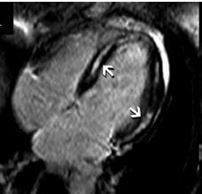
 27-year-old professionnal rugby player: asymptomatic Benefits and limits of cardiac MRI in the young athlete with a suspected heart disease. Philippe PAULE Service de Cardiologie, HIA Clermont Tonnerre,
LV geometric and functional changes in VHD: How to assess? Mi-Seung Shin M.D., Ph.D. Gachon University Gil Hospital
 LV geometric and functional changes in VHD: How to assess? Mi-Seung Shin M.D., Ph.D. Gachon University Gil Hospital LV inflow across MV LV LV outflow across AV LV LV geometric changes Pressure overload
Myocardial Strain Imaging in Cardiac Diseases and Cardiomyopathies.
 Myocardial Strain Imaging in Cardiac Diseases and Cardiomyopathies. Session: Cardiomyopathy Tarun Pandey MD, FRCR. Associate Professor University of Arkansas for Medical Sciences Disclosures No relevant
Right Ventricular Strain in Normal Healthy Adult Filipinos: A Retrospective, Cross- Sectional Pilot Study
 Right Ventricular Strain in Normal Healthy Adult Filipinos: A Retrospective, Cross- Sectional Pilot Study By Julius Caesar D. de Vera, MD Jonnah Fatima B. Pelat, MD Introduction Right ventricle contributes
Heart Failure in Women: Dr Goh Ping Ping Cardiologist Asian Heart & Vascular Centre
 Heart Failure in Women: More than EF? Dr Goh Ping Ping Cardiologist Asian Heart & Vascular Centre Overview Review pathophysiology as it relates to diagnosis and management Rational approach to workup:
Imaging and heart failure
 Imaging and heart failure Jeroen J Bax Dept of Cardiology Leiden Univ Medical Center The Netherlands Davos, feb 2013 Research grants: Medtronic, Biotronik, Boston, St Jude, BMS imaging, GE Healthcare,
Dialysis-Dependent Cardiomyopathy Patients Demonstrate Poor Survival Despite Reverse Remodeling With Cardiac Resynchronization Therapy
 Dialysis-Dependent Cardiomyopathy Patients Demonstrate Poor Survival Despite Reverse Remodeling With Cardiac Resynchronization Therapy Evan Adelstein, MD, FHRS John Gorcsan III, MD Samir Saba, MD, FHRS
좌심실수축기능평가 Cardiac Function
 Basic Echo Review Course 좌심실수축기능평가 Cardiac Function Seonghoon Choi Cardiology Hallym university LV systolic function Systolic function 좌심실수축기능 - 심근의수축으로심실에서혈액을대동맥으로박출하는기능 실제임상에서 LV function 의의미 1Diagnosis
Restrictive Cardiomyopathy
 ESC Congress 2011, Paris Imaging Unusual Causes of Cardiomyopathy Restrictive Cardiomyopathy Kazuaki Tanabe, MD, PhD Professor of Medicine Chair, Division of Cardiology Izumo, Japan I Have No Disclosures
Cardiac MRI in ACHD What We. ACHD Patients
 Cardiac MRI in ACHD What We Have Learned to Apply to ACHD Patients Faris Al Mousily, MBChB, FAAC, FACC Consultant, Pediatric Cardiology, KFSH&RC/Jeddah Adjunct Faculty, Division of Pediatric Cardiology
Aortic stenosis aetiology: morphology of calcific AS,
 How to improve patient selection in aortic stenosis? Fausto J. Pinto, FESC Aortic stenosis aetiology: morphology of calcific AS, bicuspid valve, and rheumatic AS (Adapted from C. Otto, Principles of
Evalua&on)of)Le-)Ventricular)Diastolic) Dysfunc&on)by)Echocardiography:) Role)of)Ejec&on)Frac&on)
of)Le-)Ventricular)Diastolic) Dysfunc&on)by)Echocardiography:) Role)of)Ejec&on)Frac&on)") Evalua&on)of)Le-)Ventricular)Diastolic) Dysfunc&on)by)Echocardiography:) Role)of)Ejec&on)Frac&on) N.Koutsogiannis) Department)of)Cardiology) University)Hospital)of)Patras)! I have no conflicts of interest
Evalua&on)of)Le-)Ventricular)Diastolic) Dysfunc&on)by)Echocardiography:) Role)of)Ejec&on)Frac&on) N.Koutsogiannis) Department)of)Cardiology) University)Hospital)of)Patras)! I have no conflicts of interest
Echocardiographie ou IRM dans la cardiomyopathie dilatée? Olivier Huttin
 Echocardiographie ou IRM dans la cardiomyopathie dilatée? Olivier Huttin Disclosure Statement of Financial Interest I currently have, or have had over the last two years, an affiliation or financial interests
Evaluation of the Right Ventricle and Risk Stratification for Sudden Cardiac Death
 Evaluation of the Right Ventricle and Risk Stratification for Sudden Cardiac Death Presenters: Sabrina Phillips, MD FACC FASE Director, Adult Congenital Heart Disease Services The University of Oklahoma
Importance of CRT team for optimization of the results: a European point of view
 Importance of CRT team for optimization of the results: a European point of view Matteo Bertini, MD, PhD Arcispedale S. Anna Azienda Ospedaliero-Universitaria Cona-Ferrara No conflict of interest to declare
Strain and Strain Rate Imaging How, Why and When?
 Strain and Strain Rate Imaging How, Why and When? João L. Cavalcante, MD Advanced Cardiac Imaging Fellow Cleveland Clinic Foundation Disclosures: No conflicts of interest Movement vs Deformation Movement
Left ventricular non-compaction: the New Cardiomyopathy on the Block
 Left ventricular non-compaction: the New Cardiomyopathy on the Block Aamir Jeewa MB BCh, FAAP, FRCPC Section Head, Cardiomyopathy & Heart Function Program The Hospital for Sick Children Assistant Professor
Alicia Armour, MA, BS, RDCS
 Alicia Armour, MA, BS, RDCS No disclosures Review 2D Speckle Strain (briefly) Discuss some various patient populations & disease pathways where Strain can be helpful Discuss how to acquire images for Strain
4/11/2017. Cardiomyopathy. John Steuter, MD Bryan Heart. Disclosures. No Conflicts. Cardiomyopathy. WHO Classification
 Cardiomyopathy John Steuter, MD Bryan Heart Disclosures No Conflicts Cardiomyopathy WHO Classification Anatomy & physiology of the LV 1. Dilated Enlarged Systolic dysfunction 2. Hypertrophic Thickened
Valvular Regurgitation: Can We Do Better Than Colour Doppler?
 Valvular Regurgitation: Can We Do Better Than Colour Doppler? A/Prof David Prior St Vincent s Hospital Melbourne Sports Cardiology Valvular Regurgitation Valve regurgitation volume loads the ventricles
Congestive Heart Failure or Heart Failure
 Congestive Heart Failure or Heart Failure Dr Hitesh Patel Ascot Cardiology Group Heart Failure Workshop April, 2014 Question One What is the difference between congestive heart failure and heart failure?
Mechanisms of False Positive Exercise Electrocardiography: Is False Positive Test Truly False?
 Mechanisms of False Positive Exercise Electrocardiography: Is False Positive Test Truly False? Masaki Izumo a, Kengo Suzuki b, Hidekazu Kikuchi b, Seisyo Kou b, Keisuke Kida b, Yu Eguchi b, Nobuyuki Azuma
DECLARATION OF CONFLICT OF INTEREST. None
 DECLARATION OF CONFLICT OF INTEREST None Hot Topics in Echocardiography: The position of the EAE EAE / ASE recommendation about Echo Assessment of Cardiac Mechanics Jens-Uwe Voigt Dpt. of Cardiovascular
VECTORS OF CONTRACTION
 1/3/216 Strain, Strain Rate, and Torsion: Myocardial Mechanics Simplified and Applied VECTORS OF CONTRACTION John Gorcsan, MD University of Pittsburgh, Pittsburgh, PA Shortening Thickening Twisting No
A Square Peg in a Round Hole: CRT IN PAEDIATRICS AND CONGENITAL HEART DISEASE
 A Square Peg in a Round Hole: CRT IN PAEDIATRICS AND CONGENITAL HEART DISEASE Adele Greyling Dora Nginza Hospital, Port Elizabeth SA Heart November 2017 What are the guidelines based on? MADIT-II Size:
LA Function analysis Marcia Barbosa Vice Presidente - Brazilian Soc of Cardiology President-elect - Interamerican Soc of Cardiology
 LA Function analysis Marcia Barbosa Vice Presidente - Brazilian Soc of Cardiology President-elect - Interamerican Soc of Cardiology Belo Horizonte Brazil DECLARATION OF CONFLICT OF INTEREST Nothing to
Value of echocardiography in chronic dyspnea
 Value of echocardiography in chronic dyspnea Jahrestagung Schweizerische Gesellschaft für /Schweizerische Gesellschaft für Pneumologie B. Kaufmann 16.06.2016 Chronic dyspnea Shortness of breath lasting
Novel echocardiographic modalities: 3D echo, speckle tracking and strain rate imaging. Potential roles in sports cardiology. Stefano Caselli, MD, PhD
 Novel echocardiographic modalities: 3D echo, speckle tracking and strain rate imaging. Potential roles in sports cardiology. Stefano Caselli, MD, PhD Ospedale San Pietro Fatebenefratelli Rome, Italy Differential
RIGHT VENTRICULAR SIZE AND FUNCTION
 RIGHT VENTRICULAR SIZE AND FUNCTION Edwin S. Tucay, MD, FPCC, FPCC, FPSE Philippine Society of Echocardiography Quezon City, Philippines Echo Mission, BRTTH, Legaspi City, July 1-2, 2016 NO DISCLOSURE
Νεότερα ςτην Υπερηχοκαρδιογραφία. Βαςίλειοσ Καμπερίδησ Clinical research fellow in Cardiology
 Νεότερα ςτην Υπερηχοκαρδιογραφία Βαςίλειοσ Καμπερίδησ Clinical research fellow in Cardiology Disclosures ESC training grant EACVI research grant HCS training grant ELIKAR research grant Evolution of Echocardiography
CT for Myocardial Characterization of Cardiomyopathy. Byoung Wook Choi, Yonsei University Severance Hospital, Seoul, Korea
 CT for Myocardial Characterization of Cardiomyopathy Byoung Wook Choi, Yonsei University Severance Hospital, Seoul, Korea Cardiomyopathy Elliott P et al. Eur Heart J 2008;29:270-276 The European Society
Cardiac magnetic resonance imaging in rheumatoid arthritis: promising or misleading? Sophie Mavrogeni MD FESC
 Cardiac magnetic resonance imaging in rheumatoid arthritis: promising or misleading? Sophie Mavrogeni MD FESC Onassis Cardiac Surgery Center Athens Greece Nothing to disclose Financial disclosure Cardiac
Advanced imaging of the left atrium - strain, CT, 3D, MRI -
 Advanced imaging of the left atrium - strain, CT, 3D, MRI - Monica Rosca, MD Carol Davila University of Medicine and Pharmacy, Bucharest, Romania Declaration of interest: I have nothing to declare Case
Echocardiography for the Electrophysiologist: Day-to-day practice. Emmanuel Fares, MD
 Echocardiography for the Electrophysiologist: Day-to-day practice Emmanuel Fares, MD EP and pacing service, Department of Cardiovascular Medicine, Cairo University Agenda Role of echo in arrhythmia management:
The new Guidelines: Focus on Chronic Heart Failure
 The new Guidelines: Focus on Chronic Heart Failure Petros Nihoyannopoulos MD, FRCP, FESC Professor of Cardiology Imperial College London and National & Kapodistrian University of Athens 2 3 4 The principal
Steel vs Alcohol. Or Neither. Management of Hypertrophic Cardiomyopathy. Josh Doll, MD January 24, 2015
 Steel vs Alcohol Or Neither Management of Hypertrophic Cardiomyopathy Josh Doll, MD January 24, 2015 47yo Male, Mr. L Severe progressive dyspnea on exertion and weight gain Previous avid Cross-Fit participant
Role of echocardiography in the assessment of ischemic heart disease 분당서울대학교병원윤연이
 Role of echocardiography in the assessment of ischemic heart disease 분당서울대학교병원윤연이 Outline Evaluation of Chest pain Evaluation of MI complications Prediction of Outcomes Evaluation of Chest pain Evaluation
The importance of left atrium in LV diastolic function
 II Baltic Heart Failure Meeting and Congress of Latvian Society of Cardiology The importance of left atrium in LV diastolic function Dr. Artem Kalinin Eastern Clinical University Hospital Riga 30.09.2010.
Evaluation of the Right Ventricle in Candidates for Right Ventricular Assist Device Implantation.
 Evaluation of the Right Ventricle in Candidates for Right Ventricular Assist Device Implantation. Evaluation of RVAD Function. Ioannis A Paraskevaidis Attikon University Hospital Historical Perspective
HFpEF. April 26, 2018
 HFpEF April 26, 2018 (J Am Coll Cardiol 2017;70:2476 86) HFpEF 50% or more (40-71%) of patients with CHF have preserved LV systolic function. HFpEF is an increasingly frequent hospital discharge. Outcomes
10/7/2013. Systolic Function How to Measure, How Accurate is Echo, Role of Contrast. Thanks to our Course Director: Neil J.
 Systolic Function How to Measure, How Accurate is Echo, Role of Contrast Neil J. Weissman, MD MedStar Health Research Institute & Professor of Medicine Georgetown University Washington, D.C. No Disclosures
2/2/2011. Strain and Strain Rate Imaging How, Why and When? Movement vs Deformation. Doppler Myocardial Velocities. Movement. Deformation.
 Strain and Strain Rate Imaging How, Why and When? João L. Cavalcante, MD Advanced Cardiac Imaging Fellow Cleveland Clinic Foundation Disclosures: No conflicts of interest Movement vs Deformation Movement
PRESENTER DISCLOSURE INFORMATION. There are no potential conflicts of interest regarding current presentation
 PRESENTER DISCLOSURE INFORMATION There are no potential conflicts of interest regarding current presentation Better synchrony and diastolic function for septal versus apical right ventricular permanent
Association between RV Function in PPCM and LV Recovery & Clinical Outcome
 Association between RV Function in PPCM and LV Recovery & Clinical Outcome Lori A Blauwet, MD, MA Associate Professor of Medicine Co-Director, Cardio-OB Clinic Mayo Clinic Rochester, MN USA 2016 MFMER
Case based learning: CMR in Heart Failure
 Case based learning: CMR in Heart Failure Milind Y Desai, MD FACC FAHA FESC Associate Professor of Medicine Heart and Vascular Institute, Cleveland Clinic Cleveland, OH Disclosures: none Use of Gadolinium
Tissue Doppler and Strain Imaging
 Tissue Doppler and Strain Imaging Steven J. Lester MD, FRCP(C), FACC, FASE Relevant Financial Relationship(s) None Off Label Usage None 1 Objective way with which to quantify the minor amplitude and temporal
Μαρία Μπόνου Διευθύντρια ΕΣΥ, ΓΝΑ Λαϊκό
 Μαρία Μπόνου Διευθύντρια ΕΣΥ, ΓΝΑ Λαϊκό Diastolic HF DD: Diastolic Dysfunction DHF: Diastolic HF HFpEF: HF with preserved EF DD Pathophysiologic condition: impaired relaxation, LV compliance, LV filling
Δυναμική υπερηχοκαρδιογραφία στις μυοκαρδιοπάθειες : έχει θέση και ποια ;
 Ελληνική Καρδιολογική Εταιρεία Σεμινάρια ομάδων εργασίας Θεσσαλονίκη, 8-10 Φεβρουαρίου 2018 Ομάδα εργασίας Ηχωκαρδιολογίας Δυναμική υπερηχοκαρδιογραφία στις μυοκαρδιοπάθειες : έχει θέση και ποια ; ΑΓΑΘΗ-ΡΟΖΑ
Bi-Ventricular pacing after the most recent studies
 Seminars of the Hellenic Working Groups February 18th-20 20,, 2010, Thessaloniki, Greece Bi-Ventricular pacing after the most recent studies Maurizio Lunati MD Director EP Lab & Unit Cardiology Dpt. Niguarda
When is strain assessment mandatory?
 When is strain assessment mandatory? Geneviève Derumeaux University of Lyon France Presenter Disclosure Information Geneviève Derumeaux When is strain assessment mandatory? DISCLOSURE INFORMATION: None
Cardiomyopathy. ACOI IM Board Review 2018 Martin C. Burke DO, FACOI
 Cardiomyopathy ACOI IM Board Review 2018 Martin C. Burke DO, FACOI No Disclosures Cardiomyopathies Definition: diseases of heart muscle 1980 WHO: unknown causes Not clinically relevant 1995 WHO: diseases
Echocardiographic assessment of the right ventricle in paediatric pulmonary hypertension.
 Echocardiographic assessment of the right ventricle in paediatric pulmonary hypertension. Mark K. Friedberg, MD No disclosures Outline RV response to increased afterload Echo assessment of RV function
Inherited Arrhythmia Syndromes
 Inherited Arrhythmia Syndromes When to perform Genetic testing? Arthur AM Wilde February 4, 2017 Which pts should undergo genetic testing? SCD victims with a likely diagnosis Pts diagnosed with an inherited
How to assess ischaemic MR?
 ESC 2012 How to assess ischaemic MR? Luc A. Pierard, MD, PhD, FESC, FACC Professor of Medicine Head, Department of Cardiology University Hospital Sart Tilman, Liège ESC 2012 No conflict of interest Luc
Effect of Ventricular Pacing on Myocardial Function. Inha University Hospital Sung-Hee Shin
 Effect of Ventricular Pacing on Myocardial Function Inha University Hospital Sung-Hee Shin Contents 1. The effect of right ventricular apical pacing 2. Strategies for physiologically optimal ventricular
How NOT to miss Hypertrophic Cardiomyopathy? Adaya Weissler-Snir, MD University Health Network, University of Toronto
 How NOT to miss Hypertrophic Cardiomyopathy? Adaya Weissler-Snir, MD University Health Network, University of Toronto Introduction Hypertrophic cardiomyopathy is the most common genetic cardiomyopathy,
Contribution of genetics for sudden death risk stratification in dilated cardiomyopathy
 Contribution of genetics for sudden death risk stratification in dilated cardiomyopathy Pr Philippe Charron Centre de Référence pour les maladies cardiaques héréditaires (1) Hôpital Ambroise Paré, Boulogne
Imaging congestive heart failure: role of coronary computed tomography angiography (CCTA)
") Imaging congestive heart failure: role of coronary computed tomography angiography (CCTA) Gianluca Pontone, MD, PhD, FESC, FSCCT Director of MR Unit Deputy Director of Cardiovascul CT Unit Clinical Cardiology
Imaging congestive heart failure: role of coronary computed tomography angiography (CCTA) Gianluca Pontone, MD, PhD, FESC, FSCCT Director of MR Unit Deputy Director of Cardiovascul CT Unit Clinical Cardiology
Nancy Goldman Cutler, MD Beaumont Children s Hospital Royal Oak, Mi
 Nancy Goldman Cutler, MD Beaumont Children s Hospital Royal Oak, Mi Identify increased LV wall thickness (WT) Understand increased WT in athletes Understand hypertrophic cardiomyopathy (HCM) Enhance understanding
Genotype Positive/ Phenotype Negative: Is It a Disease?
 Genotype Positive/ Phenotype Negative: Is It a Disease? Michelle Michels MD, PhD Center of Inherited Cardiovascular Diseases Erasmus MC, Rotterdam, the Netherlands No disclosures What is phenotype negative
Cardiac Resynchronization Therapy for Heart Failure
 Cardiac Resynchronization Therapy for Heart Failure Ventricular Dyssynchrony vs Resynchronization Ventricular Dysynchrony Ventricular Dysynchrony 1 Electrical: Inter- or Intraventricular conduction delays
Site of Latest Mechanical Activation, LV Lead Position and Response to Cardiac Resynchronization Therapy
 Site of Latest Mechanical Activation, LV Lead Position and Response to Cardiac Resynchronization Therapy J.M.J. Boogers Department of Cardiology Leiden University Medical Center Leiden, The Netherlands
Dilated Cardiomyopathy
 EAE EDUCATIONAL COURSE, Bucharest 2010 Dilated Cardiomyopathy Diagnosis, Prognosis, F/U: The Role of Echo? Aleksandar N. Neskovic Clinical Hospital Zemun Belgrade University School of Medicine Echo in
Cardiac Devices CRT,ICD: Who is and is not a Candidate? Who Decides
 Cardiac Devices CRT,ICD: Who is and is not a Candidate? Who Decides Colette Seifer MB(Hons) FRCP(UK) Associate Professor, University of Manitoba, Cardiologist, Cardiac Sciences Program, St Boniface Hospital
Three-dimensional Wall Motion Tracking:
 Three-dimensional Wall Motion Tracking: A Novel Echocardiographic Method for the Assessment of Ventricular Volumes, Strain and Dyssynchrony Jeffrey C. Hill, BS, RDCS, FASE Jennifer L. Kane, RCS Gerard
Tissue Doppler and Strain Imaging
 Tissue Doppler and Strain Imaging Steven J. Lester MD, FRCP(C), FACC, FASE Relevant Financial Relationship(s) None Off Label Usage None 1 Objective way with which to quantify the minor amplitude and temporal
ECHO HAWAII. Role of Stress Echo in Valvular Heart Disease. Not only ischemia! Cardiomyopathy. Prosthetic Valve. Diastolic Dysfunction
 Role of Stress Echo in Valvular Heart Disease ECHO HAWAII January 15 19, 2018 Kenya Kusunose, MD, PhD, FASE Tokushima University Hospital Japan Not only ischemia! Cardiomyopathy Prosthetic Valve Diastolic
Management of Heart Failure in Adult with Congenital Heart Disease
 Management of Heart Failure in Adult with Congenital Heart Disease Ahmed Krimly Interventional and ACHD consultant King Faisal Cardiac Center National Guard Jeddah Background 0.4% of adults have some form
Strain/Untwisting/Diastolic Suction
 What Is Diastole and How to Assess It? Strain/Untwisting/Diastolic Suction James D. Thomas, M.D., F.A.C.C. Cardiovascular Imaging Center Department of Cardiology Cleveland Clinic Foundation Cleveland,
Etiology, Classification & Management. Sheba Medical Center Cardiology Department Matthew Wright St. George s University of London
 Etiology, Classification & Management Sheba Medical Center Cardiology Department Matthew Wright St. George s University of London Introduction World Health Organization (1995): Diseases of myocardium (heart
Comprehensive Echo Assessment of Aortic Stenosis
 Comprehensive Echo Assessment of Aortic Stenosis Smonporn Boonyaratavej, MD, MSc King Chulalongkorn Memorial Hospital Bangkok, Thailand Management of Valvular AS Medical and interventional approaches to
Tissue Doppler and Strain Imaging. Steven J. Lester MD, FRCP(C), FACC, FASE
, FACC, FASE") Tissue Doppler and Strain Imaging Steven J. Lester MD, FRCP(C), FACC, FASE Relevant Financial Relationship(s) None Off Label Usage None a. Turn the wall filters on and turn down the receiver gain. b. Turn
Tissue Doppler and Strain Imaging Steven J. Lester MD, FRCP(C), FACC, FASE Relevant Financial Relationship(s) None Off Label Usage None a. Turn the wall filters on and turn down the receiver gain. b. Turn
How to Assess Dyssynchrony
 How to Assess Dyssynchrony Otto A. Smiseth, Professor, MD, PhD Oslo University Hospital None Conflicts of interest Cardiac resynchronization therapy effect on mortality Cleland JG et al, N Engl J Med
Quantitation of right ventricular dimensions and function
 SCCS Basics of cardiac assessment Quantitation of right ventricular dimensions and function Tomasz Kukulski, MD PhD Dept of Cardiology, Congenital Heart Disease and Electrotherapy Silesian Medical University
Η ηχωκαρδιολογία στην διάγνωση κα πρόγνωση της καρδιακής ανεπάρκειας µε µειωµένο και φυσιολογικό κλάσµα εξώθησης
 Η ηχωκαρδιολογία στην διάγνωση κα πρόγνωση της καρδιακής ανεπάρκειας µε µειωµένο και φυσιολογικό κλάσµα εξώθησης Βασίλειος Σαχπεκίδης Επιµελητής Β Καρδιολογίας Γ.Ν. Παπαγεωργίου Θεσσαλονίκη ESC Guidelines
SONOGRAPHER & NURSE LED VALVE CLINICS
 SONOGRAPHER & NURSE LED VALVE CLINICS Frequency of visits and alerts AORTIC STENOSIS V max > 4.0 m/s or EOA < 1.0 cm 2 V max 3.5 4.0 m/s + Ca+ V max 3.0 4.0 m/s or EOA 1.0-1.5 cm 2 V max 2.5 3.0 m/s every
Familial DilatedCardiomyopathy Georgios K Efthimiadis, MD
 Familial DilatedCardiomyopathy Georgios K Efthimiadis, MD Dilated Cardiomyopathy Dilated LV/RV, reduced EF, in the absence of CAD valvulopathy pericardial disease Prevalence:40/100.000 persons Natural
Load and Function - Valvular Heart Disease. Tom Marwick, Cardiovascular Imaging Cleveland Clinic
 Load and Function - Valvular Heart Disease Tom Marwick, Cardiovascular Imaging Cleveland Clinic Indications for surgery in common valve lesions Risks Operative mortality Failed repair - to MVR Operative
Assessing the Impact on the Right Ventricle
 Advances in Tricuspid Regurgitation Congress of the European Society of Cardiology (ESC) Munich, August 25-29, 2012 Assessing the Impact on the Right Ventricle Stephan Rosenkranz, MD Clinic III for Internal
This is What I do to Improve CRT Response for CRT Non-Responders
 This is What I do to Improve CRT Response for CRT Non-Responders Michael R Gold, MD, PhD Medical University of South Carolina Charleston, SC Disclosures: Steering Committees (unpaid) and Clinical Trials,
Hypertrophic Cardiomyopathy
 Hypertrophic Cardiomyopathy From Genetics to ECHO Alexandra A Frogoudaki Second Cardiology Department ATTIKON University Hospital Athens University Athens, Greece EUROECHO 2010, Copenhagen, 11/12/2010
Hypertrophic Cardiomyopathy: beyond gradient and wall thickness
 Hypertrophic Cardiomyopathy: beyond gradient and wall thickness Michael H. Picard, M.D. Massachusetts General Hospital Harvard Medical School no disclosures special thanks to A. Baggish 1 Hypertrophic
Echocardiography. Guidelines for Valve and Chamber Quantification. In partnership with
 Echocardiography Guidelines for Valve and Chamber Quantification In partnership with Explanatory note & references These guidelines have been developed by the Education Committee of the British Society
Stress, strain and contrast. UK available agents. Safety 13/06/2018. Which enhancing agent do you use? Ultrasound enhancing agents.
 Stress, strain and contrast Stephen Glen Ultrasound enhancing agents Safety Effectiveness during stress Perfusion / myocardial contrast UK available agents Which enhancing agent do you use? Name Bubble
THE NEW PLACE OF CARDIAC MRI IN AERONAUTICAL FITNESS
 88 th ASMA ANNUAL SCIENTIFIC MEETING DENVER - CO April 30- May 4, 2017 THE NEW PLACE OF CARDIAC MRI IN AERONAUTICAL FITNESS S. BISCONTE (1), J. MONIN (2), N. HUIBAN (3), G. GUIU (2), S. NGUYEN (1), O.
HYPERTROPHY: Behind the curtain. V. Yotova St. Radboud Medical University Center, Nijmegen
 HYPERTROPHY: Behind the curtain V. Yotova St. Radboud Medical University Center, Nijmegen Disclosure of interest: none Relative wall thickness (cm) M 0.22 0.42 0.43 0.47 0.48 0.52 0.53 F 0.24 0.42 0.43
DELAYED ENHANCEMENT IMAGING IN CHILDREN
 NASCI 38 TH ANNUAL MEENG, SEATLE October 3-5, 21 1. DELAYED ENHANCEMENT IN CHILDREN Shi-Joon Yoo, MD Lars Grosse-Wortmann, MD University of Toronto Canada -1. 1. 1. Magnitude image Magnitude images -1.