Matters of the Heart: Comprehensive Cardiology SARAH BEANLANDS RN BSCN MSC
|
|
|
- Heather Watts
- 5 years ago
- Views:
Transcription

1 Matters of the Heart: Comprehensive Cardiology SARAH BEANLANDS RN BSCN MSC


2 Who am I?

3 Class Outline Gross anatomy of the heart Trip around the heart Micro anatomy: cellular and tissue level Introduction to electrophysiology ECG interpretation

4 What is the heart? Organ and a muscle Size of your fist Pump Circulates blood: oxygen, nutrients, metabolic waste products

5 Where is the heart? Chest- mediastinum Protected by the ribs and the spine Pleural Cavity- heart and lungs Pleural sac- pleural fluid

6 Position of the heart Base of the heart= portion of the heart closest to your head Wider, in the centre of your chest underneath your sternum/ breast bone Apex of the heart= portion of the heart closest to your toes Between the 4 th & 5 th rib on the left side Pericardium- heart s own fluid filled sac
7 A look inside the heart Atria (upper chambers) Ventricles (lower chambers) Divided by the septum

8 Systole and Diastole Different phases of the heart Systole = contraction Diastole = relaxation


9 Valves Held in place by chordae tendinae aka heart strings

10 Heart Sounds Video: outube.com/ watch?v=ftx NnmifbhE

11 Trip around the heart RA receives deoxygenated blood from the vena cava Through tricuspid to LV From LV, through pulmonic to Pulmonary artery From pulmonary artery to lungs


12 Gas exchange Occurs in the lungs and in the tissues Cells pick up oxygen in the lungs and deposit it in the tissues Gas exchange done at the level of the capillaries
13 Trip around the heart From lungs back to LA From LA, through the mitral valve and into the LV From the LV, through the aortic valve, into the aorta and through to the rest of the body

14 The trip around the heart Video: atch?v=ohmmtqkgs50 1:50

15 Coronary Arteries The heart s blood supply Coronary = Crown Blood is diffused during diastole
, splits into Left Circumflex (LCX) and the Left Anterior Descending (LAD)")
16 Coronary Arteries Branch from the Aorta into left and Right Right: Right coronary Artery (RCA) Left: Left Main (LM), splits into Left Circumflex (LCX) and the Left Anterior Descending (LAD)
17 Summary: Parts of the heart/ trip around the heart

18 Microanatomy of the heart

19 Muscle cells: Smooth Skeletal Cardiac

20 Differences in muscle cells Single vs Multinucleic Striations Speed Automaticity


21 The layers of the heart: Pericardium Peri= outside From the outside in- fibrous pericardium Attached to the diaphragm and the sternum Densely woven connective tissue Serous pericardium 2 layers- visceral and parietal Parietal layer makes pericardial fluid Visceral layer aka epicardium

22 The layers of the heart: epi to endo Epicardium: also known as the visceral pericardium Myocardium: muscular tissue of the heart Endocardium: inner most layer of the heart- inside of the chambers

23 Assessing the heart

24 Visual exam Cyanosis Respiratory condition Heaves/ thrills

25 Assessing by touch Temperature Pulse Capillary Refill
 or invasively (arterial")
26 Blood pressure Systolic (top number) over Diastolic (bottom number) Normal- around 120/ 80 Measures the pressure exerted by the heart against the arterial system Can be measured noninvasively (sphygmomanometer) or invasively (arterial catheterization)

27 Auscultation Moving the stethoscope to hear different valve locations S1: Tricuspid and Mitral Valves S2: Aortic and Pulmonic Valves Splitting of heart sounds may occur related to stethoscope placement
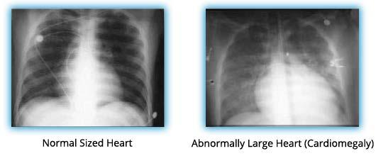
28 X-Ray

29 Echocardiograph Ultrtrasound of the heart More descriptive than an Xray Better view of heart chambers/ movement

30 Cardiac MRI Most descriptive Several slices of the heart can be seen Good for visualizing more discreet structures

31 Introduction to electrophysiology

32 The heart s electrical route Sinoatrial (SA) Node: The heart s pacemaker Intrinsic rate: bpm Internodal Pathways Atrioventricular (AV) Node: Gate keeper/ back up pacemaker Intrinsic rate: 40-60bpm Bundle of His Left and Right Bundle Branches Purkinje Fibres Intrinsic rate: 20-40bpm

33 How do we measure the heart s conduction system? Non invasive test- ECG Gives practitioners important information about several aspects of a patient s heartrate, force of conduction, regularity of beats/ arrythmias Used to diagnose serious medical conditions such as myocardial infarctions (heart attacks)

34 The ECG Single unit represents a heart beat Recorded on grid paper Height= amplitude Measured in mvolts Length= time Large square represents 0.20 seconds Small square represents 0.04 seconds
35 Depolarization and Repolarization Depolarization= contraction Repolarization= relxation Movement captured on the ECG as changes in the waveform from the isoelectric (flat) line

36 What do the different waves represent? P wave: atrial depolarization QRS complex: ventricular depolarization T wave: ventricular repolarization
37 Basic ECG interpretation concepts Look at a 6 second strip and identify/ determine the following: 1. Are P waves present, upright, and identical? 2. Is there a QRS complex following every P wave? 3. What is the PR interval? 4. How long does the QRS complex last? 5. What is the distance between R s/ is the rhythm regular? 6. What is the heart rate? 7. INTERPRET THE RHYTHM!
38 1. P waves Represent atrial depolarization Normally 0.06 to 0.12 seconds long Are p waves present? Are they all facing the same direction (upright vs downward)? Are they all the same shape?
39 2. Is there a QRS complex following every P wave? SA node as pacemaker vs AV node as pacemaker Cannot calculate the PR interval if there s no QRS
40 3. PR interval Distance from the beginning of the P wave to the beginning of the QRS complex Normally, seconds long
41 4. QRS Complex Normal length is seconds Same shape/ length throughout? Too long or too short can lead to problems
42 5. Regular vs. Irregular How far apart are the beats? Same distance every time? R-R intervals
43 6. Heart Rate 6 second strip: multiply # of beats by 10 for approximate rate Normal heart rate= bpm
44 7. Interpret! If everything is normal (P, PR, QRS, R-R, rate)= Normal Sinus rhythm If any of the above is not true, we must classify the rhythm as something else
45 Let s practice
46 #1 P s present/ upright/ identical: QRS for every P: PR interval: QRS width R-R distance/ regularity Rate: Interpretation:
47 #2 P s present/ upright/ identical: QRS for every P: PR interval: QRS width R-R distance/ regularity Rate: Interpretation:
48 #3 P s present/ upright/ identical: QRS for every P: PR interval: QRS width R-R distance/ regularity Rate: Interpretation:
49 All strips Normal Sinus! They look different, but are all considered normal, as long as they: Have P waves that are present, identical, and upright Have a QRS complex for every P wave Have a consistent PR interval Have consistent QRS widths Have regular r-r intervals Are beats/ minute

50 Let s practice some more!


51 #4 P s present/ upright/ identical: QRS for every P: PR interval: QRS width R-R distance/ regularity Rate: Interpretation:
52 #5 P s present/ upright/ identical: QRS for every P: PR interval: QRS width R-R distance/ regularity Rate: Interpretation:
53 #6 P s present/ upright/ identical: QRS for every P: PR interval: QRS width R-R distance/ regularity Rate: Interpretation:
54 Variations on Normal Sinus Rhythm Sinus Bradycardia: < 60bpm Sinus Tachycardia: >100bpm Sinus Arrythmia: regularly irregular


55 Sinus Block vs Sinus Arrest Block= one missed beat Arrest= pause is not regular Generally longer than a block


56 Atrial arrythmias Atrial flutter: regularly irregular Atrial fibrillation Symptoms: shortness of breath, palpitations, feeling faint/ tired Increased risk of stroke

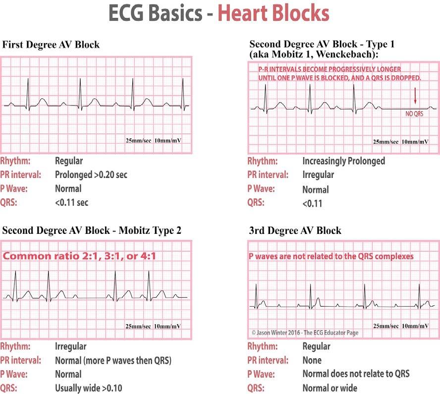
57 Heart Blocks
58 Deadly arrythmias: VT and VF Ventricular tachycardia: wide complex QRS Fast but regular Person may still have a pulse Ventricular fibrillation Fast and irregular No pulse Lethal

59 Pulseless Electrical Activity and Asystole Person is considered to be clinically dead No pulse for either rhythm
60 Next week: Cardiac conditions and pharmacology Part 1
The Heart. Happy Friday! #takeoutyournotes #testnotgradedyet
 The Heart Happy Friday! #takeoutyournotes #testnotgradedyet Introduction Cardiovascular system distributes blood Pump (heart) Distribution areas (capillaries) Heart has 4 compartments 2 receive blood (atria)
Cardiovascular System
 Cardiovascular System The Heart Cardiovascular System The Heart Overview What does the heart do? By timed muscular contractions creates pressure gradients blood moves then from high pressure to low pressure
The Cardiovascular System (Heart)
") The Cardiovascular System The Cardiovascular System (Heart) A closed system of the heart and blood vessels The heart pumps blood Blood vessels allow blood to circulate to all parts of the body The function
The Cardiovascular System The Cardiovascular System (Heart) A closed system of the heart and blood vessels The heart pumps blood Blood vessels allow blood to circulate to all parts of the body The function
37 1 The Circulatory System
 H T H E E A R T 37 1 The Circulatory System The circulatory system and respiratory system work together to supply cells with the nutrients and oxygen they need to stay alive. a) The respiratory system:
The Cardiovascular System. Chapter 15. Cardiovascular System FYI. Cardiology Closed systemof the heart & blood vessels. Functions
 Chapter 15 Cardiovascular System FYI The heart pumps 7,000 liters (4000 gallons) of blood through the body each day The heart contracts 2.5 billion times in an avg. lifetime The heart & all blood vessels
Chapter 14. The Cardiovascular System
 Chapter 14 The Cardiovascular System Introduction Cardiovascular system - heart, blood and blood vessels Cardiac muscle makes up bulk of heart provides force to pump blood Function - transports blood 2
Cardiovascular System Notes: Physiology of the Heart
 Cardiovascular System Notes: Physiology of the Heart Interesting Heart Fact Capillaries are so small it takes ten of them to equal the thickness of a human hair. Review What are the 3 parts of the cardiovascular
the Cardiovascular System I
 the Cardiovascular System I By: Dr. Nabil A Khouri MD, MsC, Ph.D MEDIASTINUM 1. Superior Mediastinum 2. inferior Mediastinum Anterior mediastinum. Middle mediastinum. Posterior mediastinum Anatomy of
THE HEART. A. The Pericardium - a double sac of serous membrane surrounding the heart
 THE HEART I. Size and Location: A. Fist-size weighing less than a pound (250 to 350 grams). B. Located in the mediastinum between the 2 nd rib and the 5 th intercostal space. 1. Tipped to the left, resting
The HEART. What is it???? Pericardium. Heart Facts. This muscle never stops working It works when you are asleep
 This muscle never stops working It works when you are asleep The HEART It works when you eat It really works when you exercise. What is it???? Located between the lungs in the mid thoracic region Apex
The Cardiovascular System
 11 PART A The Cardiovascular System PowerPoint Lecture Slide Presentation by Jerry L. Cook, Sam Houston University ESSENTIALS OF HUMAN ANATOMY & PHYSIOLOGY EIGHTH EDITION ELAINE N. MARIEB The Cardiovascular
4. The two inferior chambers of the heart are known as the atria. the superior and inferior vena cava, which empty into the left atrium.
 Answer each statement true or false. If the statement is false, change the underlined word to make it true. 1. The heart is located approximately between the second and fifth ribs and posterior to the
11/10/2014. Muscular pump Two atria Two ventricles. In mediastinum of thoracic cavity 2/3 of heart's mass lies left of midline of sternum
 It beats over 100,000 times a day to pump over 1,800 gallons of blood per day through over 60,000 miles of blood vessels. During the average lifetime, the heart pumps nearly 3 billion times, delivering
Cardiovascular System Notes: Heart Disease & Disorders
 Cardiovascular System Notes: Heart Disease & Disorders Interesting Heart Facts The Electrocardiograph (ECG) was invented in 1902 by Willem Einthoven Dutch Physiologist. This test is still used to evaluate
Pearson's Comprehensive Medical Assisting Administrative and Clinical Competencies
 Pearson's Comprehensive Medical Assisting Administrative and Clinical Competencies THIRD EDITION CHAPTER 27 The Cardiovascular System Lesson 1: Overview of the Cardiovascular System Lesson Objectives Upon
Circulation. Circulation = is a process used for the transport of oxygen, carbon! dioxide, nutrients and wastes through-out the body
 Circulation Circulation = is a process used for the transport of oxygen, carbon! dioxide, nutrients and wastes through-out the body Heart = muscular organ about the size of your fist which pumps blood.
Ch 19: Cardiovascular System - The Heart -
 Ch 19: Cardiovascular System - The Heart - Give a detailed description of the superficial and internal anatomy of the heart, including the pericardium, the myocardium, and the cardiac muscle. Trace the
Large Arteries of Heart
 Cardiovascular System (Part A-2) Module 5 -Chapter 8 Overview Arteries Capillaries Veins Heart Anatomy Conduction System Blood pressure Fetal circulation Susie Turner, M.D. 1/5/13 Large Arteries of Heart
The Cardiovascular System
 The Cardiovascular System The Cardiovascular System A closed system of the heart and blood vessels The heart pumps blood Blood vessels allow blood to circulate to all parts of the body The function of
The Cardiovascular System
 The Cardiovascular System The Manila Times College of Subic Prepared by: Stevens B. Badar, RN, MANc THE HEART Anatomy of the Heart Location and Size approx. the size of a person s fist, hollow and cone-shaped,
Unit 6: Circulatory System. 6.2 Heart
 Unit 6: Circulatory System 6.2 Heart Functions of Circulatory System 1. The heart is the pump necessary to circulate blood to all parts of the body 2. Arteries, veins and capillaries are the structures
The Heart. Size, Form, and Location of the Heart. 1. Blunt, rounded point; most inferior part of the heart.
 12 The Heart FOCUS: The heart is composed of cardiac muscle cells, which are elongated, branching cells that appear striated. Cardiac muscle cells behave as a single electrical unit, and the highly coordinated
CARDIOVASCULAR SYSTEM
 CARDIOVASCULAR SYSTEM Overview Heart and Vessels 2 Major Divisions Pulmonary Circuit Systemic Circuit Closed and Continuous Loop Location Aorta Superior vena cava Right lung Pulmonary trunk Base of heart
INTRODUCTORY REMARKS:
 INTRODUCTORY REMARKS: The circulatory system provides a way for the blood to be transported throughout the body. This provides nutrients to the cells and allows wastes to be removed. Open vs. Closed Circulatory
THE HEART OBJECTIVES: LOCATION OF THE HEART IN THE THORACIC CAVITY CARDIOVASCULAR SYSTEM
 BIOLOGY II CARDIOVASCULAR SYSTEM ACTIVITY #3 NAME DATE HOUR THE HEART OBJECTIVES: Describe the anatomy of the heart and identify and give the functions of all parts. (pp. 356 363) Trace the flow of blood
The Cardiovascular System
 Essentials of Human Anatomy & Physiology Elaine N. Marieb Seventh Edition Chapter 11 The Cardiovascular System Slides 11.1 11.19 Lecture Slides in PowerPoint by Jerry L. Cook The Cardiovascular System
Cardiovascular System
 Cardiovascular System Purpose Transport oxygen and nutrients Take waste products away from tissues & organs Things we learned Blood pressure: the force of blood pushing against the walls of blood vessels
10/23/2017. Muscular pump Two atria Two ventricles. In mediastinum of thoracic cavity 2/3 of heart's mass lies left of midline of sternum
 It beats over 100,000 times a day to pump over 1,800 gallons of blood per day through over 60,000 miles of blood vessels. During the average lifetime, the heart pumps nearly 3 billion times, delivering
2. right heart = pulmonary pump takes blood to lungs to pick up oxygen and get rid of carbon dioxide
 A. location in thorax, in inferior mediastinum posterior to sternum medial to lungs superior to diaphragm anterior to vertebrae orientation - oblique apex points down and to the left 2/3 of mass on left
CV Anatomy Quiz. Dr Ella Kim Dr Pip Green
 CV Anatomy Quiz Dr Ella Kim Dr Pip Green Q1 The location of the heart is correctly described as A) lateral to the lungs. B) medial to the sternum. C) superior to the diaphragm. D) posterior to the spinal
10. Thick deposits of lipids on the walls of blood vessels, called, can lead to serious circulatory issues. A. aneurysm B. atherosclerosis C.
 Heart Student: 1. carry blood away from the heart. A. Arteries B. Veins C. Capillaries 2. What is the leading cause of heart attack and stroke in North America? A. alcohol B. smoking C. arteriosclerosis
Chapter 18 - Heart. I. Heart Anatomy: size of your fist; located in mediastinum (medial cavity)
") Chapter 18 - Heart I. Heart Anatomy: size of your fist; located in mediastinum (medial cavity) A. Coverings: heart enclosed in double walled sac called the pericardium 1. Fibrous pericardium: dense connective
Chapter 18 - Heart I. Heart Anatomy: size of your fist; located in mediastinum (medial cavity) A. Coverings: heart enclosed in double walled sac called the pericardium 1. Fibrous pericardium: dense connective
STRUCTURES OF THE CARDIOVASCULAR SYSTEM
 STRUCTURES OF THE CARDIOVASCULAR SYSTEM CARDIOVASCULAR SYSTEM Also called the circulatory system Consists of the heart, arteries, veins, and capillaries Main function is to pump/circulate oxygenated blood
Test Review Circulatory System Chapters
 Test Review Circulatory System Chapters 13-2010 1. The tissue that forms the tight fitting sac around the heart is the a. parietal pericardium c. myocardium b. visceral pericardium d. endocardium 2. Which
Anatomy of the Heart. Figure 20 2c
 Anatomy of the Heart Figure 20 2c Pericardium & Myocardium Remember, the heart sits in it s own cavity, known as the mediastinum. The heart is surrounded by the Pericardium, a double lining of the pericardial
Atlantic Health System
 Atlantic Health System Morristown Medical Center Newton Medical Center Overlook Medical Center Basic Dysrhythmia Course Day 1 1 2 Chapter 1 Anatomy and Physiology Learning Objectives 1) Identify electrophysiology
CIRCULATORY SYSTEM BLOOD VESSELS
 Name: Block: CIRCULATORY SYSTEM Multicellular organisms (above the level of roundworms) rely on a circulatory system to bring nutrients to, and take wastes away from, cells. In higher organisms such as
The Heart. The Heart A muscular double pump. The Pulmonary and Systemic Circuits
 C H A P T E R 19 The Heart The Heart A muscular double pump circuit takes blood to and from the lungs Systemic circuit vessels transport blood to and from body tissues Atria receive blood from the pulmonary
Health Science 20 Circulatory System Notes
 Health Science 20 Circulatory System Notes Functions of the Circulatory System The circulatory system functions mainly as the body s transport system. It transports: o Oxygen o Nutrients o Cell waste o
Cardiovascular System. I. Structures of the heart A. : Pericardium sack that surrounds the heart
 Cardiovascular System I. Structures of the heart A. : Pericardium sack that surrounds the heart 1. : Pericardial Cavity serous fluid filled space between the heart and the pericardium B. Heart Wall 1.
The Heart and Heart Disease
 The Heart and Heart Disease Illustration of the heart by Leonardo DaVinci heart-surgeon.com/ history.html 2/14/2010 1 I. Location, Size and Position of the Heart A. Triangular organ located 1. of mass
THE CARDIOVASCULAR SYSTEM. Part 1
 THE CARDIOVASCULAR SYSTEM Part 1 CARDIOVASCULAR SYSTEM Blood Heart Blood vessels What is the function of this system? What other systems does it affect? CARDIOVASCULAR SYSTEM Functions Transport gases,
Human Anatomy, First Edition
 Human Anatomy, First Edition McKinley & O'Loughlin Chapter 22 : Heart 1 Functions of the Heart Center of the cardiovascular system, the heart. Connects to blood vessels that transport blood between the
The Heart and Cardiovascular System
 The Heart and Cardiovascular System What you will learn The location of the heart 3 layers and covering of the heart Explain the function of the heart as 2 separate pumps Identify the 4 chambers of the
Lab 16. The Cardiovascular System Heart and Blood Vessels. Laboratory Objectives
 Lab 16 The Cardiovascular System Heart and Blood Vessels Laboratory Objectives Describe the anatomical structures of the heart to include the pericardium, chambers, valves, and major vessels. Describe
Chapter 20: Cardiovascular System: The Heart
 Chapter 20: Cardiovascular System: The Heart I. Functions of the Heart A. List and describe the four functions of the heart: 1. 2. 3. 4. II. Size, Shape, and Location of the Heart A. Size and Shape 1.
Cardiovascular System
 Cardiovascular System angio BELLWORK Day One: Define using technology hemo/hema cardio Medical Therapeutics Standards 11) Outline the gross normal structure and function of all body systems and summarize
Section 5.1 The heart and heart disease
 Section 5.1 The heart and heart disease Mammals are too large to rely on diffusion. They need a circulatory system to move substances around the body. Blood moves down pressure gradients, from high to
The Circulatory System. The Heart, Blood Vessels, Blood Types
 The Circulatory System The Heart, Blood Vessels, Blood Types The Closed Circulatory System Humans have a closed circulatory system, typical of all vertebrates, in which blood is confined to vessels and
Do Now. Get out work from last class to be checked
 Do Now Get out work from last class to be checked Heart Actions Cardiac Cycle: One complete heartbeat. The contraction of a heart chamber is called systole and the relaxation of a chamber is called diastole.
Function: Transportation of. Oxygen Nutrients Waste Hormones gases
 Function: Transportation of Oxygen Nutrients Waste Hormones gases Pericardium: double sac of serous membrane filled with fluid (pericardial fluid to be exact) that surrounds the heart. Parietal pericardium:
Chapter 20 (1) The Heart
 The Heart") Chapter 20 (1) The Heart Learning Objectives Describe the location and structure of the heart Describe the path of a drop of blood from the superior vena cava or inferior vena cava through the heart out
Chapter 20 (1) The Heart Learning Objectives Describe the location and structure of the heart Describe the path of a drop of blood from the superior vena cava or inferior vena cava through the heart out
The cardiovascular system is composed of the heart and blood vessels that carry blood to and from the body s organs. There are 2 major circuits:
 1 The cardiovascular system is composed of the heart and blood vessels that carry blood to and from the body s organs. There are 2 major circuits: pulmonary and systemic. The pulmonary goes out to the
Cardiac Cycle. Each heartbeat is called a cardiac cycle. First the two atria contract at the same time.
 The Heartbeat Cardiac Cycle Each heartbeat is called a cardiac cycle. First the two atria contract at the same time. Next the two ventricles contract at the same time. Then all the chambers relax. http://www.youtube.com/watch?v=frd3k6lkhws
The Cardiovascular System
 PowerPoint Lecture Slide Presentation by Patty Bostwick-Taylor, Florence-Darlington Technical College The Cardiovascular System 11 PART A The Cardiovascular System A closed system of the heart and blood
Approximately the size of your fist Location. Pericardial physiology
 Heart Anatomy Approximately the size of your fist Location Superior surface of diaphragm Left of the midline Anterior to the vertebral column, posterior to the sternum Wednesday, March 28, 2012 Muscle
Heart. Structure Physiology of blood pressure and heartbeat
 Heart Structure Physiology of blood pressure and heartbeat Location and Anatomy Location and Anatomy Pericardial cavity: surrounds, isolates, and anchors heart Parietal pericardium lined with serous membrane
Principles of Anatomy and Physiology
 Principles of Anatomy and Physiology 14 th Edition CHAPTER 20 The Cardiovascular System: The Heart Introduction The purpose of the chapter is to: 1. Learn about the components of the cardiovascular system
Blood must move! 4/15/2014. Heart Basics
 What is the CARDIOVASCULAR system? The cardiovascular system carries blood and dissolved substances to and from different places in the body. The Heart has the job of pumping these things around the body.
Major Function of the Cardiovascular System. Transportation. Structures of the Cardiovascular System. Heart - muscular pump
 Structures of the Cardiovascular System Heart - muscular pump Blood vessels - network of tubes Blood - liquid transport vehicle brachiocephalic trunk superior vena cava right pulmonary arteries right pulmonary
Cardiovascular System- Heart. Miss Wheeler Unit 8
 Cardiovascular System- Heart Miss Wheeler Unit 8 Overview CARDIOVASCULAR SYSTEM heart vessels Made up of heart, blood vessels, and blood Functions Heart- pump blood Vessels- (veins, arteries, capillaries)
IB TOPIC 6.2 THE BLOOD SYSTEM
 IB TOPIC 6.2 THE BLOOD SYSTEM THE BLOOD SYSTEM TERMS TO KNOW circulation ventricle artery vein 6.2.U1 - Arteries convey blood at high pressure from the ventricles to the tissues of the body Circulation
All About the Heart. Structures of the heart. Layers. Chambers
 All About the Heart Your heart is a muscle. It is slightly larger than your fist and weighs less than a pound. It is located to the left of the middle of your chest. Your heart pumps blood to the lungs
- what other structures, besides the heart, does the mediastinum contain?
 Basic A & P II Dr. L. Bacha Chapter Outline (Martini & Nath 2010) An Introduction to the Cardiovascular System - read the paragraphs under this heading on page 580 The Heart is a Four Chambered Organ describe
The Cardiovascular System
 Essentials of Human Anatomy & Physiology Elaine N. Marieb Slides 11.1 11.19 Seventh Edition Chapter 11 The Cardiovascular System Functions of the Cardiovascular system Function of the heart: to pump blood
This lab activity is aligned with Visible Body s A&P app. Learn more at visiblebody.com/professors
 1 This lab activity is aligned with Visible Body s A&P app. Learn more at visiblebody.com/professors 2 PRE-LAB EXERCISES: A. Watch the video 29.1 Heart Overview and make the following observations: 1.
Label Diagram #1 (Pg. 664)
") Chapter 18 The Cardiovascular System 18.1 Heart Anatomy The Pulmonary and Systemic Circuits Oxygen rich vs. Oxygen Poor Heart is a transport system consisting of two side-by-side pumps Right side receives
Chapter 18 The Cardiovascular System 18.1 Heart Anatomy The Pulmonary and Systemic Circuits Oxygen rich vs. Oxygen Poor Heart is a transport system consisting of two side-by-side pumps Right side receives
UNDERSTANDING YOUR ECG: A REVIEW
 UNDERSTANDING YOUR ECG: A REVIEW Health professionals use the electrocardiograph (ECG) rhythm strip to systematically analyse the cardiac rhythm. Before the systematic process of ECG analysis is described
Anatomy & Physiology of Cardiovascular System. Chapter 18 & 19
 Anatomy & Physiology of Cardiovascular System Chapter 18 & 19 Objectives..cont 1. Discuss the physiological stages of cardiac muscle contraction. 2. Trace a typical ECG and label each wave or complex 3.
Heart. Heart 2-Tunica media: middle layer (media ='middle') muscle fibers (smooth or cardiac).
 muscle fibers (smooth or cardiac).") t. innermost lumenal General Circulatory system heart and blood vessels walls have 3 layers (inside to outside) 1-Tunica interna: aka tunica intima layer--lumenal layer epithelium--endothelium simple squamous
t. innermost lumenal General Circulatory system heart and blood vessels walls have 3 layers (inside to outside) 1-Tunica interna: aka tunica intima layer--lumenal layer epithelium--endothelium simple squamous
1. What kind of blood is found in the rt. atrium? (oxygenated or deoxygenated)
") Carl Christennsen, PhD Chap. 19, 20, & 21 - Circulatory System Bio. 2304 Human Anatomy HEART 1. What kind of blood is found in the rt. atrium? (oxygenated or deoxygenated) Where does this blood come from?
Carl Christennsen, PhD Chap. 19, 20, & 21 - Circulatory System Bio. 2304 Human Anatomy HEART 1. What kind of blood is found in the rt. atrium? (oxygenated or deoxygenated) Where does this blood come from?
EKG Competency for Agency
 EKG Competency for Agency Name: Date: Agency: 1. The upper chambers of the heart are known as the: a. Atria b. Ventricles c. Mitral Valve d. Aortic Valve 2. The lower chambers of the heart are known as
The Cardiovascular System: The Heart
 PowerPoint Lecture Slides prepared by Meg Flemming Austin Community College C H A P T E R 12 The Cardiovascular System: The Heart Chapter 12 Learning Outcomes 12-1 12-2 Describe the anatomy of the heart,
The Cardiovascular and Lymphatic Systems Cardiovascular System Blood Vessels Blood Vessels Arteries Arteries Arteries
 CH 12 The Cardiovascular and s The Cardiovascular and s OUTLINE: Cardiovascular System Blood Vessels Blood Pressure Cardiovascular System The cardiovascular system is composed of Blood vessels This system
The Heart. Made up of 3 different tissue: cardiac muscle tissue, nerve tissue, and connective tissue.
 The Heart The Heart Made up of 3 different tissue: cardiac muscle tissue, nerve tissue, and connective tissue. Your heart pumps with a regular beat (Heart Rate) Your heart rate can change depending on
Cardiovascular System. Biology 105 Lecture 15 Chapter 12
 Cardiovascular System Biology 105 Lecture 15 Chapter 12 Outline I. Functions of cardiovascular system II. Components of the cardiovascular system: I. Blood vessels II. Heart III. Regulation of the heartbeat
Collin County Community College. ! BIOL Anatomy & Physiology! WEEK 5. The Heart
 Collin County Community College! BIOL. 2402 Anatomy & Physiology! WEEK 5 The Heart 1 (1578-1657) A groundbreaking work in the history of medicine, English physician William Harvey s Anatomical Essay on
UNIT 11: THE CARDIOVASCULAR SYSTEM
 UNIT 11: THE CARDIOVASCULAR SYSTEM Functions of the Heart PUMPS Blood Transports Oxygen and Nutrients Removes Carbon Dioxide and Metabolic Wastes Thermoregulation Immunological Function Clotting Mechanisms
ADVANCED ASSESSMENT Cardiovascular System
 ONTARIO BASE HOSPITAL GROUP QUIT ADVANCED ASSESSMENT Cardiovascular System 2007 Ontario Base Hospital Group ADVANCED ASSESSMENT Cardiovascular System AUTHORS Mike Muir AEMCA, ACP, BHSc Paramedic Program
Anatomy of the Heart
 Biology 212: Anatomy and Physiology II Anatomy of the Heart References: Saladin, KS: Anatomy and Physiology, The Unity of Form and Function 8 th (2018). Required reading before beginning this lab: Chapter
Heart Dissection. 5. Locate the tip of the heart or the apex. Only the left ventricle extends all the way to the apex.
 Heart Dissection Page 1 of 6 Background: The heart is a four-chambered, hollow organ composed primarily of cardiac muscle tissue. It is located in the center of the chest in between the lungs. It is the
The Heart A-Chapter Identify the layers of the heart wall and state the type of tissue in each layer.
 Objectives: The Heart AChapter 12 1. Identify the layers of the heart wall and state the type of tissue in each layer. Label a diagram of the heart, identifying the chambers, valves, and associated vessels.
Read Me. covering the Heart Anatomy. Labs. textbook. use. car: you
 Heart Anatomy Lab Pre-Lab Exercises Read Me These exercises should be done before coming to lab, after watching the videos covering the Heart Anatomy Labs. Answer the questions in this guide using the
CORONARY ARTERIES. LAD Anterior wall of the left vent Lateral wall of left vent Anterior 2/3 of interventricluar septum R & L bundle branches
 CORONARY ARTERIES RCA Right atrium Right ventricle SA node 55% AV node 90% Posterior wall of left ventricle in 90% Posterior third of interventricular septum 90% LAD Anterior wall of the left vent Lateral
IB TOPIC 6.2 THE BLOOD SYSTEM
 IB TOPIC 6.2 THE BLOOD SYSTEM TERMS TO KNOW circulation ventricle artery vein THE BLOOD SYSTEM 6.2.U1 - Arteries convey blood at high pressure from the ventricles to the tissues of the body Circulation
Cardiovascular System
 Cardiovascular System I. Structure of the Heart A. Average adult heart is 14 cm long and 9 cm wide. B. Lies in the mediastinum. C. Enclosed in the pericardium. 1. Fibrous pericardium- Outer, tough connective
Outline. Electrical Activity of the Human Heart. What is the Heart? The Heart as a Pump. Anatomy of the Heart. The Hard Work
 Electrical Activity of the Human Heart Oguz Poroy, PhD Assistant Professor Department of Biomedical Engineering The University of Iowa Outline Basic Facts about the Heart Heart Chambers and Heart s The
Biology Unit 3 The Human Heart P
 Biology 2201 Unit 3 The Human Heart P 314-321 Structure and Function of the Human Heart Structure of the Human Heart Has four Chambers (2 Atria and 2 Ventricles) Made of Cardiac Muscle Found in Chest Cavity
The Cardiovascular System Part I: Heart Outline of class lecture After studying part I of this chapter you should be able to:
 The Cardiovascular System Part I: Heart Outline of class lecture After studying part I of this chapter you should be able to: 1. Describe the functions of the heart 2. Describe the location of the heart,
CIRCULATION. Cardiovascular & lymphatic systems Functions. Transport Defense / immunity Homeostasis
 CIRCULATION CIRCULATION Cardiovascular & lymphatic systems Functions Transport Defense / immunity Homeostasis 2 Types of Circulatory Systems Open circulatory system Contains vascular elements Mixing of
Electrical Conduction
 Sinoatrial (SA) node Electrical Conduction Sets the pace of the heartbeat at 70 bpm AV node (50 bpm) and Purkinje fibers (25 40 bpm) can act as pacemakers under some conditions Internodal pathway from
Bio 104 Cardiovascular System
 29 Blood: Introduction (Chapter 14) A. Characteristics of Blood 1. Blood Volume Lecture Outline: Cardiovascular System Hole s HAP [Chapters 14, 15, 16] 2. Blood Composition a. Blood Cells Red blood cells
The Cardiovascular System: The Heart: Part A
 PowerPoint Lecture Slides prepared by Janice Meeking, Mount Royal College CHAPTER 18 The Cardiovascular System: The Heart: Part A Heart Anatomy Approximately the size of a fist Location In the mediastinum
The Cardiovascular System
 The Cardiovascular System https://www.youtube.com/watch?v=ohmmtqkgs50 Human Anatomy & Physiology P. Wilson 1 Introduction The functions of the cardiovascular system are: to bring oxygen & nutrients to
Unit 1: Human Systems. The Circulatory System
 Unit 1: Human Systems The Circulatory System nourish all cells with oxygen, glucose, amino acids and other nutrients and carry away carbon dioxide, urea and other wastes Purposes Transport chemical messengers
Cardiovascular system
 BIO 301 Human Physiology Cardiovascular system The Cardiovascular System: consists of the heart plus all the blood vessels transports blood to all parts of the body in two 'circulations': pulmonary (lungs)
The Cardiovascular and Lymphatic Systems
 BIOLOGY OF HUMANS Concepts, Applications, and Issues Fifth Edition Judith Goodenough Betty McGuire 12 The Cardiovascular and Lymphatic Systems Lecture Presentation Anne Gasc Hawaii Pacific University and
LECTURE 5. Anatomy of the heart
 LECTURE 5. Anatomy of the heart Main components of the CVS: Heart Blood circulatory system arterial compartment haemomicrocirculatory (=microvascular) compartment venous compartment Lymphatic circulatory
The Heart. C h a p t e r. PowerPoint Lecture Slides prepared by Jason LaPres Lone Star College - North Harris
 C h a p t e r 20 The Heart PowerPoint Lecture Slides prepared by Jason LaPres Lone Star College - North Harris Copyright 2009 Pearson Education, Inc., publishing as Pearson Benjamin Cummings Introduction