Tissue Doppler Imaging in Congenital Heart Disease
|
|
|
- Stuart Marvin Smith
- 5 years ago
- Views:
Transcription

1 Tissue Doppler Imaging in Congenital Heart Disease L. Youngmin Eun, M.D. Department of Pediatrics, Division of Pediatric Cardiology, Kwandong University College of Medicine
2 The potential advantage of ultrasound cardiography is to permit the study of the soft tissues without catheterization and the introduction of contrast media. With regard to cardiovascular diagnosis the method is still in the stage of research. from 1/2 page on ultrasonic cardiography in chapter- Phonocardiography and other graphic methods Friedberg, Diseases of the Heart, 1966

3 What is TDI? Quantitative Tissue Doppler Imaging
4 What is the Principle of TDI? TDI is based on the difference between signals returned from blood and tissue. It filters out the high velocity, low amplitude signals, leaving the tissue motion information visible. ( low velocity, high amplitude) - The familiar color coding of color Doppler signals red-to-yellow scale : tissue movement towards the transducer blue-to-green scale : movement away red and blue --- low velocities yellow and green--- high velocities
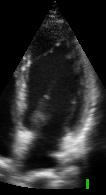
5 LV Longitudinal Shortening 2D Diastole Systole

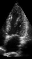
6 Why do TDI exam in Apical View? Apex epi mid RV LV endo Base 70% of fibers are longitudinal!
7 Tissue Doppler Echocardiography Simple, non-invasive and reproducible method for assessing cardiac physiology. Provide a velocity value for every point in the myocardial wall. Assessing wall motion not only regionally, but also quantitatively. This may afford the opportunity to study regional systolic and diastolic function.
8 Where can TDI be used? Quantify myocardial velocities in multiple segments of the myocardium from different echocardiographic windows Accurate estimate of LV relaxation - Insensitive to the effects of preload compensation Potential to assess regional systolic and diastolic function in both LV and RV Diastolic regional change is an early marker of ischemia (regional isovolumic relaxation time obtained by DTI) --- before the development of regional systolic dysfunction
9 How is Tissue Doppler displayed? Color B-scan : the best overall impression provide good view of a heart chambers and walls frame rate limitations : maximum frame rate frames/s M-mode : dramatically improves time resolution sampling is only performed on a single line such a line positioned on a clinically significant position PW analysis : produces the maximum amount of information highest temporal and velocity range resolution
")

10 Conventional Doppler vs TDI Conventional Doppler - velocity and direction of blood flow (RBC) Tissue Doppler - velocity and direction of myocardial tissue Blood: Mitral Flow Tissue: MV Ring Motion

11 Tissue Doppler in M-modeM
12 Advantages of Color M-mode - better temporal resolution of systolic diastolic velocity high frame rate obtained with M-mode - represent in the same image both systolic diastolic velocities - accurate quantitative information about myocardial motion during the cardiac cycle - accurately assess in one scan plane the different phases of the cardiac cycle
13 What is the Limitation of TDI? The Angle dependency The agreement between pulsed and color Doppler derived velocities has not been systematically studied pulsed Doppler derived velocities are higher. Sometimes difficult to distinguish whether the lack of color due to akinesia or to uncontrolled technical factors The Tethering effect
14 The assessment of ventricular function TDE has the potential to assess - segmental systolic and diastolic function in both LV and RV transmural velocity gradient asynchronous ventricular contraction and relaxation visualized online global and regional systolic and diastolic time interval
15 The amount of color in an image can be increased by -increasing the Doppler tissue imaging or color Doppler gain -Modifying the depth gain to take away some of the underlying gray scale image -increasing the gate size -using lower filter settings -decreasing the scale -adjusting the ROI size -changing the transducer position or orientation * Gain settings must be carefully adjusted to obtain the most homogeneous non-saturated color filling.
16 Myocardial Tissue Velocity Normal heart : endocardium moves faster than epicardium during myocardial contraction because of the change of wall thickness myocardial velocities are highest in the base of the heart and decrease toward the apex, with reversal in apical area The velocity gradient between the endo and epicardium is an indicator of the regional myocardial contraction. --- decrease in the velocity gradient should be expected in infarcted myocardium when compared to a normal one G = (V endocardium V epicardium) / W cos θ

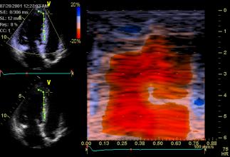
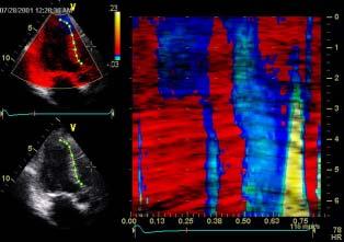
17 Myocardial Tissue Velocity

18 Myocardial Tissue Velocity Parasternal long axis view

19 Myocardial Tissue Velocity


20 Tissue Doppler Velocity & Strain Rate Tissue Velocity Strain Rate v 2 SR 1 v 1 SR 2 Expand No deformation v 1 v 2 SR 2 SR 1 Contract [m/s] [1/s]
21 Tissue Doppler Velocity Longitudinal Velocity Velocity Systole Diastole IVC Time IVR E A
 = Systolic Displacement Tissue Tracking = Color Coded Systolic")
22 Tissue Tracking (Displacement) Longitudinal Velocity Velocity IVC VTI Time IVR E A Velocity Time Integral ( VTI ) = Systolic Displacement Tissue Tracking = Color Coded Systolic Displacement
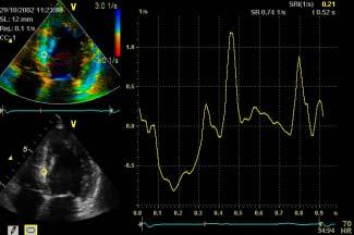
23 Strain Rate : Rate of Deformation (Spatial velocity gradient) v 2 x SR= v2 v x 1 v 1 IVC IVR E A Time Strain Rate
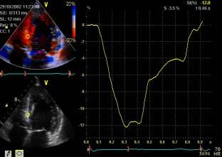
24 Strain - Local Deformation Strain Rate IVC IVR E A Time Strain

25 Color encoding based on tracked motion T Start T End
26 Longitudinal Displacement and Strain during the Systolic phase Displacement = 0 mm Systole Diastole Velocity E E A A Strain Rate Deformation = 0 %
27 Longitudinal Displacement and Strain during the Systolic phase Displacement = 6 mm Systole Diastole Velocity E E A A Strain Rate Deformation = 10 %
28 Longitudinal Displacement and Strain during the Systolic phase Displacement = 12 mm Systole Diastole Velocity E E A A Strain Rate Deformation = 20 %


29 TDI, TT, SRI, Strain imaging & Profile

30 TDI, Strain imaging

31 TDI, Tissue tracking imaging Displacement


32 TDI Curved Anatomical M-modeM Septum Apex NORMAL Curved Anatomical M-Mode Lateral Wall


33 CAMM : Apex Post systolic thickenig

34 CAMM : Strain rate imaging Normal Apical akinesia Apical dyskinesia


35 Tissue Synchronization Imaging Using Tissue Doppler to Map Synchrony Peak of Septal Wall Peak of Lateral Wall Time-to-peak TSI does this calculation for every piece of myocardium and displays time-to-peak


36 Parametric Imaging Advance Applications TVI, TTI, TSI, Strain TTI Tissue Tracking Measures Myocardial Longitudinal Displacement [mm] TSI Tissue Synchronization Imaging Measures Timing; Time-to-Peak Systolic Velocity [msec] TVI Tissue Velocity Imaging Measures Myocardial Long. Velocity [m/sec] SI Strain Imaging Measures Myocardial Longitudinal Deformation [%]
37 Clinical Application Functional assessment in Congenital Heart disease - s/p ASD device closure - Aortic Stenosis - RV function in TOF - Diastolic dysfunction - Ventricular function in Fontan physiology - Etc.

38 Strain Rate vs Strain - longitudinal

39 Longitudinal motion and deformation in RV with TOF
40 Strain Rate vs Strain - radial

41 Peri-patch regional myocardial function in VSD repair Eun, AHA 2002

42 Peri-patch regional myocardial function in VSD repair Eun, AHA 2002
43 Table 1. The Strain parameters in VSD repain patients Peripatch Remote region p-value Longitudinal SR ES ± ± 4.17 < 0.05 SR ED 4.05 ± ± 3.66 < 0.05 SR LD 3.96 ± ± 1.95 NS ε ε peak ± ± < 0.01 Radial SR ES 3.31 ± ± 1.61 NS SR ED ± ± 3.09 < 0.01 SR LD ± ± 5.49 < 0.05 ε ε peak ± ± < Eun et al. AHA 2002
44 Table 2. Time to Strain parameters in VSD patients Peripatch Remote region p-value Longitudinal SR ES ± ± < 0.01 SR ED ± ± < 0.05 SR LD ± ± NS ε ε peak ± ± < Radial SR ES ± ± NS SR ED ± ± NS SR LD ± ± NS ε ε peak ± ± < Eun et al. AHA 2002

45 Tissue velocity in Normal vs RV dysplasia

46 Tissue velocity in Normal vs RV dysplasia Herbots et al. Eur J Echocardiography 2003; 4:
47 Conventional and TDI velocity E / Ea : strong relation to invasively measured PCWP By Paelinck et al. J Am Coll Cardiol 2005;45:
48 Tissue Velocities in normal children by age group Eidem et al, J Am Soc Echocardiogr. 2004, Vol 17 (3)
49 Tissue Velocities in normal children by age group Eidem et al, J Am Soc Echocardiogr. 2004, Vol 17 (3)

50 TDI in Normal children Eun, ACC 2004

51 TDI in Heart Transplant patient Eun, ACC 2004

52 TDI in Heart Transplant patient Grade 2b Rejection Eun, ACC 2004

53 TDI in Heart Transplant patient Eun, ACC 2004

54 TDI in Heart Transplant patient Grade 2b Rejection Eun, ACC 2004

55 TDI in Heart Transplant patient Eun, ACC 2004
56 The myocardial performance index (Tei index) by TDI --- > Tei can also be obtained by TDI. Simple and noninvasive measurement for assessing global right ventricular (RV) function Sensitive indicator of RV function promising new means - global RV function with PR By Yasuoka et al. Pediatr Cardiol ; 25(2):131-6.

57 Myocardial Acceleration during Isovolumic Contraction (IVA) AT : Acceleration time of myocardial velocity during isovolumic contraction IVV : peak myocardial velocity during isovolumic contraction

58 Myocardial Acceleration during isovolumic contraction (IVA)
59 Myocardial Acceleration during isovolumic contraction (IVA) a new index of right ventricular contractile function --- unaffected by ventricular shape or loading conditions --- assess RV contractile function for patients with repaired TOF and various degrees of PR J Am Soc Echocardiogr Apr;17(4):332-7.

60 Clinical Application Congenital Heart disease - pre- operative assessment - post- operative assessment Acquired Heart disease - Myocarditis - Other infection - Kawasaki disease - Diastolic dysfunction - Systolic dysfunction - Heart failure
61 THANK YOU~*
Velocity, strain and strain rate: Doppler and Non-Doppler methods. Thoraxcentre, Erasmus MC,Rotterdam
 Velocity, strain and strain rate: Doppler and Non-Doppler methods J Roelandt J. Roelandt Thoraxcentre, Erasmus MC,Rotterdam Basics of tissue Doppler imaging Instantaneous annular velocity profiles IVCT
Quantitation of right ventricular dimensions and function
 SCCS Basics of cardiac assessment Quantitation of right ventricular dimensions and function Tomasz Kukulski, MD PhD Dept of Cardiology, Congenital Heart Disease and Electrotherapy Silesian Medical University
DISCLOSURE. Myocardial Mechanics. Relevant Financial Relationship(s) Off Label Usage
 Off Label Usage") 7th Annual Team Echocardiography: The Heart of Cardiovascular Medicine Tissue Doppler, Strain, Speckle: What? How? Christopher J Kramer RDCS Aurora Medical Group Advanced Cardiovascular Services, Aurora
7th Annual Team Echocardiography: The Heart of Cardiovascular Medicine Tissue Doppler, Strain, Speckle: What? How? Christopher J Kramer RDCS Aurora Medical Group Advanced Cardiovascular Services, Aurora
Nancy Goldman Cutler, MD Beaumont Children s Hospital Royal Oak, Mi
 Nancy Goldman Cutler, MD Beaumont Children s Hospital Royal Oak, Mi Identify increased LV wall thickness (WT) Understand increased WT in athletes Understand hypertrophic cardiomyopathy (HCM) Enhance understanding
2/2/2011. Strain and Strain Rate Imaging How, Why and When? Movement vs Deformation. Doppler Myocardial Velocities. Movement. Deformation.
 Strain and Strain Rate Imaging How, Why and When? João L. Cavalcante, MD Advanced Cardiac Imaging Fellow Cleveland Clinic Foundation Disclosures: No conflicts of interest Movement vs Deformation Movement
Evaluation of Left Ventricular Diastolic Dysfunction by Doppler and 2D Speckle-tracking Imaging in Patients with Primary Pulmonary Hypertension
 ESC Congress 2011.No 85975 Evaluation of Left Ventricular Diastolic Dysfunction by Doppler and 2D Speckle-tracking Imaging in Patients with Primary Pulmonary Hypertension Second Department of Internal
Tissue Doppler and Strain Imaging. Steven J. Lester MD, FRCP(C), FACC, FASE
, FACC, FASE") Tissue Doppler and Strain Imaging Steven J. Lester MD, FRCP(C), FACC, FASE Relevant Financial Relationship(s) None Off Label Usage None a. Turn the wall filters on and turn down the receiver gain. b. Turn
Tissue Doppler and Strain Imaging Steven J. Lester MD, FRCP(C), FACC, FASE Relevant Financial Relationship(s) None Off Label Usage None a. Turn the wall filters on and turn down the receiver gain. b. Turn
Tissue Doppler and Strain Imaging
 Tissue Doppler and Strain Imaging Steven J. Lester MD, FRCP(C), FACC, FASE Relevant Financial Relationship(s) None Off Label Usage None 1 Objective way with which to quantify the minor amplitude and temporal
좌심실수축기능평가 Cardiac Function
 Basic Echo Review Course 좌심실수축기능평가 Cardiac Function Seonghoon Choi Cardiology Hallym university LV systolic function Systolic function 좌심실수축기능 - 심근의수축으로심실에서혈액을대동맥으로박출하는기능 실제임상에서 LV function 의의미 1Diagnosis
Three-dimensional Wall Motion Tracking:
 Three-dimensional Wall Motion Tracking: A Novel Echocardiographic Method for the Assessment of Ventricular Volumes, Strain and Dyssynchrony Jeffrey C. Hill, BS, RDCS, FASE Jennifer L. Kane, RCS Gerard
Little is known about the degree and time course of
 Differential Changes in Regional Right Ventricular Function Before and After a Bilateral Lung Transplantation: An Ultrasonic Strain and Strain Rate Study Virginija Dambrauskaite, MD, Lieven Herbots, MD,
Strain imaging in children: from Tissue Doppler to 3 D
 Strain imaging in children: from Tissue Doppler to 3 D Mark kk. Friedberg Fi Outline Deformation in the fetus and neonate Deformation in pediatric cardiomyopathy y (briefly!) Deformation in Congenital
Carlos Eduardo Suaide Silva, Luiz Darcy Cortez Ferreira, Luciana Braz Peixoto, Claudia Gianini Monaco, Manuel Adán Gil, Juarez Ortiz
 Silva et al Original Article Arq Bras Cardiol Study of the Myocardial Contraction and Relaxation Velocities through Doppler Tissue Imaging Echocardiography. A New Alternative in the Assessment of the Segmental
OPTIMIZING ECHO ACQUISTION FOR STRAIN AND DIASTOLOGY
 OPTIMIZING ECHO ACQUISTION FOR STRAIN AND DIASTOLOGY October 8, 2017 Deborah Agler, ACS, RDCS, FASE Coordinator of Education and Training Cleveland Clinic General Principles Diastology Clinical Data Heart
Tissue Doppler and Strain Imaging
 Tissue Doppler and Strain Imaging Steven J. Lester MD, FRCP(C), FACC, FASE Relevant Financial Relationship(s) None Off Label Usage None 1 Objective way with which to quantify the minor amplitude and temporal
Strain/Untwisting/Diastolic Suction
 What Is Diastole and How to Assess It? Strain/Untwisting/Diastolic Suction James D. Thomas, M.D., F.A.C.C. Cardiovascular Imaging Center Department of Cardiology Cleveland Clinic Foundation Cleveland,
22 nd Annual Conference of the Saudi Heart Association Riyadh, Saudi Arabia
 22 nd Annual Conference of the Saudi Heart Association Riyadh, Saudi Arabia New Echocardiographic Modalities to Evaluate Ventricular Function in Congenital Heart Disease: Tissue Doppler & Strain Rate Imaging
Diastology Disclosures: None. Dias2011:1
 Diastology 2011 James D. Thomas, M.D., F.A.C.C. Cardiovascular Imaging Center Department of Cardiology Cleveland Clinic Foundation Cleveland, Ohio, USA Disclosures: None Dias2011:1 Is EVERYBODY a member!?!
Strain and Strain Rate Imaging How, Why and When?
 Strain and Strain Rate Imaging How, Why and When? João L. Cavalcante, MD Advanced Cardiac Imaging Fellow Cleveland Clinic Foundation Disclosures: No conflicts of interest Movement vs Deformation Movement
The rapid evolution of echocardiography during the past 25 years
 Evaluation of Myocardial Mechanics in the Fetus by Velocity Vector Imaging Adel K. Younoszai, MD, David E. Saudek, MD, Stephen P. Emery, MD, and James D. Thomas, MD, Denver, Colorado; Cleveland, Ohio;
Basic Approach to the Echocardiographic Evaluation of Ventricular Diastolic Function
 Basic Approach to the Echocardiographic Evaluation of Ventricular Diastolic Function J A F E R A L I, M D U N I V E R S I T Y H O S P I T A L S C A S E M E D I C A L C E N T E R S T A F F C A R D I O T
Incorporating the New Echo Guidelines Into Everyday Practice
 Incorporating the New Echo Guidelines Into Everyday Practice Clinical Case RIGHT VENTRICULAR FAILURE Gustavo Restrepo MD President Elect Interamerican Society of Cardiology Director Fellowship Training
Diagnostic approach to heart disease
 Diagnostic approach to heart disease Initial work up History Physical exam Chest radiographs ECG Special studies Echocardiography Cardiac catheterization Echocardiography principles Technique of producing
LV FUNCTION ASSESSMENT: WHAT IS BEYOND EJECTION FRACTION
 LV FUNCTION ASSESSMENT: WHAT IS BEYOND EJECTION FRACTION Jamilah S AlRahimi Assistant Professor, KSU-HS Consultant Noninvasive Cardiology KFCC, MNGHA-WR Introduction LV function assessment in Heart Failure:
Review Article Transthoracic Echocardiography in Children and Young Adults with Congenital Heart Disease
 International Scholarly Research Network ISRN Pediatrics Volume 2012, Article ID 753481, 15 pages doi:10.5402/2012/753481 Review Article Transthoracic Echocardiography in Children and Young Adults with
The importance of left atrium in LV diastolic function
 II Baltic Heart Failure Meeting and Congress of Latvian Society of Cardiology The importance of left atrium in LV diastolic function Dr. Artem Kalinin Eastern Clinical University Hospital Riga 30.09.2010.
Cardiac Chamber Quantification by Echocardiography
 Cardiac Chamber Quantification by Echocardiography Maryam Bokhamseen, RCS, RCDS, EACVI Echotechnologist ǁ, Non invasive Cardiac Laboratory King Abdulaziz Cardiac Center. Outline: Introduction. Background
Alicia Armour, MA, BS, RDCS
 Alicia Armour, MA, BS, RDCS No disclosures Review 2D Speckle Strain (briefly) Discuss some various patient populations & disease pathways where Strain can be helpful Discuss how to acquire images for Strain
New aspects of Echocardiography in Hypertensive Heart Disease. Fausto J. Pinto, MD, PhD, FESC, FACC, FASE
 New aspects of Echocardiography in Hypertensive Heart Disease Fausto J. Pinto, MD, PhD, FESC, FACC, FASE Progressive increase in cardiovascular morbidity (left) and all-cause mortality (right) rates
Appendix II: ECHOCARDIOGRAPHY ANALYSIS
 Appendix II: ECHOCARDIOGRAPHY ANALYSIS Two-Dimensional (2D) imaging was performed using the Vivid 7 Advantage cardiovascular ultrasound system (GE Medical Systems, Milwaukee) with a frame rate of 400 frames
Doppler Basic & Hemodynamic Calculations
 Doppler Basic & Hemodynamic Calculations August 19, 2017 Smonporn Boonyaratavej MD Division of Cardiology, Department of Medicine Chulalongkorn University Cardiac Center, King Chulalongkorn Memorial Hospital
Diastolic Function: What the Sonographer Needs to Know. Echocardiographic Assessment of Diastolic Function: Basic Concepts 2/8/2012
 Diastolic Function: What the Sonographer Needs to Know Pat Bailey, RDCS, FASE Technical Director Beaumont Health System Echocardiographic Assessment of Diastolic Function: Basic Concepts Practical Hints
Quantification of Left Ventricular Systolic Function by Tissue Doppler Echocardiography
 Quantification of Left Ventricular Systolic Function by Tissue Doppler Echocardiography Added Value of Measuring Pre- and Postejection Velocities in Ischemic Myocardium Thor Edvardsen, MD; Stig Urheim,
The Patient with Atrial Fibrilation
 Assessment of Diastolic Function The Patient with Atrial Fibrilation Assoc. Prof. Adriana Ilieşiu, FESC University of Medicine Carol Davila Bucharest, Romania Associated Conditions with Atrial Fibrillation
Tissue Doppler Imaging
 Cronicon OPEN ACCESS Hesham Rashid* Tissue Doppler Imaging CARDIOLOGY Editorial Department of Cardiology, Benha University, Egypt *Corresponding Author: Hesham Rashid, Department of Cardiology, Benha University,
Measuring cardiac tissue motion and strain
 Ultrasound Measuring cardiac tissue motion and strain Automated Cardiac Motion Quantification A.I. (acmq A.I. ) David Prater, MS, Clinical Scientist, Philips Jane Vogel, MD, Senior Product Manager, Philips
Introduction to Biomedical Imaging
 Alejandro Frangi, PhD Computational Imaging Lab Department of Information & Communication Technology Pompeu Fabra University www.cilab.upf.edu Basic principles. Comparison to X-rays Ultrasound > 20kHz
3D-stress echocardiography Bernard Cosyns, MD, PhD
 3D-stress echocardiography Bernard Cosyns, MD, PhD No Disclosure The Pro-Technology bias Sicari et al. Cardiovascular Ultrasound 2006, 4:11 Overview 2D stress echocardiography: main limitations 3D echocardiography:
B-Mode measurements protocols:
 Application Note How to Perform the Most Commonly Used Measurements from the Cardiac Measurements Package associated with Calculations of Cardiac Function using the Vevo Lab Objective The Vevo LAB offline
Advanced Multi-Layer Speckle Strain Permits Transmural Myocardial Function Analysis in Health and Disease:
 Advanced Multi-Layer Speckle Strain Permits Transmural Myocardial Function Analysis in Health and Disease: Clinical Case Examples Jeffrey C. Hill, BS, RDCS Echocardiography Laboratory, University of Massachusetts
RIGHT VENTRICULAR SIZE AND FUNCTION
 RIGHT VENTRICULAR SIZE AND FUNCTION Edwin S. Tucay, MD, FPCC, FPCC, FPSE Philippine Society of Echocardiography Quezon City, Philippines Echo Mission, BRTTH, Legaspi City, July 1-2, 2016 NO DISCLOSURE
Quantitative Assessment of Fetal Ventricular Function:
 Reprinted with permission from ECHOCARDIOGRAPHY, Volume 18, No. 1, January 2001 Copyright 2001 by Futura Publishing Company, Inc., Armonk, NY 1004-0418 Quantitative Assessment of Fetal Ventricular Function:
L ecocardiografia nello Scompenso Cardiaco Acuto e cronico: vecchi dogmi e nuovi trends.
 V SESSIONE SCOMPENSO CARDIACO 2015 Genova, 13-14 Novembre 2015 L ecocardiografia nello Scompenso Cardiaco Acuto e cronico: vecchi dogmi e nuovi trends. Gian Paolo Bezante, MD, FACC UOC Clinica di Malattie
How does the heart pump? From sarcomere to ejection volume
 How does the heart pump? From sarcomere to ejection volume Piet Claus Cardiovascular Imaging and Dynamics Department of Cardiovascular Diseases University Leuven, Leuven, Belgium Course on deformation
Assessment of LV systolic function
 Tutorial 5 - Assessment of LV systolic function Assessment of LV systolic function A knowledge of the LV systolic function is crucial in the undertanding of and management of unstable hemodynamics or a
Myocardial performance index, Tissue Doppler echocardiography
 Value of Measuring Myocardial Performance Index by Tissue Doppler Echocardiography in Normal and Diseased Heart Tarkan TEKTEN, 1 MD, Alper O. ONBASILI, 1 MD, Ceyhun CEYHAN, 1 MD, Selim ÜNAL, 1 MD, and
How to Assess Diastolic Dysfunction?
 How to Assess Diastolic Dysfunction? Fausto J Pinto, MD, PhD, FESC, FACC, FASE Lisbon University Dyastolic Dysfunction Impaired relaxation Elevated filling pressures Ischemic heart disease Cardiomyopathies
Segmental Tissue Doppler Image-Derived Tei Index in Patients With Regional Wall Motion Abnormalities
 ORIGINAL ARTICLE DOI 10.4070 / kcj.2010.40.3.114 Print ISSN 1738-5520 / On-line ISSN 1738-5555 Copyright c 2010 The Korean Society of Cardiology Open Access Segmental Tissue Doppler Image-Derived Tei Index
3/27/2014. Introduction.
 Introduction. Myocardial perfusion & contractility becomes abnormal immediately after the onset of ischaemia, even before the development of the symptoms & ST segment changes. 1 Myocardial Wall Motion
Introduction. Cardiac Imaging Modalities MRI. Overview. MRI (Continued) MRI (Continued) Arnaud Bistoquet 12/19/03
 MRI (Continued) Arnaud Bistoquet 12/19/03") Introduction Cardiac Imaging Modalities Arnaud Bistoquet 12/19/03 Coronary heart disease: the vessels that supply oxygen-carrying blood to the heart, become narrowed and unable to carry a normal amount
Introduction Cardiac Imaging Modalities Arnaud Bistoquet 12/19/03 Coronary heart disease: the vessels that supply oxygen-carrying blood to the heart, become narrowed and unable to carry a normal amount
VECTORS OF CONTRACTION
 1/3/216 Strain, Strain Rate, and Torsion: Myocardial Mechanics Simplified and Applied VECTORS OF CONTRACTION John Gorcsan, MD University of Pittsburgh, Pittsburgh, PA Shortening Thickening Twisting No
Myocardial Strain Imaging in Cardiac Diseases and Cardiomyopathies.
 Myocardial Strain Imaging in Cardiac Diseases and Cardiomyopathies. Session: Cardiomyopathy Tarun Pandey MD, FRCR. Associate Professor University of Arkansas for Medical Sciences Disclosures No relevant
British Society of Echocardiography
 British Society of Echocardiography Affiliated to the British Cardiac Society A Minimum Dataset for a Standard Adult Transthoracic Echocardiogram From the British Society of Echocardiography Education
The Fontan circulation. Folkert Meijboom
 The Fontan circulation Folkert Meijboom What to expect? Why a Fontan-circulation Indications How does it work Types of Fontan circulation Historical overview Role of echocardiography What to expect? Why
Global left ventricular circumferential strain is a marker for both systolic and diastolic myocardial function
 Global left ventricular circumferential strain is a marker for both systolic and diastolic myocardial function Toshinari Onishi 1, Samir K. Saha 2, Daniel Ludwig 1, Erik B. Schelbert 1, David Schwartzman
Pediatric Echocardiography Examination Content Outline
 Pediatric Echocardiography Examination Content Outline (Outline Summary) # Domain Subdomain Percentage 1 Anatomy and Physiology Normal Anatomy and Physiology 10% 2 Abnormal Pathology and Pathophysiology
MYOCARDIAL DEFORMATION IMAGING ON EXERCISE IN CHRONIC PRIMARY MITRAL REGURGITATION
 MYOCARDIAL DEFORMATION IMAGING ON EXERCISE IN CHRONIC PRIMARY MITRAL REGURGITATION A thesis submitted to the University of Manchester for the degree of Doctor of Medicine in the Faculty of Medical and
Patient-specific modeling of heart and circulation
 Patient-specific modeling of heart and circulation Theo Arts t.arts@bf.unimaas.nl Maastricht University, *Maastricht University Hospital **University of Tecnology, Eindhoven The Netherlands *Tammo Delhaas,
Strain rate imaging: fundamental principles and progress so far
 review Strain rate imaging: fundamental principles and progress so far Echocardiography remains the modality of choice for diagnosing heart disease in routine clinical practice. However, to date, the clinical
Pathophysiology and Current Evidence for Detection of Dyssynchrony
 Editorial Cardiol Res. 2017;8(5):179-183 Pathophysiology and Current Evidence for Detection of Dyssynchrony Michael Spartalis a, d, Eleni Tzatzaki a, Eleftherios Spartalis b, Christos Damaskos b, Antonios
Hypertrophic cardiomyopathy (HCM) of cats is the
 of cats is the") J Vet Intern Med 2006;20:65 77 Pulsed Tissue Doppler Imaging in Normal Cats and Cats with Hypertrophic Cardiomyopathy H. Koffas, J. Dukes-McEwan, B.M. Corcoran, C.M. Moran, A. French, V. Sboros, K. Simpson,
J Vet Intern Med 2006;20:65 77 Pulsed Tissue Doppler Imaging in Normal Cats and Cats with Hypertrophic Cardiomyopathy H. Koffas, J. Dukes-McEwan, B.M. Corcoran, C.M. Moran, A. French, V. Sboros, K. Simpson,
Fetal cardiac function: what to use and does it make a difference?
 17 th International Conference on Prenatal Diagnosis and Therapy Lisbon, June 2013 Fetal cardiac function: what to use and does it make a difference? Fàtima Crispi Department of Maternal-Fetal Medicine,
Grading of Myocardial Dysfunction by Tissue Doppler Echocardiography A Comparison Between Velocity, Displacement, and Strain Imaging in Acute Ischemia
 Journal of the American College of Cardiology Vol. 47, No. 8, 2006 2006 by the American College of Cardiology Foundation ISSN 0735-1097/06/$32.00 Published by Elsevier Inc. doi:10.1016/j.jacc.2006.01.051
How To Perform Strain Imaging; Step By Step Approach. Maryam Bo Khamseen Echotechnoligist II EACVI, ARDMS, RCS King Abdulaziz Cardiac Center- Riyadh
 How To Perform Strain Imaging; Step By Step Approach Maryam Bo Khamseen Echotechnoligist II EACVI, ARDMS, RCS King Abdulaziz Cardiac Center- Riyadh Outlines: Introduction Describe the basic of myocardium
Effect of loading and geometry on functional parameters
 Effect of loading and geometry on functional parameters Piet Claus Cardiovascular Imaging and Dynamics Department of Cardiovascular Diseases Leuven University, Leuven, Belgium 5 th European Echocardiography
Echo-Doppler evaluation of left ventricular diastolic function. Michel Slama Amiens France
 Echo-Doppler evaluation of left ventricular diastolic function Michel Slama Amiens France Left ventricular pressure Pressure A wave [ LVEDP LVEDP préa Congestive cardiac failure with preserved systolic
Dobutamine Stress testing In Low Flow, Low EF, Low Gradient Aortic Stenosis Case Studies
 Dobutamine Stress testing In Low Flow, Low EF, Low Gradient Aortic Stenosis Case Studies Mitral Regurgitation The New ASE Guidelines: Role of 2D/3D and CMR William A. Zoghbi MD, FASE, MACC Professor and
New approaches in small animal echocardiography: imaging the sounds of silence
 New approaches in small animal echocardiography: imaging the sounds of silence Rashmi Ram, Deanne M. Mickelsen, Catherine Theodoropoulos and Burns C. Blaxall Am J Physiol Heart Circ Physiol 301:H1765-H1780,
DECLARATION OF CONFLICT OF INTEREST. None
 DECLARATION OF CONFLICT OF INTEREST None Hot Topics in Echocardiography: The position of the EAE EAE / ASE recommendation about Echo Assessment of Cardiac Mechanics Jens-Uwe Voigt Dpt. of Cardiovascular
How to assess ischaemic MR?
 ESC 2012 How to assess ischaemic MR? Luc A. Pierard, MD, PhD, FESC, FACC Professor of Medicine Head, Department of Cardiology University Hospital Sart Tilman, Liège ESC 2012 No conflict of interest Luc
Adel Hasanin Ahmed 1
 Adel Hasanin Ahmed 1 PERICARDIAL DISEASE The pericardial effusion ends anteriorly to the descending aorta and is best visualised in the PLAX. PSAX is actually very useful sometimes for looking at posterior
ASCeXAM / ReASCE. Practice Board Exam Questions Monday Morning
 ASCeXAM / ReASCE Practice Board Exam Questions Monday Morning Ultrasound Physics Artifacts Doppler Physics Imaging, Knobology, and Artifacts Echocardiographic Evaluation of the RV Tricuspid and Pulmonary
WHAT DO ELECTROPHYSIOLOGISTS WANT TO KNOW FROM ECHOCARDIOGRAPHERS BEFORE, DURING&AFTER CARDIAC RESYNCHRONIZATION THERAPY?
 WHAT DO ELECTROPHYSIOLOGISTS WANT TO KNOW FROM ECHOCARDIOGRAPHERS BEFORE, DURING&AFTER CARDIAC RESYNCHRONIZATION THERAPY? Mary Ong Go, MD, FPCP, FPCC, FACC OUTLINE What is CRT Who needs CRT What does the
On the feasibility of speckle reduction in echocardiography using strain compounding
 Title On the feasibility of speckle reduction in echocardiography using strain compounding Author(s) Guo, Y; Lee, W Citation The 2014 IEEE International Ultrasonics Symposium (IUS 2014), Chicago, IL.,
COMPLEX CONGENITAL HEART DISEASE: WHEN IS IT TOO LATE TO INTERVENE?
 COMPLEX CONGENITAL HEART DISEASE: WHEN IS IT TOO LATE TO INTERVENE? Aurora S. Gamponia, MD, FPPS, FPCC, FPSE OBJECTIVES Identify complex congenital heart disease at high risk or too late for intervention
Ultrasound 10/1/2014. Basic Echocardiography for the Internist. Mechanical (sector) transducer Piezoelectric crystal moved through a sector sweep
 transducer Piezoelectric crystal moved through a sector sweep") Ultrasound Basic Echocardiography for the Internist Carol Gruver, MD, FACC UT Erlanger Cardiology Mechanical wave of compression and rarefaction Requires a medium for transmission Ultrasound frequency
Ultrasound Basic Echocardiography for the Internist Carol Gruver, MD, FACC UT Erlanger Cardiology Mechanical wave of compression and rarefaction Requires a medium for transmission Ultrasound frequency
Assessment of cardiac function with 3D echocardiography. Đánh giá chức năng tim bằng siêu âm tim 3D
 Assessment of cardiac function with 3D echocardiography Đánh giá chức năng tim bằng siêu âm tim 3D TS. BS. Nguyễn Thị Thu Hoài Viện Tim Mạch Quốc Gia Việt Nam TỪ SIÊU ÂM M-mode ĐẾN SIÊU ÂM 3D TỪ SIÊU ÂM
Chamber Quantitation Guidelines - Update II
 Chamber Quantitation Guidelines - Update II Right Heart Measurements Steven A. Goldstein MD FACC FASE Professor of Medicine Georgetown University Medical Center MedStar Heart Institute Washington Hospital
The background of the Cardiac Sonographer Network News masthead is a diagnostic image:
 Number 5 Welcome Number 5 Welcome to the newsletter created just for you: sonographers who perform pediatric echocardiograms in primarily adult echo labs. Each issue features tips on echocardiography of
An Integrated Approach to Study LV Diastolic Function
 An Integrated Approach to Study LV Diastolic Function Assoc. Prof. Adriana Ilieşiu, FESC University of Medicine Carol Davila Bucharest, Romania LV Diastolic Dysfunction impaired relaxation (early diastole)
Echo is Still Strong in Myocardial Viability Wook-Jin Chung, MD, PhD
 Echo is Still Strong in Myocardial Viability Wook-Jin Chung, MD, PhD Noninvasive CV Imaging Lab, HF & PAH Clinic Gachon University Gil Hospital Incheon, Korea LV dysfunction Necrosis Ischemia Stunned Hibernating
Aortic valve Stenosis: Insights in the evaluation of LV function. Erwan DONAL Cardiologie CHU Rennes
 Aortic valve Stenosis: Insights in the evaluation of LV function Erwan DONAL Cardiologie CHU Rennes erwan.donal@chu-rennes.fr Preload Afterload Myocardial Fiber Shortening Circumferential Longitudinal
Adult Echocardiography Examination Content Outline
 Adult Echocardiography Examination Content Outline (Outline Summary) # Domain Subdomain Percentage 1 2 3 4 5 Anatomy and Physiology Pathology Clinical Care and Safety Measurement Techniques, Maneuvers,
Evaluation of the Right Ventricle in Candidates for Right Ventricular Assist Device Implantation.
 Evaluation of the Right Ventricle in Candidates for Right Ventricular Assist Device Implantation. Evaluation of RVAD Function. Ioannis A Paraskevaidis Attikon University Hospital Historical Perspective
Quantification of Cardiac Chamber Size
 2017 KSE 2017-11-25 Quantification of Cardiac Chamber Size Division of Cardiology Keimyung University Dongsan Medical Center In-Cheol Kim M.D., Ph.D. LV size and function Internal linear dimensions PLX
Back to Basics: Common Errors In Quantitation In Everyday Practice
 Back to Basics: Common Errors In Quantitation In Everyday Practice Deborah Agler, ACS, RDCS, FASE October 9, 2017 ASE: Echo Florida Rebecca T. Hahn, MD Director of Interventional Echocardiography Professor
Advanced imaging of the left atrium - strain, CT, 3D, MRI -
 Advanced imaging of the left atrium - strain, CT, 3D, MRI - Monica Rosca, MD Carol Davila University of Medicine and Pharmacy, Bucharest, Romania Declaration of interest: I have nothing to declare Case
The road to successful CRT implantation: The role of echo
 The road to successful CRT implantation: The role of echo Tae-Ho Park Dong-A University Hospital, Busan, Korea Terminology Cardiac Resynchronization Therapy (CRT) = Biventricular pacing (BiV) = Left ventricular
PART II ECHOCARDIOGRAPHY LABORATORY OPERATIONS ADULT TRANSTHORACIC ECHOCARDIOGRAPHY TESTING
 PART II ECHOCARDIOGRAPHY LABORATORY OPERATIONS ADULT TRANSTHORACIC ECHOCARDIOGRAPHY TESTING STANDARD - Primary Instrumentation 1.1 Cardiac Ultrasound Systems SECTION 1 Instrumentation Ultrasound instruments
Normal TTE/TEE Examinations
 Normal TTE/TEE Examinations Geoffrey A. Rose, MD FACC FASE Sanger Heart & Vascular Institute Before you begin imaging... Obtain the patient s Height Weight BP PLAX View PLAX View Is apex @ 9-10 o clock?
Two-Dimensional Ultrasonic Strain Rate Measurement of the Human Heart in Vivo
 Two-Dimensional Ultrasonic Strain Rate Measurement of the Human Heart in Vivo Jan D hooge 1, Fadi Jamal 2, Bart Bijnens 2, Jan Thoen 3, Frans Van de Werf 2, George R. Sutherland 2, and Paul Suetens 1 1
Certificate in Clinician Performed Ultrasound (CCPU) Syllabus. Rapid Cardiac Echo (RCE)
 Syllabus. Rapid Cardiac Echo (RCE)") Certificate in Clinician Performed Ultrasound (CCPU) Syllabus Rapid Cardiac Echo (RCE) Purpose: Rapid Cardiac Echocardiography (RCE) This unit is designed to cover the theoretical and practical curriculum
Certificate in Clinician Performed Ultrasound (CCPU) Syllabus Rapid Cardiac Echo (RCE) Purpose: Rapid Cardiac Echocardiography (RCE) This unit is designed to cover the theoretical and practical curriculum
LV geometric and functional changes in VHD: How to assess? Mi-Seung Shin M.D., Ph.D. Gachon University Gil Hospital
 LV geometric and functional changes in VHD: How to assess? Mi-Seung Shin M.D., Ph.D. Gachon University Gil Hospital LV inflow across MV LV LV outflow across AV LV LV geometric changes Pressure overload
HYPERTROPHY: Behind the curtain. V. Yotova St. Radboud Medical University Center, Nijmegen
 HYPERTROPHY: Behind the curtain V. Yotova St. Radboud Medical University Center, Nijmegen Disclosure of interest: none Relative wall thickness (cm) M 0.22 0.42 0.43 0.47 0.48 0.52 0.53 F 0.24 0.42 0.43
Coronary artery disease (CAD) risk factors
 risk factors") Background Coronary artery disease (CAD) risk factors CAD Risk factors Hypertension Insulin resistance /diabetes Dyslipidemia Smoking /Obesity Male gender/ Old age Atherosclerosis Arterial stiffness precedes
Background Coronary artery disease (CAD) risk factors CAD Risk factors Hypertension Insulin resistance /diabetes Dyslipidemia Smoking /Obesity Male gender/ Old age Atherosclerosis Arterial stiffness precedes
Left atrial function. Aliakbar Arvandi MD
 In the clinic Left atrial function Abstract The left atrium (LA) is a left posterior cardiac chamber which is located adjacent to the esophagus. It is separated from the right atrium by the inter-atrial
ECHOCARDIOGRAPHY DATA REPORT FORM
 Patient ID Patient Study ID AVM - - Date of form completion / / 20 Initials of person completing the form mm dd yyyy Study period Preoperative Postoperative Operative 6-month f/u 1-year f/u 2-year f/u
Ref 1. Ref 2. Ref 3. Ref 4. See graph
 Ref 1 Ref 2 Ref 3 1. Ages 6-23 y/o 2. Significant LVM differences by gender 3. For males 95 th percentiles: a. LVM/BSA = 103 b. LVM/height = 100 4. For females 95 th percentiles: a. LVM/BSA = 84 b. LVM/height
How NOT to miss Hypertrophic Cardiomyopathy? Adaya Weissler-Snir, MD University Health Network, University of Toronto
 How NOT to miss Hypertrophic Cardiomyopathy? Adaya Weissler-Snir, MD University Health Network, University of Toronto Introduction Hypertrophic cardiomyopathy is the most common genetic cardiomyopathy,
Data Collected: June 17, Reported: June 30, Survey Dates 05/24/ /07/2010
 Job Task Analysis for ARDMS Pediatric Echocardiography Data Collected: June 17, 2010 Reported: Analysis Summary For: Pediatric Echocardiography Exam Survey Dates 05/24/2010-06/07/2010 Invited Respondents