Το ΗΚΓ στις Μυοκαρδιοπάθειες και στην Περικαρδίτιδα
|
|
|
- Moses Park
- 5 years ago
- Views:
Transcription
1 4 ο ΠΑΝΕΛΛΗΝΙΟ ΑΡΡΥΘΜΙΟΛΟΓΙΚΟ ΣΥΝΕΔΡΙΟ Φροντιστηριακό Μάθημα ΗΚΓ Το ΗΚΓ στις Μυοκαρδιοπάθειες και στην Περικαρδίτιδα Γ.Ν.Α. «Ο Ευαγγελισμός» Ξυδώνας Σωτήριος, MD, PhD, FESC Εργαστήριο Ηλεκτροφυσιολογίας και Βηματοδότησης Μονάδα Καρδιακής Ανεπάρκειας Β Καρδιολογικό Τμήμα, Γ.Ν.Α. «Ο Ευαγγελισμός»
2 Conflict of Interest Bayer, Boehringer-Ingelheim, Boston Scientific, Elpen, Medtronic, Merck, Novartis, Pfizer, Servier
3 Classification of Inherited Cardiomyopathy Morphological Features Eur Cardiol Rev 2016;11:
4 HCM Common ECG findings LVH with associated ST T wave abnormalities Deep, narrow Q waves ( dagger-like ) in the lateral > inferior leads Giant precordial Twave inversions in apical HCM LA enlargement WPW (short PR, delta wave) Arrhythmias: AF, SVT, PAC, PVC, VT

5 HCM Dagger-like Q waves E Burns, lifeinthefastlane.com

6 HCM Dagger-like Q waves ΙΑΤΡΕΙΟ ΜΥΟΚΑΡΔΙΟΠΑΘΕΙΩΝ ΑΡΡΥΘΜΙΟΛΟΓΙΚΟ ΙΑΤΡΕΙΟ

7 HCM LVH, deep narrow q waves Am J Emerg Med. 2007:25:72-9.

8 HCM LVH, deep q waves ΙΑΤΡΕΙΟ ΜΥΟΚΑΡΔΙΟΠΑΘΕΙΩΝ ΑΡΡΥΘΜΙΟΛΟΓΙΚΟ ΙΑΤΡΕΙΟ
9 HCM Am J Emerg Med. 2007:25:72-9.

10 HCM Apical Am J Roentgenol 2007:189:

11 HCM Apical E Burns, lifeinthefastlane.com

12 HCM Apical ΙΑΤΡΕΙΟ ΜΥΟΚΑΡΔΙΟΠΑΘΕΙΩΝ ΑΡΡΥΘΜΙΟΛΟΓΙΚΟ ΙΑΤΡΕΙΟ
13 HCM Apical ΙΑΤΡΕΙΟ ΜΥΟΚΑΡΔΙΟΠΑΘΕΙΩΝ ΑΡΡΥΘΜΙΟΛΟΓΙΚΟ ΙΑΤΡΕΙΟ
14 DCM Common ECG findings LA enlargement -> may progress to AF Biatrial enlargement LVH or biventricular enlargement LBBB (RBBB can also occur) Left axis deviation. Poor R wave progression with QS in V1-4 ( pseudo-infarction pattern). PVCs and ventricular bigeminy (seen with severe DCM).

15 DCM Myocarditis ΙΑΤΡΕΙΟ ΜΥΟΚΑΡΔΙΟΠΑΘΕΙΩΝ ΑΡΡΥΘΜΙΟΛΟΓΙΚΟ ΙΑΤΡΕΙΟ

16 DCM Myocarditis ΙΑΤΡΕΙΟ ΜΥΟΚΑΡΔΙΟΠΑΘΕΙΩΝ ΑΡΡΥΘΜΙΟΛΟΓΙΚΟ ΙΑΤΡΕΙΟ

17 DCM LVH, RVH, LAE E Burns, lifeinthefastlane.com

18 DCM LVH, LAE

19 DCM LVH, LAE, RAE E Burns, lifeinthefastlane.com

20 DCM LVH, LBBB like, Right axis E Burns, lifeinthefastlane.com

21 DCM AF and LBBB E Burns, lifeinthefastlane.com
22 Arrhythmogenic Right Ventricular Cardiomyopathy Inherited myocardial disease associated with paroxysmal VAs and SCD Fibro-fatty replacement of the RV 2 nd most common cause of SCD in young people (after HOCM), causing up to 20% of SCD in patients <35 yrs Autosomal dominant trait, with variable penetrance and expression Naxos Disease: autosomal recessive form associated with woolly hair and skin changes Men > women (3:1) 1 : 5.000
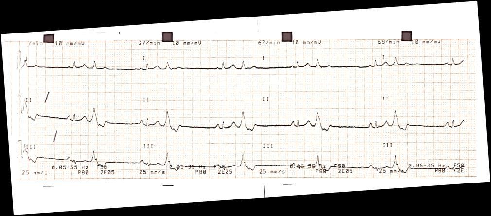
 Localized QRS widening of 110ms in V 1 -V 3 VT episodes with LBBB morphology (right ventricular")
23 Arrhythmogenic Right Ventricular Cardiomyopathy Epsilon wave (most specific finding, seen in 30% of patients) T wave inversions in V 1 -V 3 (85% of patients) Prolonged S wave upstroke of 55ms in V 1 -V 3 (95% of patients) Localized QRS widening of 110ms in V 1 -V 3 VT episodes with LBBB morphology (right ventricular VT)

24 Arrhythmogenic Right Ventricular Cardiomyopathy E Burns, lifeinthefastlane.com
25 Arrhythmogenic Right Ventricular Cardiomyopathy Br J Sports Med 2009;43:
26 Arrhythmogenic Right Ventricular Cardiomyopathy Br J Sports Med 2009;43:
27 Arrhythmogenic Right Ventricular Cardiomyopathy Br J Sports Med 2009;43:

28 Arrhythmogenic Right Ventricular Cardiomyopathy ΙΑΤΡΕΙΟ ΜΥΟΚΑΡΔΙΟΠΑΘΕΙΩΝ ΑΡΡΥΘΜΙΟΛΟΓΙΚΟ ΙΑΤΡΕΙΟ

29 Arrhythmogenic Right Ventricular Cardiomyopathy ΙΑΤΡΕΙΟ ΜΥΟΚΑΡΔΙΟΠΑΘΕΙΩΝ ΑΡΡΥΘΜΙΟΛΟΓΙΚΟ ΙΑΤΡΕΙΟ
30 Restrictive Cardiomyopathy Least common form of cardiomyopathy. It occurs in the advanced stages of myocardial infiltrative disease hemochromatosis, amyloidosis, sarcoidosis. Low voltage QRS complexes. AF may occur due to LAE Conduction disturbances due to infiltration of the cardiac conducting system septal granuloma formation in sarcoidosis may lead to BBB and AV block pseudo-infarction Q waves healing granulomas in sarcoidosis
31 RCM Common ECG findings Low voltage QRS complexes Non-specific ST / T changes Bundle branch blocks AV (3rd degree AV block may occur in sarcoidosis) Pathological Q waves Atrial and ventricular dysrhythmias
32 RCM AF and LBBB
33 Left Ventricular Non-Compaction Subtypes and ECG findings 1. Benign LVNC 2. LVNC with arrhythmias 3. Dilated LVNC 4. Hypertrophic LVNC 5. Hypertrophic Dilated LVNC 6. Restrictive LVNC 7. Right Ventricular or Biventricular LVNC hypertrophy by voltage criteria (either LVH or biventricular hypertrophy) T wave inversion ST abnormalities / strain LA enlargement left axis deviation QTc prolongation 8. LVNC with congenital heart disease Lancet 2015; 386:

34 Left ventricular non-compaction Lancet 2015; 386:

35 Left ventricular non-compaction Eur Heart J 2015:36;

36 Left ventricular non-compaction ΙΑΤΡΕΙΟ ΜΥΟΚΑΡΔΙΟΠΑΘΕΙΩΝ ΑΡΡΥΘΜΙΟΛΟΓΙΚΟ ΙΑΤΡΕΙΟ

37 Pericardial Diseases Work-up Eur Heart J 2015:36;
38 Acute Pericarditis Common ECG findings Stage 1 widespread ST and PR with reciprocal changes in avr (occurs during first 2w) Stage 2 normalization of ST changes; generalized T wave flattening (1-3w) Stage 3 flattened T waves become inverted (3 to several weeks) Stage 4 ECG returns to normal (several weeks onwards)

39 Acute Pericarditis Eur Heart J 2015:36;

40 Acute Pericarditis ΙΑΤΡΕΙΟ ΜΥΟΚΑΡΔΙΟΠΑΘΕΙΩΝ ΑΡΡΥΘΜΙΟΛΟΓΙΚΟ ΙΑΤΡΕΙΟ

41 Acute Pericarditis ΙΑΤΡΕΙΟ ΜΥΟΚΑΡΔΙΟΠΑΘΕΙΩΝ ΑΡΡΥΘΜΙΟΛΟΓΙΚΟ ΙΑΤΡΕΙΟ


42 Γ.Ν.Α. «Ο Ευαγγελισμός»

43 Takotsubo Syndrome J Med Sci 2014:83:

44 Takotsubo Syndrome J Med Sci 2014:83:

45 Takotsubo Syndrome World J Cardiol 2016;8:
46 Dilated Cardiomyopathy ICH Dilated cardiomyopathy commonly occurs following massive anterior MI due to extensive myocardial necrosis and loss of contractility Non-ICH Most cases are idiopathic Up to 25% are familial (primarily autosomal dominant, some types are X-linked) Other Viral myocarditis Alcoholism Toxins - Drugs (e.g. doxorubicin) Autoimmune disease peripartum cardiomyopathy

47 Constrictive Pericarditis vs Restrictive Cardiomyopathy Eur Heart J 2015:36;
Acute Coronary Syndromes Unstable Angina Non ST segment Elevation MI (NSTEMI) ST segment Elevation MI (STEMI)
 ST segment Elevation MI (STEMI)") Leanna R. Miller, RN, MN, CCRN-CSC, PCCN-CMC, CEN, CNRN, CMSRN, NP Education Specialist LRM Consulting Nashville, TN Objectives Evaluate common abnormalities that mimic myocardial infarction. Identify
Leanna R. Miller, RN, MN, CCRN-CSC, PCCN-CMC, CEN, CNRN, CMSRN, NP Education Specialist LRM Consulting Nashville, TN Objectives Evaluate common abnormalities that mimic myocardial infarction. Identify
ECG Underwriting Puzzler Dr. Regina Rosace AVP & Medical Director
 December 2018 ECG Underwriting Puzzler Dr. Regina Rosace AVP & Medical Director To obtain best results Select Slide Show from the ribbon at the top of your PowerPoint screen Select From Beginning on the
Office ECG Interpretation
 Office ECG Interpretation Jason Evanchan, DO Assistant Professor of Medicine Division of Cardiovascular Medicine The Ohio State University Wexner Medical Center Outline of topics High risk ischemia T wave
6/19/2018. Background Athlete s heart. Ultimate question. Applying the International Criteria for ECG
 Applying the International Criteria for ECG Interpretation in Athletes to a preparticipation screening program DAVE SIEBERT, MD, CAQSM ASSISTANT PROFESSOR DEPARTMENT OF FAMILY MEDICINE UNIVERSITY OF WASHINGTON
Section V. Objectives
 Section V Landscape of an MI Objectives At the conclusion of this presentation the participant will be able to Outline a systematic approach to 12 lead ECG interpretation Demonstrate the process for determining
Please check your answers with correct statements in answer pages after the ECG cases.
 ECG Cases ECG Case 1 Springer International Publishing AG, part of Springer Nature 2018 S. Okutucu, A. Oto, Interpreting ECGs in Clinical Practice, In Clinical Practice, https://doi.org/10.1007/978-3-319-90557-0
at least 4 8 hours per week
 ECG IN ATHLETS An athlete is defined as an individual who engages in regular exercise or training for sport or general fitness, typically with a premium on performance, and often engaged in individual
REtrive. REpeat. RElearn Design by. Test-Enhanced Learning based ECG practice E-book
 Test-Enhanced Learning Test-Enhanced Learning Test-Enhanced Learning Test-Enhanced Learning based ECG practice E-book REtrive REpeat RElearn Design by S I T T I N U N T H A N G J U I P E E R I Y A W A
PAEDIATRIC ECG Dimosthenis Avramidis, MD.
 PAEDIATRIC ECG Dimosthenis Avramidis, MD. Consultant Mitera Children s Hospital Athens Greece S. Associate 1st Cardiology Dpt Evangelismos Hospital Athens Greece 5 y/o with sinus tach Background ECG changes
ECG Interpretation. Best to have a system to methodically evaluate ECG (from Dubin) * Rate * Rhythm * Axis * Intervals * Hypertrophy * Infarction
 * Rate * Rhythm * Axis * Intervals * Hypertrophy * Infarction") ECG to save Babies ECG Interpretation Best to have a system to methodically evaluate ECG (from Dubin) * Rate * Rhythm * Axis * Intervals * Hypertrophy * Infarction Electrical Activity in the heart 5 events
ECG to save Babies ECG Interpretation Best to have a system to methodically evaluate ECG (from Dubin) * Rate * Rhythm * Axis * Intervals * Hypertrophy * Infarction Electrical Activity in the heart 5 events
Paediatric ECG Interpretation
 Paediatric ECG Interpretation Dr Sanj Fernando (thanks to http://lifeinthefastlane.com/ecg-library/paediatric-ecginterpretation/) 3 yo boy complaining of abdominal pain and chest pain Child ECG vs Adult
Αμφικοιλιακή βηματοδότηση: LBBB ή ευρύ QRS;
 ΠΑΝΕΛΛΗΝΙΑ ΣΕΜΙΝΑΡΙΑ ΟΜΑΔΩΝ ΕΡΓΑΣΙΑΣ 2016 ΟΜΑΔΑ ΕΡΓΑΣΙΑΣ ΚΑΡΔΙΑΚΗΣ ΑΝΕΠΑΡΚΕΙΑΣ Στρογγυλό τραπέζι: Χρόνια Καρδιακή Ανεπάρκεια Αμφικοιλιακή βηματοδότηση: LBBB ή ευρύ QRS; Ξυδώνας Σωτήριος, MD, FESC Β Καρδιολογικό
Return to Basics. ECG Rate and Rhythm. Management of the Hospitalized Patient September 25, 2009
 Management of the Hospitalized Patient September 25, 2009 ECG Refresher and Update 2009 Return to Basics Determine rate and rhythm Determine intervals and axes Define morphology of P-QRS-T-U Compare with
Return to Basics. Normal Intervals & Axes. ECG Rate and Rhythm
 Return to Basics Management of the Hospitalized Patient October 15, 2010 ECG Refresher and Update 2010 Determine rate and rhythm Determine intervals and axes Define morphology of P-QRS-T-U Compare with
General Introduction to ECG. Reading Assignment (p2-16 in PDF Outline )
") General Introduction to ECG Reading Assignment (p2-16 in PDF Outline ) Objectives 1. Practice the 5-step Method 2. Differential Diagnosis: R & L axis deviation 3. Differential Diagnosis: Poor R-wave progression
General Introduction to ECG Reading Assignment (p2-16 in PDF Outline ) Objectives 1. Practice the 5-step Method 2. Differential Diagnosis: R & L axis deviation 3. Differential Diagnosis: Poor R-wave progression
Myocardial Infarction. Reading Assignment (p66-78 in Outline )
") Myocardial Infarction Reading Assignment (p66-78 in Outline ) Objectives 1. Why do ST segments go up or down in ischemia? 2. STEMI locations and culprit vessels 3. Why 15-lead ECGs? 4. What s up with avr?
Myocardial Infarction Reading Assignment (p66-78 in Outline ) Objectives 1. Why do ST segments go up or down in ischemia? 2. STEMI locations and culprit vessels 3. Why 15-lead ECGs? 4. What s up with avr?
Pennsylvania Academy of Family Physicians Foundation & UPMC 43rd Refresher Course in Family Medicine CME Conference March 10-13, 2016
 Pennsylvania Academy of Family Physicians Foundation & UPMC 43rd Refresher Course in Family Medicine CME Conference March 10-13, 2016 Disclosures: EKG Workshop Louis Mancano, MD Speaker has no disclosures
Appendix D Output Code and Interpretation of Analysis
 Appendix D Output Code and Interpretation of Analysis 8 Arrhythmia Code No. Description 8002 Marked rhythm irregularity 8110 Sinus rhythm 8102 Sinus arrhythmia 8108 Marked sinus arrhythmia 8120 Sinus tachycardia
Return to Basics. ECG Rate and Rhythm. Management of the Hospitalized Patient October 4, 2007
 Management of the Hospitalized Patient October 4, 2007 ECG Refresher for the Hospitalists Return to Basics Determine rate and rhythm Determine intervals and axes Define morphology of P-QRS-T-U Compare
12-Lead ECG Interpretation. Kathy Kuznar, RN, ANP
 12-Lead ECG Interpretation Kathy Kuznar, RN, ANP The 12-Lead ECG Objectives Identify the normal morphology and features of the 12- lead ECG. Perform systematic analysis of the 12-lead ECG. Recognize abnormalities
Μη φαρμακευτική θεραπεία στην Χρόνια Καρδιακή Ανεπάρκεια Νεότερες συσκευές. Ξυδώνας Σωτήριος, MD, PhD, FESC
 ΠΑΝΕΛΛΗΝΙΑ ΣΕΜΙΝΑΡΙΑ ΟΜΑΔΩΝ ΕΡΓΑΣΙΑΣ 2017 ΟΜΑΔΑ ΕΡΓΑΣΙΑΣ ΚΑΡΔΙΑΚΗΣ ΑΝΕΠΑΡΚΕΙΑΣ Μη φαρμακευτική θεραπεία στην Χρόνια Καρδιακή Ανεπάρκεια Νεότερες συσκευές Ξυδώνας Σωτήριος, MD, PhD, FESC Καρδιολογικό Τμήμα,
Electrical System Overview Electrocardiograms Action Potentials 12-Lead Positioning Values To Memorize Calculating Rates
 Electrocardiograms Electrical System Overview James Lamberg 2/ 74 Action Potentials 12-Lead Positioning 3/ 74 4/ 74 Values To Memorize Inherent Rates SA: 60 to 100 AV: 40 to 60 Ventricles: 20 to 40 Normal
Study methodology for screening candidates to athletes risk
 1. Periodical Evaluations: each 2 years. Study methodology for screening candidates to athletes risk 2. Personal history: Personal history of murmur in childhood; dizziness, syncope, palpitations, intolerance
Ablative Therapy for Ventricular Tachycardia
 Ablative Therapy for Ventricular Tachycardia Nitish Badhwar, MD, FACC, FHRS 2 nd Annual UC Davis Heart and Vascular Center Cardiovascular Nurse / Technologist Symposium May 5, 2012 Disclosures Research
The Electrocardiogram part II. Dr. Adelina Vlad, MD PhD
 The Electrocardiogram part II Dr. Adelina Vlad, MD PhD Basic Interpretation of the ECG 1) Evaluate calibration 2) Calculate rate 3) Determine rhythm 4) Determine QRS axis 5) Measure intervals 6) Analyze
2017 EKG Workshop Advanced. Family Medicine Review Course Lou Mancano, MD, FAAFP Reading Health System Family and Community Medicine Reading, PA
 2017 EKG Workshop Advanced Family Medicine Review Course Lou Mancano, MD, FAAFP Reading Health System Family and Community Medicine Reading, PA Part II - Objective Describe a useful approach to interpreting
Abnormal electrocardiographic findings in athletes: recognising changes suggestive of cardiomyopathy
 Scan to access more free content For numbered affiliations see end of article Correspondence to Jonathan A Drezner, Department of Family Medicine, University of Washington, 1959 NE Pacific Street, Box
Dr. Schroeder has no financial relationships to disclose
 Valerie A Schroeder MD MS Assistant Professor University of Kansas Medical Center READING THE WAVES- THE HEART S ELECTRICAL MESSAGE FINANCIAL DISCLOSURE Dr. Schroeder has no financial relationships to
Etiology, Classification & Management. Sheba Medical Center Cardiology Department Matthew Wright St. George s University of London
 Etiology, Classification & Management Sheba Medical Center Cardiology Department Matthew Wright St. George s University of London Introduction World Health Organization (1995): Diseases of myocardium (heart
Cardiomyopathy. Jeff Grubbe MD FACP, Chief Medical Director, Allstate Life & Retirement
 Cardiomyopathy Jeff Grubbe MD FACP, Chief Medical Director, Allstate Life & Retirement Nebraska Home Office Life Underwriters Association March 20, 2018 1 Cardiomyopathy A myocardial disorder in which
Chapter 2 Practical Approach
 Chapter 2 Practical Approach There are beginners in electrocardiogram (ECG) analysis who are fascinated by a special pattern (e.g., a bundle-branch block or a striking Q wave) and thereby overlook other
Reading Assignment (p1-91 in Outline ) Objectives What s in an ECG?
 Objectives What s in an ECG?") Reading Assignment (p1-91 in Outline ) Objectives What s in an ECG? The 5-Step Method ECG #: Mearurements: Rhythm (s): Conduction: Waveform: Interpretation: A= V= PR= QRS= QT= Axis= 1. Compute the 5 basic
Reading Assignment (p1-91 in Outline ) Objectives What s in an ECG? The 5-Step Method ECG #: Mearurements: Rhythm (s): Conduction: Waveform: Interpretation: A= V= PR= QRS= QT= Axis= 1. Compute the 5 basic
Echocardiographic Evaluation of the Cardiomyopathies. Stephanie Coulter, MD, FACC, FASE April, 2016
 Echocardiographic Evaluation of the Cardiomyopathies Stephanie Coulter, MD, FACC, FASE April, 2016 Cardiomyopathies (CMP) primary disease intrinsic to cardiac muscle Dilated CMP Hypertrophic CMP Infiltrative
Electrocardiography for Healthcare Professionals. Chapter 14 Basic 12-Lead ECG Interpretation
 Electrocardiography for Healthcare Professionals Chapter 14 Basic 12-Lead ECG Interpretation 2012 The Companies, Inc. All rights reserved. Learning Outcomes 14.1 Discuss the anatomic views seen on a 12-lead
ECG Cases and Questions. Ashish Sadhu, MD, FHRS, FACC Electrophysiology/Cardiology
 ECG Cases and Questions Ashish Sadhu, MD, FHRS, FACC Electrophysiology/Cardiology 32 yo female Life Insurance Physical 56 yo male with chest pain Terminology Injury ST elevation Ischemia T wave inversion
La valutazione dell atleta: è una strategia salva-vita e costo-efficace?
 La valutazione dell atleta: è una strategia salva-vita e costo-efficace? Primo trattato di Medicina Wilson and Jungner s criteria In the 1960s the World Health Organization adopted the Wilson and Jungner
CARDIOMYOPATHY IN CT. Hans- Christoph Becker Professor of Radiology
 CARDIOMYOPATHY IN CT Hans- Christoph Becker Professor of Radiology 1 Cardiomyopathy Heart muscle disease Deterioration of the heart function, heart failure Dyspnea, peripheral edema Risk of arrhythmia,
Miscellaneous Stuff Keep reading the Outline
 Miscellaneous Stuff Keep reading the Outline Welcome to the 5-Step Method ECG #: Mearurements: Rhythm (s): Conduction: Waveform: Interpretation: A= V= PR= QRS= QT= Axis= 1. Compute the 5 basic measurements:
Advances in Ablation Therapy for Ventricular Tachycardia
 Advances in Ablation Therapy for Ventricular Tachycardia Nitish Badhwar, MD, FACC, FHRS Director, Cardiac Electrophysiology Training Program University of California, San Francisco For those of you who
ECG Practice Strips Discussion part 1:
 ECG Practice Strips Discussion part 1: The first 20 strips are for teaching various abnormalities of the morphology of the waves of the ECG. Strips 21 and following are for teaching some abnormalities
The Efficient and Smart Methods for Diagnosis of SVT 대구파티마병원순환기내과정병천
 The Efficient and Smart Methods for Diagnosis of SVT 대구파티마병원순환기내과정병천 Differentiation Supraventricular Origin from Ventricular Origin on ECG. QRS-Complex Width. 1. Narrow QRS-Complex Tachycardia (
12 Lead ECG. Presented by Rebecca Sevigny BSN, RN Professional Practice & Development Dept.
 12 Lead ECG Presented by Rebecca Sevigny BSN, RN Professional Practice & Development Dept. Two Main Coronary Arteries RCA LCA which branches into Left Anterior Descending Circumflex Artery Two Main Coronary
Relax and Learn at the FARM 2012: Session 8: 12 Lead ECG 401: ECG Variants
 Relax and Learn at the FARM 2012: Session 8: 12 Lead ECG 401: ECG Variants A Ship in the Harbor is Safe But that is not what ships are built for. Karen Marzlin DNP, RN, CCNS, CCRN-CMC, CHFN Cardiovascular
FANS ARVC (Arrhythmogenic Right Ventricular Cardiomyopathy) Investigation Protocol
 Investigation Protocol") Clinical Features FANS ARVC (Arrhythmogenic Right Ventricular Cardiomyopathy) Investigation Protocol History: Progressive disease, characterised by the following clinical stages: o Early concealed phase
Clinical Features FANS ARVC (Arrhythmogenic Right Ventricular Cardiomyopathy) Investigation Protocol History: Progressive disease, characterised by the following clinical stages: o Early concealed phase
ELECTROCARDIOGRAPH. General. Heart Rate. Starship Children s Health Clinical Guideline
 General Heart Rate QRS Axis T Wave Axis PR Interval according to Heart Rate & Age P Wave Duration and Amplitude QRS Duration according to Age QT Interval R & S voltages according to Lead & Age R/S ratio
All About STEMIs. Presented By: Brittney Urvand, RN, BSN, CCCC. Essentia Health Fargo Cardiovascular Program Manager.
 All About STEMIs Presented By: Brittney Urvand, RN, BSN, CCCC Essentia Health Fargo Cardiovascular Program Manager Updated 10/2/2018 None Disclosures Objectives Identify signs and symptoms of a heart attack
Section 3 and 4. Objectives. Bundle Branches 10/9/2018. LBBB, RBBB Bifascicular, Trifascicular Block
 Section 3 and 4 LBBB, RBBB Bifascicular, Trifascicular Block Objectives At the conclusion of this presentation the participant will be able to Outline a systematic approach to 12 lead ECG interpretation
Cardiomyopathy: The Good, the Bad.and the Insurable?
 Cardiomyopathy: The Good, the Bad.and the Insurable? WAHLU Spring Seminar 2014 Joy Geiger, RN, BSN, ALMI Medical Consultant The Northwestern Mutual Life Insurance Company Milwaukee, WI Objectives Overview
EVALUATION OF ELECTROCARDIOGRAPHIC FINDINGS IN ATHLETES
 EVALUATION OF ELECTROCARDIOGRAPHIC FINDINGS IN ATHLETES UNIT OF INHERITED CV DISEASES HEART CENTER OF THE YOUNG AND ATHLETES A DPT OF CARDIOLOGY UNIVERSITY OF ATHENS EVALUATION OF ELECTROCARDIOGRAPHIC
Εμφύτευση απινιδωτών για πρωτογενή πρόληψη σε ασθενείς που δεν περιλαμβάνονται στις κλινικές μελέτες
 Εμφύτευση απινιδωτών για πρωτογενή πρόληψη σε ασθενείς που δεν περιλαμβάνονται στις κλινικές μελέτες Δημήτριος M. Κωνσταντίνου Ειδικός Καρδιολόγος, MD, MSc, PhD, CCDS Πανεπιστημιακός Υπότροφος Dr. Konstantinou
Family Medicine for English language students of Medical University of Lodz ECG. Jakub Dorożyński
 Family Medicine for English language students of Medical University of Lodz ECG Jakub Dorożyński Parts of an ECG The standard ECG has 12 leads: six of them are considered limb leads because they are placed
Ekg pra pr c a tice D.HAMMOUDI.MD
 Ekg practice D.HAMMOUDI.MD Anatomy Revisited RCA (Right Coronary Artery) Right ventricle Inferior wall of LV Posterior wall of LV (75%) SA Node (60%) AV Node (>80%) LCA (Left Coronary Artery) Septal wall
3/4/2018. March Martina Frost, PA C Desert Cardiology. Electricity moving towards/away from electrode create downward/upward directions of waves
 March 2018 Martina Frost, PA C Desert Cardiology Electricity moving towards/away from electrode create downward/upward directions of waves Frontal view Limb leads: I, II, III, avl, avf, (avr) Horizontal
Masqueraders of STEMI
 Masqueraders of STEMI Steven M. Costa, M.D. Assistant Professor Department of Medicine Division of Cardiology Scott & White Memorial Hospital and Clinic Texas A&M University Health Science Center Disclosures
Acute Coronary Syndromes. Disclosures
 Acute Coronary Syndromes Disclosures I work for Virginia Garcia Memorial Health Center, Beaverton, OR. Jon Tardiff, BS, PA-C OHSU Clinical Assistant Professor And I am a medical editor for Jones & Bartlett
HR: 50 bpm (Sinus) PR: 280 ms QRS: 120 ms QT: 490 ms Axis: -70. Sinus bradycardia with one ventricular escape (*)
 PR: 280 ms QRS: 120 ms QT: 490 ms Axis: -70. Sinus bradycardia with one ventricular escape (*)") 1? HR: 50 bpm (Sinus) PR: 280 ms QRS: 120 ms QT: 490 ms Axis: -70 1 Sinus P waves? 2 sinus cycles The pause (2 sinus cycles) suggests that the sinus fired (?) but did not conduct to the atria (i.e., missing
1? HR: 50 bpm (Sinus) PR: 280 ms QRS: 120 ms QT: 490 ms Axis: -70 1 Sinus P waves? 2 sinus cycles The pause (2 sinus cycles) suggests that the sinus fired (?) but did not conduct to the atria (i.e., missing
ECG ABNORMALITIES D R. T AM A R A AL Q U D AH
 ECG ABNORMALITIES D R. T AM A R A AL Q U D AH When we interpret an ECG we compare it instantaneously with the normal ECG and normal variants stored in our memory; these memories are stored visually in
Interpretation and Consequences of Repolarisation Changes in Athletes
 Interpretation and Consequences of Repolarisation Changes in Athletes Professor Sanjay Sharma E-mail sasharma@sgul.ac.uk @SSharmacardio Disclosures: None Athlete s ECG Vagotonia Sinus bradycardia Sinus
ECG Interpretation Made Easy
 ECG Interpretation Made Easy Dr. A Tageldien Abdellah, MSc MD EBSC Lecturer of Cardiology- Hull University Hull York Medical School 2007-2008 ECG Interpretation Made Easy Synopsis Benefits Objectives Process
ECGs: Everything a finalist needs to know. Dr Amy Coulden As part of the Simply Finals series
 ECGs: Everything a finalist needs to know Dr Amy Coulden As part of the Simply Finals series Aims and objectives To be able to interpret basic ECG abnormalities To be able to recognise commonly tested
ECG CONVENTIONS AND INTERVALS
 1 ECG Waveforms and Intervals ECG waveforms labeled alphabetically P wave== represents atrial depolarization QRS complex=ventricular depolarization ST-T-U complex (ST segment, T wave, and U wave)== V repolarization.
Other 12-Lead ECG Findings
 Other 12-Lead ECG Findings Left Atrial Enlargement Left atrial enlargement is illustrated by increased P wave duration in lead II, top ECG, and by the prominent negative P terminal force in lead V1, bottom
Basic electrocardiography reading. R3 lee wei-chieh
 Basic electrocardiography reading R3 lee wei-chieh The Normal Conduction System Lead Placement avf Limb Leads Precordial Leads Interpretation Rate Rhythm Interval Axis Chamber abnormality QRST change What
Cardiology Flash Cards
 Cardiology Flash Cards EKG in a nut shell www.brain101.info Conduction System www.brain101.info 2 Analyzing EKG Step by step Steps in Analyzing ECG'S 1. Rhythm: - Regular _ Sinus, Junctional or Ventricular.
Bundle Branch & Fascicular Blocks. Reading Assignment (p53-58 in Outline )
") Bundle Branch & Fascicular Blocks Reading Assignment (p53-58 in Outline ) Objectives 1. QRS analysis of Right and Left BBB 2. Uncomplicated vs complicated BBB 3. Diagnosis of RBBB with LAFB and LPFB 4.
Bundle Branch & Fascicular Blocks Reading Assignment (p53-58 in Outline ) Objectives 1. QRS analysis of Right and Left BBB 2. Uncomplicated vs complicated BBB 3. Diagnosis of RBBB with LAFB and LPFB 4.
2/7/ LEAD ECG CASE STUDIES Lisa Riggs MSN, RN, ACNS-BC, CCRN-K CASE #1 WHAT ELSE WOULD YOU ASSESS? WHAT S YOUR DIAGNOSIS?
 12 LEAD ECG CASE STUDIES Lisa Riggs MSN, RN, ACNS-BC, CCRN-K CASE #1 31 y/o male is a direct admit from the physician s office with c/o chest pain and SOA WHAT ELSE WOULD YOU ASSESS? WHAT S YOUR DIAGNOSIS?
10 ECGs No Practitioner Can Afford to Miss. Objectives
 10 ECGs No Practitioner Can Afford to Miss Mary L. Dohrmann, MD Professor of Clinical Medicine Division of Cardiovascular Medicine University of Missouri School of Medicine No disclosures Objectives 1.
Huseng Vefali MD St. Luke s University Health Network Department of Cardiology
 Huseng Vefali MD St. Luke s University Health Network Department of Cardiology Learning Objectives Establish Consistent Approach to Interpreting ECGs Review Essential Cases for Paramedics and first responders
Paroxysmal Supraventricular Tachycardia PSVT.
 Atrial Tachycardia; is the name for an arrhythmia caused by a disorder of the impulse generation in the atrium or the AV node. An area in the atrium sends out rapid signals, which are faster than those
Restrictive Cardiomyopathy
 ESC Congress 2011, Paris Imaging Unusual Causes of Cardiomyopathy Restrictive Cardiomyopathy Kazuaki Tanabe, MD, PhD Professor of Medicine Chair, Division of Cardiology Izumo, Japan I Have No Disclosures
402 Index. B β-blockers, 4, 5 Bradyarrhythmias, 76 77
 Index A Acquired immunodeficiency syndrome (AIDS), 126, 163 Action potentials, 1, 5, 27 Acute coronary syndromes, 123t, 129 Adenosine, intravenous, 277 Alcohol abuse, as T wave inversion cause, 199 Aneurysm,
Supraventricular Arrhythmias. Reading Assignment. Chapter 5 (p17-30)
") Supraventricular Arrhythmias Reading Assignment Chapter 5 (p17-30) The Supraventricular Rhythms In Our Lives Site of Origin Single Events Slow Rates Intermediate Rates Fast Rates (>100 bpm) Sinus Sinus
Supraventricular Arrhythmias Reading Assignment Chapter 5 (p17-30) The Supraventricular Rhythms In Our Lives Site of Origin Single Events Slow Rates Intermediate Rates Fast Rates (>100 bpm) Sinus Sinus
THE ELECTROCARDIOGRAM A UBIQUITOUS AND COST-EFFECTIVE DIAGNOSTIC TOOL FOR THE FAMILY MEDICINE REFRESHER COURSE MARCH 8, 2019
 THE ELECTROCARDIOGRAM A UBIQUITOUS AND COST-EFFECTIVE DIAGNOSTIC TOOL FOR THE FAMILY MEDICINE REFRESHER COURSE MARCH 8, 2019 Major Clinical Disorders Pulmonary Embolism 69 y/o woman with dyspnea and an
The ABC of Pediatric ECG
 The ABC of Pediatric ECG Mohamed Hamdan, MD, FAAP, FACC Assistant Professor of Pediatrics Columbia University College of Physicians and Surgeons, NY, USA Consultant Pediatric Cardiologist & Co-Director
MICS OF MYOCARDIAL ISCHEMIA AND INFARCTION REVISED FOR LAS VEGAS
 ECG MIMICS OF MYOCARDIAL ISCHEMIA AND INFARCTION 102.06.05 Tzong-Luen Wang MD, PhD, JM, FESC, FACC Professor. Medical School, Fu-Jen Catholic University Chief, Emergency Department, Shin-Kong Wu Ho-Su
Conduction Problems / Arrhythmias. Conduction
 Conduction Problems / Arrhythmias Conduction Wolf-Parkinson White Syndrome (WPW) and Lown-Ganong-Levine (LGL): Atrial impulses bypass the AV node through an accessory pathway or bypass tract (bundle of
DIAGNOSIS AND MANAGEMENT OF ARRHYTHMOGENIC CARDIOMYOPATHY. David SIU MD ( 蕭頌華醫生 ) Division of Cardiology The University of Hong Kong
 Division of Cardiology The University of Hong Kong") APHRS Summit 2018 in conjunction with HKCC Heart Rhythm Refresher Course DIAGNOSIS AND MANAGEMENT OF ARRHYTHMOGENIC CARDIOMYOPATHY David SIU MD ( 蕭頌華醫生 ) Division of Cardiology The University of Hong Kong
APHRS Summit 2018 in conjunction with HKCC Heart Rhythm Refresher Course DIAGNOSIS AND MANAGEMENT OF ARRHYTHMOGENIC CARDIOMYOPATHY David SIU MD ( 蕭頌華醫生 ) Division of Cardiology The University of Hong Kong
Genetic Cardiomyopathies
 2017 HFCT Annual Scientific Meeting The Heart Failure Crosstalk Genetic Cardiomyopathies Teerapat Yingchoncharoen M.D. Ramathibodi Hospital Cardiomyopathy Group of diseases of the myocardium associated
Appendix. Table 1: Causes for abnormal axis deviation Left axis deviation
 Appendix Table 1: Causes for abnormal axis deviation Left axis deviation Normal variant (2 5%) Left anterior fascicular block Left ventricular hypertrophy Inferior wall myocardial infarction Primum atrial
Case 1. Case 2. Case 3
 Case 1 The correct answer is D. Occasionally, the Brugada syndrome can present similar morphologies to A and also change depending on the lead position but in the Brugada pattern the r is wider and ST
12 Lead EKG Interpretation. Disclosures
 12 Lead EKG Interpretation Louann B. Bailey, DNP, APRN, FAANP ACNP BC I have no disclosures Disclosures 2 1 Objectives At the conclusion of this presentation the participant will be able to Outline a systematic
PAEDIATRIC ACUTE CARE GUIDELINE. ECG Interpretation
 Princess Margaret Hospital for Children PAEDIATRIC ACUTE CARE GUIDELINE ECG Interpretation Scope (Staff): Scope (Area): All Emergency Department Clinicians Emergency Department This document should be
If the P wave > 0.12 sec( 3 mm) usually in any lead. Notched P wave usually in lead I,aVl may be lead II Negative terminal portion of P wave in V1, 1
 usually in any lead. Notched P wave usually in lead I,aVl may be lead II Negative terminal portion of P wave in V1, 1") If the P wave > 0.12 sec( 3 mm) usually in any lead. Notched P wave usually in lead I,aVl may be lead II Negative terminal portion of P wave in V1, 1 mm depth and 3 mm width( most specific) Since Mitral
If the P wave > 0.12 sec( 3 mm) usually in any lead. Notched P wave usually in lead I,aVl may be lead II Negative terminal portion of P wave in V1, 1 mm depth and 3 mm width( most specific) Since Mitral
Solutions for Every Day Problems Cardiologists and the ECG: Are We Really That Good at It? Part II Daniel José Piñeiro Profesor Titular de Medicina,
 Solutions for Every Day Problems Cardiologists and the ECG: Are We Really That Good at It? Part II Daniel José Piñeiro Profesor Titular de Medicina, Universidad de Buenos Aires, Argentina Member, Membership
Pathologic ECG. Adelina Vlad, MD PhD
 Pathologic ECG Adelina Vlad, MD PhD Basic Interpretation of the ECG 1) Evaluate calibration 2) Calculate rate 3) Determine rhythm 4) Determine QRS axis 5) Measure intervals 6) Analyze the morphology and
ECG Basics Sonia Samtani 7/2017 UCI Resident Lecture Series
 ECG Basics Sonia Samtani 7/2017 UCI Resident Lecture Series Agenda I. Introduction II.The Conduction System III.ECG Basics IV.Cardiac Emergencies V.Summary The Conduction System Lead Placement avf Precordial
How to Read an Athlete s ECG. Sanjay Sharma BSc (Hons), MD, FRCP, FESC
, MD, FRCP, FESC") How to Read an Athlete s ECG Sanjay Sharma BSc (Hons), MD, FRCP, FESC Athlete s EKG Vagotonia Sinus bradycardia Sinus arrhythmia First degree AVB ST-elevation Tall T waves Increased chamber size Left ventricular
How to Read an Athlete s ECG Sanjay Sharma BSc (Hons), MD, FRCP, FESC Athlete s EKG Vagotonia Sinus bradycardia Sinus arrhythmia First degree AVB ST-elevation Tall T waves Increased chamber size Left ventricular
Normal ECG And ECHO Findings in Athletes
 Normal ECG And ECHO Findings in Athletes Dr.Yahya Kiwan Consultant Interventional Cardiologist Head Of Departement Of Cardiology Canadian Specialist Hospital Sinus Bradycardia The normal heartbeat is initiated
ECG pre-reading manual. Created for the North West Regional EMET training program
 ECG pre-reading manual Created for the North West Regional EMET training program Author:- Dr Juan Carlos Ascencio-Lane juan.ascencio-lane@ths.tas.gov.au 1 Disclaimer This handbook has been created for
If you woke up breathing this morning, Congratulations! You get another chance. Use it wisely!
 Seton Medical Center Austin, Texas Cynthia Webner DNP, RN, CCNS, CCRN-CMC, CHFN www.cardionursing.com RULES OF LIFE If you woke up breathing this morning, Congratulations! You get another chance. Use it
ΗΚΓ ΚΑΙ ΣΝ Γ Ε Ω Ρ Γ Ι Ο Σ Γ Ι ΑΝ Ν Ο Π Ο Υ Λ Ο Σ Ε Π Ι Μ Ε Λ Η Τ Η Σ Β Κ Α Ρ Δ Ι Ο Λ Ο Γ Ι Α Σ Γ Ν Α «Γ. Γ Ε Ν Ν Η Μ Α Τ Α Σ»
 ΗΚΓ ΚΑΙ ΣΝ Γ Ε Ω Ρ Γ Ι Ο Σ Γ Ι ΑΝ Ν Ο Π Ο Υ Λ Ο Σ Ε Π Ι Μ Ε Λ Η Τ Η Σ Β Κ Α Ρ Δ Ι Ο Λ Ο Γ Ι Α Σ Γ Ν Α «Γ. Γ Ε Ν Ν Η Μ Α Τ Α Σ» ΑΝΔΡΑΣ 48 ΕΤΩΝ ΜΕ ΑΛΓΟΣ ΕΠΙΓΑΣΤΡΙΟΥ ΚΑΙ ΝΑΥΤΙΑ ECG evidence of myocardial
Current ECG interpretation guidelines in the screening of athletes
 REVIEW ARTICLE 7 How to differentiate physiological adaptation to intensive physical exercise from pathologies Current ECG interpretation guidelines in the screening of athletes Gemma Parry-Williams, Sanjay
DR QAZI IMTIAZ RASOOL OBJECTIVES
 PRACTICAL ELECTROCARDIOGRAPHY DR QAZI IMTIAZ RASOOL OBJECTIVES Recording of electrical events in heart Established electrode pattern results in specific tracing pattern Health of heart i. e. Anatomical
Sudden cardiac death: Primary and secondary prevention
 Sudden cardiac death: Primary and secondary prevention By Kai Chi Chan Penultimate Year Medical Student St George s University of London at UNic Sheba Medical Centre Definition Sudden cardiac arrest (SCA)
ΔΙΑΤΑΡΑΧΕΣ ΕΝΔΟΚΟΙΛΙΑΚΗΣ ΑΓΩΓΙΜΟΤΗΤΑΣ ΔΗΜΗΤΡΙΟΣ Δ. ΜΑΝΩΛΑΤΟΣ Β ΚΑΡΔΙΟΛΟΓΙΚΗ ΚΛΙΝΙΚΗ ΕΡΓΑΣΤΗΡΙΟ ΗΛΕΚΤΡΟΦΥΣΙΟΛΟΓΙΑΣ Γ.Ν.Α.
 ΔΙΑΤΑΡΑΧΕΣ ΕΝΔΟΚΟΙΛΙΑΚΗΣ ΑΓΩΓΙΜΟΤΗΤΑΣ ΔΗΜΗΤΡΙΟΣ Δ. ΜΑΝΩΛΑΤΟΣ Β ΚΑΡΔΙΟΛΟΓΙΚΗ ΚΛΙΝΙΚΗ ΕΡΓΑΣΤΗΡΙΟ ΗΛΕΚΤΡΟΦΥΣΙΟΛΟΓΙΑΣ Γ.Ν.Α. «ΕΥΑΓΓΕΛΙΣΜΟΣ» Intraventricular conduction delay and Blocks Right Bundle Branch
Ben Taylor, PhD, PA-C
 Ben Taylor, PhD, PA-C The patient is a 23-year-old white male with a history of polysubstance abuse who was found unresponsive, last seen the day before. Classic signs of systemic hypothermia with prominent
SUDDEN CARDIAC DEATH IN ATHLETES
 SUDDEN CARDIAC DEATH IN ATHLETES Alix Dufresne, MD, FACP, FACC, FESC Cardiology Division Chief, Interfaith Medical Center Director Cardiology Clinic, Kingsbrook Jewish Center PURPOSE AND OBJECTIVES PURPOSE
ECG S: A CASE-BASED APPROACH December 6,
 ECG S: A CASE-BASED APPROACH December 6, 2018 1 Faculty Disclosure Faculty: Lorne Gula MD, FRCPC Professor, Western University Cardiologist, Hearth Rhythm Specialist Director, Electrophysiology Laboratory,
, David Stultz, MD.
 http://www.dilbert.com EKG Rounds Handouts available at http://www.drstultz.com January 5, 2004 David Stultz, MD Cardiology Fellow, PGY 4 Overview of Topics How to read an EKG Normal EKG Determination
XVth Balkan Congress of Radiology Danubius Hotel Helia, October 2017, Budapest, Hungary
 XVth Balkan Congress of Radiology Danubius Hotel Helia, 12-14 October 2017, Budapest, Hungary Ružica Maksimović MRI in Myocarditis Faculty of Medicine, University of Belgrade, Centre for Radiology and