Ablative Therapy for Ventricular Tachycardia
|
|
|
- Erick Ira Morgan
- 6 years ago
- Views:
Transcription

1 Ablative Therapy for Ventricular Tachycardia Nitish Badhwar, MD, FACC, FHRS 2 nd Annual UC Davis Heart and Vascular Center Cardiovascular Nurse / Technologist Symposium May 5, 2012
2 Disclosures Research grant - St. Jude, Stereotaxis Honoraria - St. Jude, Boston Scientific, Janssen
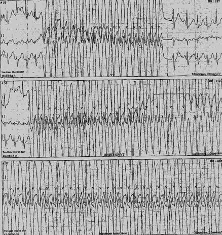
3 Ventricular Tachycardia Non-sustained ventricular tachycardia (NSVT) 3 or more consecutive QRS complexes of ventricular origin at rate of more than 100 bpm Sustained VT Lasts more than 30 seconds, usually requires intervention for termination Monomorphic VT Uniform QRS configuration Polymorphic VT Beat to beat variation in QRS configuration Electrical storm > 3 VT/VF episodes in 24 hours

4 What is the best treatment option? 1. Catheter Ablation 2. Urgent left heart catheterization 3. Start I/V Amiodarone 4. Do Nothing

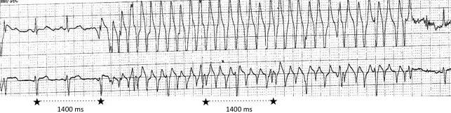
5 Wide Complex Tachycardia: Artifact
6 Ventricular Tachycardia Sudden cardiac arrest (ventricular fibrillation) Syncope, near syncope Palpitations with wide complex tachycardia (hemodynamically tolerated) or frequent PVCs
7 Ventricular Tachycardia Idiopathic VT VT associated with structural heart disease
8 Idiopathic Ventricular Tachycardia Polymorphic VT /VF Long QT syndrome Brugada syndrome Short coupled torsades Short QT syndrome Catecholamine induced polymorphic V T Monomorphic VT Outflow tract VT- RVOT VT, LVOT VT,cusp VT Fascicular VT- LAF, LPF, Septal Annular VT- Mitral, Tricuspid VT from crux of heart Idiopathic VF
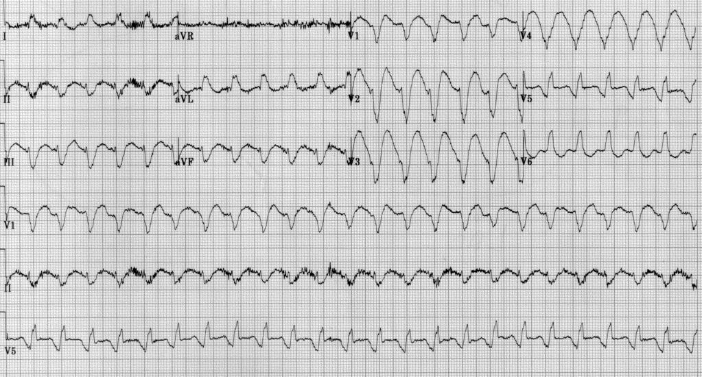
9 I II III avr avl avf V1 V2 V3 V4 V5 V6 Right Ventricular Outflow Tract VT

10 Right Ventricular Outflow Tract VT Pulmonary valve Ablation site Tricuspid valve

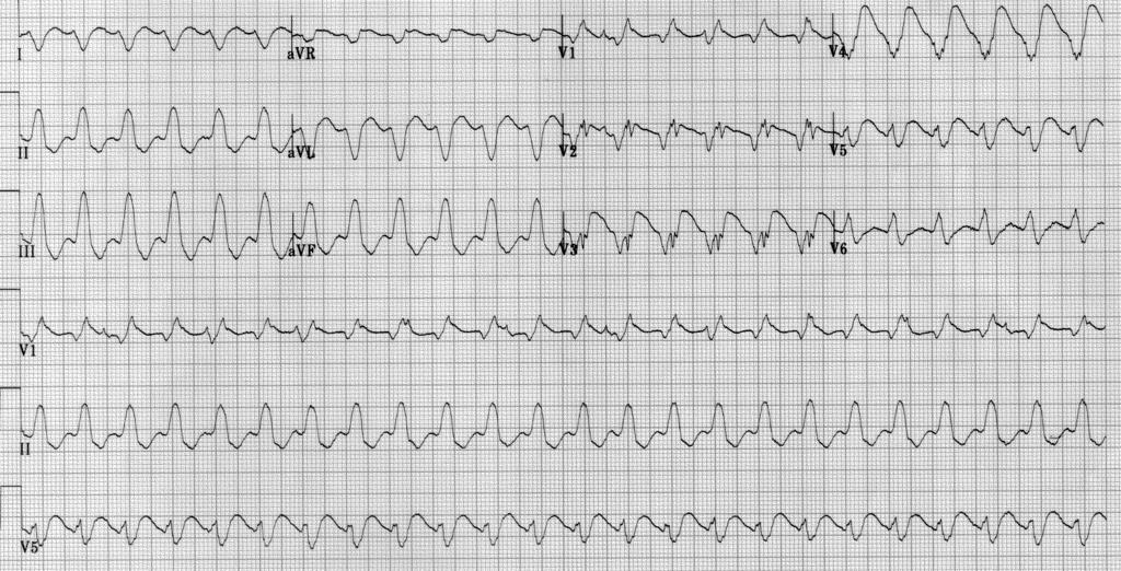
11 Outflow Tract VT


12 Outflow Tract VT requiring Epicardial Ablation Epi Epi CS CS


13 Outflow Tract VT: Ablation in the Left Aortic Cusp Left main coronary artery Ablation catheter
14 Aortic Cusp VT ECG Criteria Early transition in precordial leads (V1, V2) Notch in V5, lack of S in V5, V6 Broad R wave in V1, V2 Larger R/S amplitude in V1, V2 Notch in V1 in L cusp VT (transeptal conduction) Lead I negative in L cusp, positive in R cusp Phase analysis (as measured from earliest surface onset) Local onset in V2 7 ms Initial peak / nadir in III 120 ms Initial peak / nadir in V2 78 ms
15 Fascicular VT Induction with atrial pacing RBBB, LAD No structural heart disease (Zipes, 1979) Verapamil sensitive (Belhassen, 1981) RBBB, RAD (Ohe, 1988) Upper septal (Shimoike, 2000)

16 Left Posterior Fascicular VT Badhwar N, Scheinman MM. Curr Probl Cardiol. 2007; 32(1): 7-43.

17 Left Posterior Fascicular VT

18 Left Anterior Fascicular PVCs

19 Mitral Annular PVCs / VT a. Anterolateral b. Posterior c. Posteroseptal

20 Tricuspid Annular PVCs / VT 1. Septal a. Qs in V1 b. Narrower c. No notching 2. Lateral a. rs in V1 b. Wider c. Notching
21 VT arising from the Crux of the Heart


22 VT arising from the Crux of the Heart LAO RAO
23 Idiopathic VT VT associated with structural heart disease
24

25 3D mapping of LV showing scar in a patient with ischemic cardiomyopathy
26 Ventricular Tachycardia with Structural Heart Disease Coronary artery disease Idiopathic dilated cardiomyopathy Hypertrophic cardiomyopathy (HOCM) Arrhythmogenic right ventricular cardiomyopathy (ARVC) Infiltrative cardiomyopathy- amyloidosis, sarcoidosis Chagas disease
27 Ventricular Tachycardia with Structural Heart Disease Congenital heart disease Tetrology of Fallot, aortic stenosis Valvular heart disease Pre surgery Post surgical repair Mitral valve prolapse Right sided AV bundle, fibrotic scars in septum, degenerative changes in conduction system Myotonic dystrophy AV block more common Familial VT Genetic abnormality of conduction system
28 Arrhythmogenic Right Ventricular Cardiomyopathy (ARVC)
29 ARVC: VT arising from Right Ventricle 29

30 Role of imaging modalities in the diagnosis of ARVC Holter Carto RV angio MRI 30
31 Non ischemic Dilated Cardiomyopathy Annular scar VT arising from the conduction system (bundle branch reentry, fascicular, interfascicular Epicardial scar

32 Bundle Branch Reentry VT

33 Catheter Ablation of Right Bundle Branch I II V 1 RA Current Voltage

34 Epicardial VT in Patient with Dilated Cardiomyopathy
35 Chagas 30-40% Epicardial VT Non ischemic cardiomyopathy 25-50% Ischemic cardiomyopathy 10-15% ARVC 5-10% LV aneurysm, Sarcoid, Non compaction Idiopathic VT 10% (mainly around epicardial arteries)
36 Epicardial Mapping
37 Epicardial VT: EKG criteria QRS duration > 200 ms Pseudo delta wave > 34 ms Intrinsicoid deflection > 85 ms Shortest RS > 121 ms Precordial MDI > 0.55 ms Non ischemic cardiomyopathy: lack of q wave in inferior leads, positive q wave in lead I
38 Ventricular Tachycardia Ventricular tachycardia is an important cause of sudden cardiac arrest ECG characteristics can localize the site and origin of VT Idiopathic VT Monomorphic VT / Frequent PVCs curable with catheter ablation Polymorphic VT treated with ICD and drugs VT associated with structural heart disease ICD and antiarrhythmic drugs Catheter ablation is mainly palliative, improved efficacy with epicardial mapping, impella/echmo/iabp


39 Thank you
Advances in Ablation Therapy for Ventricular Tachycardia
 Advances in Ablation Therapy for Ventricular Tachycardia Nitish Badhwar, MD, FACC, FHRS Director, Cardiac Electrophysiology Training Program University of California, San Francisco For those of you who
Catheter Ablation of VT Without Structural Heart Disease 성균관의대 온영근
 Catheter Ablation of VT Without Structural Heart Disease 성균관의대 온영근 Idiopathic Monomorphic Ventricular Tachycardia Adenosine-sensitive Verapamil-sensitive Propranolol-sensitive Mech (Triggered activity)
Arrhythmias (II) Ventricular Arrhythmias. Disclosures
 Ventricular Arrhythmias. Disclosures") Arrhythmias (II) Ventricular Arrhythmias Amy Leigh Miller, MD, PhD Cardiovascular Electrophysiology, Brigham & Women s Hospital Disclosures None Rhythms and Mortality Implantable loop recorder post-mi
Arrhythmias (II) Ventricular Arrhythmias Amy Leigh Miller, MD, PhD Cardiovascular Electrophysiology, Brigham & Women s Hospital Disclosures None Rhythms and Mortality Implantable loop recorder post-mi
Idiopathic Ventricular Tachycardia Need for an Update in EHRA/HRS Consensus?
 Idiopathic Ventricular Tachycardia Need for an Update in EHRA/HRS Consensus? Arash Arya, M.D. Department of Interventional Electrophysiology Heart Center University of Leipzig Disclosures: NONE Idiopathic
Mapping and Ablation of Challenging Outflow Tract VTs: Pulmonary Artery, LVOT, Epicardial
 Mapping and Ablation of Challenging Outflow Tract VTs: Pulmonary Artery, LVOT, Epicardial Samuel J. Asirvatham, MD Mayo Clinic Rochester California Heart Rhythm Symposium San Francisco, CA September 8,
Technique of Epicardial VT Ablation
 CARTO Club Jan 2014 Technique of Epicardial VT Ablation Amir AbdelWahab, MD Electrophysiology and Pacing Service Department of Cardiovascular Medicine, Cairo University Need for Epicardial VT ablation
NAAMA s 24 th International Medical Convention Medicine in the Next Decade: Challenges and Opportunities Beirut, Lebanon June 26 July 2, 2010
 NAAMA s 24 th International Medical Convention Medicine in the Next Decade: Challenges and Opportunities Beirut, Lebanon June 26 July 2, 2010 I have a financial interest/arrangement or affiliation with
Sudden cardiac death: Primary and secondary prevention
 Sudden cardiac death: Primary and secondary prevention By Kai Chi Chan Penultimate Year Medical Student St George s University of London at UNic Sheba Medical Centre Definition Sudden cardiac arrest (SCA)
VENTRICULAR TACHYCARDIA IN THE ABSENCE OF STRUCTURAL HEART DISEASE
 VENTRICULAR TACHYCARDIA IN THE ABSENCE OF STRUCTURAL HEART DISEASE Dimosthenis Avramidis, MD. Consultant Mitera Children s Hospital Athens Greece Scientific Associate 1st Cardiology Dpt Evangelismos Hospital
INTRODUCTION. left ventricular non-compaction is a sporadic or familial cardiomyopathy characterized by
 A Rare Case of Arrhythmogenic Right Ventricular Cardiomyopathy Co-existing with Isolated Left Ventricular Non-compaction NS Yelgeç, AT Alper, Aİ Tekkeşin, C Türkkan INTRODUCTION Arrhythmogenic right ventricular
12-Lead ECG Interpretation. Kathy Kuznar, RN, ANP
 12-Lead ECG Interpretation Kathy Kuznar, RN, ANP The 12-Lead ECG Objectives Identify the normal morphology and features of the 12- lead ECG. Perform systematic analysis of the 12-lead ECG. Recognize abnormalities
Urgent VT Ablation in a Patient with Presumed ARVC
 Urgent VT Ablation in a Patient with Presumed ARVC Mr Alex Cambridge, Chief Cardiac Physiologist, St. Barts Hospital, London, UK The patient, a 52 year-old male, attended the ICD clinic without an appointment
Acute Coronary Syndromes Unstable Angina Non ST segment Elevation MI (NSTEMI) ST segment Elevation MI (STEMI)
 ST segment Elevation MI (STEMI)") Leanna R. Miller, RN, MN, CCRN-CSC, PCCN-CMC, CEN, CNRN, CMSRN, NP Education Specialist LRM Consulting Nashville, TN Objectives Evaluate common abnormalities that mimic myocardial infarction. Identify
Leanna R. Miller, RN, MN, CCRN-CSC, PCCN-CMC, CEN, CNRN, CMSRN, NP Education Specialist LRM Consulting Nashville, TN Objectives Evaluate common abnormalities that mimic myocardial infarction. Identify
ECG Cases and Questions. Ashish Sadhu, MD, FHRS, FACC Electrophysiology/Cardiology
 ECG Cases and Questions Ashish Sadhu, MD, FHRS, FACC Electrophysiology/Cardiology 32 yo female Life Insurance Physical 56 yo male with chest pain Terminology Injury ST elevation Ischemia T wave inversion
December 2018 Tracings
 Tracings Tracing 1 Tracing 4 Tracing 1 Answer Tracing 4 Answer Tracing 2 Tracing 5 Tracing 2 Answer Tracing 5 Answer Tracing 3 Tracing 6 Tracing 3 Answer Tracing 6 Answer Questions? Contact Dr. Nelson
Treatment of VT of Purkinje fiber origin: ablation targets and outcome
 Treatment of VT of Purkinje fiber origin: ablation targets and outcome Ch. Piorkowski University Leipzig - Heart Center - Dept. of Electrophysiology Leipzig, Germany Presenter Disclosure Information Gerhard
Ventricular arrhythmias
 Ventricular arrhythmias Assoc.Prof. Lucie Riedlbauchová, MD, PhD Department of Cardiology University HospitalMotol and2nd FacultyofMedicine, Charles University in Prague Definition and classification Ventricular
Office ECG Interpretation
 Office ECG Interpretation Jason Evanchan, DO Assistant Professor of Medicine Division of Cardiovascular Medicine The Ohio State University Wexner Medical Center Outline of topics High risk ischemia T wave
Managing Hypertrophic Cardiomyopathy with Imaging. Gisela C. Mueller University of Michigan Department of Radiology
 Managing Hypertrophic Cardiomyopathy with Imaging Gisela C. Mueller University of Michigan Department of Radiology Disclosures Gadolinium contrast material for cardiac MRI Acronyms Afib CAD Atrial fibrillation
Ventricular Tachycardia in Normal Heart: Approach and Management
 ndian Journal of Cardiology SSN-0972-1622 20 ] 2012 by the ndian Society of Cardiology Vol. 15, (3-4), 20-26 Review Article Ventricular Tachycardia in Normal Heart: Approach and Management S.K. Chutani
Map-Guided Ablation of Non-ischemic VT. Takashi Nitta Cardiovascular Surgery, Nippon Medical School Tokyo, JAPAN
 Map-Guided Ablation of Non-ischemic VT Takashi Nitta Cardiovascular Surgery, Nippon Medical School Tokyo, JAPAN nothing Declaration of Interest Catheter Ablation of Non-ischemic VT Sarcoidosis, 13, 6%
Tachycardias II. Štěpán Havránek
 Tachycardias II Štěpán Havránek Summary 1) Supraventricular (supraventricular rhythms) Atrial fibrillation and flutter Atrial ectopic tachycardia / extrabeats AV nodal reentrant a AV reentrant tachycardia
Medicine. Dynamic Changes of QRS Morphology of Premature Ventricular Contractions During Ablation in the Right Ventricular Outflow Tract
 Medicine CLINICAL CASE REPORT Dynamic Changes of QRS Morphology of Premature Ventricular Contractions During Ablation in the Right Ventricular Outflow Tract A Case Report Li Yue-Chun, MD, Lin Jia-Feng,
Ablation of Ventricular Tachycardia in Non-Ischemic Cardiomyopathy
 Ablation of Ventricular Tachycardia in Non-Ischemic Cardiomyopathy Fermin C Garcia, MD University of Pennsylvania Cardiac Electrophysiology Philadelphia, PA Nothing to disclose No conflict of interest
REtrive. REpeat. RElearn Design by. Test-Enhanced Learning based ECG practice E-book
 Test-Enhanced Learning Test-Enhanced Learning Test-Enhanced Learning Test-Enhanced Learning based ECG practice E-book REtrive REpeat RElearn Design by S I T T I N U N T H A N G J U I P E E R I Y A W A
Premature ventricular complexes or contractions
 CLINICAL STUDY Analysis of Morphological Characteristics and Origins of Idiopathic Premature Ventricular Contractions Under a 12-Lead Electrocardiogram in Children with Structurally Normal Hearts Jianbin
Ventricular tachycardia Ventricular fibrillation and ICD
 EKG Conference Ventricular tachycardia Ventricular fibrillation and ICD Samsung Medical Center CCU D.I. Hur Ji Won 2006.05.20 Ventricular tachyarrhythmia ventricular tachycardia ventricular fibrillation
General Introduction to ECG. Reading Assignment (p2-16 in PDF Outline )
") General Introduction to ECG Reading Assignment (p2-16 in PDF Outline ) Objectives 1. Practice the 5-step Method 2. Differential Diagnosis: R & L axis deviation 3. Differential Diagnosis: Poor R-wave progression
General Introduction to ECG Reading Assignment (p2-16 in PDF Outline ) Objectives 1. Practice the 5-step Method 2. Differential Diagnosis: R & L axis deviation 3. Differential Diagnosis: Poor R-wave progression
Section V. Objectives
 Section V Landscape of an MI Objectives At the conclusion of this presentation the participant will be able to Outline a systematic approach to 12 lead ECG interpretation Demonstrate the process for determining
Conduction Problems / Arrhythmias. Conduction
 Conduction Problems / Arrhythmias Conduction Wolf-Parkinson White Syndrome (WPW) and Lown-Ganong-Levine (LGL): Atrial impulses bypass the AV node through an accessory pathway or bypass tract (bundle of
What s New in IV Conduction? (Quadrafascicular, not Trifascicular)
") What s New in IV Conduction? (Quadrafascicular, not Trifascicular) Frank Yanowitz, MD Professor, University of Utah School of Medicine Medical Director, IHC ECG Services (Urban Central Region) http://ecg.utah.edu
What s New in IV Conduction? (Quadrafascicular, not Trifascicular) Frank Yanowitz, MD Professor, University of Utah School of Medicine Medical Director, IHC ECG Services (Urban Central Region) http://ecg.utah.edu
Ventricular Tachycardia Ablation. Saverio Iacopino, MD, FACC, FESC
 Ventricular Tachycardia Ablation Saverio Iacopino, MD, FACC, FESC ü Ventricular arrhythmias, both symptomatic and asymptomatic, are common, but syncope and SCD are infrequent initial manifestations of
The Efficient and Smart Methods for Diagnosis of SVT 대구파티마병원순환기내과정병천
 The Efficient and Smart Methods for Diagnosis of SVT 대구파티마병원순환기내과정병천 Differentiation Supraventricular Origin from Ventricular Origin on ECG. QRS-Complex Width. 1. Narrow QRS-Complex Tachycardia (
The Electrocardiogram part II. Dr. Adelina Vlad, MD PhD
 The Electrocardiogram part II Dr. Adelina Vlad, MD PhD Basic Interpretation of the ECG 1) Evaluate calibration 2) Calculate rate 3) Determine rhythm 4) Determine QRS axis 5) Measure intervals 6) Analyze
Outflow Tract Ventricular Tachycardia Always Benign?
 Outflow Tract Ventricular Tachycardia Always Benign? Arash Arya, M.D. Department of Interventional Electrophysiology Heart Center University of Leipzig Disclosures: NONE Outflow Ventricular Tachycardia
Bundle Branch & Fascicular Blocks. Reading Assignment (p53-58 in Outline )
") Bundle Branch & Fascicular Blocks Reading Assignment (p53-58 in Outline ) Objectives 1. QRS analysis of Right and Left BBB 2. Uncomplicated vs complicated BBB 3. Diagnosis of RBBB with LAFB and LPFB 4.
Bundle Branch & Fascicular Blocks Reading Assignment (p53-58 in Outline ) Objectives 1. QRS analysis of Right and Left BBB 2. Uncomplicated vs complicated BBB 3. Diagnosis of RBBB with LAFB and LPFB 4.
ECG Workshop. Nezar Amir
 ECG Workshop Nezar Amir Myocardial Ischemia ECG Infarct ECG in STEMI is dynamic & evolving Common causes of ST shift Infarct Localisation Left main artery occlusion: o diffuse ST-depression with ST elevation
ECG Interpretation Made Easy
 ECG Interpretation Made Easy Dr. A Tageldien Abdellah, MSc MD EBSC Lecturer of Cardiology- Hull University Hull York Medical School 2007-2008 ECG Interpretation Made Easy Synopsis Benefits Objectives Process
12 Lead ECG Skills: Building Confidence for Clinical Practice. Presented By: Cynthia Webner, BSN, RN, CCRN-CMC. Karen Marzlin, BSN, RN,CCRN-CMC
 12 Lead ECG Skills: Building Confidence for Clinical Practice NTI 2009 Preconference Session 803 Presented By: Karen Marzlin, BSN, RN,CCRN-CMC 1 12 Lead ECG Fundamentals: The Starting Place for Linking
Basic electrocardiography reading. R3 lee wei-chieh
 Basic electrocardiography reading R3 lee wei-chieh The Normal Conduction System Lead Placement avf Limb Leads Precordial Leads Interpretation Rate Rhythm Interval Axis Chamber abnormality QRST change What
Cardiology Flash Cards
 Cardiology Flash Cards EKG in a nut shell www.brain101.info Conduction System www.brain101.info 2 Analyzing EKG Step by step Steps in Analyzing ECG'S 1. Rhythm: - Regular _ Sinus, Junctional or Ventricular.
ECGs on the acute admission ward. - Cardiology Update -
 ECGs on the acute admission ward - Cardiology Update - Dr Simon Fynn Consultant Cardiologist Papworth Hospital, Cambridge RCP London Oct 2017 ECG 1 1. AF with BBB 2. Pre-excited AF 3. SVT with BBB 4.
ECG Workshop. Carolyn Shepherd And Anya Horne UWE Principles of Cardiac Care
 ECG Workshop Carolyn Shepherd And Anya Horne UWE Principles of Cardiac Care ECG workshop case study1 44 Year old male. Reports SOB, Lethargy, tiredness. PMH: Hypertension, nil else. What tests? What treatment?
Study methodology for screening candidates to athletes risk
 1. Periodical Evaluations: each 2 years. Study methodology for screening candidates to athletes risk 2. Personal history: Personal history of murmur in childhood; dizziness, syncope, palpitations, intolerance
ECG Basics Sonia Samtani 7/2017 UCI Resident Lecture Series
 ECG Basics Sonia Samtani 7/2017 UCI Resident Lecture Series Agenda I. Introduction II.The Conduction System III.ECG Basics IV.Cardiac Emergencies V.Summary The Conduction System Lead Placement avf Precordial
Index. cardiacep.theclinics.com. Note: Page numbers of article titles are in boldface type.
 Note: Page numbers of article titles are in boldface type. A AEDs. See Automated external defibrillators (AEDs) AF. See Atrial fibrillation (AF) Age as factor in SD in marathon runners, 45 Antiarrhythmic
Case Report Coexistence of Atrioventricular Nodal Reentrant Tachycardia and Idiopathic Left Ventricular Outflow-Tract Tachycardia
 www.ipej.org 149 Case Report Coexistence of Atrioventricular Nodal Reentrant Tachycardia and Idiopathic Left Ventricular Outflow-Tract Tachycardia Majid Haghjoo, M.D, Arash Arya, M.D, Mohammadreza Dehghani,
Biventricular Enlargement/ Hypertrophy
 Biventricular Enlargement/ Hypertrophy Keywords congenital heart disease left ventricular hypertrophy right ventricular hypertrophy SR MITTAL Abstract Electrocardiographic diagnosis of early biventricular
Φαρμακεσηική αγωγή ζηις ιδιοπαθείς κοιλιακές αρρσθμίες. Άννα Κωζηοπούλοσ Επιμελήηρια Α Ωνάζειο Καρδιοτειροσργικό Κένηρο
 Φαρμακεσηική αγωγή ζηις ιδιοπαθείς κοιλιακές αρρσθμίες Άννα Κωζηοπούλοσ Επιμελήηρια Α Ωνάζειο Καρδιοτειροσργικό Κένηρο Όλες οι κοιλιακές αρρσθμίες δεν είναι ίδιες Υπάρτοσν διαθορές ζηον πληθυσμό, ηον μηχανισμό
EHRA Accreditation Exam - Sample MCQs Invasive cardiac electrophysiology
 EHRA Accreditation Exam - Sample MCQs Invasive cardiac electrophysiology Dear EHRA Member, Dear Colleague, As you know, the EHRA Accreditation Process is becoming increasingly recognised as an important
12 Lead ECG Interpretation
 12 Lead ECG Interpretation Julie Zimmerman, MSN, RN, CNS, CCRN Significant increase in mortality for every 15 minutes of delay! N Engl J Med 2007;357:1631-1638 Who should get a 12-lead ECG? Also include
Implantable Cardioverter Defibrillator (ICD)
") Medical Coverage Policy Effective Date... 3/15/2018 Next Review Date... 3/15/2019 Coverage Policy Number... 0181 Implantable Cardioverter Defibrillator (ICD) Table of Contents Related Coverage Resources
Medical Coverage Policy Effective Date... 3/15/2018 Next Review Date... 3/15/2019 Coverage Policy Number... 0181 Implantable Cardioverter Defibrillator (ICD) Table of Contents Related Coverage Resources
402 Index. B β-blockers, 4, 5 Bradyarrhythmias, 76 77
 Index A Acquired immunodeficiency syndrome (AIDS), 126, 163 Action potentials, 1, 5, 27 Acute coronary syndromes, 123t, 129 Adenosine, intravenous, 277 Alcohol abuse, as T wave inversion cause, 199 Aneurysm,
Epicardial Approach to Mapping and Ablation of VT: Clinical and ECG Predictors of Epicardial Location
 Epicardial Approach to Mapping and Ablation of VT: Clinical and ECG Predictors of Epicardial Location Mathew D. Hutchinson, MD, FACC, FHRS Perelman School of Medicine University of Pennsylvania Presenter
Fast & Slow Tachy & Brady Arrhythmias DAVID STULTZ, MD, FACC KPN HEART & VASCULAR AUGUST 7, 2017
 Fast & Slow Tachy & Brady Arrhythmias DAVID STULTZ, MD, FACC KPN HEART & VASCULAR AUGUST 7, 2017 Normal EKG EKG boxes Heart Rate 1 big box = 200ms 1 small box = 40ms Big Boxes Between QRS complexes Heart
Myocardial Infarction. Reading Assignment (p66-78 in Outline )
") Myocardial Infarction Reading Assignment (p66-78 in Outline ) Objectives 1. Why do ST segments go up or down in ischemia? 2. STEMI locations and culprit vessels 3. Why 15-lead ECGs? 4. What s up with avr?
Myocardial Infarction Reading Assignment (p66-78 in Outline ) Objectives 1. Why do ST segments go up or down in ischemia? 2. STEMI locations and culprit vessels 3. Why 15-lead ECGs? 4. What s up with avr?
that number is extremely high. It s 16 episodes, or in other words, it s 14, one-four, ICD shocks per patient per day.
 Doctor Karlsner, Doctor Schumosky, ladies and gentlemen. It s my real pleasure to participate in this session on controversial issues in the management of ventricular tachycardia and I m sure that will
Ventricular Tachycardia Substrate. For the ablationist. Stanley Tung, MD FRCPC Arrhythmia Service/St Paul Hospital University of British Columbia
 Ventricular Tachycardia Substrate For the ablationist Stanley Tung, MD FRCPC Arrhythmia Service/St Paul Hospital University of British Columbia Two Attitudes of Ventricular Tachycardia Ablation 1 2C:\Documents
Epicardial VT Ablation The Cleveland Clinic Experience
 Epicardial VT Ablation The Cleveland Clinic Experience Walid Saliba, MD, FHRS Director, EP Lab Cardiac Electrophysiology Heart and Vascular Institute Epicardial Access in the EP Lab Why Epicardial Special
Electrocardiogram A valuable diagnostic tool. Jean Vorster Netcare Unitas Hospital STEMI Early Reperfusion Initiative 2015
 Electrocardiogram A valuable diagnostic tool Jean Vorster Netcare Unitas Hospital STEMI Early Reperfusion Initiative 2015 Overview 1. Arrhythmias 2. Structural heart disease 3. Ischaemia Arrhythmias Sinus
ΔΠΔΜΒΑΣΙΚΗ ΘΔΡΑΠΔΙΑ ΚΟΙΛΙΑΚΩΝ ΑΡΡΤΘΜΙΩΝ
 ΔΠΔΜΒΑΣΙΚΗ ΘΔΡΑΠΔΙΑ ΚΟΙΛΙΑΚΩΝ ΑΡΡΤΘΜΙΩΝ ΣΔΛΙΟ ΠΑΡΑΚΔΤΑÏΓΗ ΓΙΔΤΘΤΝΣΗ ΔΤ Α Καρδιολογική Κλινική ΑΠΘ, Νοζοκομείο ΑΧΕΠΑ, Θεζζαλονίκη NO CONFLICT OF INTEREST INTRODUCTION Sustained VT is an important cause
Cardiac MRI: Cardiomyopathy
 Cardiac MRI: Cardiomyopathy Laura E. Heyneman, MD I do not have any relevant financial relationships with any commercial interests Cardiac MRI: Cardiomyopathy Laura E. Heyneman, MD Duke University Medical
TACHYARRHYTHMIAs. Pawel Balsam, MD, PhD
 TACHYARRHYTHMIAs Pawel Balsam, MD, PhD SupraVentricular Tachycardia Atrial Extra Systole Sinus Tachycardia Focal A. Tachycardia AVRT AVNRT Atrial Flutter Atrial Fibrillation Ventricular Tachycardia Ventricular
Return to Basics. ECG Rate and Rhythm. Management of the Hospitalized Patient September 25, 2009
 Management of the Hospitalized Patient September 25, 2009 ECG Refresher and Update 2009 Return to Basics Determine rate and rhythm Determine intervals and axes Define morphology of P-QRS-T-U Compare with
Please check your answers with correct statements in answer pages after the ECG cases.
 ECG Cases ECG Case 1 Springer International Publishing AG, part of Springer Nature 2018 S. Okutucu, A. Oto, Interpreting ECGs in Clinical Practice, In Clinical Practice, https://doi.org/10.1007/978-3-319-90557-0
Reentrant Ventricular Tachycardia Originating in the Right Ventricular Outflow Tract
 Circ J 2008; 72: 855 860 Reentrant Ventricular Tachycardia Originating in the Right Ventricular Outflow Tract Slow Conduction Identified by Right Coronary Artery Ostium Pacing Emi Nakano, MD; Tomoo Harada,
ΔΙΑΤΑΡΑΧΕΣ ΕΝΔΟΚΟΙΛΙΑΚΗΣ ΑΓΩΓΙΜΟΤΗΤΑΣ ΔΗΜΗΤΡΙΟΣ Δ. ΜΑΝΩΛΑΤΟΣ Β ΚΑΡΔΙΟΛΟΓΙΚΗ ΚΛΙΝΙΚΗ ΕΡΓΑΣΤΗΡΙΟ ΗΛΕΚΤΡΟΦΥΣΙΟΛΟΓΙΑΣ Γ.Ν.Α.
 ΔΙΑΤΑΡΑΧΕΣ ΕΝΔΟΚΟΙΛΙΑΚΗΣ ΑΓΩΓΙΜΟΤΗΤΑΣ ΔΗΜΗΤΡΙΟΣ Δ. ΜΑΝΩΛΑΤΟΣ Β ΚΑΡΔΙΟΛΟΓΙΚΗ ΚΛΙΝΙΚΗ ΕΡΓΑΣΤΗΡΙΟ ΗΛΕΚΤΡΟΦΥΣΙΟΛΟΓΙΑΣ Γ.Ν.Α. «ΕΥΑΓΓΕΛΙΣΜΟΣ» Intraventricular conduction delay and Blocks Right Bundle Branch
Cardiovascular Nursing Practice: A Comprehensive Resource Manual and Study Guide for Clinical Nurses 2 nd Edition
 Cardiovascular Nursing Practice: A Comprehensive Resource Manual and Study Guide for Clinical Nurses 2 nd Edition Table of Contents Volume 1 Chapter 1: Cardiovascular Anatomy and Physiology Basic Cardiac
Positive QRS Complex in Lead I as a Malignant Sign in Right Ventricular Outflow Tract Tachycardia
 Circulation Journal Official Journal of the Japanese Circulation Society http://www.j-circ.or.jp ORIGINAL ARTICLE Arrhythmia/Electrophysiology Positive QRS Complex in Lead I as a Malignant Sign in Right
Ventricular Tachycardia in Structurally Normal Hearts (Idiopathic VT) Patient Information
 Patient Information") Melbourne Heart Rhythm Ventricular Tachycardia in Structurally Normal Hearts (Idiopathic VT) Patient Information What is Ventricular Tachycardia? Ventricular tachycardia (VT) is an abnormal rapid heart
Melbourne Heart Rhythm Ventricular Tachycardia in Structurally Normal Hearts (Idiopathic VT) Patient Information What is Ventricular Tachycardia? Ventricular tachycardia (VT) is an abnormal rapid heart
DIAGNOSIS AND MANAGEMENT OF ARRHYTHMOGENIC CARDIOMYOPATHY. David SIU MD ( 蕭頌華醫生 ) Division of Cardiology The University of Hong Kong
 Division of Cardiology The University of Hong Kong") APHRS Summit 2018 in conjunction with HKCC Heart Rhythm Refresher Course DIAGNOSIS AND MANAGEMENT OF ARRHYTHMOGENIC CARDIOMYOPATHY David SIU MD ( 蕭頌華醫生 ) Division of Cardiology The University of Hong Kong
APHRS Summit 2018 in conjunction with HKCC Heart Rhythm Refresher Course DIAGNOSIS AND MANAGEMENT OF ARRHYTHMOGENIC CARDIOMYOPATHY David SIU MD ( 蕭頌華醫生 ) Division of Cardiology The University of Hong Kong
15 16 September Seminar W10O. ECG for General Practice
 15 16 September 2012 Seminar W10O ECG for General Practice Speaker: Ms Natasha Eaton ECG for General Practice Speaker: Natasha Eaton Cardiac CNC Executive Representative Electrocardiography The graphic
BSH Heart Failure Nurse and Healthcare Professional Study Day 2017
 BSH Heart Failure Nurse and Healthcare Professional Study Day 2017 Presentation title: Malignant Arrhythmias Speaker: Derek Connelly Conflicts of interest: Speakers fees / advisory boards: Boehringer Ingelheim,
Ekg pra pr c a tice D.HAMMOUDI.MD
 Ekg practice D.HAMMOUDI.MD Anatomy Revisited RCA (Right Coronary Artery) Right ventricle Inferior wall of LV Posterior wall of LV (75%) SA Node (60%) AV Node (>80%) LCA (Left Coronary Artery) Septal wall
Purkinje-related Arrhythmias
 J Arrhythmia Vol 27 No 1 2011 Review Article Purkinje-related Arrhythmias Akihiko Nogami MD Department of Heart Rhythm Management, Yokohama Rosai Hospital, Yokohama, Japan The Purkinje system has been
Clinical Cardiac Electrophysiology
 Clinical Cardiac Electrophysiology Certification Examination Blueprint Purpose of the exam The exam is designed to evaluate the knowledge, diagnostic reasoning, and clinical judgment skills expected of
SUDDEN CARDIAC DEATH(SCD): Definition
: Definition") SUDDEN CARDIAC DEATH EPIDEMIOLOGY, PATHOPHYSIOLOGY, PREVENTION & THERAPY Hasan Garan, M.D. Columbia University Medical Center SUDDEN CARDIAC DEATH(SCD): Definition DEATH DUE TO A CARDIAC CAUSE IN A CLINICALLY
SUDDEN CARDIAC DEATH EPIDEMIOLOGY, PATHOPHYSIOLOGY, PREVENTION & THERAPY Hasan Garan, M.D. Columbia University Medical Center SUDDEN CARDIAC DEATH(SCD): Definition DEATH DUE TO A CARDIAC CAUSE IN A CLINICALLY
ECG ABNORMALITIES D R. T AM A R A AL Q U D AH
 ECG ABNORMALITIES D R. T AM A R A AL Q U D AH When we interpret an ECG we compare it instantaneously with the normal ECG and normal variants stored in our memory; these memories are stored visually in
THE ELECTROCARDIOGRAM A UBIQUITOUS AND COST-EFFECTIVE DIAGNOSTIC TOOL FOR THE FAMILY MEDICINE REFRESHER COURSE MARCH 8, 2019
 THE ELECTROCARDIOGRAM A UBIQUITOUS AND COST-EFFECTIVE DIAGNOSTIC TOOL FOR THE FAMILY MEDICINE REFRESHER COURSE MARCH 8, 2019 Major Clinical Disorders Pulmonary Embolism 69 y/o woman with dyspnea and an
Catheter Ablation of Idiopathic Premature Ventricular Contractions and Ventricular Tachycardias Originating from Right Ventricular Septum
 Catheter Ablation of Idiopathic Premature Ventricular Contractions and Ventricular Tachycardias Originating from Right Ventricular Septum Wu Lian-Pin., Li Yue-Chun., Zhao Jing-Lin, Zheng Cheng, Chen Jun-Hua,
Clinical Policy: Holter Monitors Reference Number: CP.MP.113
 Clinical Policy: Reference Number: CP.MP.113 Effective Date: 05/18 Last Review Date: 04/18 Coding Implications Revision Log Description Ambulatory electrocardiogram (ECG) monitoring provides a view of
Return to Basics. Normal Intervals & Axes. ECG Rate and Rhythm
 Return to Basics Management of the Hospitalized Patient October 15, 2010 ECG Refresher and Update 2010 Determine rate and rhythm Determine intervals and axes Define morphology of P-QRS-T-U Compare with
Theroleofcatheterablationinthemanagement of ventricular tachycardia
 European Heart Journal Advance Access published August 31, 2015 European Heart Journal doi:10.1093/eurheartj/ehv421 REVIEW Novel therapeutic concepts Theroleofcatheterinthemanagement of ventricular tachycardia
PVCs: Do they cause Cardiomyopathy? Raed Abu Sham a, M.D.
 PVCs: Do they cause Cardiomyopathy? Raed Abu Sham a, M.D. Cardiologist and Electrophysiologist No conflict of interest related to this presentation Objectives 1. PVCs are benign. What is the Evidence?
SUDDEN CARDIAC DEATH(SCD): Definition
: Definition") SUDDEN CARDIAC DEATH EPIDEMIOLOGY, PATHOPHYSIOLOGY, PREVENTION & THERAPY Hasan Garan, M.D. Columbia University Medical Center SUDDEN CARDIAC DEATH(SCD): Definition DEATH DUE TO A CARDIAC CAUSE IN A CLINICALLY
SUDDEN CARDIAC DEATH EPIDEMIOLOGY, PATHOPHYSIOLOGY, PREVENTION & THERAPY Hasan Garan, M.D. Columbia University Medical Center SUDDEN CARDIAC DEATH(SCD): Definition DEATH DUE TO A CARDIAC CAUSE IN A CLINICALLY
Chapter 2 Practical Approach
 Chapter 2 Practical Approach There are beginners in electrocardiogram (ECG) analysis who are fascinated by a special pattern (e.g., a bundle-branch block or a striking Q wave) and thereby overlook other
All About STEMIs. Presented By: Brittney Urvand, RN, BSN, CCCC. Essentia Health Fargo Cardiovascular Program Manager.
 All About STEMIs Presented By: Brittney Urvand, RN, BSN, CCCC Essentia Health Fargo Cardiovascular Program Manager Updated 10/2/2018 None Disclosures Objectives Identify signs and symptoms of a heart attack
Case 1. Case 2. Case 3
 Case 1 The correct answer is D. Occasionally, the Brugada syndrome can present similar morphologies to A and also change depending on the lead position but in the Brugada pattern the r is wider and ST
Sudden Cardiac Death What an electrophysiologist thinks a cardiologist should know
 Sudden Cardiac Death What an electrophysiologist thinks a cardiologist should know Steven J. Kalbfleisch, M.D. Medical Director Electrophysiology Laboratory Ross Heart Hospital Wexner Medical Center Sudden
Use of Catheter Ablation in the Treatment of Ventricular Tachycardia Triggered by Premature Ventricular Contraction
 J Arrhythmia Vol 22 No 3 2006 Case Report Use of Catheter Ablation in the Treatment of Ventricular Tachycardia Triggered by Premature Ventricular Contraction sao Kato MD, Toru wa MD, Yasushi Suzuki MD,
SIMPLY ECGs. Dr William Dooley
 SIMPLY ECGs Dr William Dooley 1 No anatomy just interpretation 2 Setting up an ECG 3 Setting up an ECG 1 V1-4 th Right intercostal space at sternal border 2 V2-4 th Left intercostal space at sternal border
If the P wave > 0.12 sec( 3 mm) usually in any lead. Notched P wave usually in lead I,aVl may be lead II Negative terminal portion of P wave in V1, 1
 usually in any lead. Notched P wave usually in lead I,aVl may be lead II Negative terminal portion of P wave in V1, 1") If the P wave > 0.12 sec( 3 mm) usually in any lead. Notched P wave usually in lead I,aVl may be lead II Negative terminal portion of P wave in V1, 1 mm depth and 3 mm width( most specific) Since Mitral
If the P wave > 0.12 sec( 3 mm) usually in any lead. Notched P wave usually in lead I,aVl may be lead II Negative terminal portion of P wave in V1, 1 mm depth and 3 mm width( most specific) Since Mitral
Ben Taylor, PhD, PA-C
 Ben Taylor, PhD, PA-C The patient is a 23-year-old white male with a history of polysubstance abuse who was found unresponsive, last seen the day before. Classic signs of systemic hypothermia with prominent
, David Stultz, MD.
 http://www.dilbert.com EKG Rounds Handouts available at http://www.drstultz.com January 5, 2004 David Stultz, MD Cardiology Fellow, PGY 4 Overview of Topics How to read an EKG Normal EKG Determination
KNOW YOUR ECG. G. Somasekhar MD DM FEp Consultant Electro physiologist, Aayush Hospital, Vijayawada
 KNOW YOUR ECG G. Somasekhar MD DM FEp Consultant Electro physiologist, Aayush Hospital, Vijayawada CASE DETAILS A 48-year-old female non hypertensive, non diabetic presented with history of shortness of
Το ΗΚΓ στις Μυοκαρδιοπάθειες και στην Περικαρδίτιδα
 4 ο ΠΑΝΕΛΛΗΝΙΟ ΑΡΡΥΘΜΙΟΛΟΓΙΚΟ ΣΥΝΕΔΡΙΟ Φροντιστηριακό Μάθημα ΗΚΓ Το ΗΚΓ στις Μυοκαρδιοπάθειες και στην Περικαρδίτιδα Γ.Ν.Α. «Ο Ευαγγελισμός» Ξυδώνας Σωτήριος, MD, PhD, FESC Εργαστήριο Ηλεκτροφυσιολογίας
6/19/2018. Background Athlete s heart. Ultimate question. Applying the International Criteria for ECG
 Applying the International Criteria for ECG Interpretation in Athletes to a preparticipation screening program DAVE SIEBERT, MD, CAQSM ASSISTANT PROFESSOR DEPARTMENT OF FAMILY MEDICINE UNIVERSITY OF WASHINGTON
Return to Basics. ECG Rate and Rhythm. Management of the Hospitalized Patient October 4, 2007
 Management of the Hospitalized Patient October 4, 2007 ECG Refresher for the Hospitalists Return to Basics Determine rate and rhythm Determine intervals and axes Define morphology of P-QRS-T-U Compare
Arrhythmic Complications of MI. Teferi Mitiku, MD Assistant Clinical Professor of Medicine University of California Irvine
 Arrhythmic Complications of MI Teferi Mitiku, MD Assistant Clinical Professor of Medicine University of California Irvine Objectives Brief overview -Pathophysiology of Arrhythmia ECG review of typical
Two Years Living with the EHRA/HRS Consensus Document of VT Ablation: Need for an Update?
 Two Years Living with the EHRA/HRS Consensus Document of VT Ablation: Need for an Update? David Wilber MD Loyola University of Chicago Disclosures: Biosense / Webster: Consultant, Investigator; Boston