EFAST. Extended Focussed Assessment with Sonography for Trauma. Ultrasound Logbook. Name
|
|
|
- Regina Gilbert
- 6 years ago
- Views:
Transcription

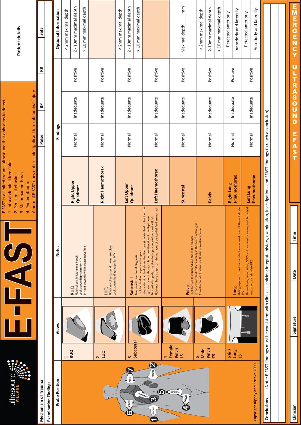
1 EFAST Extended Focussed Assessment with Sonography for Trauma Ultrasound Logbook ame Contents EFAST Accreditation Requirements 25 Abdominal Aorta Report Forms 3 Formative Assessments 1 Summative Assessment
2 E-FAST Accreditation Extended Focussed Assessment with Sonography for Trauma Accreditation requires (as a minimum) 1. Completion of Introductory US course Physics, artefacts, how to use the machine and perform a scan 2. Completion of a FAST or E-FAST course With theoretical and hands on components Including integration of EFAST into the clinical setting 3. Completion of an ultrasound logbook 25 scans with recording of images Half indicated 5 positive for free fluid Scans all checked by a supervisor (may simply view images retrospectively) Ideally scans compared to a gold standard (CT / Serial clinical exam / Formal ultrasound / Operative findings / Post mortem) 4. Completion of 3 Formative Assessments (Ultrasound Village recommendation) Detailed and directed E-FAST examinations with a supervisor, going through the attached work sheet. 5. Summative Assessment (Ultrasound Village recommendation) A formative assessment with no help / feedback, where the competence of the sonographer is completely assessed by a supervisor. 6. Testing of Knowledge Ideally a test of image interpretation and clinical decision making ability to test knowledge rather than ultrasound ability.
3 Indicated?
4 Indicated?
5 Indicated?
6 Indicated?
7 Indicated?
8 Indicated?
9 Indicated?
10 Indicated?
11 Indicated?
12 Indicated?
13 Indicated?
14 Indicated?
15 Indicated?
16 Indicated?
17 Indicated?
18 Indicated?
19 Indicated?
20 Indicated?
21 Indicated?
22 Indicated?
23 Indicated?
24 Indicated?
25 Indicated?
26 Indicated?
27 Indicated?
28 Formative Assessment E-FAST Trainee: Tutor: Date: A Formative Assessment is a structured teaching process. The student is led through a complete ultrasound examination by their tutor. The tutor may direct, prompt and teach as they see appropriate. At least 3 Formative Assessments are required before attempting the final Summative Assessment. The Summative Assessment is a structured assessment process where the candidate may be prompted through the ultrasound examination process, is asked questions but should not be instructed. Competent Required Instruction Preparation Prepare patient Position Consent / Explanation Prepare environment Lights dimmed if possible Prepare machine Correct position Turn machine on Probe selection Can change transducer Selects appropriate transducer for indication Preset selection Select correct preset Data entry Enter patient / study details
29 Competent Required Instruction Image acquisition RUQ Optimisation Adjusts depth Understands frequency adjustment Adjusts focus if on machine Adjusts gain & TGC Liver Morrisons pouch Kidney Diaphragm Lung Gallbladder (if seen) IVC (if seen) Bowel Duodenum (if seen) Where intraabdominal blood collects Where pleural blood collects LUQ Optimises image Spleen Kidney Diaphragm Can identify bowel / stomach Where intraabdominal blood collects Where pleural blood collects Pelvis Optimises image Adjusts gain appropriately Bladder Iliac vessels Prostate / Uterus & Vagina Rectum Scans through in TS / LS appropriately Where free fluid collects
30 Competent Required Instruction Pericardium Subcostal view Optimises image Adjusts depth appropriately Liver Lung Heart R Ventricle L Ventricle Septum Atria Pericardium Where pericardial fluid collects Long axis parasternal view (optional) Optimises image Heart RV LV LA MV AV Pericardium Where pericardial fluid collects Lung (optional) Optimises image High resolution (abdo or linear probe) Shallow depth Rib Pleura Comet tail artifact Sliding sign Appearance of pneumothorax Assessment of pneumothorax size Other (optional) Sternum fracture assessment IVC size and variation assessment Competent Required Instruction
31 Essential Clinical Knowledge Acts on ultrasound findings appriopriately Free fluid ormal scan Indeterminate Incidental findings Record Keeping Stores / prints appropriate images Writes appropriate report Machine Maintenance Cleans ultrasound probe Can replace printer paper (if printer attached) Stores machine and probes safely and correctly Trainee Signature Trainee's ame Tutor Signature Tutor's ame A copy of this completed formative assessment form should be kept by the trainee. Copyright Dr James Rippey
32 Formative Assessment E-FAST Trainee: Tutor: Date: A Formative Assessment is a structured teaching process. The student is led through a complete ultrasound examination by their tutor. The tutor may direct, prompt and teach as they see appropriate. At least 3 Formative Assessments are required before attempting the final Summative Assessment. The Summative Assessment is a structured assessment process where the candidate may be prompted through the ultrasound examination process, is asked questions but should not be instructed. Competent Required Instruction Preparation Prepare patient Position Consent / Explanation Prepare environment Lights dimmed if possible Prepare machine Correct position Turn machine on Probe selection Can change transducer Selects appropriate transducer for indication Preset selection Select correct preset Data entry Enter patient / study details
33 Competent Required Instruction Image acquisition RUQ Optimisation Adjusts depth Understands frequency adjustment Adjusts focus if on machine Adjusts gain & TGC Liver Morrisons pouch Kidney Diaphragm Lung Gallbladder (if seen) IVC (if seen) Bowel Duodenum (if seen) Where intraabdominal blood collects Where pleural blood collects LUQ Optimises image Spleen Kidney Diaphragm Can identify bowel / stomach Where intraabdominal blood collects Where pleural blood collects Pelvis Optimises image Adjusts gain appropriately Bladder Iliac vessels Prostate / Uterus & Vagina Rectum Scans through in TS / LS appropriately Where free fluid collects
34 Competent Required Instruction Pericardium Subcostal view Optimises image Adjusts depth appropriately Liver Lung Heart R Ventricle L Ventricle Septum Atria Pericardium Where pericardial fluid collects Long axis parasternal view (optional) Optimises image Heart RV LV LA MV AV Pericardium Where pericardial fluid collects Lung (optional) Optimises image High resolution (abdo or linear probe) Shallow depth Rib Pleura Comet tail artifact Sliding sign Appearance of pneumothorax Assessment of pneumothorax size Other (optional) Sternum fracture assessment IVC size and variation assessment Competent Required Instruction
35 Essential Clinical Knowledge Acts on ultrasound findings appriopriately Free fluid ormal scan Indeterminate Incidental findings Record Keeping Stores / prints appropriate images Writes appropriate report Machine Maintenance Cleans ultrasound probe Can replace printer paper (if printer attached) Stores machine and probes safely and correctly Trainee Signature Trainee's ame Tutor Signature Tutor's ame A copy of this completed formative assessment form should be kept by the trainee. Copyright Dr James Rippey
36 Formative Assessment E-FAST Trainee: Tutor: Date: A Formative Assessment is a structured teaching process. The student is led through a complete ultrasound examination by their tutor. The tutor may direct, prompt and teach as they see appropriate. At least 3 Formative Assessments are required before attempting the final Summative Assessment. The Summative Assessment is a structured assessment process where the candidate may be prompted through the ultrasound examination process, is asked questions but should not be instructed. Competent Required Instruction Preparation Prepare patient Position Consent / Explanation Prepare environment Lights dimmed if possible Prepare machine Correct position Turn machine on Probe selection Can change transducer Selects appropriate transducer for indication Preset selection Select correct preset Data entry Enter patient / study details
37 Competent Required Instruction Image acquisition RUQ Optimisation Adjusts depth Understands frequency adjustment Adjusts focus if on machine Adjusts gain & TGC Liver Morrisons pouch Kidney Diaphragm Lung Gallbladder (if seen) IVC (if seen) Bowel Duodenum (if seen) Where intraabdominal blood collects Where pleural blood collects LUQ Optimises image Spleen Kidney Diaphragm Can identify bowel / stomach Where intraabdominal blood collects Where pleural blood collects Pelvis Optimises image Adjusts gain appropriately Bladder Iliac vessels Prostate / Uterus & Vagina Rectum Scans through in TS / LS appropriately Where free fluid collects
38 Competent Required Instruction Pericardium Subcostal view Optimises image Adjusts depth appropriately Liver Lung Heart R Ventricle L Ventricle Septum Atria Pericardium Where pericardial fluid collects Long axis parasternal view (optional) Optimises image Heart RV LV LA MV AV Pericardium Where pericardial fluid collects Lung (optional) Optimises image High resolution (abdo or linear probe) Shallow depth Rib Pleura Comet tail artifact Sliding sign Appearance of pneumothorax Assessment of pneumothorax size Other (optional) Sternum fracture assessment IVC size and variation assessment Competent Required Instruction
39 Essential Clinical Knowledge Acts on ultrasound findings appriopriately Free fluid ormal scan Indeterminate Incidental findings Record Keeping Stores / prints appropriate images Writes appropriate report Machine Maintenance Cleans ultrasound probe Can replace printer paper (if printer attached) Stores machine and probes safely and correctly Trainee Signature Trainee's ame Tutor Signature Tutor's ame A copy of this completed formative assessment form should be kept by the trainee. Copyright Dr James Rippey
40 Summative Assessment E-FAST Candidate: Examiner: Date: A Summative Assessment is a structured assessment process. The student is led through a complete ultrasound examination by their examiner. At least 3 Formative Assessments are required before attempting the final Summative Assessment. The candidate may be prompted through the ultrasound examination process and is asked questions but should not be instructed. Failure to complete any one element changes the Summative Assessment into a Formative Assessment and the examination is completed as a teaching exercise, not a final assessment. A further Summative Assessment is required prior to accreditation. Competent Fail Preparation Prepare patient Position Consent / Explanation Prepare environment Lights dimmed if possible Prepare machine Correct position Turn machine on Probe selection Can change transducer Selects appropriate transducer for indication Preset selection Select correct preset Data entry Enter patient / study details
41 Competent Fail Image acquisition RUQ Optimisation Adjusts depth Understands frequency adjustment Adjusts focus if on machine Adjusts gain & TGC Liver Morrisons pouch Kidney Diaphragm Lung Gallbladder (if seen) IVC (if seen) Bowel Duodenum (if seen) Where intraabdominal blood collects Where pleural blood collects LUQ Optimises image Spleen Kidney Diaphragm Can identify bowel / stomach Where intraabdominal blood collects Where pleural blood collects Pelvis Optimises image Adjusts gain appropriately Bladder Iliac vessels Prostate / Uterus & Vagina Rectum Scans through in TS / LS appropriately Where free fluid collects
42 Competent Fail Pericardium Subcostal view Optimises image Adjusts depth appropriately Liver Lung Heart R Ventricle L Ventricle Septum Atria Pericardium Where pericardial fluid collects Long axis parasternal view (optional) Optimises image Heart RV LV LA MV AV Pericardium Where pericardial fluid collects Lung (optional) Optimises image High resolution (abdo or linear probe) Shallow depth Rib Pleura Comet tail artifact Sliding sign Appearance of pneumothorax Assessment of pneumothorax size Other (optional) Sternum fracture assessment IVC size and variation assessment
43 Competent Fail Essential Clinical Knowledge Acts on ultrasound findings appriopriately Free fluid ormal scan Indeterminate Incidental findings Record Keeping Stores / prints appropriate images Writes appropriate report Machine Maintenance Cleans ultrasound probe Can replace printer paper (if printer attached) Stores machine and probes safely and correctly Candidate's Signature Candidate's ame Examiner's Signature Examiner's ame A copy of this completed summative assessment form should be kept by the trainee. If the department has a Dierctor of Emergency Ultrasound they should keep a copy of this document. Copyright Dr James Rippey
Abdominal Aortic Aneurysm. Ultrasound Logbook. Name
 Abdominal Aortic Aneurysm Ultrasound Logbook ame Contents AAA Accreditation Requirements 15 Abdominal Aorta Report Records 3 Formative Assessments 1 Summative Assessment AAA Accreditation Abdominal Aortic
Certificate in Clinician Performed Ultrasound (CCPU) Syllabus. Extended Focussed Abdominal Scan for Trauma (E-FAST)
 Syllabus. Extended Focussed Abdominal Scan for Trauma (E-FAST)") Certificate in Clinician Performed Ultrasound (CCPU) Syllabus Extended Focussed Abdominal Scan for Trauma (E-FAST) Page 1 of 6 01/17 ACN 001 679 161 ABN 64 001 679 Extended Focussed Abdominal Scan for
Certificate in Clinician Performed Ultrasound (CCPU) Syllabus Extended Focussed Abdominal Scan for Trauma (E-FAST) Page 1 of 6 01/17 ACN 001 679 161 ABN 64 001 679 Extended Focussed Abdominal Scan for
Certificate in Clinician Performed Ultrasound (CCPU) Syllabus
 Syllabus") Certificate in Clinician Performed Ultrasound (CCPU) Syllabus Abdominal Aortic Aneurysm (AAA) Page 1 of 6 12/18 Abdominal Aortic Aneurysm (AAA) Syllabus Purpose: This unit is designed to cover the theoretical
Certificate in Clinician Performed Ultrasound (CCPU) Syllabus Abdominal Aortic Aneurysm (AAA) Page 1 of 6 12/18 Abdominal Aortic Aneurysm (AAA) Syllabus Purpose: This unit is designed to cover the theoretical
Certificate in Clinician Performed Ultrasound (CCPU) Syllabus. Hepatic Procedural
 Syllabus. Hepatic Procedural") Certificate in Clinician Performed Ultrasound (CCPU) Syllabus Hepatic Procedural Page 1 of 6 12/18 Hepatic Procedural Syllabus Purpose: This unit is designed to cover the theoretical and practical curriculum
Certificate in Clinician Performed Ultrasound (CCPU) Syllabus Hepatic Procedural Page 1 of 6 12/18 Hepatic Procedural Syllabus Purpose: This unit is designed to cover the theoretical and practical curriculum
Certificate in Clinician Performed Ultrasound (CCPU) Syllabus. Basic Echocardiography in Life Support
 Syllabus. Basic Echocardiography in Life Support") Certificate in Clinician Performed Ultrasound (CCPU) Syllabus Basic Echocardiography in Life Support Page 1 of 7 05/18 ACN 001 679 161 ABN 64 001 679 Basic Echocardiography in Life Support (BELS) Syllabus
Certificate in Clinician Performed Ultrasound (CCPU) Syllabus Basic Echocardiography in Life Support Page 1 of 7 05/18 ACN 001 679 161 ABN 64 001 679 Basic Echocardiography in Life Support (BELS) Syllabus
Certificate in Clinician Performed Ultrasound (CCPU) Syllabus. Abdominal Aortic Aneurysm (AAA)
 Syllabus. Abdominal Aortic Aneurysm (AAA)") Certificate in Clinician Performed Ultrasound (CCPU) Syllabus Abdominal Aortic Aneurysm (AAA) Purpose: Prerequisites: Training: Assessments: Course Objectives Abdominal Aortic Aneurysm (AAA) This unit
Certificate in Clinician Performed Ultrasound (CCPU) Syllabus Abdominal Aortic Aneurysm (AAA) Purpose: Prerequisites: Training: Assessments: Course Objectives Abdominal Aortic Aneurysm (AAA) This unit
Certificate in Clinician Performed Ultrasound (CCPU) Syllabus. Rapid Cardiac Echo (RCE)
 Syllabus. Rapid Cardiac Echo (RCE)") Certificate in Clinician Performed Ultrasound (CCPU) Syllabus Rapid Cardiac Echo (RCE) Purpose: Rapid Cardiac Echocardiography (RCE) This unit is designed to cover the theoretical and practical curriculum
Certificate in Clinician Performed Ultrasound (CCPU) Syllabus Rapid Cardiac Echo (RCE) Purpose: Rapid Cardiac Echocardiography (RCE) This unit is designed to cover the theoretical and practical curriculum
Certificate in Clinician Performed Ultrasound (CCPU) Syllabus. Lung
 Syllabus. Lung") Certificate in Clinician Performed Ultrasound (CCPU) Syllabus Lung Page 1 of 8 01/17 Lung Syllabus Purpose: This unit is designed to cover the theoretical and practical curriculum for lung ultrasound in
Certificate in Clinician Performed Ultrasound (CCPU) Syllabus Lung Page 1 of 8 01/17 Lung Syllabus Purpose: This unit is designed to cover the theoretical and practical curriculum for lung ultrasound in
Certificate in Clinician Performed Ultrasound
 Certificate in Clinician Performed Ultrasound (CCPU) Syllabus Hepatic Procedural Hepatic Procedural Purpose: Prerequisites: Training: Assessments: This unit is designed to cover the theoretical and practical
Certificate in Clinician Performed Ultrasound (CCPU) Syllabus. Renal Hydronephrosis & Calculi
 Syllabus. Renal Hydronephrosis & Calculi") Certificate in Clinician Performed Ultrasound (CCPU) Syllabus Renal Hydronephrosis & Calculi Page 1 of 6 01/17 Renal Hydronephrosis and Calculi Syllabus Purpose: This unit is designed to cover the theoretical
Certificate in Clinician Performed Ultrasound (CCPU) Syllabus Renal Hydronephrosis & Calculi Page 1 of 6 01/17 Renal Hydronephrosis and Calculi Syllabus Purpose: This unit is designed to cover the theoretical
Certificate in Clinician Performed Ultrasound (CCPU) Syllabus. Lung
 Syllabus. Lung") Certificate in Clinician Performed Ultrasound (CCPU) Syllabus Lung Page 1 of 8 12/15 Lung Syllabus Purpose: This unit is designed to cover the theoretical and practical curriculum for lung ultrasound in
Certificate in Clinician Performed Ultrasound (CCPU) Syllabus Lung Page 1 of 8 12/15 Lung Syllabus Purpose: This unit is designed to cover the theoretical and practical curriculum for lung ultrasound in
Certificate in Clinician Performed Ultrasound (CCPU) Syllabus. Biliary
 Syllabus. Biliary") Certificate in Clinician Performed Ultrasound (CCPU) Syllabus Biliary Page 1 of 6 12/18 Biliary Syllabus Purpose: This unit is designed to cover the theoretical and practical curriculum for basic ultrasound
Certificate in Clinician Performed Ultrasound (CCPU) Syllabus Biliary Page 1 of 6 12/18 Biliary Syllabus Purpose: This unit is designed to cover the theoretical and practical curriculum for basic ultrasound
Point of Care Ultrasound (PoCUS)
") Point of Care Ultrasound (PoCUS) Competency Assessment Forms AORTA Competency A Focussed Assessment of the Aorta (AAA) Guidance Please follow this guidance as closely as possible to ensure consistency
Point of Care Ultrasound (PoCUS) Competency Assessment Forms AORTA Competency A Focussed Assessment of the Aorta (AAA) Guidance Please follow this guidance as closely as possible to ensure consistency
Certificate in Clinician Performed Ultrasound (CCPU) Syllabus. Lung
 Syllabus. Lung") Certificate in Clinician Performed Ultrasound (CCPU) Syllabus Lung ASUM Quality CCPU Syllabi Released: 21 March 2013 Approved by: CEO Lung Purpose: This unit is designed to cover the theoretical and practical
Certificate in Clinician Performed Ultrasound (CCPU) Syllabus Lung ASUM Quality CCPU Syllabi Released: 21 March 2013 Approved by: CEO Lung Purpose: This unit is designed to cover the theoretical and practical
Certificate in Clinician Performed Ultrasound (CCPU) Syllabus. Basic Soft Tissue Ultrasound
 Syllabus. Basic Soft Tissue Ultrasound") Certificate in Clinician Performed Ultrasound (CCPU) Syllabus Basic Soft Tissue Ultrasound Page 1 of 7 07/16 Basic Soft Tissue Ultrasound Syllabus Purpose: This unit is designed to cover the theoretical
Certificate in Clinician Performed Ultrasound (CCPU) Syllabus Basic Soft Tissue Ultrasound Page 1 of 7 07/16 Basic Soft Tissue Ultrasound Syllabus Purpose: This unit is designed to cover the theoretical
Certificate in Allied Health Performed Ultrasound (CAHPU) Syllabus. Basic Soft Tissue Ultrasound for ED
 Syllabus. Basic Soft Tissue Ultrasound for ED") Certificate in Allied Health Performed Ultrasound (CAHPU) Syllabus Basic Soft Tissue Ultrasound for ED Page 1 of 7 01/16 CAHPU Basic Soft Tissue Ultrasound for ED Syllabus Purpose: This unit is designed
Certificate in Allied Health Performed Ultrasound (CAHPU) Syllabus Basic Soft Tissue Ultrasound for ED Page 1 of 7 01/16 CAHPU Basic Soft Tissue Ultrasound for ED Syllabus Purpose: This unit is designed
Certificate in Allied Health Performed Ultrasound (CAHPU) Syllabus. Soft Tissue Ultrasound for Physiotherapy
 Syllabus. Soft Tissue Ultrasound for Physiotherapy") Certificate in Allied Health Performed Ultrasound (CAHPU) Syllabus Soft Tissue Ultrasound for Physiotherapy Page 1 of 7 12/18 Soft Tissue Ultrasound for Physiotherapy Syllabus Purpose: This unit is designed
Certificate in Allied Health Performed Ultrasound (CAHPU) Syllabus Soft Tissue Ultrasound for Physiotherapy Page 1 of 7 12/18 Soft Tissue Ultrasound for Physiotherapy Syllabus Purpose: This unit is designed
Extended FAST Exam. Goal of Trauma Care. Golden Hour of Trauma
 Extended FAST Exam Goal of Trauma Care Golden Hour of Trauma Best INITIAL screening modality in trauma efast 2014 LLSA Article (ACEP Policy Statement) Level B Recommendation: In hemodynamically unstable
Certificate in Clinician Performed Ultrasound (CCPU) Syllabus
 Syllabus") Certificate in Clinician Performed Ultrasound (CCPU) Syllabus Proximal Deep Vein Thrombosis (DVT) Page 1 of 6 03/17 Deep Vein Thrombosis (DVT) Syllabus Purpose: This unit is designed to cover the theoretical
Certificate in Clinician Performed Ultrasound (CCPU) Syllabus Proximal Deep Vein Thrombosis (DVT) Page 1 of 6 03/17 Deep Vein Thrombosis (DVT) Syllabus Purpose: This unit is designed to cover the theoretical
Focused Assessment Sonography of Trauma (FAST) Scanning Protocol
 Scanning Protocol") Focused Assessment Sonography of Trauma (FAST) Scanning Protocol Romolo Gaspari CHAPTER 3 GOAL OF THE FAST EXAM Demonstrate free fluid in abdomen, pleural space, or pericardial space. EMERGENCY ULTRASOUND
Focused Assessment Sonography of Trauma (FAST) Scanning Protocol Romolo Gaspari CHAPTER 3 GOAL OF THE FAST EXAM Demonstrate free fluid in abdomen, pleural space, or pericardial space. EMERGENCY ULTRASOUND
Certificate in Clinician Performed Ultrasound (CCPU) Syllabus. Vascular Access (venous (peripheral and central) and arterial)
 Syllabus. Vascular Access (venous (peripheral and central) and arterial)") Certificate in Clinician Performed Ultrasound (CCPU) Syllabus Vascular Access (venous (peripheral and central) and arterial) Page 1 of 8 04/16 Vascular Access (venous (peripheral and central) and arterial)
Certificate in Clinician Performed Ultrasound (CCPU) Syllabus Vascular Access (venous (peripheral and central) and arterial) Page 1 of 8 04/16 Vascular Access (venous (peripheral and central) and arterial)
Archiving in Qpath Defining Adequate
 General Archiving Information for QPath Users As you become familiar with Qpath and how to archive your clips you will want to be sure you are capturing good quality clips for review. The properly captured,
Focused Assessment with Sonography in Trauma (FAST) UC Irvine School of Medicine
 UC Irvine School of Medicine") Focused Assessment with Sonography in Trauma (FAST) UC Irvine School of Medicine Purpose of FAST exam Quickly evaluate patient s status in emergency situations Blunt or penetrating trauma Visualize fluid
Focused Assessment with Sonography in Trauma (FAST) UC Irvine School of Medicine Purpose of FAST exam Quickly evaluate patient s status in emergency situations Blunt or penetrating trauma Visualize fluid
Ultrasound. FAST Focused Assessment with Sonography in Trauma
 Ultrasound FAST Focused Assessment with Sonography in Trauma Rohit Patel, MD University of Florida Health Director, Critical Care Ultrasound Surgical ICU Center for Intensive Care Gainesville, Florida
Certificate in Clinician Performed Ultrasound (CCPU) Syllabus. Above Knee Deep Vein Thrombosis (DVT)
 Syllabus. Above Knee Deep Vein Thrombosis (DVT)") Certificate in Clinician Performed Ultrasound (CCPU) Syllabus Above Knee Deep Vein Thrombosis (DVT) Deep Vein Thrombosis (DVT) Purpose: Prerequisites: Training: Assessments: This unit is designed to cover
Certificate in Clinician Performed Ultrasound (CCPU) Syllabus Above Knee Deep Vein Thrombosis (DVT) Deep Vein Thrombosis (DVT) Purpose: Prerequisites: Training: Assessments: This unit is designed to cover
The faculty will include physicians with international reputations as outstanding ultrasound educators.
 Ultrasound Courses Course Description Whether you re a beginner or a seasoned sonographer, this year s AAEM pre-conference ultrasound course will be worth your time. We will be offering a half day course
Point-of-Care Ultrasound Guide for Landmarks, Recording, and Report Content. TJUH/MHD EM Ultrasound Division 2012
 Point-of-Care Ultrasound Guide for Landmarks, Recording, and Report Content TJUH/MHD EM Ultrasound Division 2012 Table of Contents 1 - Objectives 2 - Procedural 3 - AAA 4 - Abdominal OB 5 - Transvaginal
Background & Indications
 Teresa S. Wu, MD, FACEP Director, EM Ultrasound Program & Fellowship Co-Director, Simulation Based Training Program & Fellowship Maricopa Medical Center Simulation Curriculum Director Associate Professor,
Ultrasound in the ICU
 Ultrasound in the ICU Kristine E. W. Breyer, MD Assistant Professor Anesthesia & Critical Care Medicine UCSF DISCLOSURES: NONE Definition The Ultrasound Exam Types & Uses Training Clinical Examples Objectives
L o o k L i s t e n F e e l S c a n. Your Pocus Cards For Your Every Day Scanning.
 L o o k L i s t e n F e e l S c a n Your Pocus Cards For Your Every Day Scanning E-FAST Extended Focused Assessment by Sonography in Trauma Subcostal Heart View Pleural Sliding on M-mode (Sea-shore sign)
Basic of Ultrasound Physics E FAST & Renal Examination. Dr Muhammad Umer Ihsan MBBS,MD, DCH CCPU,DDU1,FACEM
 Basic of Ultrasound Physics E FAST & Renal Examination Dr Muhammad Umer Ihsan MBBS,MD, DCH CCPU,DDU1,FACEM What is Sound? Sound is Mechanical pressure waves What is Ultrasound? Ultrasounds are sound waves
Background Focused Assessment with Sonography in Trauma. Johann Baptist Dormagen, MD, PhD
 Focused Assessment with Sonography in Trauma Johann Baptist Dormagen, MD, PhD Unit of Abdominal and Oncologic Radiology Department of Radiology and Nuclear Medicine Oslo University Hospital, Norway 8 th
Objectives. The Extended FAST Exam. Focused Assessment e With Sonography In. Trauma (FAST)
") Northern California Emergency Ultrasound Course Objectives The Extended FAST Exam Rimon Bengiamin, MD, RDMS UC SF Discuss the components of the EFAST exam Evaluate the utility of the EFAST Review how to
Northern California Emergency Ultrasound Course Objectives The Extended FAST Exam Rimon Bengiamin, MD, RDMS UC SF Discuss the components of the EFAST exam Evaluate the utility of the EFAST Review how to
Advanced Bedside Ultrasound Course for Primary Care Clinicians MUSE 2.0
 M U S E McGill UltraSound Evaluation Program Advanced Bedside Ultrasound Course for Primary Care Clinicians MUSE 2.0 Table of Contents Course description... 2 Introduction... 2 Accreditation... 2 Course
Abdominal Ultrasonography
 Abdominal Ultrasonography David A. Masneri, DO, FACEP, FAAEM Assistant Professor of Emergency Medicine Assistant Director, Emergency Medicine Residency Medical Director, Operational Medicine Division Center
The Human Body: An Overview of Anatomy. Anatomy. Physiology. Anatomy - Study of internal and external body structures
 C H A P T E R 1 The Human Body: An Orientation An Overview of Anatomy Anatomy The study of the structure of the human body Physiology The study of body function Anatomy - Study of internal and external
FAST Focused Assessment with Sonography in Trauma
 FAST Focused Assessment with Sonography in Trauma Wilma Rodriguez Mojica,MD,FACR Professor of Radiology UPR School of Medicine Ultrasound Section - Radiological Sciences Department OBJECTIVES Understand
Abdominal Ultrasound
 Abdominal Ultrasound Imaging Control Buttons Depth The organ imaged should take up 3/4 of the screen Frequency = Penetration Use high frequencies (harmonics) for fluid filled and superficial structures
The FAST Exam! Dr. David Easton MD FRCPC Critical Care and Emergency Medicine University of Manitoba Canada
 The FAST Exam! Dr. David Easton MD FRCPC Critical Care and Emergency Medicine University of Manitoba Canada Dr. David Easton MD FRCPC Assistant Professor Section of Critical Care and Emergency Medicine
Clinical Guideline. Thoracic Society of Australia and New Zealand. November Approved by Board: 22nd September 2016
 Thoracic Society of Australia and New Zealand Recognition of Competency in Thoracic Ultrasound Clinical Guideline November 2016 Approved by Board: 22nd September 2016 Document Review Date: November 2021
Ultrasound basics Part 1
 Ultrasound basics Part 1 'Ultrasound enhanced critical care medicine' Rohit Patel, MD University of Florida Health Director, Critical Care Ultrasound Surgical ICU Center for Intensive Care Gainesville,
ICCUME All Domain recommendations Montreal, October 14,
 Domain 1 (Scope) ICCUME All Domain recommendations Montreal, October 14, 2017 10 1.1 Goal: The ICC will produce consensus recommendations on An integrated ultrasound curriculum for undergraduate medical
Appendix 9: Endoscopic Ultrasound in Gastroenterology
 Appendix 9: Endoscopic Ultrasound in Gastroenterology This curriculum is intended for clinicians who perform endoscopic ultrasonography (EUS) in gastroenterology. It includes standards for theoretical
Advanced Imaging Practice CSB068
 Advanced Imaging Practice CSB068 Week 1 Peer Review - Evaluation of work by one or more people of similar competence to the producer - A form of self-regulation about improving quality and upholding standards
AAENP US WORKSHOP 2/25/17
 Know the components of the Rapid Ultrasound for Shock & Hypotension & Extended Focused Assessment Sonography in Trauma & how they can help quickly determine diagnosis. Be comfortable obtaining and interpreting
Emergency Ultrasound Educational Objectives
 Emergency Ultrasound Educational Objectives 1) Basics of Technique A) Machine Operation Administrative Demonstrate basic workflow ordering, performing, reporting studies Explain how to enter pt info if
The 2 nd Cambridge Advanced Emergency Ultrasound Course
 The 2 nd Cambridge Advanced Emergency Ultrasound Course Addenbrooke s Hospital Cambridge Sept 2008 1 2 Faculty! UK! USA! Australia! Toshiba! Emergency Medicine! Radiology 3 Programme! Day 1 Introduction
Background & Indications Probe Selection
 Teresa S. Wu, MD, FACEP Director, EM Ultrasound Program & Fellowship Co-Director, Simulation Based Training Program & Fellowship Associate Program Director, EM Residency Program Maricopa Medical Center
Sonography. 1. Introduction. 2. Documentation of Compliance. 3. Didactic Competency Requirements. 4. Clinical Competency Requirements
 PRIMARY CERTIFICATION Sonography 1. Introduction Candidates for certification and registration are required to meet the Professional Education Requirements specified in the ARRT Rules and Regulations.
The Role of the FAST exam in the EDRU
 The Role of the FAST exam in the EDRU A. Robb McLean, MD, MHCM Vice Chair of Clinical Operations, Department of Emergency Medicine Joint Trauma Conference June 20, 2017 Disclosures Goals Describe the performance,
Date Lab Pd. Lecture Notes (57)
") Name SECTION OBJECTIVES Describe the locations of the major body cavities List the organs located in each major body cavity Name the membranes associated with the thoracic and abdominopelvic cavities Name
Name SECTION OBJECTIVES Describe the locations of the major body cavities List the organs located in each major body cavity Name the membranes associated with the thoracic and abdominopelvic cavities Name
JFICMI Basic Critical Care Echocardiography (BCCE)
") JFICMI Basic Critical Care Echocardiography (BCCE) 2017 Introduction The International expert statement on training standards for critical care ultrasonography position paper published in Intensive Care
JFICMI Basic Critical Care Echocardiography (BCCE) 2017 Introduction The International expert statement on training standards for critical care ultrasonography position paper published in Intensive Care
FOCUSED INTENSIVE CARE ECHOCARDIOGRAPHY. Accreditation Pack.
 FOCUSED INTENSIVE CARE ECHOCARDIOGRAPHY Accreditation Pack. V2. Revised Sept 2017 1 Table of Contents Focused Intensive Care Echo... 3 The FICE training programme... 3 Identification of a Mentor... 4 FICE
AAFP s Point of Care Ultrasound Recommended Curriculum Guidelines & The SonoSim Ultrasound Training Solution
 AAFP s Point of Care Ultrasound Recommended Curriculum Guidelines & The SonoSim Ultrasound Training Solution In 2018, the American Academy of Family Physicians (AAFP) released recommended guidelines for
Ultrasound Principles cycle Frequency Wavelength Period Velocity
 ! Teresa S. Wu, MD, FACEP Director, EM Ultrasound Program & Fellowship Co-Director, Simulation Based Training Program & Fellowship Associate Program Director, EM Residency Program Maricopa Medical Center
ASSESSING THE PLAIN ABDOMINAL RADIOGRAPH M A A M E F O S U A A M P O F O
 ASSESSING THE PLAIN ABDOMINAL RADIOGRAPH M A A M E F O S U A A M P O F O Introduction The abdomen (less formally called the belly, stomach, is that part of the body between the thorax (chest) and pelvis,
Image optimization for critical care US
 Image optimization for critical care US 1 Although we assume you are already familiar with focused US in the ED, it might not hurt to revise the basics: Machines & transducers US appearance of normal tissues
EFSUMB EUROPEAN FEDERATION OF SOCIETIES FOR ULTRASOUND IN MEDICINE AND BIOLOGY Building a European Ultrasound Community
 MINIMUM TRAINING REQUIREMENTS FOR THE PRACTICE OF MEDICAL ULTRASOUND IN EUROPE Appendix 9: Endoscopic Ultrasound in Gastroenterology This curriculum is intended for clinicians who perform endoscopic ultrasonography
Abdomen and Retroperitoneum Ultrasound Protocols
 Abdomen and Retroperitoneum Ultrasound Protocols Reviewed By: Anna Ellermeier, MD Last Reviewed: March 2018 Contact: (866) 761-4200, Option 1 **NOTE for all examinations: 1. If documenting possible flow
MEDICAL TERMINOLOGY. Complete! Second Edition CHAPTER. The Human Body in Health and Disease Content Review Slides
 MEDICAL TERMINOLOGY Complete! Second Edition CHAPTER 4 The Human Body in Health and Disease Content Review Slides Learning Objectives Define and spell the word parts used to create terms for the human
General Ultrasound. What is General Ultrasound Imaging?
 Scan for mobile link. General Ultrasound What is General Ultrasound Imaging? Ultrasound is safe and painless, and produces pictures of the inside of the body using sound waves. Ultrasound imaging, also
A Practical Approach to Ultrasound Assessment of Respiratory Distress
 A Practical Approach to Ultrasound Assessment of Respiratory Distress Yanick Beaulieu, MD, FRCPC Director, Bedside Ultrasound Curriculum Division of Cardiology and Critical Care Hôpital du Sacré-Coeur
Normal TTE Examination, Doppler Echocardiography and Normal Antegrade Flow Patterns
 Normal TTE Examination, Doppler Echocardiography and Normal Antegrade Flow Patterns Pravin Patil, MD FACC FASE Associate Professor of Medicine Director, Cardiovascular Disease Training Program Lewis Katz
POCUS for the Internist: Lungs & Pericardial Effusions
 POCUS for the Internist: Lungs & Pericardial Effusions Jeremy S. Boyd, MD, FACEP Asst. Professor of Emergency Medicine Vanderbilt University Medical Illustrations courtesy of Robinson Ferre, MD, FACEP
FOR APPOINTMENT: MULTIMEDIA HEALTH EDUCATION
 P R E S E N T S MULTIMEDIA HEALTH EDUCATION FOR APPOINTMENT: Tel: 215-997-1660 Email: info@colmarimaging.com Location: 182 BETHLEHEM PIKE COLMAR, PA 18915 MULTIMEDIA HEALTH EDUCATION MANUAL TABLE OF CONTENTS
Small animal point of care ultrasound techniques
 Small animal point of care ultrasound techniques The role of veterinary point of care ultrasound in determining the presence or absence of specific pathologies is examined by Jantina McMurray DVM; Søren
POLICY ON CREDENTIALING FOR FOCUSSED ECHOCARDIOGRAPHY IN LIFE SUPPORT
 POLICY Document No: P61 Approved: Jul 2000 Last Revised: Feb 2016 Version No: 03 POLICY ON CREDENTIALING FOR FOCUSSED ECHOCARDIOGRAPHY IN LIFE SUPPORT 1. PURPOSE AND SCOPE This document is a policy of
Anatomy. Contents Brain (Questions)
") Anatomy 12 Contents 12.1 Brain (Questions).................................................... 683 12.2 Head and Neck (Questions)............................................. 685 12.3 Thorax (Questions)...................................................
Anatomy 12 Contents 12.1 Brain (Questions).................................................... 683 12.2 Head and Neck (Questions)............................................. 685 12.3 Thorax (Questions)...................................................
Abdomen Sonography Examination Content Outline
 Abdomen Sonography Examination Content Outline (Outline Summary) # Domain Subdomain Percentage 1 2 3 Anatomy, Perfusion, and Function Pathology, Vascular Abnormalities, Trauma, and Postoperative Anatomy
ULTRASOUND OF THE FETAL HEART
 ULTRASOUND OF THE FETAL HEART Cameron A. Manbeian, MD Disclosure Statement Today s faculty: Cameron Manbeian, MD does not have any relevant financial relationships with commercial interests or affiliations
FHS Appendicitis US Protocol
 FHS Appendicitis US Protocol Reviewed By: Shireen Khan, MD; Sarah Farley, MD; Anna Ellermeier, MD Last Reviewed: May 2018 Contact: (866) 761-4200 **NOTE for all examinations: 1. If documenting possible
CAEP Emergency Ultrasound Committee- Curriculum Working Group Members. Vancouver General Hospital. Lions Gate Hospital. Royal Columbian Hospital
 Appendix A CAEP Emergency Ultrasound Committee- Curriculum Working Group Members Daniel Kim Donna Lee Maja Stachura Justin Ahn Oron Frenkel Vancouver General Hospital Vancouver General Hospital Lions Gate
General Ultrasound. What is General Ultrasound Imaging?
 Scan for mobile link. General Ultrasound Ultrasound imaging uses sound waves to produce pictures of the inside of the body. It is used to help diagnose the causes of pain, swelling and infection in the
Realistic simulation in Point of Care Ultrasound
 Realistic simulation in Point of Care Ultrasound 2 Preparing for PoCUS... BodyWorks Eve is an ultra-realistic female patient simulator designed for interactive and immersive Point of Care Ultrasound (PoCUS)
Ex. 1 :Language of Anatomy
 Collin College BIOL 2401 : Human Anatomy & Physiology Ex. 1 :Language of Anatomy The Anatomical Position Used as a reference point when referring to specific areas of the human body Body erect Head and
RADIOLOGIC TECHNOLOGY (526)
") RADIOLOGIC TECHNOLOGY (526) 526-133 DMS General Procedures 2 Radiologic Technology (526) 1 526-130 Introduction to Diagnostic Medical Sonography This course introduces the student to the history of ultrasound
RADIOLOGIC TECHNOLOGY (526) 526-133 DMS General Procedures 2 Radiologic Technology (526) 1 526-130 Introduction to Diagnostic Medical Sonography This course introduces the student to the history of ultrasound
DIAGNOSTIC MEDICAL SONOGRAPHY CLINICAL SONOGRAPHY I. (See Clinical Rotation Schedule for hours)
") DIAGNOSTIC MEDICAL SONOGRAPHY CLINICAL SONOGRAPHY I 201220 Year I, Spring Term SON1804L 2 Credit Hours, 16 Contact Hours Time: Tuesday Thursday (See Clinical Rotation Schedule for hours) Prerequisites:
DIAGNOSTIC MEDICAL SONOGRAPHY CLINICAL SONOGRAPHY I 201220 Year I, Spring Term SON1804L 2 Credit Hours, 16 Contact Hours Time: Tuesday Thursday (See Clinical Rotation Schedule for hours) Prerequisites:
Abdominal Ultrasound
 Abdominal Ultrasound What is Ultrasound Imaging of the Abdomen? What are some common uses of the procedure? How should I prepare? What does the equipment look like? How does the procedure work? How is
Medical Sonography Program Information PowerPoint
 Medical Sonography Program 2019 Information PowerPoint Sonographers are skilled health care professional who uses high frequency sound waves to produce dynamic visual images of organs, tissues, or blood
Appendix 5. EFSUMB Newsletter. Gastroenterological Ultrasound
 EFSUMB Newsletter 87 Examinations should encompass the full range of pathological conditions listed below A log book listing the types of examinations undertaken should be kept Training should usually
Ultrasound in critical care
 Stephen Wilson Bsc MBChB MRCP FRCA Andrew Mackay MBChB, FRCA, EDIC, FFICM Matrix Reference 2C01 Key points Focused ultrasound (US) studies can supplement physical examination of critically ill patients.
Putting it all together: 1. The arrested patient 2. The shocked patient 3. The breathless patient
 Putting it all together: 1. The arrested patient 2. The shocked patient 3. The breathless patient 1 The Arrested Patient Adapted from Lichtenstein's SESAME protocol, with permission 2 Summary 1. (Ongoing
Breakout Session: Transesophageal Echocardiography
 Breakout Session: Transesophageal Echocardiography Doris Ockert, MD Andrew Schroeder, MD University of Wisconsin School of Medicine and Public Health Jutta Novalija, MD, PhD Medical College of Wisconsin
Human Systems. Technology - Ultrasounds
 Human Systems Technology - Ultrasounds What is General Ultrasound Imaging? Ultrasound imaging, also called ultrasound scanning or sonography, involves exposing part of the body to high-frequency sound
Guidelines, Policies and Statements D5 Statement on Abdominal Scanning
 Guidelines, Policies and Statements D5 Statement on Abdominal Scanning Disclaimer and Copyright The ASUM Standards of Practice Board have made every effort to ensure that this Guideline/Policy/Statement
Intro to Bedside Ultrasound. Cardiac Ultrasound
 Intro to Bedside Ultrasound Cardiac Ultrasound TEACHERS University of California-Irvine School of Medicine Nathan Molina nathan.d.molina@gmail.com Trevor Plescia taplescia90@gmail.com Jack Silva jpsilva42@gmail.com
Dissection Lab Manuals: Required Content
 Dissection Lab Manuals: Required Content 1. Introduction a. Basic terminology (directions) b. External features of the cat c. Adaptations to predatory niche d. How to skin a cat e. How to make the incisions
Ultrasound (2 weeks,) v PGY 1 & 3
 v PGY 1 & 3") Ultrasound (2 weeks,) v 11.14.10 PGY 1 & 3 PLEASE READ THESE GUIDELI ES CAREFULLY. YOU ARE RESPO SIBLE FOR FAMILIARITY WITH THIS CO TE T A D FOR ACHIEVI G THE GOALS OF THE ROTATIO. PLEASE CO TACT A ULTRASOU
Ultrasound (2 weeks,) v 11.14.10 PGY 1 & 3 PLEASE READ THESE GUIDELI ES CAREFULLY. YOU ARE RESPO SIBLE FOR FAMILIARITY WITH THIS CO TE T A D FOR ACHIEVI G THE GOALS OF THE ROTATIO. PLEASE CO TACT A ULTRASOU
Internal Injury Documentation Guidelines
 Internal Injury Documentation Guidelines General Open Wound of Thorax Injury to Heart Identify episode of care Initial Subsequent Sequela Laterality Sequela of injury Place of occurrence of injury Activity
Diagnostic Imaging
 www.fisiokinesiterapia.biz Diagnostic Imaging Diagnostic Imaging is no longer limited to radiography. Major technological advancements have lead to the use of new and improved imaging technologies. The
Perioperative Ultrasonography Ehab Farag, MD, FRCA Hesham Elsharkawy David G. Anthony, M.D.
 Perioperative Ultrasonography Ehab Farag, MD, FRCA Hesham Elsharkawy David G. Anthony, M.D. Cleveland Clinic, Cleveland OH 1 Complications during central venous catheterization (CVC) occur 2% -15% of the
Intro Case. Outline What We ll Cover. What we won t cover. Cardiac Ultrasound and The RUSH Exam: Bedside Ultrasound in Resuscitation and Shock
 Cardiac Ultrasound and The RUSH Exam: Bedside Ultrasound in Resuscitation and Shock Justin Davis, MD, MPH, RDMS Associate Physician Subchief for Emergency Ultrasound Services Kaiser Oakland Medical Center
An abdominal ultrasound produces a picture of the organs and other structures in the upper abdomen.
 Scan for mobile link. Ultrasound - Abdomen Ultrasound imaging of the abdomen uses sound waves to produce pictures of the structures within the upper abdomen. It is used to help diagnose pain or distention
Caudal Edge of the Liver in the Right Upper Quadrant (RUQ) View Is the Most Sensitive Area for Free Fluid on the FAST Exam
 View Is the Most Sensitive Area for Free Fluid on the FAST Exam") Original Research Caudal Edge of the Liver in the Right Upper Quadrant (RUQ) View Is the Most Sensitive Area for Free Fluid on the FAST Exam Viveta Lobo, MD* Michelle Hunter-Behrend, MD* Erin Cullnan,
Original Research Caudal Edge of the Liver in the Right Upper Quadrant (RUQ) View Is the Most Sensitive Area for Free Fluid on the FAST Exam Viveta Lobo, MD* Michelle Hunter-Behrend, MD* Erin Cullnan,
Disclosures. Cardiac Ultrasound. Introductory Case. 80 y/o male Syncope at home Emesis x 3 in ambulance Looks sick. No pain.
 Disclosures Cardiac Ultrasound Justin A Davis, MD MPH RDMS Subchief for Emergency Ultrasound Kaiser Permanente East Bay Medical Center I have nothing to disclose. Introductory Case HR 118 BP 65/43 RR 27
Lung sonography in the diagnosis of pneumothorax.
 Lung sonography in the diagnosis of pneumothorax. Poster No.: C-0526 Congress: ECR 2011 Type: Educational Exhibit Authors: K. Stefanidis, K. Vintzilaios, D. D. Cokkinos, E. Antypa, S. Dimopoulos, S. Nanas,
ULTRASOUND NOMENCLATURE
 Chapter 1: Ultrasound Nomenclature, Image Orientation, and Basic Instrumentation CYNTHIA SIKOWSKI Ultrasound waves are sound waves that have a frequency exceeding 20,000 Hz. When sound waves are transmitted
Muscle spasm Diminished bowel sounds Nausea/vomiting
 3 4 5 6 7 8 9 0 Chapter 8: Abdomen and Genitalia Injuries Abdominal Injuries Abdomen is major body cavity extending from to pelvis. Contains organs that make up digestive, urinary, and genitourinary systems.