POCUS for the Internist: Lungs & Pericardial Effusions
|
|
|
- Shauna Waters
- 6 years ago
- Views:
Transcription
1 POCUS for the Internist: Lungs & Pericardial Effusions Jeremy S. Boyd, MD, FACEP Asst. Professor of Emergency Medicine Vanderbilt University Medical Illustrations courtesy of Robinson Ferre, MD, FACEP Vanderbilt University
2 No Financial Disclosures
3 One Federal Grant Disclosure
4 Lots of Cognitive Disclosures
5 I think its great, and I m sure for those techies who can use it that it will revolutionize their practice in the right clinical circumstances. That being said, I am extremely doubtful that it will ever come into general use. To do it well takes too much time, and it can be a hassle for both the physician and the patient. -Unknown (or at least to be revealed)
6 POCUS Rule #1: Ultrasound is a tool.
7 POCUS Rule #2: Ultrasound is not a substitute for clinical judgment.
8

9
10 Ultrasound Basics

11

12

13 top = closest to probe t brightness

14
15 From Soni et al, Point-of-Care Ultrasound, 1 st edition, 2014

16

17

18 Acoustic Impedance (Tissues Reflect Soundwaves Differently)

19

20
21

22
23
24

25

26

27

28

29

30

31

32

33 POCUS

34 POCUS
35 40yo patient presents to the office with the complaint of post-prandial abdominal pain, especially after eating at her favorite hot chicken establishment (even when ordered mild). Does she have gallstones?

36
37 72yo m with COPD, CHF, 1ppd smoker complains of worsened dyspnea with exertion, orthopnea. Breath sounds with faint wheezing and diminished bilaterally, R > L. Does he have a pleural effusion?

38
39 Lung Ultrasound
40 The lungs should be seen and not heard. -Keith Wrenn, MD, FACP, FAAEM
41 The lungs should be seen (with soundwaves). -never said by Keith Wrenn, MD, FACP, FAAEM
42 Lung Anatomy
43 Chest Wall Skin Rib Parietal Pleura Intercostal Muscle Slide 43 Visceral Pleura

44 Chest Wall Rib Skin Parietal Pleura Intercostal Muscle Slide 44 Visceral Pleura
45 Costophrenic Angle

46 Costophrenic Angle
47 Probe Placement
48 Depends on the Clinical Question
49 Pneumothorax
50 Pleural Effusion


51 Right Left Pulmonary Edema
52 Understanding Ultrasound Artifacts of the Lung
53 1.Mirror Artifact 2.Comet Tail Artifact

54
55 Requires 2 Conditions 1. Smooth surface 2. Doesn t transmit sound Lung Bone Air Filled Structure
56 Slide 56

57
58 A-lines A A A

59 Dry Lung A A A


60 Comet Tails
61 water air
62 air H2O H2O air H2O H2O air H2O air air H2O air H2O air

63 many returning echos 1 returning echo air H2O H2O air H2O air air H2O air H2O air air H2O air H2O air H2O H2O H2O H2O H2O air
64 Slide 64


65 Comet Tail Artifact
66 B-lines B B B
67 Wet Lung B B B

68 The B-Line 1. Arises from the pleura 2. Laser Like 3. Reaches the bottom of the ultrasound screen 4. Moves with respiration 5. Erases A-lines
69 Lung Pathology
70 Potential Pathology 1. Pneumothorax = Lung Sliding = Aerated lung is touching the chest wall 2. Interstitial Edema = A lines (Dry Lung) or B Lines (Wet Lung) 3. Consolidation = Fluid/Puss filled Alveoli 4. Pleural Fluid
71 Potential Pathology 1. Pneumothorax = Lung Sliding = Aerated lung is touching the chest wall 2. Interstitial Edema = A lines (Dry Lung) or B Lines (Wet Lung) 3. Consolidation = Fluid/Puss filled Alveoli 4. Pleural Fluid


72 Lung Sliding Parietal Pleura Visceral Pleura


73 Lung Sliding

74 Lung Point


75 Lung Point = Edge of the Pneumothorax Air Slide 75 Lung


76 Lung Air

77 Lung Point
78 Potential Pathology 1. Pneumothorax = Lung Sliding = Aerated lung is touching the chest wall 2. Interstitial Edema = A lines (Dry Lung) or B Lines (Wet Lung) 3. Consolidation = Fluid/Puss filled Alveoli 4. Pleural Fluid
79 A-Lines

80 A Lines with PTX

81 B-Lines

82 B-Lines
83 B-Lines
84 The Meaning of B-lines B-lines are not necessarily pathologic 3 or more is abnormal Scanning protocols help to understand significance probe placement matters
85 Pathology (3 or More B-Lines) Increased Lung Water ARDS/ALI Cardiogenic Pulmonary Edema Infection PNA (viral or bacterial) Pneumonitis (i.e. HAPE) Contusion Pulmonary Fibrosis
86 Location & Number


87 1 or 2 here can be normal Zone 1 Zone 2 Zone 3

88 B-Lines = Lung Water
89 Scanning Protocols: Volpicelli
90 Scanning Protocols: Lichtenstein Ant Pos Lat Ant
91 Potential Pathology 1. Pneumothorax = Lung Sliding = Aerated lung is touching the chest wall 2. Interstitial Edema = A lines (Dry Lung) or B Lines (Wet Lung) 3. Consolidation = Fluid/Puss filled Alveoli 4. Pleural Fluid

92 Consolidation

93 Consolidation

94 Consolidation
95 Consolidation
96 Potential Pathology 1. Pneumothorax = Lung Sliding = Aerated lung is touching the chest wall 2. Interstitial Edema = A lines (Dry Lung) or B Lines (Wet Lung) 3. Consolidation = Fluid/Puss filled Alveoli 4. Pleural Fluid

97 Normal CPA
98 Pleural Effusion Spine Sign

99 Pleural Fluid Normal

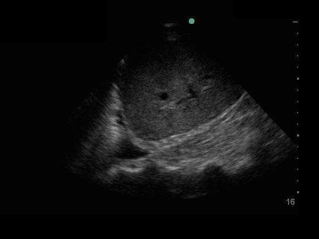
100 Pleural Effusion

101 Pleural Effusion with Consolidation

102 Pleural Effusion
103 Clinical Application

104 Included in systematic review History and physical: n=31 ECG: n=11 CXR: n=18 BNP/NT-proBNP*: n=41 Bedside echo: n=4 Lung ultrasound: n=8 Bioimpedance: n=4

105 SENS SPEC
106 +Test -Test

107 Pericardial Effusion


108 Anatomy Pericardium Wraps around heart ends at major vessels Pericardial fluid Normal = cc Best Windows Sitting = Subcostal Supine = PSLA
109 Top 5 Causes 1. Malignancy 30% 2. Idiopathic 15% 3. Acute MI / Post cath 15% 4. Infectious 15% 5. Uremia 10%


110 No Pericardial Effusion
111
112 Small Pericardial Effusion

113
114 Large Pericardial Effusion
115
116 Pericardial Effusion Size < 1 cm = Small 1-2 cm = Moderate > 2 cm = Large

117 Pericardial Effusion Size Measure in Diastole = when the effusion is the smallest
118 If the heart is surrounded by fluid how do I tell if it is pleural or pericardial fluid?
119
120

121

122
123 Tamponade?
124
125 5 Sonographic Signs 1. Pericardial effusion 2. Enlarged and plethoric IVC 3. Right atrial systolic collapse 4. RV Diastolic Collapse 5. All chambers underfilled & hyperdynamic
126


127 Tale of Two IVCs
128 5 Sonographic Signs 1. Pericardial effusion 2. Enlarged and plethoric IVC 3. Right atrial systolic collapse 4. RV Diastolic Collapse 5. All chambers underfilled & hyperdynamic
129 Pulsus Paradoxus?
130 LR+ = 5.9 LR- = 0.03 Roy et al, JAMA 2007
131 (Remember Rule #1)
132 I think its great, and I m sure for those techies who can use it that it will revolutionize their practice in the right clinical circumstances. That being said, I am extremely doubtful that it will ever come into general use. To do it well takes too much time, and it can be a hassle for both the physician and the patient. -Unknown (or at least to be revealed)
133 " I have no doubt whatever, from my own experience of its value, that it will be acknowledged to be one of the greatest discoveries in medicine by all those who are of a temper, and in circumstances, that will enable them to give it a fair trial. That it will ever come into general use, notwithstanding its value, I am extremely doubtful; because its beneficial application requires much time, and gives a good deal of trouble both to the patient and the practitioner -John Forbes, FRCP, FRS

134
135 Ultrasound is a tool.
136 Ultrasound is a tool. Use it wisely.
137 Thank you.
138
139
140
141
142
143
144
145
146 E-FAST Exam


147

148

149

150
151

152

153
154
155

156

157
158

159

160
161

162

163

164

165
166

167

168

169

170

171
172
173 But wait. There s more

174 Vascular Access

175 Vascular Access

176 Vascular Access

177 Vascular Access

178 Cardiac Arrest


179 Intubation
A Practical Approach to Ultrasound Assessment of Respiratory Distress
 A Practical Approach to Ultrasound Assessment of Respiratory Distress Yanick Beaulieu, MD, FRCPC Director, Bedside Ultrasound Curriculum Division of Cardiology and Critical Care Hôpital du Sacré-Coeur
Ultrasound. FAST Focused Assessment with Sonography in Trauma
 Ultrasound FAST Focused Assessment with Sonography in Trauma Rohit Patel, MD University of Florida Health Director, Critical Care Ultrasound Surgical ICU Center for Intensive Care Gainesville, Florida
This appendix was part of the submitted manuscript and has been peer reviewed. It is posted as supplied by the authors.
 This appendix was part of the submitted manuscript and has been peer reviewed. It is posted as supplied by the authors. - Figure S1: The four quadrant approach lung ultrasound at the bedside. * The anterolateral
Background & Indications Probe Selection
 Teresa S. Wu, MD, FACEP Director, EM Ultrasound Program & Fellowship Co-Director, Simulation Based Training Program & Fellowship Associate Program Director, EM Residency Program Maricopa Medical Center
THE PERICARDIUM: LOOKING OUTSIDE THE HEART
 THE PERICARDIUM: LOOKING OUTSIDE THE HEART DISCLOSURE Relevant relationships with commercial entities none Potential for conflicts of interest within this presentation none Steps taken to review and mitigate
Ultrasound in the ICU
 Ultrasound in the ICU Kristine E. W. Breyer, MD Assistant Professor Anesthesia & Critical Care Medicine UCSF DISCLOSURES: NONE Definition The Ultrasound Exam Types & Uses Training Clinical Examples Objectives
Chest Ultrasound: Pneumothorax
 WINFOCUS BASIC ECHO (WBE) Chest Ultrasound: Pneumothorax Mark Hamlin, MD, MS Associate Professor of Anesthesiology and Surgery University of Vermont College of Medicine Co-Director of Surgical Critical
ASSESSMENT OF LUNG PARENCHYMAL ABNORMALITIES
 2016 by the author Thank you for viewing this presentation. We would like to remind you that this material is the property of the author. It is provided to you by the ERS for your personal use only, as
The Shocked Patient. Adapted from Lichtenstein's FALLS protocol, with permission
 The Shocked Patient Adapted from Lichtenstein's FALLS protocol, with permission 1 Summary 1. (Ongoing resus) Clinical assessment: formulate the question 2. Rapid shock screen 3. Form a working diagnosis
Definitions and diagnostic implications of terms used in the chest radiograph and lung ultrasound diagnoses of pneumonia.
 Supplementary 1 Definitions and diagnostic implications of terms used in the chest radiograph and lung ultrasound diagnoses of pneumonia. Imaging finding Definition Implication CR Consolidation Interstitial
For more information about how to cite these materials visit
 Project: Ghana Emergency Medicine Collaborative Document Title: Approach to the Dyspenic Adult Patient Author(s): Randall Ellis, MD MPH (Vanderbilt University) License: Unless otherwise noted, this material
Initially for cardiac echo Subsequent studies non-cardiac applications
 No disclosures But Heavy accent Initially for cardiac echo Subsequent studies non-cardiac applications 1973: Goldberg et al in JCUS 30 mediastinal masses in pts. age 1-84 yrs. 1977: Kangarloo et al in
Contents& & & 1.! Ultrasound&basics& 1! 2.! Image&generation& 15!
 A l i n e press é % % % Contents& & & 1. Ultrasound&basics& 1 1.1. What,is,ultrasound?, 1 1.2. Ultrasound,probes,send,and,receive,ultrasound, 3 1.3. How,does,ultrasound,behave,travelling,through,tissue?,
Lung ultrasound in the critically ill patient BASICS
 Lung ultrasound in the critically ill patient BASICS Rohit Patel, MD University of Florida Health Director, Critical Care Ultrasound Surgical ICU Center for Intensive Care Gainesville, Florida Introduction
Pericardial Diseases/Tamponade Illustrative Cases
 Pericardial Diseases/Tamponade Illustrative Cases Jae K. Oh, MD Echo Hawaii 2017 2012 MFMER 3200268v3(2010)-1 Case #1 47 year old man Chest pain Not exertional Normal Examination 2016 MFMER slide-2 1 47
Extended FAST Exam. Goal of Trauma Care. Golden Hour of Trauma
 Extended FAST Exam Goal of Trauma Care Golden Hour of Trauma Best INITIAL screening modality in trauma efast 2014 LLSA Article (ACEP Policy Statement) Level B Recommendation: In hemodynamically unstable
OVERVIEW. Need for USG. Weaning assessment. Mechanics of USG. Pneumonia / VAP. Principles of lung USG. Prone position ventilation assessment
 OVERVIEW Need for USG Mechanics of USG Principles of lung USG BLUE protocol Alveolar syndrome Interstitial syndrome Weaning assessment Pneumonia / VAP Prone position ventilation assessment ETT positioning
Case Study #2. Case Study #1 cont 9/28/2011. CAPA 2011 Christy Wilson PA C. LH is 78 yowf with PMHx of metz breast CA presents
 Case Study #1 CAPA 2011 Christy Wilson PA C 46 yo female presents with community acquired PNA (CAP). Her condition worsened and she was transferred to the ICU and placed on mechanical ventilation. Describe
Pediatric Lung Ultrasound (PLUS) In Diagnosis of Community Acquired Pneumonia (CAP)
 In Diagnosis of Community Acquired Pneumonia (CAP)") Pediatric Lung Ultrasound (PLUS) In Diagnosis of Community Acquired Pneumonia (CAP) Dr Neetu Talwar Senior Consultant, Pediatric Pulmonology Fortis Memorial Research Institute, Gurugram Study To compare
Pediatric Lung Ultrasound (PLUS) In Diagnosis of Community Acquired Pneumonia (CAP) Dr Neetu Talwar Senior Consultant, Pediatric Pulmonology Fortis Memorial Research Institute, Gurugram Study To compare
Certificate in Clinician Performed Ultrasound (CCPU) Syllabus. Lung
 Syllabus. Lung") Certificate in Clinician Performed Ultrasound (CCPU) Syllabus Lung Page 1 of 8 01/17 Lung Syllabus Purpose: This unit is designed to cover the theoretical and practical curriculum for lung ultrasound in
Certificate in Clinician Performed Ultrasound (CCPU) Syllabus Lung Page 1 of 8 01/17 Lung Syllabus Purpose: This unit is designed to cover the theoretical and practical curriculum for lung ultrasound in
Lung ultrasound in the critically ill patient Pleural Effusions
 Lung ultrasound in the critically ill patient Pleural Effusions Rohit Patel, MD University of Florida Health Director, Critical Care Ultrasound Surgical ICU Center for Intensive Care Gainesville, Florida
Point-of-care lung ultrasound
 Ultrasound Point-of-care lung ultrasound Philips tutorial Michael B. Stone, MD, RDMS Director, Division of Emergency Ultrasound Department of Emergency Medicine Brigham and Women s Hospital, Boston, MA
Intro Case. Outline What We ll Cover. What we won t cover. Cardiac Ultrasound and The RUSH Exam: Bedside Ultrasound in Resuscitation and Shock
 Cardiac Ultrasound and The RUSH Exam: Bedside Ultrasound in Resuscitation and Shock Justin Davis, MD, MPH, RDMS Associate Physician Subchief for Emergency Ultrasound Services Kaiser Oakland Medical Center
Putting it all together: 1. The arrested patient 2. The shocked patient 3. The breathless patient
 Putting it all together: 1. The arrested patient 2. The shocked patient 3. The breathless patient 1 The Arrested Patient Adapted from Lichtenstein's SESAME protocol, with permission 2 Summary 1. (Ongoing
NON INVASIVE LIFE SAVERS. Ultrasound in PICU
 VOL 1 NO.1 Jan March 2014 54 Table 1. Selected Applications of Point-of-Care Ultrasonography, According to Medical Specialty. Specialty Ultrasound Applications Anesthesia Cardiology Guidance for vascular
Disclosures. Cardiac Ultrasound. Introductory Case. 80 y/o male Syncope at home Emesis x 3 in ambulance Looks sick. No pain.
 Disclosures Cardiac Ultrasound Justin A Davis, MD MPH RDMS Subchief for Emergency Ultrasound Kaiser Permanente East Bay Medical Center I have nothing to disclose. Introductory Case HR 118 BP 65/43 RR 27
Objectives. The Extended FAST Exam. Focused Assessment e With Sonography In. Trauma (FAST)
") Northern California Emergency Ultrasound Course Objectives The Extended FAST Exam Rimon Bengiamin, MD, RDMS UC SF Discuss the components of the EFAST exam Evaluate the utility of the EFAST Review how to
Northern California Emergency Ultrasound Course Objectives The Extended FAST Exam Rimon Bengiamin, MD, RDMS UC SF Discuss the components of the EFAST exam Evaluate the utility of the EFAST Review how to
Pericardial diseases
 Pericardial diseases Anatomy of the pericardium Consists of parietal and visceral membranes. The space between them(pericardial space is normally filled by a lymph like fluid. The fluid s normal quantity
Outline. Echocardiographic Assessment of Pericardial Effusion/Tamponade: The Essentials
 Echocardiographic Assessment of Pericardial Effusion/Tamponade: The Essentials John R Schairer DO FACC Henry Ford Heart and Vascular Institute No Disclosures Outline Normal Anatomy and Physiology Pathophysiology
Certificate in Clinician Performed Ultrasound (CCPU) Syllabus. Basic Echocardiography in Life Support
 Syllabus. Basic Echocardiography in Life Support") Certificate in Clinician Performed Ultrasound (CCPU) Syllabus Basic Echocardiography in Life Support Page 1 of 7 05/18 ACN 001 679 161 ABN 64 001 679 Basic Echocardiography in Life Support (BELS) Syllabus
Certificate in Clinician Performed Ultrasound (CCPU) Syllabus Basic Echocardiography in Life Support Page 1 of 7 05/18 ACN 001 679 161 ABN 64 001 679 Basic Echocardiography in Life Support (BELS) Syllabus
BELLWORK page 343. Apnea Dyspnea Hypoxia pneumo pulmonary Remember the structures of the respiratory system 1
 BELLWORK page 343 Apnea Dyspnea Hypoxia pneumo pulmonary respiratory system 1 STANDARDS 42) Review case studies that involve persons with respiratory disorders, diseases, or syndromes. Citing information
Certificate in Clinician Performed Ultrasound (CCPU) Syllabus. Extended Focussed Abdominal Scan for Trauma (E-FAST)
 Syllabus. Extended Focussed Abdominal Scan for Trauma (E-FAST)") Certificate in Clinician Performed Ultrasound (CCPU) Syllabus Extended Focussed Abdominal Scan for Trauma (E-FAST) Page 1 of 6 01/17 ACN 001 679 161 ABN 64 001 679 Extended Focussed Abdominal Scan for
Certificate in Clinician Performed Ultrasound (CCPU) Syllabus Extended Focussed Abdominal Scan for Trauma (E-FAST) Page 1 of 6 01/17 ACN 001 679 161 ABN 64 001 679 Extended Focussed Abdominal Scan for
DIASTOLOGY DON T BE SUCH A STIFF
 DIASTOLOGY DON T BE SUCH A STIFF Michael Mallin, MD University of Utah Director Emergency Ultrasound www.ultrasoundpodcast.com Warning: The education found within this presentation is not approved by anyone
Certificate in Clinician Performed Ultrasound (CCPU) Syllabus. Lung
 Syllabus. Lung") Certificate in Clinician Performed Ultrasound (CCPU) Syllabus Lung Page 1 of 8 12/15 Lung Syllabus Purpose: This unit is designed to cover the theoretical and practical curriculum for lung ultrasound in
Certificate in Clinician Performed Ultrasound (CCPU) Syllabus Lung Page 1 of 8 12/15 Lung Syllabus Purpose: This unit is designed to cover the theoretical and practical curriculum for lung ultrasound in
Using Lung Ultrasound to Diagnose and Manage Acute Heart Failure
 Using Lung Ultrasound to Diagnose and Manage Acute Heart Failure Jennifer Martindale, MD Assistant Professor Department of Emergency Medicine SUNY Downstate/Kings County Hospital Brooklyn, NY What is acute
Echocardiography as a diagnostic and management tool in medical emergencies
 Echocardiography as a diagnostic and management tool in medical emergencies Frank van der Heusen MD Department of Anesthesia and perioperative Care UCSF Medical Center Objective of this presentation Indications
Constrictive Pericarditis Pitfalls in MR Diagnosis Cylen Javidan-Nejad Associate Professor Mallinckrodt Institute of Radiology Washington University
 Constrictive Pericarditis Pitfalls in MR Diagnosis Cylen Javidan-Nejad Associate Professor Mallinckrodt Institute of Radiology Washington University in St. Louis Goal o To review the imaging criteria of
Cardiac Radiography. Jared D. Christensen, M.D.
 Cardiac Radiography Jared D. Christensen, M.D. Cardiac radiography Jared D. Christensen, M.D. Overview Basic Concepts Technique Normal anatomy Cases Technique 3 Standard Views Posterior-Anterior (PA) Anterior-Posterior
Lung sonography in the diagnosis of pneumothorax.
 Lung sonography in the diagnosis of pneumothorax. Poster No.: C-0526 Congress: ECR 2011 Type: Educational Exhibit Authors: K. Stefanidis, K. Vintzilaios, D. D. Cokkinos, E. Antypa, S. Dimopoulos, S. Nanas,
10/17/2016. Nuts and Bolts of Thoracic Radiology. Objectives. Techniques
 Nuts and Bolts of Thoracic Radiology October 20, 2016 Carleen Risaliti Objectives Understand the basics of chest radiograph Develop a system for interpreting chest radiographs Correctly identify thoracic
Objectives. What is a Chest X Ray? CXR Workshop. Definition (diagnostic tool/internal PE) Types. Cost
 Types. Cost") Objectives CAPA 2011 Christy Wilson, PA C Georgia Lung Associates Identify the radiographic landmarks on a chest radiograph Recognize identifiers of poor quality on the chest radiograph Outline an approach
Objectives CAPA 2011 Christy Wilson, PA C Georgia Lung Associates Identify the radiographic landmarks on a chest radiograph Recognize identifiers of poor quality on the chest radiograph Outline an approach
Pericardial Disease: Case Examples. Echo Fiesta 2017
 Pericardial Disease: Case Examples Echo Fiesta 2017 2014 2014 MFMER MFMER 3346252-1 slide-1 Objectives Have a systematic approach to evaluation of constriction 2014 MFMER 3346252-2 CASE 1 2013 MFMER 3248567-3
POCUS is the future of the physical exam
 Diagnostic Point of Care Ultrasound For Hospitalists Nima Afshar MD Associate Professor Trevor Jensen MD MS Assistant Professor Department of Medicine, UCSF Oct 2018 POCUS is the future of the physical
Certificate in Clinician Performed Ultrasound (CCPU) Syllabus. Lung
 Syllabus. Lung") Certificate in Clinician Performed Ultrasound (CCPU) Syllabus Lung ASUM Quality CCPU Syllabi Released: 21 March 2013 Approved by: CEO Lung Purpose: This unit is designed to cover the theoretical and practical
Certificate in Clinician Performed Ultrasound (CCPU) Syllabus Lung ASUM Quality CCPU Syllabi Released: 21 March 2013 Approved by: CEO Lung Purpose: This unit is designed to cover the theoretical and practical
TAMPONADE CARDIAQUE. Dr Cédrick Zaouter TUSAR 15 décembre 2015
 TAMPONADE CARDIAQUE Dr Cédrick Zaouter TUSAR 15 décembre 2015 OUTLINE History Incidence Definition Pathophysiology Aetiologies Investigations - Echocardiography Treatment of cardiac tamponade Pericardial
Cath Lab Essentials: Pericardial effusion & tamponade
 Cath Lab Essentials: Pericardial effusion & tamponade Pranav M. Patel, MD, FACC, FSCAI Chief, Division of Cardiology Director, Cardiac Cath Lab & CCU University of California, Irvine Division of Cardiology
EFAST. Extended Focussed Assessment with Sonography for Trauma. Ultrasound Logbook. Name
 EFAST Extended Focussed Assessment with Sonography for Trauma Ultrasound Logbook ame Contents EFAST Accreditation Requirements 25 Abdominal Aorta Report Forms 3 Formative Assessments 1 Summative Assessment
4/16/2017. Learning Objectives. Interpretation of the Chest Radiograph. Components. Production of the Radiograph. Density & Appearance
 Interpretation of the Arthur Jones, EdD, RRT Learning Objectives Identify technical defects in chest radiographs Identify common radiographic abnormalities This Presentation is Approved for 1 CRCE Credit
Case 1. A 35-year-old male presented with fever, cough, and purulent sputum for one week. This was his CXR (Fig. 1.1). What is the diagnosis?
. What is the diagnosis?") 1 Interpreting Chest X-Rays CASE 1 Fig. 1.1 Case 1. A 35-year-old male presented with fever, cough, and purulent sputum for one week. This was his CXR (Fig. 1.1). What is the diagnosis? CASE 1 Interpreting
1 Interpreting Chest X-Rays CASE 1 Fig. 1.1 Case 1. A 35-year-old male presented with fever, cough, and purulent sputum for one week. This was his CXR (Fig. 1.1). What is the diagnosis? CASE 1 Interpreting
Pitfalls in Shortness of Breath
 Pitfalls in Shortness of Breath Stuart Swadron, MD FRCPC FACEP Vice-Chair of Education and Program Director Department of Emergency Medicine Los Angeles County-University of Southern California Medical
Diagnostic Bedside Ultrasound for the Hospitalist
 Diagnostic Bedside Ultrasound for the Hospitalist Trevor Jensen MD MS Assistant Professor, UCSF Nima Afshar MD Associate Professor, UCSF Diagnostic Bedside Ultrasound AKA Point-of-Care Ultrasound (POCUS)
Focused Assessment Sonography of Trauma (FAST) Scanning Protocol
 Scanning Protocol") Focused Assessment Sonography of Trauma (FAST) Scanning Protocol Romolo Gaspari CHAPTER 3 GOAL OF THE FAST EXAM Demonstrate free fluid in abdomen, pleural space, or pericardial space. EMERGENCY ULTRASOUND
Focused Assessment Sonography of Trauma (FAST) Scanning Protocol Romolo Gaspari CHAPTER 3 GOAL OF THE FAST EXAM Demonstrate free fluid in abdomen, pleural space, or pericardial space. EMERGENCY ULTRASOUND
Percutaneous Mechanical Circulatory Support Devices
 Percutaneous Mechanical Circulatory Support Devices Daniel Vazquez RN, RCIS Miami Cardiac & Vascular Institute FINANCIAL DISCLOSURES none CASE STUDY CASE STUDY 52 year old gentlemen Complaining of dyspnea
AAENP US WORKSHOP 2/25/17
 Know the components of the Rapid Ultrasound for Shock & Hypotension & Extended Focused Assessment Sonography in Trauma & how they can help quickly determine diagnosis. Be comfortable obtaining and interpreting
Chest X-ray (CXR) Interpretation Brent Burbridge, MD, FRCPC
 Interpretation Brent Burbridge, MD, FRCPC") Chest X-ray (CXR) Interpretation Brent Burbridge, MD, FRCPC An approach to reviewing a chest x-ray will create a foundation that will facilitate the detection of abnormalities. You should create your own
Chest X-ray (CXR) Interpretation Brent Burbridge, MD, FRCPC An approach to reviewing a chest x-ray will create a foundation that will facilitate the detection of abnormalities. You should create your own
Point of Care Ultrasound in the ICU
 Point of Care Ultrasound in the ICU JENNIFER P. KANAAN, M.D. ASSISTANT PROFESSOR OF MEDICINE UNIVERSITY OF CONNECTICUT I have no disclosures 1 Ultrasound Ultrasound imaging is among the fastest, safest
We are now going to review the diagnosis and management of pericardial collections and tamponade
 We are now going to review the diagnosis and management of pericardial collections and tamponade FEEL COURSE PAGE 1 Paying particular attention to the difference between a collection and cardiac tamponade
Unusual new signs of pneumothorax at lung ultrasound
 Unusual new signs of pneumothorax at lung ultrasound Volpicelli et al. Volpicelli et al. Critical Ultrasound Journal 2013, 5:10 Volpicelli et al. Critical Ultrasound Journal 2013, 5:10 SHORT COMMUNICATION
Chris Fox, MD Professor and Interim Chair Department of Emergency
 Close Calls with the Executioner: Where POCUS Avoided Misdiagnosis Chris Fox, MD Professor and Interim Chair Department of Emergency Medicine En route with 22 year old female Asthma attack witnessed by
Objectives: Students: Residents Pediatric Fellows Nurses Paramedics Pediatric Faculty Adult Faculty Move EM ultrasound forward Around the World
 Cardiac Arrest Dave Spear, MD, FACEP Parkland / Children s Medical Center Dallas UT Southwestern School of Medicine Copyright All Rights Reserved 2002 Objectives: Cardiac Arrest History of ER Cardiac Cardiac
Bedside ultrasound - Lung ultrasound in the Intensive Care Unit
 Bedside ultrasound - Lung ultrasound in the Intensive Care Unit Kishore K. Pichamuthu, Professor, Department of Critical Care, Christian Medical College, Vellore. Summary In an ICU setting, ultrasonographic
Bedside Sonographic Diagnosis of Pneumothorax in Pediatric Patients: A Preliminary Report Chia-Wang Tang 1, Kai-Sheng Hsieh 1 1
 ORIGINAL ARTICLE Bedside Sonographic Diagnosis of in Pediatric Patients: A Preliminary Report Chia-Wang Tang 1, Kai-Sheng Hsieh 1 1 Division of Pediatric Pulmonology, Department of Pediatrics, Kaohsiung
NT-proBNP: Evidence-based application in primary care
 NT-proBNP: Evidence-based application in primary care Associate Professor Rob Doughty The University of Auckland, Auckland City Hospital, Auckland Heart Group NT-proBNP: Evidence in Primary Care The problem
Palpable Pulsus Paradoxus in the Setting of Malignant Pericardial Effusion and Tamponade Akshay Pendyal, MD
 Palpable Pulsus Paradoxus in the Setting of Malignant Pericardial Effusion and Tamponade Akshay Pendyal, MD University of Colorado Department of Internal Medicine None Conflicts of Interest Objectives
Non Invasive Hemodynamic Monitoring in the ED
 Is haemodinamic monitoring useful in the ED? Non Invasive Hemodynamic Monitoring in the ED Roberta PETRINO Director Emergency Medicine Unit S. Andrea Hospital, Vercelli - Italy EuSEM Vice-president Helps
POLICY ON CREDENTIALING FOR FOCUSSED ECHOCARDIOGRAPHY IN LIFE SUPPORT
 POLICY Document No: P61 Approved: Jul 2000 Last Revised: Feb 2016 Version No: 03 POLICY ON CREDENTIALING FOR FOCUSSED ECHOCARDIOGRAPHY IN LIFE SUPPORT 1. PURPOSE AND SCOPE This document is a policy of
Lung Ultrasound in Small Animals: The Vet BLUE SM
 Lung Ultrasound in Small Animals: The Vet BLUE SM Gregory R. Lisciandro, DVM, Dipl. ABVP, Dipl. ACVECC Hill Country Veterinary Specialists & FASTVet.com, San Antonio, Texas USA FASTVet TM and FAST Saves
Problem Based Learning Session. Mr Robinson is a 67 year old man. He visits the GP as he has had a cough and fever for 5 days.
 Problem Based Learning Session Mr Robinson is a 67 year old man. He visits the GP as he has had a cough and fever for 5 days. The GP takes a history from him and examines his chest. Over the left base
Internal Medicine Ultrasound Curriculum Outline. Mike Wagner, MD, FACP,
 Internal Medicine Ultrasound Curriculum Outline Mike Wagner, MD, FACP, RDMS @sonointernist Before Curriculum Implementation Bolus Training No Image QA outside of the ED No formal requirements Competency
Focused Assessment with Sonography in Trauma (FAST) UC Irvine School of Medicine
 UC Irvine School of Medicine") Focused Assessment with Sonography in Trauma (FAST) UC Irvine School of Medicine Purpose of FAST exam Quickly evaluate patient s status in emergency situations Blunt or penetrating trauma Visualize fluid
Focused Assessment with Sonography in Trauma (FAST) UC Irvine School of Medicine Purpose of FAST exam Quickly evaluate patient s status in emergency situations Blunt or penetrating trauma Visualize fluid
CARDIOLOGY SAUDI BOARD PROGRAM SAUDI BOARD FINAL CLINICAL EXAMINATION OF CARDIOLOGY (2018)
") CARDIOLOGY SAUDI BOARD PROGRAM SAUDI BOARD FINAL CLINICAL EXAMINATION OF CARDIOLOGY (2018) I Objectives a. Determine the ability of the candidate to practice as a specialist and provide consultation in
CARDIOLOGY SAUDI BOARD PROGRAM SAUDI BOARD FINAL CLINICAL EXAMINATION OF CARDIOLOGY (2018) I Objectives a. Determine the ability of the candidate to practice as a specialist and provide consultation in
Imaging of Thoracic Trauma: Tips and Traps. Arun C. Nachiappan, MD Associate Professor of Clinical Radiology University of Pennsylvania
 Imaging of Thoracic Trauma: Tips and Traps Arun C. Nachiappan, MD Associate Professor of Clinical Radiology University of Pennsylvania None Disclosures Objectives Describe blunt and penetrating traumatic
Pulmonary Ultrasound in Emergency Medicine and Critical Care
 Pulmonary Ultrasound in Emergency Medicine and Critical Care www.rmgultrasound.com Author: Virginia M Stewart, MD RDMS RDCS RDMSK Dr Stewart is a practicing Emergency Physician in Eastern Virginia, USA.
Radiology of the respiratory/cardiac diseases (part 2)
") Cardiology Cycle - Lecture 6 436 Teams Radiology of the respiratory/cardiac diseases (part 2) Objectives Done By Team Leaders: Khalid Alshehri Hanin Bashaikh Team Members: Leena Alwakeel Aroob Alhuthail
Cardiology Cycle - Lecture 6 436 Teams Radiology of the respiratory/cardiac diseases (part 2) Objectives Done By Team Leaders: Khalid Alshehri Hanin Bashaikh Team Members: Leena Alwakeel Aroob Alhuthail
Pericardial Diseases. Smonporn Boonyaratavej, MD. Division of Cardiology, Department of Medicine Chulalongkorn University
 Pericardial Diseases Smonporn Boonyaratavej, MD Division of Cardiology, Department of Medicine Chulalongkorn University Cardiac Center, King Chulalongkorn Memorial Hospital 21 AUGUST 2016 Pericardial
Point of Care Ultrasound (PoCUS)
") Point of Care Ultrasound (PoCUS) Competency Assessment Forms AORTA Competency A Focussed Assessment of the Aorta (AAA) Guidance Please follow this guidance as closely as possible to ensure consistency
Point of Care Ultrasound (PoCUS) Competency Assessment Forms AORTA Competency A Focussed Assessment of the Aorta (AAA) Guidance Please follow this guidance as closely as possible to ensure consistency
Resident Case Review CHEST. Daria Manos CAR 2016
 Resident Case Review CHEST CAR 2016 Daria Manos Disclosure Speakers bureau, Roche CAR 2016 Daria Manos 1. Recognize common and critical chest radiograph and computed tomography signs and use these clues
Pericardial disease. Se-Jung Yoon Cardiology division NHIS Ilsan hospital
 Pericardial disease Se-Jung Yoon Cardiology division NHIS Ilsan hospital Normal pericardial effusion Normal pericardium Normal pericardium Pericardial Layers: Visceral layer Parietal layer Fibrous pericardium
Matters of the Heart: Comprehensive Cardiology SARAH BEANLANDS RN BSCN MSC
 Matters of the Heart: Comprehensive Cardiology SARAH BEANLANDS RN BSCN MSC Who am I? Class Outline Gross anatomy of the heart Trip around the heart Micro anatomy: cellular and tissue level Introduction
Emergency Ultrasound. Case ECG. Potential diagnoses. An 80-year-old man in shock. ED patient with non traumatic undifferentiated hypotension
 Emergency Ultrasound An 80-year-old man in shock Case 80/M Present with chest pain Found low BP 62/44 at triage Resus. Room Pulse 86 Temp 35.6, SpO2 95% (RA) No history of drug overdose Past health PAF,
Dr. Rami M. Adil Al-Hayali Assistant Professor in Medicine
 Dr. Rami M. Adil Al-Hayali Assistant Professor in Medicine Venous thromboembolism: pulmonary embolism (PE) deep vein thrombosis (DVT) 1% of all patients admitted to hospital 5% of in-hospital mortality
Heart Failure Dr ahmed almutairi Assistant professor internal medicin dept
 Heart Failure Dr ahmed almutairi Assistant professor internal medicin dept (MBBS)(SBMD) Introduction Epidemiology Pathophysiology diastolic/systolic Risk factors Signs and symptoms Classification of HF
Pericardial effusion, Cardiac Tamponade, and echo guided pericardiocentesis
 KSC 2017 Echo5- Myocardial and Pericardial disease Pericardial effusion, Cardiac Tamponade, and echo guided pericardiocentesis Ji-Hyun Jung Division of Cardiology Sejong Hospital KSC 2017 The 61 th Annual
Point-of-Care Ultrasound Closer look at the Inferior Vena Cavae &
 Point-of-Care Ultrasound Closer look at the Inferior Vena Cavae & Brief Introduction to Gross Systolic Function Omar S. Darwish, MS, DO Certified in Point-of-Care Ultrasound Hospitalist University of California,
Choose the grading of diastolic function in 82 yo woman
 Question #1 Choose the grading of diastolic function in 82 yo woman E= 80 cm/s A= 70 cm/s LAVI < 34 ml/m 2 1= Grade 1 2= Grade 2 3= Grade 3 4= Normal 5= Indeterminate 2018 MFMER 3712003-1 Choose the grading
Paramedic Rounds. Pre-Hospital Continuous Positive Airway Pressure (CPAP)
") Paramedic Rounds Pre-Hospital Continuous Positive Airway Pressure (CPAP) Morgan Hillier MD Class of 2011 Dr. Mike Peddle Assistant Medical Director SWORBHP Objectives Outline evidence for pre-hospital
Paramedic Rounds Pre-Hospital Continuous Positive Airway Pressure (CPAP) Morgan Hillier MD Class of 2011 Dr. Mike Peddle Assistant Medical Director SWORBHP Objectives Outline evidence for pre-hospital
USE OF LUNG ULTRASOUND IN SMALL ANIMALS - THE VET BLUE THE BASICS OF VET BLUE
 Ultrasound in the Respiratory Distress Patient: Using Vet Blue International Veterinary Emergency and Critical Care Symposium 2018 Gregory R. Lisciandro, DVM, DABVP, DACVECC Hill Country Veterinary Specialists
Constrictive Pericarditis
 Constrictive Pericarditis Never Confused with Anything Else Jae K. Oh, MD 2018 MFMER 3712003-1 ARS #1 CP Which of following patients has constrictive pericarditis? 1 2 3 Medial e 13 cm/s Medial e 3 cm/s
UERMMMC Department of Radiology. Basic Chest Radiology
 UERMMMC Department of Radiology Basic Chest Radiology PHYSICS DENSITIES BONE SOFT TISSUES WATER FAT AIR TELEROENTGENOGRAM Criteria for an Ideal Chest Radiograph 1. Upright 2. Posteroanterior View 3. Full
Bedside RUQ Ultrasound. Replace Formal ULS? Why Bedside ULS RUQ? RUQ Ultrasound. Bedside ULS is Limited, Goal-Directed
 Bedside RUQ Ultrasound RUQ Ultrasound Why do it How to do it Elizabeth Kwan UCSF Emergency Ultrasound Fellow Why Bedside ULS RUQ? Dx or Rule Out Acute Cholecystitis Cholelithiasis, Choledocolithiasis Earlier
The faculty will include physicians with international reputations as outstanding ultrasound educators.
 Ultrasound Courses Course Description Whether you re a beginner or a seasoned sonographer, this year s AAEM pre-conference ultrasound course will be worth your time. We will be offering a half day course
X-rays. Dr Will Dooley
 X-rays Dr Will Dooley Plan Chest X-Rays Abdominal X-Rays Exam approach Presentation skills EMQ EMQ- answers Chest X-Ray - Systematic Approach D R Details RIP Image Quality +/- OBVIOUS ABNORMALITY A B C
Ultrasonography for Novices
 module 03 Ultrasonography for Novices Stephanie J. Doniger, MD, RDMS, FAAP, FACEP Lei Chen, MD, FAAP Objectives 1Understand the basic principles of ultrasound physics. 2Be familiar with basic controls
Don t Panic! Dr. Karau s Guide to Respiratory Emergencies November 4, 2018
 Don t Panic! Dr. Karau s Guide to Respiratory Emergencies November 4, 2018 Objectives Oxygen delivery methods Emergent diagnostic tests Differentiating between upper and lower respiratory disease Respiratory
Interactive Lecture. Lecture 7 - Interactive. Radiology of cardiorespiratory disease. Editing File. Done By. Color Coding Important Notes Extra
 Lecture 7 - Interactive 436 Teams Interactive Lecture Radiology of cardiorespiratory disease Done By Team Leaders: Khalid Alshehri Hanin Bashaikh Team Members: Ghaida Alsaeed Maha Alissa Nawwaf AlHarbi
INTERNAL MEDICINE SAUDI BOARD PROGRAM SAUDI BOARD FINAL CLINICAL EXAMINATION OF INTERNAL MEDICINE (2016)
") INTERNAL MEDICINE SAUDI BOARD PROGRAM SAUDI BOARD FINAL CLINICAL EXAMINATION OF INTERNAL MEDICINE (206) I Objectives a. Determine the ability of the candidate to practice as a specialist and provide consultation
INTERNAL MEDICINE SAUDI BOARD PROGRAM SAUDI BOARD FINAL CLINICAL EXAMINATION OF INTERNAL MEDICINE (206) I Objectives a. Determine the ability of the candidate to practice as a specialist and provide consultation
Cardiology. Objectives. Chapter
 1:44 M age 1121 Chapter Cardiology Objectives art 1: Cardiovascular natomy and hysiology, ECG Monitoring, and Dysrhythmia nalysis (begins on p. 1127) fter reading art 1 of this chapter, you should be able
ACUTE HEART FAILURE in the ED. Pr. Samir Nouira Emergency Department Fattouma Bourguiba University Hospital Monastir Tunisia
 ACUTE HEART FAILURE in the ED Pr. Samir Nouira Emergency Department Fattouma Bourguiba University Hospital Monastir Tunisia ACUTE HEART FAILURE 80% Acute Heart Failure Syndrome Sensitivity Specificity
Pulmonary-Vascular Disease. Howard J. Sachs, MD.
 Pulmonary-Vascular Disease Howard J. Sachs, MD www.12daysinmarch.com Dyspnea Cardiac Pulmonary CAD Pump Failure Chest Wall Airways Valve Disease Pericardial Disease Alveoli Interstitium Rhythm Disturbance
Medical NREMT-PTE. NREMT Paramedic Trauma Exam.
 Medical NREMT-PTE NREMT Paramedic Trauma Exam https://killexams.com/pass4sure/exam-detail/nremt-pte Question: 41 Which of the following most accurately describes the finding of jugular venous distension