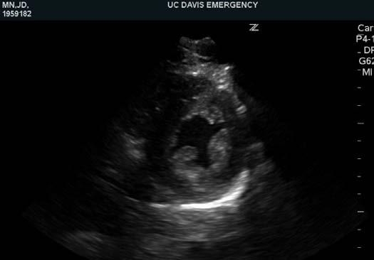
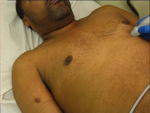
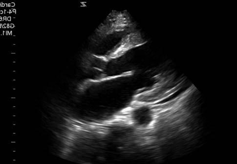
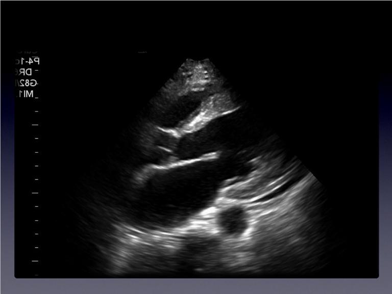
Disclosures. Cardiac Ultrasound. Introductory Case. 80 y/o male Syncope at home Emesis x 3 in ambulance Looks sick. No pain.
|
|
|
- Shawn Logan
- 5 years ago
- Views:
Transcription

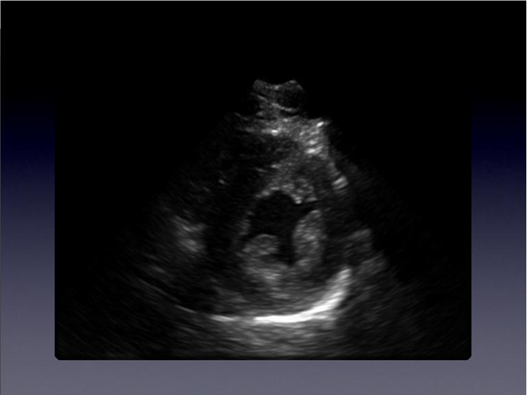
1 Disclosures Cardiac Ultrasound Justin A Davis, MD MPH RDMS Subchief for Emergency Ultrasound Kaiser Permanente East Bay Medical Center I have nothing to disclose. Introductory Case HR 118 BP 65/43 RR 27 O 2 99% Talking fine 80 y/o male Syncope at home Emesis x 3 in ambulance Looks sick. No pain. Clear Lungs Pulses weak No Edema No murmurs Soft, non-tender No pulsatile Mass 1


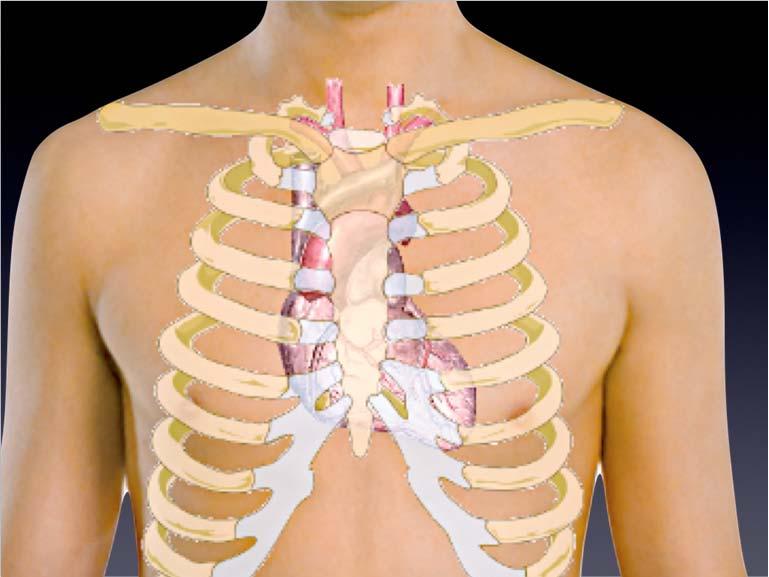
2 Distal Aorta Transvers e Apical Long Axis Learning Objectives Understand cardiac anatomy Understand image acquisition Recognize common findings and pitfalls Understand basic clinical applications Recognize a few advanced applications 2
3 Outline Information Gained and its Applications Cardiac Anatomy & Image Acquisition The Basics: Effusions, Function, Advanced: Tamponade, RV Strain, Aortic Root Dilation Information Provided By Bedside Ultrasound The Basics: Pericardial Effusion Cardiac Function Central Venous Pressure Pericardial Effusion Trauma Cardiac Arrest Hypotension Chest Pain Applications Cardiac Function Central Venous Pressure Dyspnea Sepsis Fluid Resuscitation Diuresis Outline Information Gained and its Applications Cardiac Anatomy & Image Acquisition The Basics: Effusions, Function, Advanced: Tamponade, RV Strain, Aortic Root Dilation 3


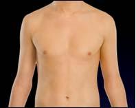
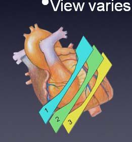
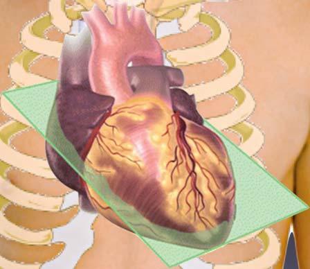
4 Echocardiogram Anatomy Windows + = Views Planes 3 Windows Parasternal Apical Bedside Echo: Sonographic Windows Subxiphoid Bedside Echo: Cardiac Planes 3 Primary Planes Long Axis Short Axis Four Chamber 4 Echocardiogram Views Parasternal Long Axis Parasternal Short Axis Apical 4 Chamber Subxiphoid 4 Chamber 4


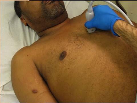
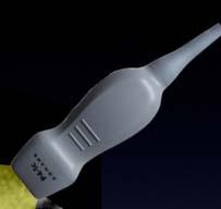
5 Image Small footprint Acquisition & Probe Selection Echocardiogram AnatomyWindow Differences Low frequency COPD, Barrel Chest, Tall and Thin Cardiomegaly, Large Abdomen Echocardiogram Anatomy Axis Differences Echocardiogram AnatomyWindows and Axes Windows & axes vary Vertical Axis Horizontal Axis First: Find your Window THEN: Adjust the Axis 5

 Scan from pts")


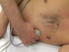
6 Controversy: Probe Orientation General Radiology/EM Cardiology Parasternal Long Axis View (The only one that differs) Scan from pts RIGHT Scan from pts LEFT Indicator Screen LEFT Indicator Screen RIGHT Moore, C. Current issues with emergency cardiac ultrasound probe and image conventions. Acad Emerg Med 2008; 15: What setting does my machine use? Choose cardiac probe and preset Look for the indicator Can L/R invert Can save default Parasternal Long Axis View Probe Indicator Toward right shoulder 6


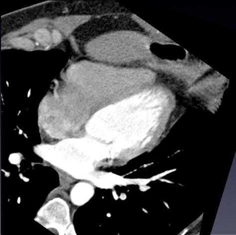
7 Cardiac Planes Long Axis Plane Long Axis Short Axis Ao RV LV LA DTA Parasternal Long Axis View RV LV Ao RV LV Mitral Valve Leaflets Parasternal Long Axis View Tips: Stay close to sternum End-expiratory hold Difficult in COPD DTA 7


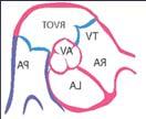
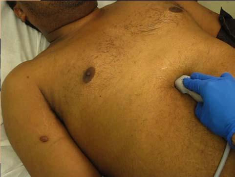
8 Parasternal Short Axis Indicator 90º CCW from Long Axis Short Axis Plane RV LV Chest Wall Back View Parasternal Short Axis View Tips: Try to maintain circular LV End-expiratory hold View varies depending on level of heart 8


9 Apical 4 Chamber View Apical 4 Chamber View Indicator similar to Short Axis, Perpendicular plane Plane is 90º from Short Axis, Window is at the PMI 4 Chamber Plane Apical Window 4 Chamber Plane RA LA RV LV 9


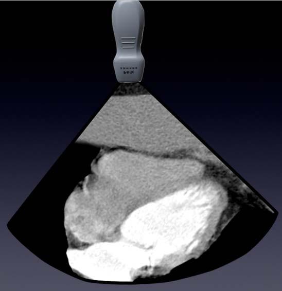

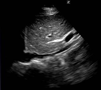
10 RV RA Apical 4 Chamber View LV LA Apical 4 Chamber View Tips: Left lateral decubitus End-expiratory hold Under the breast fold Aim sound waves toward right scapula Subxiphoid 4 Chamber View Subxiphoid 4 Chamber View LA RA LV RV Liver RV RA LV LA 10


11 Liver RV RA LA Subxiphoid 4 Chamber View LV Subxiphoid 4 Chamber View Tips: Firm pressure Inspiratory hold Bowel Gas? Try right of midline Indicator toward chin Aim towards thoracic spine 11


12 Image the entering Right Atrium RA Goals & CVP Assess for fullness Assess % collapse with spontaneous inspiration Just inferior to hepatic vein junction 12


 Will appear dilated")
13 Pitfalls: vs Aorta Transverse View Empties into heart heart Flows deep to Flows through liver liver Undulating Pulsation Pulsation Flows deep to Bounding Spine Aorta Avoiding Pitfalls: Do NOT scan from the far lateral torso ( collapses Ant-Post, not laterally) Will appear dilated with minimal variation X 13


14 Tips: Maintain axis along upper May need to scan through right anterior ribs Differentiate vs Aorta Scanning Flow Parasternal Long Parasternal Short Apical 4 SubXiphoid Outline Information Gained and its Applications Cardiac Anatomy & Image Acquisition The Basics: Effusions, Function, Advanced: Tamponade, RV Strain, Aortic Root Dilation 14


15 Basics: Pericardial Effusions Anechoic signal (Black) Between myocardium and pericardium Generally dependent Except in trauma or post-op, clinically significant effusions are circumferential Pericardial Effusions Parasternal Long Axis Pericardial Effusions Subxiphoid 4 Chamber Pericardial Effusions False Positives Epicardial fat pad Left pleural effusion Ascites 15


16 False Positive: Fat Pad Pericardial Effusions False Positive: Fat Pad Echogenic Moves with myocardium Not displaced by heart motion Usually not dependent False Positive: Fat Pad False Positive: L Pleural Effusion Pericardium DTA 16


17 Pericardial Effusions False Positive: L Pleural Effusion Only seen posterior/lateral views In parasternal long axis, extends deep to the descending thoracic aorta (not between DTA and heart) Use FAST splenorenal view to confirm False Positive: L Pleural Pericardial Effusion Effusion DTA Pleural Effusion False Positive: L Pleural Use FAST LUQ view to confirm Effusion False Positive: Ascites 17
18 Pericardial Effusions False Positive: Ascites Only seen in subxiphoid view Will often disappear with deep inspiration Confirm ascites in abdominal views Pericardial Effusions False Negative: Blood Clot Clotting blood can appear from anechoic to hyperechoic, to mixed. Look for your landmarks Check multiple views False Negative: Clot Outline Information Gained and its Applications Cardiac Anatomy & Image Acquisition The Basics: Effusions, Function, Advanced: Tamponade, RV Strain, Aortic Root Dilation 18
 (should come within 8mm of septal wall) General contraction of LV E-Point")

.")
 PS long axis Image center of LV (No visible chordae) M-mode through")
19 Basics: LV Function General estimate Dead to Hyperdynamic Parasternal long and short axes, look at Anterior mitral valve leaflet (EPSS) (should come within 8mm of septal wall) General contraction of LV E-Point Septal Separation (EPSS) Shortest distance from anterior mitral valve leaflet to LV septum Strong inverse correlation with LVEF Elagha, Abdalla, and Anthon Fuisz. Mitral Valve E-Point to Septal Separation (EPSS) Measurement by Cardiac Magnetic Resonance Imaging as a Quantitative Surrogate of Left Ventricular Ejection Fraction (LVEF). Journal of Cardiovascular Magnetic Resonance 14.Suppl 1 (2012): P154. PMC. Web. 20 Mar E-Point Septal Separation (EPSS) PS long axis Image center of LV (No visible chordae) M-mode through anterior mitral valve tip Measure minimum distance to LV Septum Normal < 8mm Septum Mitral Valve LV Function STANDSTILL 19


20 LV Function LV Function Agonal Severely Depressed LV Function LV Function Moderately Depressed Moderately Depressed 20

 Minimal 15-20cm H20 Large (>2.")
21 LV Function LV Function Normal Hyperdynamic Outline Information Gained and its Applications Cardiac Anatomy & Image Acquisition The Basics: Effusions, Function, Advanced: Tamponade, RV Strain, Aortic Root Dilation and CVP Distension Inspiratory collapse CVP Small Complete <5cm H20 Moderate to Full >50% 5-10 Moderate to Full <50% Large (>2.5cm) Minimal 15-20cm H20 Large (>2.5cm) None >20cm H20 21

22 and CVP However, don t have to use numbers Give a general estimate, trend is more important than single measurement Is the CVP... low, moderate, high, or extremely high? Nearly empty, with complete collapse Full, with complete collapse Full, with partial collapse 22

 M-Mode to visualize and")


23 Fill the Tank: In hypotension, Give fluids until it collapses less than 50% & CVP Distended, with no variation >50% Collapse = CVP < 8mmHg (10cmH20) M-Mode to visualize and Quantify Collapse and CVP M-Mode Outline Information Gained and its Applications Cardiac Anatomy & Image Acquisition The Basics: Effusions, Function, Advanced: Tamponade, RV Strain, Aortic Root Dilation 23
 distention w/o resp.")

24 Advanced Finding: Impending Tamponade (Clinical Diagnosis) What does RA or RV collapse look like? 1) distention w/o resp. variation (ALMOST ALWAYS) 2) Diastolic RA or RV Collapse RA Diastolic Collapse Seen in 75% RV Diastolic Collapse Seen in 25% 24


25 RV Collapse? Tamponade M-Mode Is it collapsing in Diastole? In Diastole the Mitral Valve is open M-Mode Parasternal long, short, or subxiphoid M - Mode RV Collapse RV Free Wall Ant. Mitral Valve RV wall moving inward while mitral valve is open Pulsus Paradoxus Pulsed Wave Doppler In tamponade, exaggerated drop in stroke volume and BP with inspiration Apical 4 or 5 chamber view Mitral valve inflow, LV outflow, Tricuspid inflow Doppler gate distal distal to valve tips Look for drop >25% with inspiration 25


 RV distended and hardly squeezing Sometimes")

26 Pulsus Paradoxus Outline > 25% drop Information Gained and its Applications Cardiac Anatomy & Image Acquisition The Basics: Effusions, Function, Advanced: Tamponade, RV Strain, Aortic Root Dilation Advanced Finding: RV Strain When RV is pushing against high pressure (eg. massive PE) RV distended and hardly squeezing Sometimes LV is compressed/empty is plethoric (full) Normal RV - Large & Hypokinetic Parasternal Long Axis LV - Small & Hyperkinetic 26


 Need to image both")


27 Normal Parasternal Short Axis RV - Large & Hypokinetic Normal Apical 4 Chamber RV - Large & Hypokinetic D -Shaped D Left Ventricle (Septal Wall Flattening) LV - Small & Hyperkinetic LV - Small & Hyperkinetic RV:LV >1 (normal<1) Need to image both tricuspid and mitral valves well to comment on RV:LV ratio = Plethoric (Full, Stiff) RV Dysfunction: TAPSE Tricuspid Annular Plane Systolic Excursion Apical 4 Chamber M-mode Tricuspid Annulus at RV free wall Normal excursion > 16mm 27


28 RV Dysfunction: TAPSE RV Dysfunction: TAPSE M-Mode RV Dysfunction: Tissue Doppler Select TDI mode on your Doppler Focuses on tissue velocity, not fluid velocity Upward systolic motion is S 1 wave Normal S 1 > 10 cm/s S E A Outline Information Gained and its Applications Cardiac Anatomy & Image Acquisition The Basics: Effusions, Function, Advanced: Tamponade, RV Strain, Aortic Root Dilation 28
 Parasternal long axis and 1-2 rib spaces")


29 Advanced Finding Dilated Aortic Root 90% of Ascending aortic dissection have dilated ascending aorta (>4cm) Parasternal long axis and 1-2 rib spaces superior Image 3-5 cm length of ascending Ao Neither sensitive nor specific, but may push you along towards the diagnosis Aortic Root Dilation Aortic Root Dilation Aortic Root Dilation 5.4cm Parasternal Long Axis Aortic Valve Annulus is at end of septum, Anything in aorta distal to that is a dissection flap, not a leaflet 29
30 Outline Information Gained and its Applications Cardiac Anatomy & Image Acquisition The Basics: Effusions, Function, Advanced: Tamponade, RV Strain, Aortic Root Dilation Bedside Echo Summary The Basics: Significant Pericardial Effusion: Yes/No Circumferential hypoechoic fluid displaced by heart motion LV Function: Gestalt estimate Note LV contraction and Anterior Mitral Valve leaflet approaching the septum : Gestalt CVP estimation Note size and collapse with respiration Bedside Echo Summary Advanced Findings: Impending Tamponade: Large effusion, plethoric, +/- RA/RV collapse RV Strain: RV appears enlarged and poorly contracting LV is D-shaped on short axis Aortic Root Dilation: High parasternal long axis, normal <4cm 30
Intro Case. Outline What We ll Cover. What we won t cover. Cardiac Ultrasound and The RUSH Exam: Bedside Ultrasound in Resuscitation and Shock
 Cardiac Ultrasound and The RUSH Exam: Bedside Ultrasound in Resuscitation and Shock Justin Davis, MD, MPH, RDMS Associate Physician Subchief for Emergency Ultrasound Services Kaiser Oakland Medical Center
Echocardiography as a diagnostic and management tool in medical emergencies
 Echocardiography as a diagnostic and management tool in medical emergencies Frank van der Heusen MD Department of Anesthesia and perioperative Care UCSF Medical Center Objective of this presentation Indications
Certificate in Clinician Performed Ultrasound (CCPU) Syllabus. Rapid Cardiac Echo (RCE)
 Syllabus. Rapid Cardiac Echo (RCE)") Certificate in Clinician Performed Ultrasound (CCPU) Syllabus Rapid Cardiac Echo (RCE) Purpose: Rapid Cardiac Echocardiography (RCE) This unit is designed to cover the theoretical and practical curriculum
Certificate in Clinician Performed Ultrasound (CCPU) Syllabus Rapid Cardiac Echo (RCE) Purpose: Rapid Cardiac Echocardiography (RCE) This unit is designed to cover the theoretical and practical curriculum
Intro to Bedside Ultrasound. Cardiac Ultrasound
 Intro to Bedside Ultrasound Cardiac Ultrasound TEACHERS University of California-Irvine School of Medicine Nathan Molina nathan.d.molina@gmail.com Trevor Plescia taplescia90@gmail.com Jack Silva jpsilva42@gmail.com
Adel Hasanin Ahmed 1
 Adel Hasanin Ahmed 1 PERICARDIAL DISEASE The pericardial effusion ends anteriorly to the descending aorta and is best visualised in the PLAX. PSAX is actually very useful sometimes for looking at posterior
Pericardial Diseases. Smonporn Boonyaratavej, MD. Division of Cardiology, Department of Medicine Chulalongkorn University
 Pericardial Diseases Smonporn Boonyaratavej, MD Division of Cardiology, Department of Medicine Chulalongkorn University Cardiac Center, King Chulalongkorn Memorial Hospital 21 AUGUST 2016 Pericardial
Appendix II: ECHOCARDIOGRAPHY ANALYSIS
 Appendix II: ECHOCARDIOGRAPHY ANALYSIS Two-Dimensional (2D) imaging was performed using the Vivid 7 Advantage cardiovascular ultrasound system (GE Medical Systems, Milwaukee) with a frame rate of 400 frames
The role of bedside ultrasound in the diagnosis of pericardial effusion and cardiac tamponade
 Symposium The role of bedside ultrasound in the diagnosis of pericardial effusion and cardiac tamponade Adam Goodman, Phillips Perera, Thomas Mailhot, Diku Mandavia Department of Emergency Medicine, Los
Outline. Echocardiographic Assessment of Pericardial Effusion/Tamponade: The Essentials
 Echocardiographic Assessment of Pericardial Effusion/Tamponade: The Essentials John R Schairer DO FACC Henry Ford Heart and Vascular Institute No Disclosures Outline Normal Anatomy and Physiology Pathophysiology
Focused Assessment Sonography of Trauma (FAST) Scanning Protocol
 Scanning Protocol") Focused Assessment Sonography of Trauma (FAST) Scanning Protocol Romolo Gaspari CHAPTER 3 GOAL OF THE FAST EXAM Demonstrate free fluid in abdomen, pleural space, or pericardial space. EMERGENCY ULTRASOUND
Focused Assessment Sonography of Trauma (FAST) Scanning Protocol Romolo Gaspari CHAPTER 3 GOAL OF THE FAST EXAM Demonstrate free fluid in abdomen, pleural space, or pericardial space. EMERGENCY ULTRASOUND
Certificate in Clinician Performed Ultrasound (CCPU) Syllabus. Basic Echocardiography in Life Support
 Syllabus. Basic Echocardiography in Life Support") Certificate in Clinician Performed Ultrasound (CCPU) Syllabus Basic Echocardiography in Life Support Page 1 of 7 05/18 ACN 001 679 161 ABN 64 001 679 Basic Echocardiography in Life Support (BELS) Syllabus
Certificate in Clinician Performed Ultrasound (CCPU) Syllabus Basic Echocardiography in Life Support Page 1 of 7 05/18 ACN 001 679 161 ABN 64 001 679 Basic Echocardiography in Life Support (BELS) Syllabus
Copyright 2017 American College of Emergency Physicians. All rights reserved.
 POLICY Approved April 2017 Guidelines for the Use of Transesophageal Echocardiography (TEE) in the ED for Cardiac Arrest Approved by the ACEP Board of Directors April 2017 1. Introduction The American
Point of Care Ultrasound (PoCUS)
") Point of Care Ultrasound (PoCUS) Competency Assessment Forms AORTA Competency A Focussed Assessment of the Aorta (AAA) Guidance Please follow this guidance as closely as possible to ensure consistency
Point of Care Ultrasound (PoCUS) Competency Assessment Forms AORTA Competency A Focussed Assessment of the Aorta (AAA) Guidance Please follow this guidance as closely as possible to ensure consistency
Cardiac ultrasound protocols
 Cardiac ultrasound protocols IDEXX Telemedicine Consultants Two-dimensional and M-mode imaging planes Right parasternal long axis four chamber Obtained from the right side Displays the relative proportions
M-Mode Echocardiography Is it still Alive? Itzhak Kronzon, MD,FASE. Sampling Rate M-Mode: 1800 / sec 2D: 30 / sec
 M-Mode Echocardiography Is it still Alive? Itzhak Kronzon, MD,FASE Honoraria: Philips Classical M-mode Echocardiography M-Mode offers better time and image resolution. Sampling Rate M-Mode: 1800 / sec
Normal TTE/TEE Examinations
 Normal TTE/TEE Examinations Geoffrey A. Rose, MD FACC FASE Sanger Heart & Vascular Institute Before you begin imaging... Obtain the patient s Height Weight BP PLAX View PLAX View Is apex @ 9-10 o clock?
PROSTHETIC VALVE BOARD REVIEW
 PROSTHETIC VALVE BOARD REVIEW The correct answer D This two chamber view shows a porcine mitral prosthesis with the typical appearance of the struts although the leaflets are not well seen. The valve
US Applications. Case Based Wrap-Up 1. Case 1 E-FAST. Case presentations E-FAST Abdominal. Pearls for each indication
 Case Based Wrap-Up 1 Stephanie J. Doniger MD RDMS FAAP FACEP Associate Director, Pediatric Emergency Ultrasound Stanford University Medical Center US Applications Case presentations E-FAST Abdominal Aorta
PRACTICAL GUIDE TO FETAL ECHOCARDIOGRAPHY IC Huggon and LD Allan
 PRACTICAL GUIDE TO FETAL ECHOCARDIOGRAPHY IC Huggon and LD Allan Fetal Cardiology Unit, Harris Birthright Research Centre for Fetal Medicine, King's College Hospital, London, UK IMPORTANCE OF PRENATAL
COMPREHENSIVE EVALUATION OF FETAL HEART R. GOWDAMARAJAN MD
 COMPREHENSIVE EVALUATION OF FETAL HEART R. GOWDAMARAJAN MD Disclosure No Relevant Financial Relationships with Commercial Interests Fetal Echo: How to do it? Timing of Study -optimally between 22-24 weeks
PERICARDIAL DIAESE. Kaijun Cui Associated professor Sichuan University
 PERICARDIAL DIAESE Kaijun Cui Associated professor Sichuan University CLASSIFICATION acute pericarditis pericardial effusion cardiac tamponade constrictive pericarditis congenitally absent pericardium
Echocardiography Conference
 Echocardiography Conference David Stultz, MD Cardiology Fellow, PGY-6 September 20, 2005 Atrial Septal Aneurysm Bulging of Fossa Ovalis Associated commonly with Atrial septal defect or small perforations
Incorporating the New Echo Guidelines Into Everyday Practice
 Incorporating the New Echo Guidelines Into Everyday Practice Clinical Case RIGHT VENTRICULAR FAILURE Gustavo Restrepo MD President Elect Interamerican Society of Cardiology Director Fellowship Training
Emergency Ultrasound. Case ECG. Potential diagnoses. An 80-year-old man in shock. ED patient with non traumatic undifferentiated hypotension
 Emergency Ultrasound An 80-year-old man in shock Case 80/M Present with chest pain Found low BP 62/44 at triage Resus. Room Pulse 86 Temp 35.6, SpO2 95% (RA) No history of drug overdose Past health PAF,
Background & Indications Probe Selection
 Teresa S. Wu, MD, FACEP Director, EM Ultrasound Program & Fellowship Co-Director, Simulation Based Training Program & Fellowship Associate Program Director, EM Residency Program Maricopa Medical Center
Practical Echocardiography: ECHOES in the REAL WORLD Know When to Hold Em and When to Fold Em
 Practical Echocardiography: ECHOES in the REAL WORLD Know When to Hold Em and When to Fold Em Introduction The use of ultrasound in private veterinary practice is continuing to grow. The popularity of
Pericardial Disease: Case Examples. Echo Fiesta 2017
 Pericardial Disease: Case Examples Echo Fiesta 2017 2014 2014 MFMER MFMER 3346252-1 slide-1 Objectives Have a systematic approach to evaluation of constriction 2014 MFMER 3346252-2 CASE 1 2013 MFMER 3248567-3
Adult Echocardiography Examination Content Outline
 Adult Echocardiography Examination Content Outline (Outline Summary) # Domain Subdomain Percentage 1 2 3 4 5 Anatomy and Physiology Pathology Clinical Care and Safety Measurement Techniques, Maneuvers,
TAMPONADE CARDIAQUE. Dr Cédrick Zaouter TUSAR 15 décembre 2015
 TAMPONADE CARDIAQUE Dr Cédrick Zaouter TUSAR 15 décembre 2015 OUTLINE History Incidence Definition Pathophysiology Aetiologies Investigations - Echocardiography Treatment of cardiac tamponade Pericardial
ASCeXAM / ReASCE. Practice Board Exam Questions Monday Morning
 ASCeXAM / ReASCE Practice Board Exam Questions Monday Morning Ultrasound Physics Artifacts Doppler Physics Imaging, Knobology, and Artifacts Echocardiographic Evaluation of the RV Tricuspid and Pulmonary
The Doppler Examination. Katie Twomley, MD Wake Forest Baptist Health - Lexington
 The Doppler Examination Katie Twomley, MD Wake Forest Baptist Health - Lexington OUTLINE Principles/Physics Use in valvular assessment Aortic stenosis (continuity equation) Aortic regurgitation (pressure
Index. K Knobology, TTE artifact, image resolution, ultrasound, 14
 A Acute aortic regurgitation (AR), 124 128 Acute aortic syndrome (AAS) classic aortic dissection diagnosis, 251 263 evolutive patterns, 253 255 pathology, 250 251 classifications, 247 248 incomplete aortic
좌심실수축기능평가 Cardiac Function
 Basic Echo Review Course 좌심실수축기능평가 Cardiac Function Seonghoon Choi Cardiology Hallym university LV systolic function Systolic function 좌심실수축기능 - 심근의수축으로심실에서혈액을대동맥으로박출하는기능 실제임상에서 LV function 의의미 1Diagnosis
HISTORY. Question: What category of heart disease is suggested by this history? CHIEF COMPLAINT: Heart murmur present since early infancy.
 HISTORY 18-year-old man. CHIEF COMPLAINT: Heart murmur present since early infancy. PRESENT ILLNESS: Although normal at birth, a heart murmur was heard at the six week check-up and has persisted since
Abdominal Ultrasonography
 Abdominal Ultrasonography David A. Masneri, DO, FACEP, FAAEM Assistant Professor of Emergency Medicine Assistant Director, Emergency Medicine Residency Medical Director, Operational Medicine Division Center
We are now going to review the diagnosis and management of pericardial collections and tamponade
 We are now going to review the diagnosis and management of pericardial collections and tamponade FEEL COURSE PAGE 1 Paying particular attention to the difference between a collection and cardiac tamponade
Imaging Guide Echocardiography
 Imaging Guide Guide to Small Animal Echocardiography using the Vevo Imaging Systems System Compatibility: This guide contains instructions and suggestions for work on the Vevo2100, VevoLAZR, Vevo 3100
RIGHT VENTRICULAR SIZE AND FUNCTION
 RIGHT VENTRICULAR SIZE AND FUNCTION Edwin S. Tucay, MD, FPCC, FPCC, FPSE Philippine Society of Echocardiography Quezon City, Philippines Echo Mission, BRTTH, Legaspi City, July 1-2, 2016 NO DISCLOSURE
LV FUNCTION ASSESSMENT: WHAT IS BEYOND EJECTION FRACTION
 LV FUNCTION ASSESSMENT: WHAT IS BEYOND EJECTION FRACTION Jamilah S AlRahimi Assistant Professor, KSU-HS Consultant Noninvasive Cardiology KFCC, MNGHA-WR Introduction LV function assessment in Heart Failure:
HISTORY. Question: How do you interpret the patient s history? CHIEF COMPLAINT: Dyspnea of two days duration. PRESENT ILLNESS: 45-year-old man.
 HISTORY 45-year-old man. CHIEF COMPLAINT: Dyspnea of two days duration. PRESENT ILLNESS: His dyspnea began suddenly and has been associated with orthopnea, but no chest pain. For two months he has felt
Atrial Septal Defects
 Supplementary ACHD Echo Acquisition Protocol for Atrial Septal Defects The following protocol for echo in adult patients with atrial septal defects (ASDs) is a guide for performing a comprehensive assessment
PART II ECHOCARDIOGRAPHY LABORATORY OPERATIONS ADULT TRANSTHORACIC ECHOCARDIOGRAPHY TESTING
 PART II ECHOCARDIOGRAPHY LABORATORY OPERATIONS ADULT TRANSTHORACIC ECHOCARDIOGRAPHY TESTING STANDARD - Primary Instrumentation 1.1 Cardiac Ultrasound Systems SECTION 1 Instrumentation Ultrasound instruments
Echocardiographic assessment of the right ventricle in paediatric pulmonary hypertension.
 Echocardiographic assessment of the right ventricle in paediatric pulmonary hypertension. Mark K. Friedberg, MD No disclosures Outline RV response to increased afterload Echo assessment of RV function
What are the best diagnostic tools to quantify aortic regurgitation?
 What are the best diagnostic tools to quantify aortic regurgitation? Agnès Pasquet, MD, PhD Pôle de Recherche Cardiovasculaire Institut de Recherche Expérimentale et Clinique Université catholique de Louvain
Cardiac Ausculation in the Elderly
 Cardiac Ausculation in the Elderly 박성하 신촌세브란스병원심장혈관병원심장내과 Anatomy Surface projection of the Heart and Great Vessels Evaluating pulsation Superior vena cava Rt. pulmonary artery Right atrium Right ventricle
Constrictive Pericarditis Pitfalls in MR Diagnosis Cylen Javidan-Nejad Associate Professor Mallinckrodt Institute of Radiology Washington University
 Constrictive Pericarditis Pitfalls in MR Diagnosis Cylen Javidan-Nejad Associate Professor Mallinckrodt Institute of Radiology Washington University in St. Louis Goal o To review the imaging criteria of
JFICMI Basic Critical Care Echocardiography (BCCE)
") JFICMI Basic Critical Care Echocardiography (BCCE) 2017 Introduction The International expert statement on training standards for critical care ultrasonography position paper published in Intensive Care
JFICMI Basic Critical Care Echocardiography (BCCE) 2017 Introduction The International expert statement on training standards for critical care ultrasonography position paper published in Intensive Care
Ιπποκράτειες μέρες καρδιολογίας Θεσσαλονίκη, 9-10 Μαρτίου Φωτεινή Α. Λαζαρίδου Επιμελήτρια Α Γενικό Νοσοκομείο Αγιος Παύλος, Θεσσαλονίκη
 Ιπποκράτειες μέρες καρδιολογίας Θεσσαλονίκη, 9-10 Μαρτίου 2018 Φωτεινή Α. Λαζαρίδου Επιμελήτρια Α Γενικό Νοσοκομείο Αγιος Παύλος, Θεσσαλονίκη RV shape Triangular shape in frontal plane crescent shape in
ULTRASOUND OF THE FETAL HEART
 ULTRASOUND OF THE FETAL HEART Cameron A. Manbeian, MD Disclosure Statement Today s faculty: Cameron Manbeian, MD does not have any relevant financial relationships with commercial interests or affiliations
ISUOG Basic Training. Obtaining & Interpreting Heart Views Correctly Alfred Abuhamad, USA. Basic training. Editable text here
 ISUOG Basic Training Obtaining & Interpreting Heart Views Correctly Alfred Abuhamad, USA Learning Objectives 6, 7 & 8 At the end of the lecture you will be able to: describe how to assess cardiac situs
VMS Quick Reference Guide
 VMS Quick Reference Guide Connecting to the VMS Workstation Ensure Ultrasound system is up and running prior to starting VMS. Connect the video cable from the ultrasound machine to the VMS Integrated Station.
Putting it all together: 1. The arrested patient 2. The shocked patient 3. The breathless patient
 Putting it all together: 1. The arrested patient 2. The shocked patient 3. The breathless patient 1 The Arrested Patient Adapted from Lichtenstein's SESAME protocol, with permission 2 Summary 1. (Ongoing
AAENP US WORKSHOP 2/25/17
 Know the components of the Rapid Ultrasound for Shock & Hypotension & Extended Focused Assessment Sonography in Trauma & how they can help quickly determine diagnosis. Be comfortable obtaining and interpreting
Echocardiographic Cardiovascular Risk Stratification: Beyond Ejection Fraction
 Echocardiographic Cardiovascular Risk Stratification: Beyond Ejection Fraction October 4, 2014 James S. Lee, M.D., F.A.C.C. Associates in Cardiology, P.A. Silver Spring, M.D. Disclosures Financial none
Point of Care Ultrasound in the ICU
 Point of Care Ultrasound in the ICU JENNIFER P. KANAAN, M.D. ASSISTANT PROFESSOR OF MEDICINE UNIVERSITY OF CONNECTICUT I have no disclosures 1 Ultrasound Ultrasound imaging is among the fastest, safest
MITRAL STENOSIS. Joanne Cusack
 MITRAL STENOSIS Joanne Cusack BSE Breakdown Recognition of rheumatic mitral stenosis Qualitative description of valve and sub-valve calcification and fibrosis Measurement of orifice area by planimetry
Right Heart Evaluation ASE Guidelines Review. Chris Mann RDCS, RCS, FASE Faculty, Echocardiography Pitt Community College Greenville, NC
 Right Heart Evaluation ASE Guidelines Review Chris Mann RDCS, RCS, FASE Faculty, Echocardiography Pitt Community College Greenville, NC Objectives Briefly review right atrial and right ventricular anatomy
ECHOCARDIOGRAPHY DATA REPORT FORM
 Patient ID Patient Study ID AVM - - Date of form completion / / 20 Initials of person completing the form mm dd yyyy Study period Preoperative Postoperative Operative 6-month f/u 1-year f/u 2-year f/u
British Society of Echocardiography
 British Society of Echocardiography Affiliated to the British Cardiac Society A Minimum Dataset for a Standard Adult Transthoracic Echocardiogram From the British Society of Echocardiography Education
Basic of Ultrasound Physics E FAST & Renal Examination. Dr Muhammad Umer Ihsan MBBS,MD, DCH CCPU,DDU1,FACEM
 Basic of Ultrasound Physics E FAST & Renal Examination Dr Muhammad Umer Ihsan MBBS,MD, DCH CCPU,DDU1,FACEM What is Sound? Sound is Mechanical pressure waves What is Ultrasound? Ultrasounds are sound waves
Pulmonary Embolism. Thoracic radiologist Helena Lauri
 Pulmonary Embolism Thoracic radiologist Helena Lauri 8.5.2017 Statistics 1-2 out of 1000 adults annually are diagnosed with deep vein thrombosis (DVT) and/or pulmonary embolism (PE) About half of patients
Aortic Stenosis: Spectrum of Disease, Low Flow/Low Gradient and Variants
 Aortic Stenosis: Spectrum of Disease, Low Flow/Low Gradient and Variants Martin G. Keane, MD, FASE Professor of Medicine Lewis Katz School of Medicine at Temple University Basic root structure Parasternal
Emergency Ultrasound Educational Objectives
 Emergency Ultrasound Educational Objectives 1) Basics of Technique A) Machine Operation Administrative Demonstrate basic workflow ordering, performing, reporting studies Explain how to enter pt info if
5 Working With Measurements
 5 Working With Measurements Measurement Overview Measurements accompanying ultrasound images supplement other clinical procedures available to the attending physician. Accuracy of the measurements is determined
Fig.1 Normal appearance of RV in SAX:
 Tutorial 7 - Assessment of the right heart Assessment of the Right heart The right heart assessment clinically and echocardiographically is not a very important part of mainstream cardiology. In the ICU,
ORIGINAL ARTICLE. Role of Ultrasound in Evaluation of Undifferentiated Shock in ICU Settings
 Journal of The Association of Physicians of India Vol. 66 August 2018 13 Role of Ultrasound in Evaluation of Undifferentiated Shock in ICU Settings Tanvi Vaidya 1*, Pradeep D costa 2, Satish Pande 3 ORIGINAL
Diastolic Function: What the Sonographer Needs to Know. Echocardiographic Assessment of Diastolic Function: Basic Concepts 2/8/2012
 Diastolic Function: What the Sonographer Needs to Know Pat Bailey, RDCS, FASE Technical Director Beaumont Health System Echocardiographic Assessment of Diastolic Function: Basic Concepts Practical Hints
The background of the Cardiac Sonographer Network News masthead is a diagnostic image:
 Number 5 Welcome Number 5 Welcome to the newsletter created just for you: sonographers who perform pediatric echocardiograms in primarily adult echo labs. Each issue features tips on echocardiography of
B-Mode measurements protocols:
 Application Note How to Perform the Most Commonly Used Measurements from the Cardiac Measurements Package associated with Calculations of Cardiac Function using the Vevo Lab Objective The Vevo LAB offline
MR Advance Techniques. Cardiac Imaging. Class IV
 MR Advance Techniques Cardiac Imaging Class IV Heart The heart is a muscular organ responsible for pumping blood through the blood vessels by repeated, rhythmic contractions. Layers of the heart Endocardium
Heart and Lungs. LUNG Coronal section demonstrates relationship of pulmonary parenchyma to heart and chest wall.
 Heart and Lungs Normal Sonographic Anatomy THORAX Axial and coronal sections demonstrate integrity of thorax, fetal breathing movements, and overall size and shape. LUNG Coronal section demonstrates relationship
Quantification of Cardiac Chamber Size
 2017 KSE 2017-11-25 Quantification of Cardiac Chamber Size Division of Cardiology Keimyung University Dongsan Medical Center In-Cheol Kim M.D., Ph.D. LV size and function Internal linear dimensions PLX
CARDIOVASCULAR CASE-BASED SMALL GROUP DISCUSSION
 MHD I, Session 7, STUDENT Copy Page 1 CARDIOVASCULAR CASE-BASED SMALL GROUP DISCUSSION MHD I SESSION 7 OCTOBER 16, 2015 Helpful Resource McPhee, SJ, Hammer GD. Pathophysiology of Disease: An Introduction
A Cardiologist s Approach to Thoracic Radiology. Outline. Technique. Technique. Principles of interpretation. Case Examples. Optimize image quality
 A Cardiologist s Approach to Thoracic Radiology Kacie Schmitt Felber, DVM, DACVIM Cardiology Thursday, May 17 th, 2018 Mid Atlantic States Veterinary Clinic Conference Outline Technique Principles of interpretation
AIMI-HF PROCEDURE MANUAL TECHNICAL GUIDE FOR ECHOCARDIOGRAPHY. MHI Core Laboratory E. O Meara - J.C. Tardif J. Vincent, G. Grenier, C.
 AIMI-HF PROCEDURE MANUAL TECHNICAL GUIDE FOR ECHOCARDIOGRAPHY MHI Core Laboratory E. O Meara - J.C. Tardif J. Vincent, G. Grenier, C. Roy February 2016 Montreal Heart Institute HF Research Aude Turgeon,
THE PERICARDIUM: LOOKING OUTSIDE THE HEART
 THE PERICARDIUM: LOOKING OUTSIDE THE HEART DISCLOSURE Relevant relationships with commercial entities none Potential for conflicts of interest within this presentation none Steps taken to review and mitigate
TAVR: Echo Measurements Pre, Post And Intra Procedure
 2017 ASE Florida, Orlando, FL October 10, 2017 8:00 8:25 AM 25 min TAVR: Echo Measurements Pre, Post And Intra Procedure Muhamed Sarić MD, PhD, MPA Director of Noninvasive Cardiology Echo Lab Associate
Department of Cardiac, Thoracic and Vascular Sciences University of Padua Cardiac Tamponade. Echocardiography in Diagnosis and Management
 Department of Cardiac, Thoracic and Vascular Sciences University of Padua Cardiac Tamponade. Echocardiography in Diagnosis and Management Luigi P. Badano, MD, FESC, FACC Declaration of interest **Dr. Badano
Index. Note: Page numbers of article titles are in boldface type.
 Index Note: Page numbers of article titles are in boldface type. A Acute coronary syndrome(s), anticoagulant therapy in, 706, 707 antiplatelet therapy in, 702 ß-blockers in, 703 cardiac biomarkers in,
HISTORY. Question: What category of heart disease is suggested by the fact that a murmur was heard at birth?
 HISTORY 23-year-old man. CHIEF COMPLAINT: Decreasing exercise tolerance of several years duration. PRESENT ILLNESS: The patient is the product of an uncomplicated term pregnancy. A heart murmur was discovered
POCUS for the Internist: Lungs & Pericardial Effusions
 POCUS for the Internist: Lungs & Pericardial Effusions Jeremy S. Boyd, MD, FACEP Asst. Professor of Emergency Medicine Vanderbilt University Medical Illustrations courtesy of Robinson Ferre, MD, FACEP
Case 47 Clinical Presentation
 93 Case 47 C Clinical Presentation 45-year-old man presents with chest pain and new onset of a murmur. Echocardiography shows severe aortic insufficiency. 94 RadCases Cardiac Imaging Imaging Findings C
L o o k L i s t e n F e e l S c a n. Your Pocus Cards For Your Every Day Scanning.
 L o o k L i s t e n F e e l S c a n Your Pocus Cards For Your Every Day Scanning E-FAST Extended Focused Assessment by Sonography in Trauma Subcostal Heart View Pleural Sliding on M-mode (Sea-shore sign)
Doppler Basic & Hemodynamic Calculations
 Doppler Basic & Hemodynamic Calculations August 19, 2017 Smonporn Boonyaratavej MD Division of Cardiology, Department of Medicine Chulalongkorn University Cardiac Center, King Chulalongkorn Memorial Hospital
Objectives: Students: Residents Pediatric Fellows Nurses Paramedics Pediatric Faculty Adult Faculty Move EM ultrasound forward Around the World
 Cardiac Arrest Dave Spear, MD, FACEP Parkland / Children s Medical Center Dallas UT Southwestern School of Medicine Copyright All Rights Reserved 2002 Objectives: Cardiac Arrest History of ER Cardiac Cardiac
Diastolic Heart Function: Applying the New Guidelines Case Studies
 Diastolic Heart Function: Applying the New Guidelines Case Studies Mitral Regurgitation The New ASE William Guidelines: A. Zoghbi Role MD, of FASE, 2D/3D MACCand CMR Professor and Chairman, Department
37 1 The Circulatory System
 H T H E E A R T 37 1 The Circulatory System The circulatory system and respiratory system work together to supply cells with the nutrients and oxygen they need to stay alive. a) The respiratory system:
Extended FAST Exam. Goal of Trauma Care. Golden Hour of Trauma
 Extended FAST Exam Goal of Trauma Care Golden Hour of Trauma Best INITIAL screening modality in trauma efast 2014 LLSA Article (ACEP Policy Statement) Level B Recommendation: In hemodynamically unstable
Echocardiography Volume assessment. Justin Mandeville 2014
 Echocardiography Volume assessment Justin Mandeville 2014 Volume assessment and the intensivist Hypovolaemic shock Fluid tolerance Optimising cardiac output Avoiding overloading Guided fluid removal Add
Echo Emergencies. Outline. Michael H. Picard, MD Massachusetts General Hospital Harvard Medical School No disclosures
 Echo Emergencies Michael H. Picard, MD Massachusetts General Hospital Harvard Medical School No disclosures Outline Common emergency / on call scenarios Tamponade Pulmonary embolism/rv strain Cardiogenic
Session 2: Ultrasonography for Primary Care Clinicians Learning Objectives
 Session 2: Ultrasonography for Primary Care Clinicians Learning Objectives 1. Assess the main components and functions of a portable ultrasound unit. 2. Identify three clinical applications of portable
Top 10 Facts in Contrast Echocardiography. Pamela R. Burgess, BS, RDCS, RDMS, RVT, FASE
 Top 10 Facts in Contrast Echocardiography Pamela R. Burgess, BS, RDCS, RDMS, RVT, FASE Presenter Disclosure The following relationship exist related to this presentation: Pamela R. Burgess, BS, RDCS, RDMS,
Case # 1. Page: 8. DUKE: Adams
 Case # 1 Page: 8 1. The cardiac output in this patient is reduced because of: O a) tamponade physiology O b) restrictive physiology O c) coronary artery disease O d) left bundle branch block Page: 8 1.
Looking Outside the Box: Incidental Extracardiac Finding in Echo
 Looking Outside the Box: Incidental Extracardiac Finding in Echo Dr. Aijaz Shah Head of Division, Adult Echocardiography Laboratory Prince Sultan Cardiac Centre Riyadh Case 1 17 year old boy presented
AP2 Lab 3 Coronary Vessels, Valves, Sounds, and Dissection
 AP2 Lab 3 Coronary Vessels, Valves, Sounds, and Dissection Project 1 - BLOOD Supply to the Myocardium (Figs. 18.5 &18.10) The myocardium is not nourished by the blood while it is being pumped through the
Heart Dissection. 5. Locate the tip of the heart or the apex. Only the left ventricle extends all the way to the apex.
 Heart Dissection Page 1 of 6 Background: The heart is a four-chambered, hollow organ composed primarily of cardiac muscle tissue. It is located in the center of the chest in between the lungs. It is the
Chest Ultrasound: Pneumothorax
 WINFOCUS BASIC ECHO (WBE) Chest Ultrasound: Pneumothorax Mark Hamlin, MD, MS Associate Professor of Anesthesiology and Surgery University of Vermont College of Medicine Co-Director of Surgical Critical
Quantitation of right ventricular dimensions and function
 SCCS Basics of cardiac assessment Quantitation of right ventricular dimensions and function Tomasz Kukulski, MD PhD Dept of Cardiology, Congenital Heart Disease and Electrotherapy Silesian Medical University
cardiac imaging planes planning basic cardiac & aortic views for MR
 cardiac imaging planes planning basic cardiac & aortic views for MR Dianna M. E. Bardo, M. D. Assistant Professor of Radiology & Cardiovascular Medicine Director of Cardiac Imaging cardiac imaging planes
THE HEART. A. The Pericardium - a double sac of serous membrane surrounding the heart
 THE HEART I. Size and Location: A. Fist-size weighing less than a pound (250 to 350 grams). B. Located in the mediastinum between the 2 nd rib and the 5 th intercostal space. 1. Tipped to the left, resting