Pericardial disease. Se-Jung Yoon Cardiology division NHIS Ilsan hospital
|
|
|
- Lionel Collins
- 6 years ago
- Views:
Transcription

1 Pericardial disease Se-Jung Yoon Cardiology division NHIS Ilsan hospital
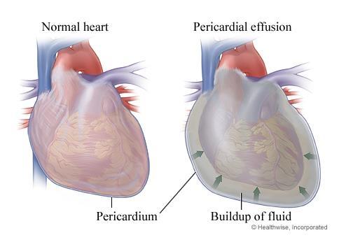
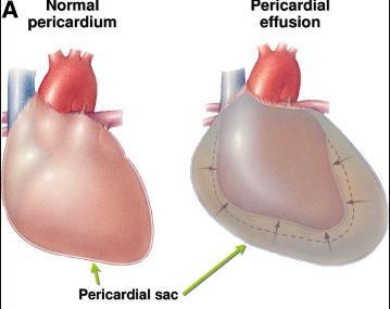
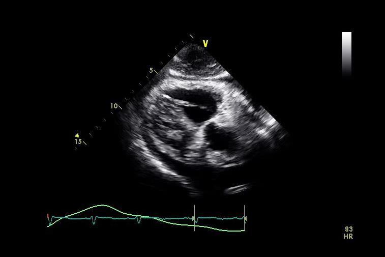
2 Normal pericardial effusion

3 Normal pericardium

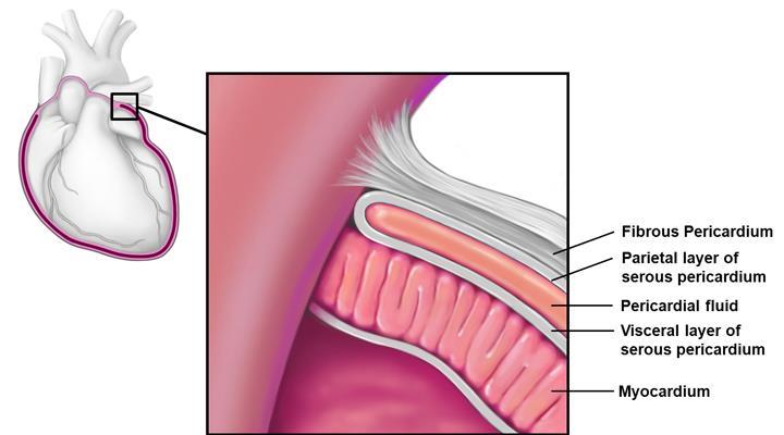
4 Normal pericardium Pericardial Layers: Visceral layer Parietal layer Fibrous pericardium
5
6 Anatomy of pericardium thin avascular sac comprising two layers ; the visceral and parietal pericardium encompass the heart and a portion of the adjoining great vessels Sagristà-Sauleda J, et al. N Engl J Med 2004; 350: Faisal FS, et al. Heart Fail Rev 2013; 18:
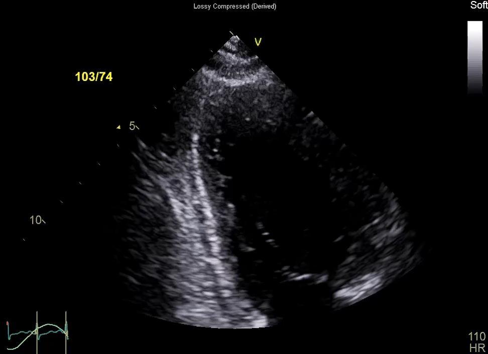
7 Normal pericardium
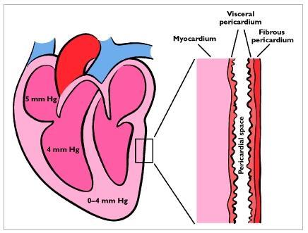
8 Normal pericardium Normally <2 mm thick normal amount of pericardial fluid is < 50 ml transudative with a low protein content pressure differential between the pericardium and the cardiac chambers (transmural pressure) is about 3 mm Hg
9 The role of pericardium 1. provides structural support 2. optimizes cardiac pressure volume relationships 3. act as barrier to infection, metastasis.. Spodick DH Marcel Dekker, New York Faisal FS, et al. Heart Fail Rev 2013; 18:
10 Pericardial Agenesis
11 Pericardial Disease The common underlying physiologic abnormality impaired diastolic filling of the heart Significant constrictive pericarditis right-sided heart failure Pericardial tamponade systemic hypotension Combinations of the disease effusive-constrictive pericarditis
12 Pericardial Effusion in Echo? 1. Amount 2. Location 3. Character 4. Hemodynamic significance
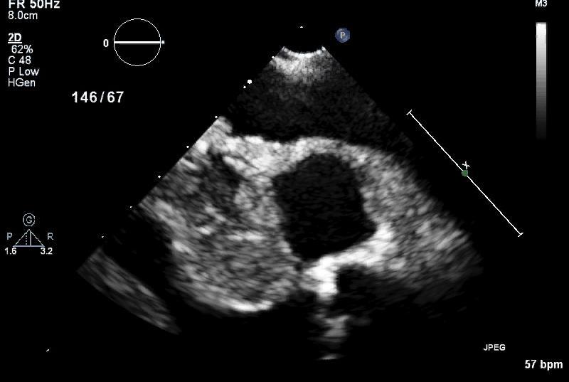
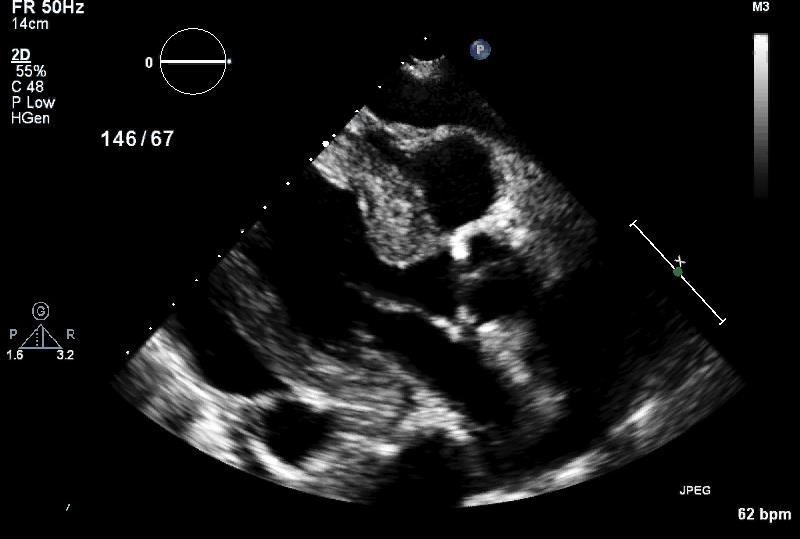
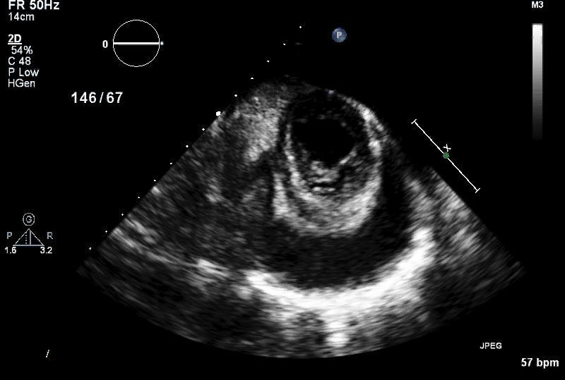
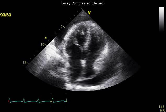
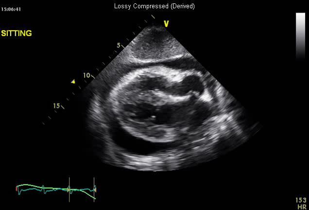
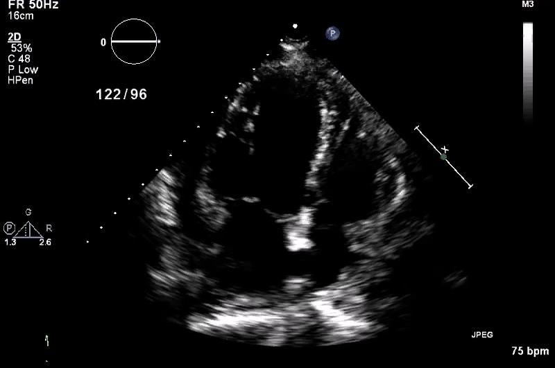
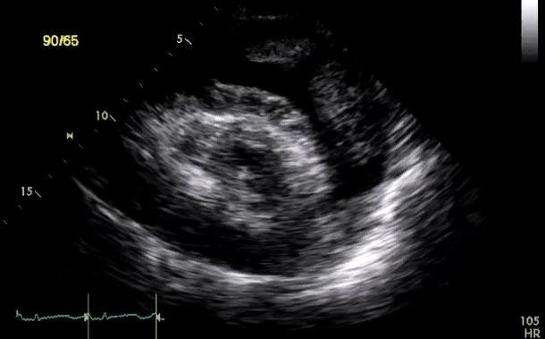
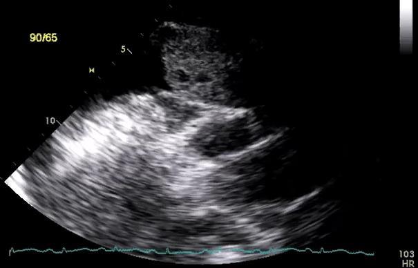
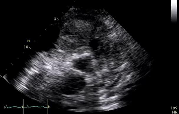
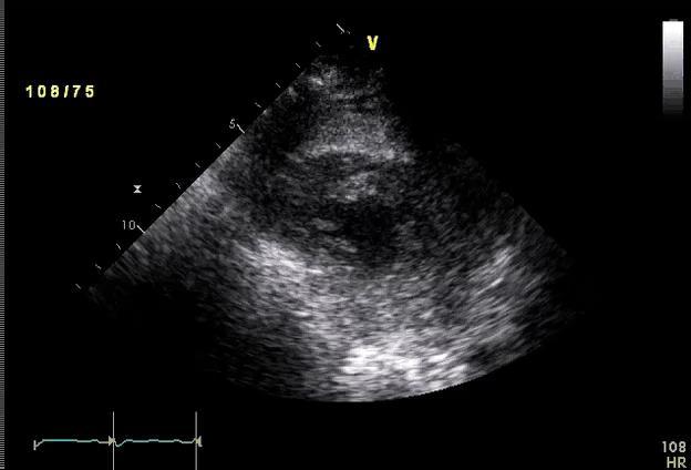
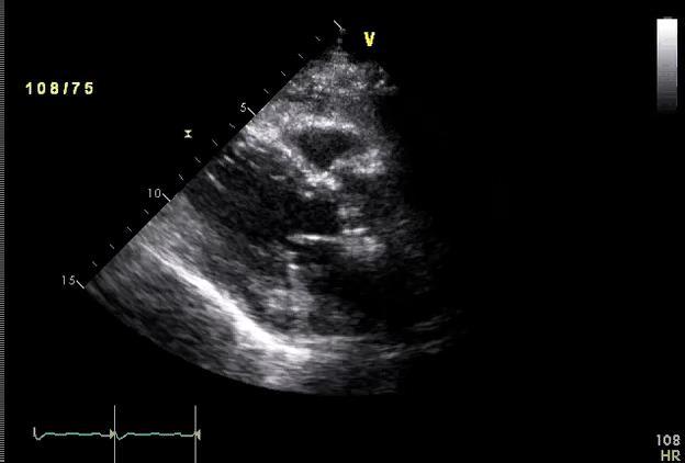
13 Effusion
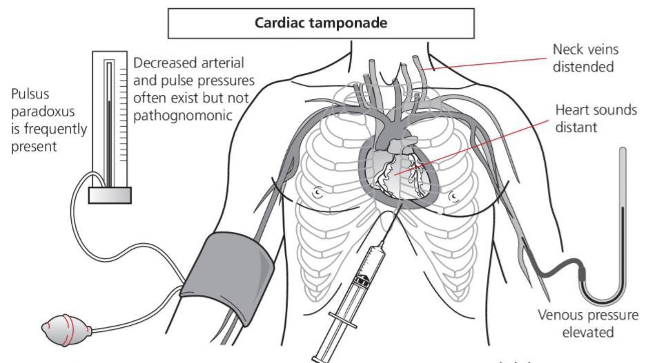
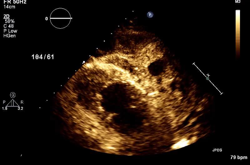
14 Cardiac Tamponade
15
16
17

18
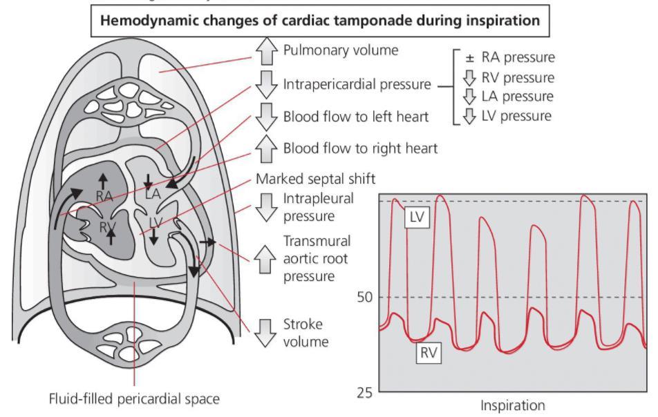
19 Variation of cardiac pressure
20
21
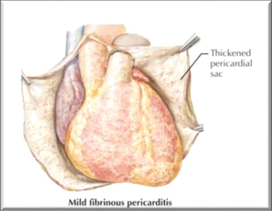
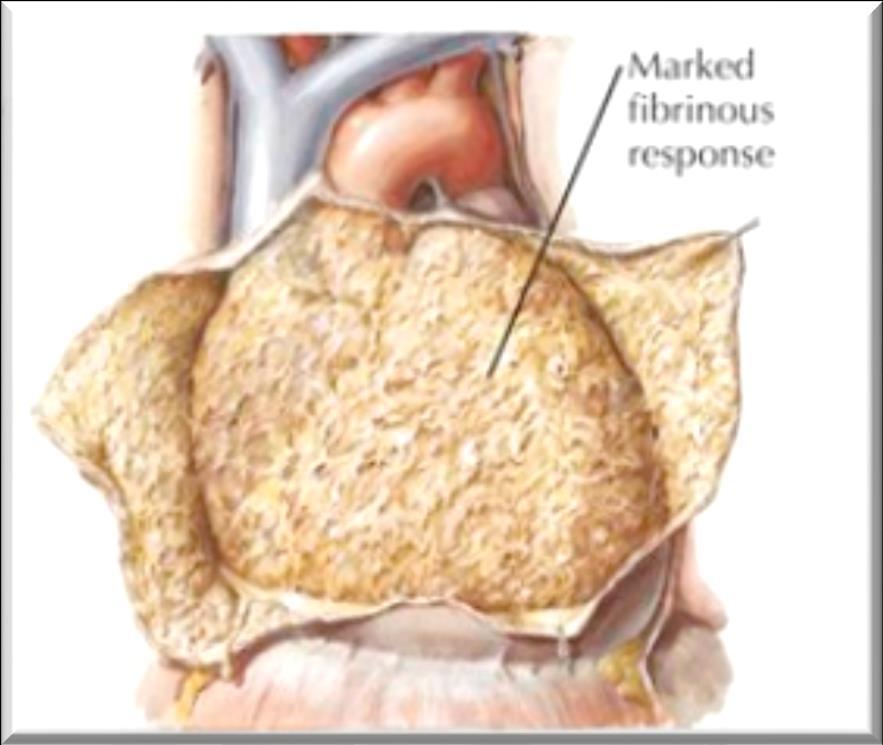
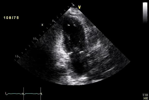
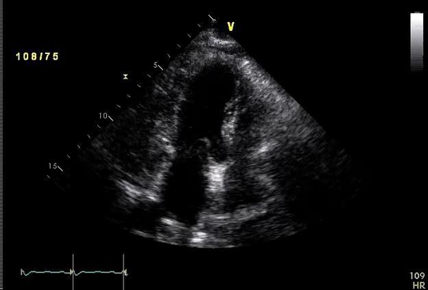
22 Pericarditis

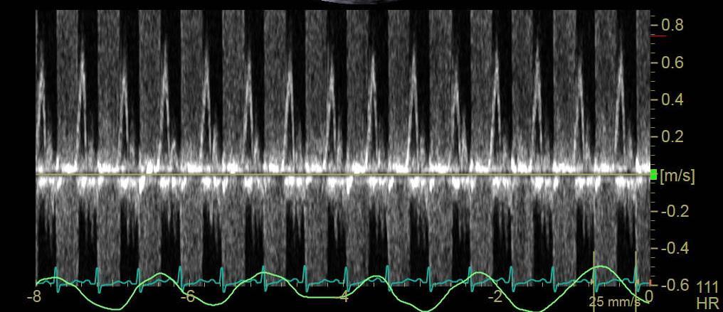
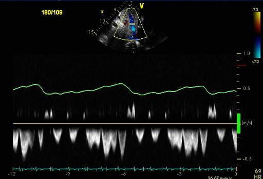
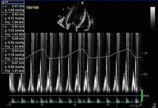
23 Pericarditis
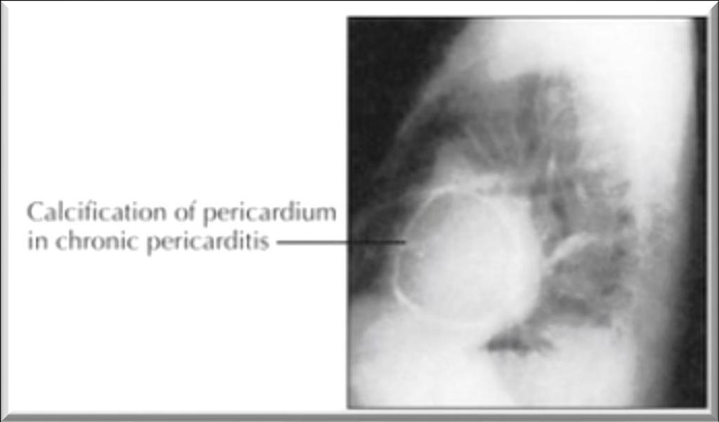
24 Constrictive pericarditis Constrictive pericarditis was recognized at autopsy in the 19th century and described as a chronic fibrous callous thickening of the wall of the pericardial sac that is so contracted that the normal diastolic filling of the heart is prevented
25 The difference between the subacute & chronic forms of constrictive pericarditis whether only the visceral pericardium is fused to the epicardium of the heart (subacute) both the visceral and the parietal pericardial layers are fused together (chronic) In both instances, the diastolic pressures in the atria are elevated due to the restriction of ventricular diastolic inflow.
26
27 Constriction? Tamponade? and the Effect?

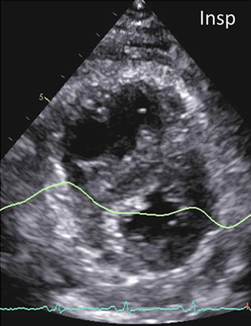
28 Flow pattern in constrictive pericarditis Inspiration Expiration
29

30 Significant respiratory variations in the mitral E velocities
31 Perimyocarditis Myopericarditis Myocarditis Pericarditis ACS-like Myocarditis
32 Etiology Acute Pericarditis Infectious Viral : Coxsackie, Echo, EBV, Influenza, HIV Bacterial: TB, staph, hemophillus, pneumococcal, salmonella Fungal/other: histo/blasto/coccidio, rickettsia Rheumatologic SLE, Sarcoid, RA, Dermatomyositis, Ankylosing Spondylitis, Scleroderma, PAN Neoplastic Primary: angiosarcoma, mesothelioma Metastatic: breast, lung, lymphoma, melanoma, leukemia Immunologic Celiac sprue, Inflammatory Bowel Disease Drug Hydralizine, Procainamide Other MI, Dressler s, Post Pericardiotomy, Chest Trauma, Aortic dissection Uremic, Post Radiation IDIOPATHIC
33 Etiology Acute Pericarditis Common causes T = Trauma, TUMOR U = Uremia M = Myocardial infarction (acute, post) Medications (hydralazine, procain) O = Other infections (bacterial, fungal, TB) R = Rheumatoid, autoimmune disorder Radiation


34 Tuberculous pericarditis F/55 C.C ; chest discomfort, palpitation, fever for 2 months EKG ; a fib
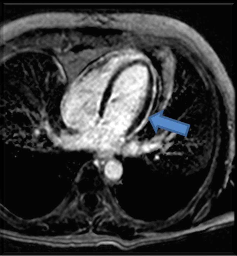
35
36
37 2 months later : After window formation and NSAID

38 Initial 2 months 1yr
39 Effusive Constrictive Pericarditis
40 C.C ; DOE, fever for 2 weeks M/54

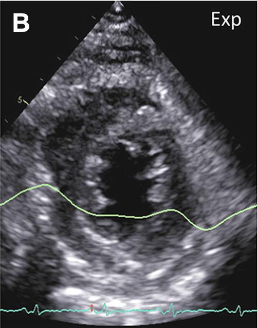
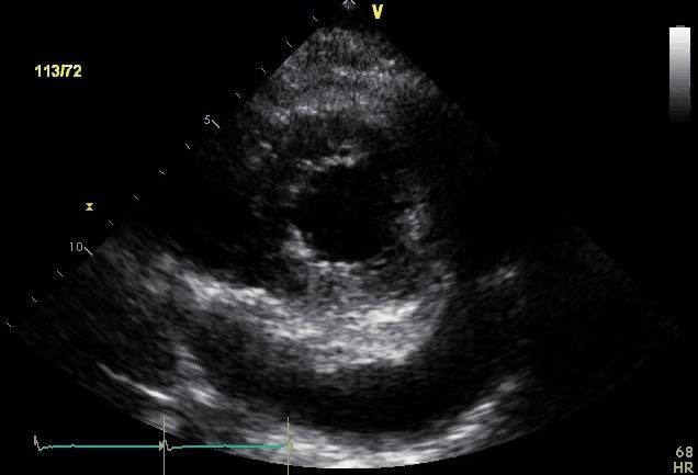
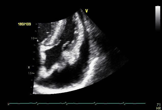
41 After window formation

42 Pericardial inflammation in tuberculous effusive-constrictive pericarditis on MR imaging Russell JB, et al. Cardiovasc J Afr 2008; 19(4):200 1
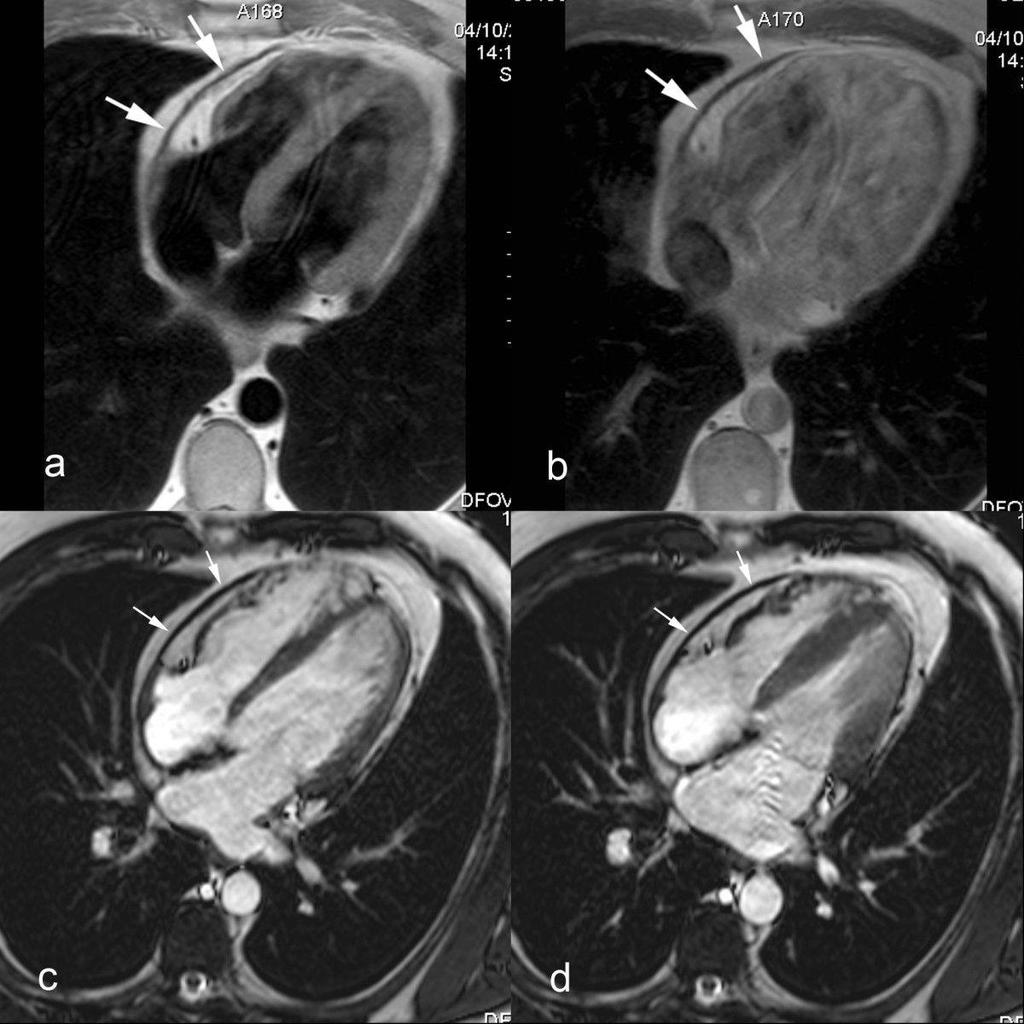
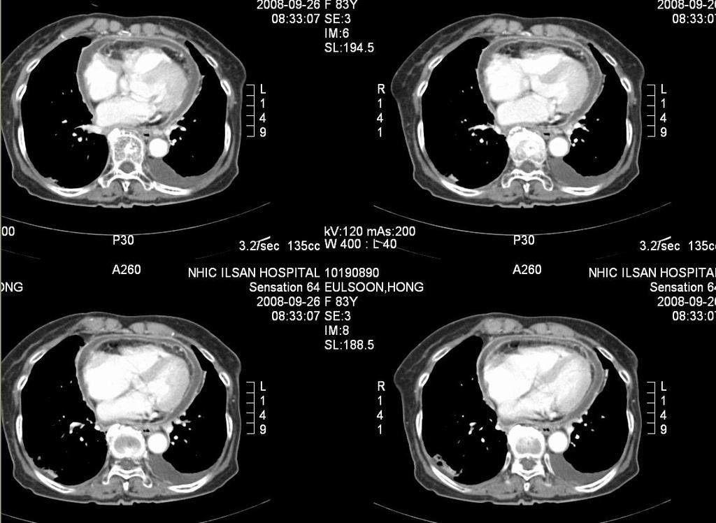
43 The reported prevalence of ECP among patients with a pericardial effusion has ranged widely, from 1% to 2% to more than 50% Sagrista-Sauleda J, et al. N Engl J Med 2004; 350: Nugue O, et al. Circulation 1996;94: Ntsekhe M, et al. Cardiovasc J Afr 2012;23: van der Bijl P, et al. J Cardiovasc Ultrasound 2016;24:
44 Effusive Constrictive Pericarditis Clinical hemodynamic syndrome in which constriction of the heart by the visceral pericardium occurs in the presence of tense effusion in a free pericardial space. Sagristà-Sauleda J, et al. N Engl J Med 2004; 350:
45 The hallmark of effusive constrictive pericarditis persistence of elevated RA pressure after intrapericardial pressure has been reduced to normal levels by removal of pericardial fluid Sagristà-Sauleda J, et al. N Engl J Med 2004; 350: Faisal FS, et al. Heart Fail Rev 2013; 18:
46 Definitions of effusive constrictive pericarditis On the basis of hemodynamic findings during combined pericardiocentesis and cardiac catheterization. The diagnostic criterion was tamponade that evolved into constriction (with failure of the RA pressure to fall by 50 % or more or to a level below 10 mm Hg) after intrapericardial pressure was lowered to near 0 mm Hg by the removal of pericardial fluid. Sagristà-Sauleda J, et al. N Engl J Med 2004; 350: Faisal FS, et al. Heart Fail Rev 2013; 18: Ntsekhe M, et al. J Am Coll Cardiol 2009; 53(10):A169
47 The component of effusive constrictive pericarditis Pericardial inflammation Constriction Pericardial effusion under pressure Sagristà-Sauleda J, et al. N Engl J Med 2004; 350: Faisal FS, et al. Heart Fail Rev 2013; 18:


48 Echo in EPC persistence of IVC dilatation Significant respiratory variations in the mitral E velocities Expiratory diastolic flow reversals in the hepatic veins

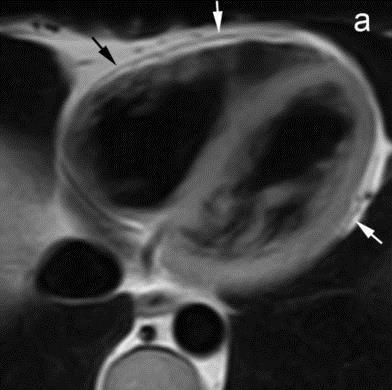
49 Marked respirophasic shift in ECP Expiration Inspiration


50 Pericardial effusion in CT and MR moderate pericardial effusion Pericardial thickening in late Ga enhancement


51 Pericardial effusion in CT and MR CT T1-weighted MR balanced SSFP CMR Large oval-shaped pericardial effusion
52 Pericarditis in CT

53 Pericardial effusion in MR Systolic collapse of the right atrial wall during systole

54 Pericarditis in MR : delayed enhancement imaging initial F/U

55 Acute viral pericarditis in MR Diffuse hyperintense appearance of the pericardium on T2-weighted image

56 Constrictive pericarditis in MR Short-axis T1- weighted spin-echo CMR LGE CMR Diffusely thickened pericardium, with strong enhancement of the pericardial layers after Ga enhancement


57 Effusive pericardial constriction Little WC, et al. Circulation 2006;113:
58 Others


59 Dressler s syndrome F/85 Cc. : dyspnea PHx : STEMI (1w ago), CAD 1vd, s/p PCI c stent at mlad, A-fib, Asthma, Old CVA
60


61 Post window formation


62 Uremic pericarditis M/58 Cc. : dyspnea PHx : DM, HTN, hypothyroidism, ESRD on HD
63
64 Metastasis F/86

65 Post window formation
66 Metastasis F/85

67 Respiratory variation of Mitral E velocity

68 Chest CT and PET CT
69 Take Home Message 1. The understanding of pericardium 2. The significance of hemodynamics
70 Thank you!
71

72 Normal cardiac blood flow during inspiration and expiration
73
74

75 Post op adhesion
Pericardial Diseases. Smonporn Boonyaratavej, MD. Division of Cardiology, Department of Medicine Chulalongkorn University
 Pericardial Diseases Smonporn Boonyaratavej, MD Division of Cardiology, Department of Medicine Chulalongkorn University Cardiac Center, King Chulalongkorn Memorial Hospital 21 AUGUST 2016 Pericardial
Pericardial diseases
 Pericardial diseases Anatomy of the pericardium Consists of parietal and visceral membranes. The space between them(pericardial space is normally filled by a lymph like fluid. The fluid s normal quantity
THE PERICARDIUM: LOOKING OUTSIDE THE HEART
 THE PERICARDIUM: LOOKING OUTSIDE THE HEART DISCLOSURE Relevant relationships with commercial entities none Potential for conflicts of interest within this presentation none Steps taken to review and mitigate
Pericardial Diseases/Tamponade Illustrative Cases
 Pericardial Diseases/Tamponade Illustrative Cases Jae K. Oh, MD Echo Hawaii 2017 2012 MFMER 3200268v3(2010)-1 Case #1 47 year old man Chest pain Not exertional Normal Examination 2016 MFMER slide-2 1 47
Outline. Echocardiographic Assessment of Pericardial Effusion/Tamponade: The Essentials
 Echocardiographic Assessment of Pericardial Effusion/Tamponade: The Essentials John R Schairer DO FACC Henry Ford Heart and Vascular Institute No Disclosures Outline Normal Anatomy and Physiology Pathophysiology
Constrictive Pericarditis Pitfalls in MR Diagnosis Cylen Javidan-Nejad Associate Professor Mallinckrodt Institute of Radiology Washington University
 Constrictive Pericarditis Pitfalls in MR Diagnosis Cylen Javidan-Nejad Associate Professor Mallinckrodt Institute of Radiology Washington University in St. Louis Goal o To review the imaging criteria of
An Uncommon Cardiac Etiology of Liver Cirrhosis, Recurrent Ascites, Atrial Fibrillation and Congestive Heart Failure
 Cronicon OPEN ACCESS EC CARDIOLOGY Case Report An Uncommon Cardiac Etiology of Liver Cirrhosis, Recurrent Ascites, Atrial Fibrillation and Congestive Heart Failure Montaser Y Ismail 1 *, Mohammed I Nassar
Integrative Clinical Hemodyamics
 Integrative Clinical Hemodyamics James A. Goldstein, MD Director, Research & Education Division of Cardiology William Beaumont Hospital Disclosure Information Integrative Clinical Hemodyamics James A.
Pericardial Diseases 2015 Update
 Pericardial Diseases 2015 Update BRUCE W. USHER, MD PROFESSOR OF MEDICINE CARDIOLOGY DIVISION MEDICAL UNIVERSITY SOUTH CAROLINA CHARLESTON, SOUTH CAROLINA GUIDELINES: PERICARDIAL DISEASES Ø European Society
Constrictive/Restrictive Cardiomyopathies: Diagnosis and Management Update; Radiation Induced Heart Disease. Alexander (Sandy) Dick, MD
 Dick, MD") Constrictive/Restrictive Cardiomyopathies: Diagnosis and Management Update; Radiation Induced Heart Disease Alexander (Sandy) Dick, MD Outline Pericardial Constriction Diagnosis: Imaging, Hemodynamics
Constrictive/Restrictive Cardiomyopathies: Diagnosis and Management Update; Radiation Induced Heart Disease Alexander (Sandy) Dick, MD Outline Pericardial Constriction Diagnosis: Imaging, Hemodynamics
Adel Hasanin Ahmed 1
 Adel Hasanin Ahmed 1 PERICARDIAL DISEASE The pericardial effusion ends anteriorly to the descending aorta and is best visualised in the PLAX. PSAX is actually very useful sometimes for looking at posterior
PERICARDIAL DIAESE. Kaijun Cui Associated professor Sichuan University
 PERICARDIAL DIAESE Kaijun Cui Associated professor Sichuan University CLASSIFICATION acute pericarditis pericardial effusion cardiac tamponade constrictive pericarditis congenitally absent pericardium
Pericarditis. Marquette University. James F. Ginter Aurora Cardiovascular Services
 Marquette University e-publications@marquette Physician Assistant Studies Faculty Research and Publications Physician Assistant Studies, Department 4-23-2012 Pericarditis James F. Ginter Aurora Cardiovascular
10/1/2016. Constrictive Pericarditis Unique Hemodynamics. What s New in Pericardial Disease? Case-based Discussion
 Mayo Clinic Department of Cardiovascular Diseases Mayo Clinic Echocardiography Review Course for Boards and Recertification What s New in Pericardial Disease? Case-based Discussion Jae K. Oh, MD Samsung
Cardiovascular manifestations of HIV
 Cardiovascular manifestations of HIV Prabhakar Rajiah, MBBS, MD, FRCR Associate Professor of Radiology Associate Director, Cardiac CT and MRI University of Texas Southwestern Medical Center, Dallas, USA
Pericardial Disease: Case Examples. Echo Fiesta 2017
 Pericardial Disease: Case Examples Echo Fiesta 2017 2014 2014 MFMER MFMER 3346252-1 slide-1 Objectives Have a systematic approach to evaluation of constriction 2014 MFMER 3346252-2 CASE 1 2013 MFMER 3248567-3
Περικαρδίτιδα στην καρδιακή ανεπάρκεια
 ΣΕΜΙΝΑΡΙΑ ΟΜΑΔΩΝ ΕΡΓΑΣΙΑΣ, ΙΩΑΝΝΙΝΑ 2015 Περικαρδίτιδα στην καρδιακή ανεπάρκεια Γεώργιος Λάζαρος Α Πανεπιστηµιακή Καρδιολογική Κλινική Ιπποκράτειο Γ.Ν. Αθηνών I declare that I have no conflict of interest
Constrictive Pericarditis
 Constrictive Pericarditis Never Confused with Anything Else Jae K. Oh, MD 2018 MFMER 3712003-1 ARS #1 CP Which of following patients has constrictive pericarditis? 1 2 3 Medial e 13 cm/s Medial e 3 cm/s
Carditis: Inside and Out. Derek Brown, MD SAUSHEC Military EMS & Disaster Medicine Fellow
 Carditis: Inside and Out Derek Brown, MD SAUSHEC Military EMS & Disaster Medicine Fellow Pericarditis Bruce Blaus https://commons.wikimedia.org/wiki/file:pericarditis.png Pericarditis 0.1% of Hospitalized
Transient Constrictive Pericarditis: Causes and Natural History
 Journal of the American College of Cardiology Vol. 43, No. 2, 2004 2004 by the American College of Cardiology Foundation ISSN 0735-1097/04/$30.00 Published by Elsevier Inc. doi:10.1016/j.jacc.2003.08.032
Normal Pericardial Physiology
 Normal Pericardial Physiology Normal pericardium contains 20-30 ml of lymphoid fluid lubricating function that facilitates normal myocardial rotation and translation during each cardiac cycle in that the
ΚΑΡΔΙΟΛΟΓΟΣ EUROPEAN ACCREDITATION IN TRANSTHORACIC AND TRANSESOPHAGEAL ECHOCARDIOGRAPHY
 1 ΚΑΡΔΙΟΛΟΓΟΣ EUROPEAN ACCREDITATION IN TRANSTHORACIC AND TRANSESOPHAGEAL ECHOCARDIOGRAPHY 2 Constrictive pericarditis (CP) is characterized by impaired ventricular filling due to a stiffened or noncompliant
Νόσοι του περικαρδίου. Γεώργιος Λάζαρος Α Πανεπιστημιακή Καρδιολογική Κλινική Ιπποκράτειο Γ.Ν. Αθηνών
 Νόσοι του περικαρδίου Γεώργιος Λάζαρος Α Πανεπιστημιακή Καρδιολογική Κλινική Ιπποκράτειο Γ.Ν. Αθηνών I do not have any conflict of interests relevant to this presentation to declare. Pericardial syndromes
A Case of Impending Cardiac Tamponade Caused by Effusive Constrictive Pericarditis
 Archives of Clinical and Medical Case Reports doi: 10.26502/acmcr.96550038 Volume 2, Issue 5 Case Report A Case of Impending Cardiac Tamponade Caused by Effusive Constrictive Pericarditis Catalina Sanchez-Alvarez
Choose the grading of diastolic function in 82 yo woman
 Question #1 Choose the grading of diastolic function in 82 yo woman E= 80 cm/s A= 70 cm/s LAVI < 34 ml/m 2 1= Grade 1 2= Grade 2 3= Grade 3 4= Normal 5= Indeterminate 2018 MFMER 3712003-1 Choose the grading
Pericardial effusion, Cardiac Tamponade, and echo guided pericardiocentesis
 KSC 2017 Echo5- Myocardial and Pericardial disease Pericardial effusion, Cardiac Tamponade, and echo guided pericardiocentesis Ji-Hyun Jung Division of Cardiology Sejong Hospital KSC 2017 The 61 th Annual
Objectives. Highlight typical feature of TB pericarditis. How to make a diagnosis. How to treat TB pericarditis
 Dr. Conteh Objectives Highlight typical feature of TB pericarditis How to make a diagnosis How to treat TB pericarditis New evidence for adjunctive corticosteroid Introduction TB pericarditis occurs in
The Cardiovascular System Part I: Heart Outline of class lecture After studying part I of this chapter you should be able to:
 The Cardiovascular System Part I: Heart Outline of class lecture After studying part I of this chapter you should be able to: 1. Describe the functions of the heart 2. Describe the location of the heart,
Case 5 15-year-old male
 Case 5 15-year-old male Present illness: Six months ago, abnormality of ECG was incidentally detected by annual health check. His blood level of γ-gtp, HbA1c and norepinephrine were elevated; however,
Imaging in Heart Failure: A Multimodality Approach. Thomas Ryan, MD
 Imaging in Heart Failure: A Multimodality Approach Thomas Ryan, MD Heart Failure HFrEF HFpEF EF50% Lifetime risk 20% Prevalence 6M Americans Societal costs - $30B 50% 5-year survival 1 Systolic
We are now going to review the diagnosis and management of pericardial collections and tamponade
 We are now going to review the diagnosis and management of pericardial collections and tamponade FEEL COURSE PAGE 1 Paying particular attention to the difference between a collection and cardiac tamponade
TAMPONADE CARDIAQUE. Dr Cédrick Zaouter TUSAR 15 décembre 2015
 TAMPONADE CARDIAQUE Dr Cédrick Zaouter TUSAR 15 décembre 2015 OUTLINE History Incidence Definition Pathophysiology Aetiologies Investigations - Echocardiography Treatment of cardiac tamponade Pericardial
Cardiac MRI: Clinical Application to Disease
 Cardiac MRI: Clinical Application to Disease Jessi Smith, MD Cardiothoracic imaging, Indiana University Slides courtesy of Stacy Rissing, MD Outline Imaging planes Disease findings Pulse sequences used
For more information about how to cite these materials visit
 Project: Ghana Emergency Medicine Collaborative Document Title: Case Presentation- Pericarditis Author(s): Kwaku Nyame License: Unless otherwise noted, this material is made available under the terms of
The Cardiovascular System
 The Cardiovascular System The Manila Times College of Subic Prepared by: Stevens B. Badar, RN, MANc THE HEART Anatomy of the Heart Location and Size approx. the size of a person s fist, hollow and cone-shaped,
efferent fibers from t.. Heart Surface anatomy and heart sounds -Dry lecture -Gray s 169,
 A patient is diagnosed with ischemia (i.e., lack of blood flow) in a left lobar pulmonary vein. The attending physician determines that the ischemia is due to a vasospastic episode. Constriction of this
Echocardiography as a diagnostic and management tool in medical emergencies
 Echocardiography as a diagnostic and management tool in medical emergencies Frank van der Heusen MD Department of Anesthesia and perioperative Care UCSF Medical Center Objective of this presentation Indications
Essentials of Pericardial Diseases
 Essentials of Pericardial Diseases 1 Nikolaos Skubas MD, 2 Manuel Fontes MD The pericardial diseases result in cardiovascular perturbations ranging from asymptomatic electrocardiographic findings (in pericarditis
Cardiovascular Nursing Practice: A Comprehensive Resource Manual and Study Guide for Clinical Nurses 2 nd Edition
 Cardiovascular Nursing Practice: A Comprehensive Resource Manual and Study Guide for Clinical Nurses 2 nd Edition Table of Contents Volume 1 Chapter 1: Cardiovascular Anatomy and Physiology Basic Cardiac
Effusive Constrictive Pericarditis
 original article Effusive Constrictive Pericarditis Jaume Sagristà-Sauleda, M.D., Juan Angel, M.D., Antonio Sánchez, M.D., Gaietà Permanyer-Miralda, M.D., and Jordi Soler-Soler, M.D. abstract background
4/11/2017. Cardiomyopathy. John Steuter, MD Bryan Heart. Disclosures. No Conflicts. Cardiomyopathy. WHO Classification
 Cardiomyopathy John Steuter, MD Bryan Heart Disclosures No Conflicts Cardiomyopathy WHO Classification Anatomy & physiology of the LV 1. Dilated Enlarged Systolic dysfunction 2. Hypertrophic Thickened
UPDATE ON CONSTRICTIVE PERICARDITIS ECHOCARDIOGRAPHY AND CARDIAC CATHETERISATION
 Arsen D. Ristić, MD, PhD, FESC (no conflicts of interest to disclose regarding this presentation) UPDATE ON CONSTRICTIVE PERICARDITIS ECHOCARDIOGRAPHY AND CARDIAC CATHETERISATION Department of Cardiology,
Pericardial effusion with emphasis on the electrocardiographic aspects By Andrés Ricardo Pérez-Riera MDPhD
 Pericardial effusion with emphasis on the electrocardiographic aspects By Andrés Ricardo Pérez-Riera MDPhD ECG In pericarditis Pericardial effusion Pericardium The pericardium is a double sheet made up
2/4/2011. Nathan Kerner, M.D.
 Nathan Kerner, M.D. Definition Elevated pressures - cut off usually >40 mmhg pulmonary artery systolic pressure (PASP) Usually associated with elevated pulmonary vascular resistance (PVR) measured in dynessec/cm
Pericarditis and Myocarditis. Sheba Medical Center Cardiology Department Carlyn Wallis
 Pericarditis and Myocarditis Sheba Medical Center Cardiology Department Carlyn Wallis Outline Pericarditis Normal function of the pericardium Pathophysiology + Etiology Clinical Presentation Differential
Cardiac Radiography. Jared D. Christensen, M.D.
 Cardiac Radiography Jared D. Christensen, M.D. Cardiac radiography Jared D. Christensen, M.D. Overview Basic Concepts Technique Normal anatomy Cases Technique 3 Standard Views Posterior-Anterior (PA) Anterior-Posterior
Looking Outside the Box: Incidental Extracardiac Finding in Echo
 Looking Outside the Box: Incidental Extracardiac Finding in Echo Dr. Aijaz Shah Head of Division, Adult Echocardiography Laboratory Prince Sultan Cardiac Centre Riyadh Case 1 17 year old boy presented
Acute Viral Myopericarditis Presenting as a Transient Effusive-Constrictive Pericarditis Caused by Coinfection with Coxsackieviruses A4 and B3
 case report korean j intern med 2012;27:216-220 ORIGINL RTICLE pissn 1226-3303 eissn 2005-6648 cute Viral Myopericarditis Presenting as a Transient Effusive-Constrictive Pericarditis Caused y Coinfection
Department of Cardiac, Thoracic and Vascular Sciences University of Padua Cardiac Tamponade. Echocardiography in Diagnosis and Management
 Department of Cardiac, Thoracic and Vascular Sciences University of Padua Cardiac Tamponade. Echocardiography in Diagnosis and Management Luigi P. Badano, MD, FESC, FACC Declaration of interest **Dr. Badano
Constrictive pericarditis: Morphological, functional and haemodynamic evaluation
 Constrictive pericarditis: Morphological, functional and haemodynamic evaluation Poster No.: C-0743 Congress: ECR 2010 Type: Educational Exhibit Topic: Cardiac Authors: B. Graca, P. Donato, M. Ferreira,
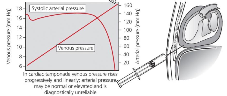
Low-pressure cardiac tamponade has been described as a
 Pericardial Disease Low-Pressure Cardiac Tamponade Clinical and Hemodynamic Profile Jaume Sagristà-Sauleda, MD; Juan Angel, MD; Antonia Sambola, MD; Joan Alguersuari, MD; Gaietà Permanyer-Miralda, MD;
10/16/2014. CCRN Review - Cardiovascular. CCRN Review - Cardiovascular. CCRN Review - Cardiovascular
 Hypertrophic (IHSS) Diagnosis Chest x ray cardiomegaly Electrocardiography LV hypertrophy, ST segment T was changes, Q waves in inferior & precordial leads Atrial & ventricular dysrhythmias Hypertrophic
Cardiac magnetic resonance imaging in rheumatoid arthritis: promising or misleading? Sophie Mavrogeni MD FESC
 Cardiac magnetic resonance imaging in rheumatoid arthritis: promising or misleading? Sophie Mavrogeni MD FESC Onassis Cardiac Surgery Center Athens Greece Nothing to disclose Financial disclosure Cardiac
THE HEART. A. The Pericardium - a double sac of serous membrane surrounding the heart
 THE HEART I. Size and Location: A. Fist-size weighing less than a pound (250 to 350 grams). B. Located in the mediastinum between the 2 nd rib and the 5 th intercostal space. 1. Tipped to the left, resting
Unit 6: Circulatory System. 6.2 Heart
 Unit 6: Circulatory System 6.2 Heart Functions of Circulatory System 1. The heart is the pump necessary to circulate blood to all parts of the body 2. Arteries, veins and capillaries are the structures
Constriction vs Restriction Case-based Discussion
 Mayo Clinic Department of Cardiovascular Diseases Mayo Clinic Echocardiography Review Course for Boards and Recertification Constriction vs Restriction Case-based Discussion Jae K. Oh, MD Samsung Professor
Inflammatory Cardiovascular Diseases
 Inflammatory Cardiovascular Diseases Presented By: Cynthia Webner, BSN, RN, CCRN-CMC www.cardionursing.com CNEA 2009 1 Pericarditis Inflammatory process involving the visceral or parietal pericardium Multiple
Cardiac MRI: Clinical Application to Disease
 Cardiac MRI: Clinical Application to Disease Stacy Rissing, MD! Cardiothoracic imaging, Indiana University! Outline Imaging planes Disease findings Pulse sequences used for each indication Pathophysiology
Echocardiography Conference
 Echocardiography Conference David Stultz, MD Cardiology Fellow, PGY-6 September 20, 2005 Atrial Septal Aneurysm Bulging of Fossa Ovalis Associated commonly with Atrial septal defect or small perforations
Current Indications for Cardiac MRI: What You See is What You Get?
 Current Indications for Cardiac MRI: What You See is What You Get? Javier Ganame, MD, PhD, FASE No disclosures Cardiology Update, Niagara, Sept 24th, 2016 The Ideal Diagnostic Technique Easy to apply Accurate
Palpable Pulsus Paradoxus in the Setting of Malignant Pericardial Effusion and Tamponade Akshay Pendyal, MD
 Palpable Pulsus Paradoxus in the Setting of Malignant Pericardial Effusion and Tamponade Akshay Pendyal, MD University of Colorado Department of Internal Medicine None Conflicts of Interest Objectives
BEDSIDE ASSESSMENT OF PATIENTS WITH STEMI
 BEDSIDE ASSESSMENT OF PATIENTS WITH STEMI Prof. Maria Dorobantu, PhD, FESC, FACC Emergency Hospital of Bucharest, Romania Presenter Disclosures There are no conflicts/ grants/ disclosures for this presentation.
Clinical Anatomy Series Cardiac Anatomy
 2012 Scottish Universities Medical Journal, Dundee Published online: Feb 2012 Vol 1 Issue 1: page 76-80 Kennedy J Clinical Anatomy Cardiac Anatomy Clinical Anatomy Series Cardiac Anatomy John Kennedy (5
Ch 19: Cardiovascular System - The Heart -
 Ch 19: Cardiovascular System - The Heart - Give a detailed description of the superficial and internal anatomy of the heart, including the pericardium, the myocardium, and the cardiac muscle. Trace the
RADIATION HEART DISEASE: MANAGEMENT STRATEGIES
 RADIATION HEART DISEASE: MANAGEMENT STRATEGIES AMMAR CHAUDHARY MBChB, ABIM, FRCPC ASSOCIATE CONSULTANT CARDIOLOGIST KING FAISAL SPECIALIST HOSPITAL & RESEARCH CENTER - JEDDAH Scope of the Problem ~ 50
Πνευμονική υπέρταση και περικαρδιακή συλλογή. Τρόποι αντιμετώπισης
 Πνευμονική υπέρταση και περικαρδιακή συλλογή. Τρόποι αντιμετώπισης Γεώργιος Λάζαρος Καρδιολόγος, Διευθυντής ΕΣΥ Α Πανεπιστημιακή Καρδιολογική Κλινική Ιπποκράτειο Γ.Ν. Αθηνών Pericardial syndromes o Acute
CARDIOVASCULAR SYSTEM
 CARDIOVASCULAR SYSTEM Overview Heart and Vessels 2 Major Divisions Pulmonary Circuit Systemic Circuit Closed and Continuous Loop Location Aorta Superior vena cava Right lung Pulmonary trunk Base of heart
Acute pericarditis is the most
 Electrocardiographic Manifestations and Differential Diagnosis of Acute Pericarditis MARK A. MARINELLA, M.D., Wright State University School of Medicine, Dayton, Ohio Acute pericarditis has many potential
Heart Anatomy. 7/5/02 Stephen G Davenport 1
 Heart Anatomy Copyright 1999, Stephen G. Davenport, No part of this publication may be reproduced, stored in a retrieval system, or transmitted, in any form without prior written permission. 7/5/02 Stephen
ΙΟΛΟΓΙΑΣ. Virology of the cardiovascular system
 ΙΟΛΟΓΙΑΣ Virology of the cardiovascular system Of an importance to know the following: This project includes tow lectures of the virology considering the cardiovascular system. We will talk about this
Study of etiological and clinical profile of pericardial effusion in a tertiary care hospital in Kosi region of Bihar, India
 International Journal of Advances in Medicine Jamal Uddin MD et al. Int J Adv Med. 2016 Aug;3(3):514-518 http://www.ijmedicine.com pissn 2349-3925 eissn 2349-3933 Research Article DOI: http://dx.doi.org/10.18203/2349-3933.ijam20161844
Conflict Disclosures. Vermont Cardiac Network. Outline. Series Learning Objectives 4/27/2016. Scott E. Friedman April 28, 2016
 Conflict Disclosures Vermont Cardiac Network The Speaker has reported no significant financial relationship with any companies whose product may be germane to the content of their presentations or who
THE HEART OBJECTIVES: LOCATION OF THE HEART IN THE THORACIC CAVITY CARDIOVASCULAR SYSTEM
 BIOLOGY II CARDIOVASCULAR SYSTEM ACTIVITY #3 NAME DATE HOUR THE HEART OBJECTIVES: Describe the anatomy of the heart and identify and give the functions of all parts. (pp. 356 363) Trace the flow of blood
Rotation: Echocardiography: Transthoracic Echocardiography (TTE)
") Rotation: Echocardiography: Transthoracic Echocardiography (TTE) Rotation Format and Responsibilities: Fellows rotate in the echocardiography laboratory in each clinical year. Rotations during the first
Rotation: Echocardiography: Transthoracic Echocardiography (TTE) Rotation Format and Responsibilities: Fellows rotate in the echocardiography laboratory in each clinical year. Rotations during the first
Pathophysiology: Heart Failure
 Pathophysiology: Heart Failure Mat Maurer, MD Irving Assistant Professor of Medicine Outline Definitions and Classifications Epidemiology Muscle and Chamber Function Pathophysiology Heart Failure: Definitions
HFpEF. April 26, 2018
 HFpEF April 26, 2018 (J Am Coll Cardiol 2017;70:2476 86) HFpEF 50% or more (40-71%) of patients with CHF have preserved LV systolic function. HFpEF is an increasingly frequent hospital discharge. Outcomes
Case Report Chronic Constrictive Pericarditis in Association with End-Stage Renal Disease
 SAGE-Hindawi Access to Research International Nephrology Volume 2011, Article ID 469602, 4 pages doi:10.4061/2011/469602 Case Report Chronic Constrictive Pericarditis in Association with End-Stage Renal
Controversial issues in the management of pericardial diseases
 Controversial issues in the management of pericardial diseases Massimo Imazio, a David Spodick, b Antonio Brucato, c Rita Trinchero, a Yehuda Adler d a Cardiology Department, Maria Vittoria Hospital, Torino,
Miscellaneous Cardiology Topics pregnancy - congenital - myocarditis - pericardial disease. Pregnancy and Cardiovascular Disease MCQ
 Miscellaneous Cardiology Topics pregnancy - congenital - myocarditis - pericardial disease Maan Jokhadar, MD, FACC Emory Center for Advanced Heart Failure Therapy Emory Adult Congenital Heart Center Pregnancy
When An MI Is Not An MI. Morning Report July 30, 2003 Ryan Mattison, MD
 When An MI Is Not An MI Morning Report July 30, 2003 Ryan Mattison, MD Confounding Factors In This Patient WPW Abnormality Dynamic EKG Changes With Symptoms Myocarditis: Definition As the name implies,
Rhythm Disorders 2017 TazKai LLC and NRSNG.com
 Rhythm Disorders 1. Outline the conduction system of the heart. 2. What do the different portions of the EKG represent? 3. Define the following terms: a. Automaticity b. Conductivity c. Excitability d.
CARDIOLOGY SAUDI BOARD PROGRAM SAUDI BOARD FINAL CLINICAL EXAMINATION OF CARDIOLOGY (2018)
") CARDIOLOGY SAUDI BOARD PROGRAM SAUDI BOARD FINAL CLINICAL EXAMINATION OF CARDIOLOGY (2018) I Objectives a. Determine the ability of the candidate to practice as a specialist and provide consultation in
CARDIOLOGY SAUDI BOARD PROGRAM SAUDI BOARD FINAL CLINICAL EXAMINATION OF CARDIOLOGY (2018) I Objectives a. Determine the ability of the candidate to practice as a specialist and provide consultation in
NOT ANOTHER TALK ABOUT A - FIB
 NOT ANOTHER TALK ABOUT A - FIB CASES KUDOS AND A CHALLENGE Case 1 67 y/o female s/p R mastectomy 3 months earlier Second course of adjuvant chemotherapy Muga scan E.F. 35% What do we do next? Case 1 Cardiology
Review of Cardiac Imaging Modalities in the Renal Patient. George Youssef
 Review of Cardiac Imaging Modalities in the Renal Patient George Youssef ECHO Left ventricular hypertrophy (LVH) assessment Diastolic dysfunction Stress ECHO Cardiac CT angiography Echocardiography - positives
correlated). 4. It prevents excessive cardiac dilatation.
. 4. It prevents excessive cardiac dilatation.") Anatomy of Pericardium Functions of Pericardium Pericardial disease 1. Fibrous layer 2. Serous layer: Filled with ~ 50 ml Epicardium: visceral layer that covers the heart Parietal pericardium: reflection
Anatomy of Pericardium Functions of Pericardium Pericardial disease 1. Fibrous layer 2. Serous layer: Filled with ~ 50 ml Epicardium: visceral layer that covers the heart Parietal pericardium: reflection
The Cardiovascular System
 The Cardiovascular System https://www.youtube.com/watch?v=ohmmtqkgs50 Human Anatomy & Physiology P. Wilson 1 Introduction The functions of the cardiovascular system are: to bring oxygen & nutrients to
Advanced Imaging MRI and CTA
 Advanced Imaging MRI and CTA Who and why may benefit. Matthew W. Martinez, M.D. FACC Lehigh Valley Health Network Director, Cardiovascular Imaging Learning Objectives Review basics of CMR and CTA Review
STRUCTURES OF THE CARDIOVASCULAR SYSTEM
 STRUCTURES OF THE CARDIOVASCULAR SYSTEM CARDIOVASCULAR SYSTEM Also called the circulatory system Consists of the heart, arteries, veins, and capillaries Main function is to pump/circulate oxygenated blood
Pericardial Effusion
 Pericardial Effusion How does the heart work? The heart is the organ responsible for pumping blood to and from all tissues of the body. The heart is divided into right and left sides. The job of the right
Ve V rmont rmon Card Car iac d Netw Ne ork tw Scott E. Friedman April 28, 2016
 Vermont Cardiac Network Scott E. Friedman April 28, 2016 Conflict Disclosures Th S k h d i ifi fi i l l i hi ih The Speaker has reported no significant financial relationship with any companies whose product
Index. K Knobology, TTE artifact, image resolution, ultrasound, 14
 A Acute aortic regurgitation (AR), 124 128 Acute aortic syndrome (AAS) classic aortic dissection diagnosis, 251 263 evolutive patterns, 253 255 pathology, 250 251 classifications, 247 248 incomplete aortic
Cardiothoracic Manifestations of Connective Tissue Disease
 Cardiothoracic Manifestations of Connective Tissue Disease Carole Dennie MD FRCPC Professor of Radiology and Medicine Head, Thoracic and Cardiac Imaging Sections The Ottawa Hospital Co-director Cardiac
Right-Sided Congestive Heart Failure Basics
 Right-Sided Congestive Heart Failure Basics OVERVIEW Failure of the right side of the heart to pump blood at a sufficient rate to meet the needs of the body or to prevent blood from pooling within the
Outline. Pathophysiology: Heart Failure. Heart Failure. Heart Failure: Definitions. Etiologies. Etiologies
 Outline Pathophysiology: Mat Maurer, MD Irving Assistant Professor of Medicine Definitions and Classifications Epidemiology Muscle and Chamber Function Pathophysiology : Definitions An inability of the
Decompensated cardiac tamponade is a medical emergency
 Heart Failure Hemodynamic Effects of Volume Expansion in Patients With Cardiac Tamponade Jaume Sagristà-Sauleda, MD; Juan Angel, MD; Antonia Sambola, MD; G. Permanyer-Miralda, MD Background Volume expansion
EAE Teaching Course. Magnetic Resonance Imaging. Competitive or Complementary? Sofia, Bulgaria, 5-7 April F.E. Rademakers
 EAE Teaching Course Magnetic Resonance Imaging Competitive or Complementary? Sofia, Bulgaria, 5-7 April 2012 F.E. Rademakers Complementary? Of Course N Engl J Med 2012;366:54-63 Clinical relevance Treatment
Pericarditis in a Swiss regional hospital
 ORIGINAL ARTICLE 300 A retrospective analysis of real-world data on pericarditis patients Pericarditis in a Swiss regional hospital Eliane Schwegler a, Marta Bachmann a,b,c, Nazmi Krasniqi a,c, Urs Eriksson
Anatomy of the Heart. Figure 20 2c
 Anatomy of the Heart Figure 20 2c Pericardium & Myocardium Remember, the heart sits in it s own cavity, known as the mediastinum. The heart is surrounded by the Pericardium, a double lining of the pericardial
Tuberculous Pericarditis: A multimodality imaging approach
 Tuberculous Pericarditis: A multimodality imaging approach Poster No.: C-1612 Congress: ECR 2011 Type: Authors: Educational Exhibit A. S. Udare 1, P. K. Mondel 1, A. A. Raut 2 ; 1 Mumbai, Maharastra/IN,