Neurovascular Ultrasound Course
|
|
|
- Lora Lester
- 5 years ago
- Views:
Transcription
 Father of Neurosonology Founder, Neurosonology Course, WFUSM Welcome to Winston-Salem, NC, Wake")


1 Neurovascular Ultrasound Course William M. McKinney (6/6/30-10/24/03) Father of Neurosonology Founder, Neurosonology Course, WFUSM Welcome to Winston-Salem, NC, Wake Forest School of Medicine, and the Program for Medical Ultrasound Course Overview Schedule for the week Sign slips for CME Introductions Textbooks Food, restrooms, bookstore, phones Applications for ASN, NSRG, AIUM Special needs Carotid Interpretation Charles H. Tegeler, MD McKinney-Avant Professor of Neurology Director, Comprehensive Stroke Center Director, Ward A. Riley Ultrasound Center Medical Director, Neurosonology Lab WFUSM Carotid Interpretation CEA Specimen: ICA Plaque Review clinical imperative Doppler velocity B-mode imaging Duplex and color flow imaging Select ancillary testing 1
2 Carotid Ultrasound Clinical Imperatives Carotid/Vertebral disease is the most commonly identified stroke mechanism Carotid/Vertebral atherosclerosis/stenosis is marker of increased stroke risk Established surgical benefit for tight symptomatic carotid stenosis (NASCET) and tight asymptomatic stenosis (ACAS, ACST) Carotid Ultrasound Clinical Imperatives Results influence use of additional testing Influences choice of medical Rx Marker of increased risk for other atherosclerotic events (MI and PAD) Carotid Ultrasound Clinical Decision-Making First decide if any testing is needed Will it help with Dx or influence management decisions? If so, use safe, non-invasive, low risk, less costly methods initially, saving more costly, risky, invasive methods for specific needs If not, don t do it. Carotid/Vertebral Ultrasound Clinical Decision-Making Carotid ultrasound now part of initial vascular evaluation for patients with Stroke or TIA, or at risk for the same. Safe, accurate, portable, relatively less expensive, and readily available. If CUS negative, usually don t pursue Ideal for serial follow up for progression Carotid/Vertebral Ultrasound Clinical Questions to Answer Is any carotid stenosis present? If so, where, what is the distribution, how bad is it, and is it accessible? Most Rx decisions still made based on hemodynamic effect (% stenosis) Plaque features can influence decision Carotid Ultrasound Indications Stroke, TIA, Cerebral ischemia Bruit evaluation (Sx or Asx) Serial follow up of CVD Serial follow up of CVD Pre-op study, or perioperative in CEA Pulsatile neck masses/abnormal structures High risk groups for CS or stroke/screening? 2


")


3 Doppler Spectral Analysis Normal vessels have laminar flow Multiple speeds & directions of flow in any sample volume Doppler Spectral Analysis At any point in time, there is a spectrum of different speeds and directions i of flows (frequency shifts or velocities) Doppler Spectral Analysis FFT Spectral Display Vascular Doppler Spectral Analysis Parameters Flow direction Peak systolic velocity End-diastolic velocity Spectral pattern (vessel fingerprint) Spectral broadening Doppler Spectral Analysis Parameters Doppler Spectral Analysis Spectral Fingerprints 3


 Brain tries to")
4 Doppler Velocity Spectrum: ICA Doppler Velocity Spectrum: ECA CCA/BIF ECA CCA/BIF ICA Doppler sample volume CCA Doppler Spectral Analysis Sampling Across Vessel Velocity scale PSV Doppler velocity spectrum: CCA EDV Hemodynamic Principles Key Factors Affecting Flow Hemodynamic Effect Of Stenosis Pressure difference Resistance Tube/stenosis length Fluid viscosity Radius (residual lumen) Brain tries to maintain flow 4


5 Hemodynamic Effect of Stenosis Doppler Velocity Spectral Analysis Normal ICA Velocity Doppler Spectral Analysis Changes with Moderate Stenosis Doppler Spectral Analysis Moderate Stenosis ICA BIF Increased systolic velocity shows 50-75% ICA stenosis Doppler Spectral Analysis Severe Stenosis Doppler Spectral Analysis Severe Stenosis/Near Occlusion 5


6 Doppler Spectral Analysis Severe Stenosis/Near Occlusion Vascular Doppler Correlating with Stenosis Use velocity and spectral pattern to determine presence/severity of stenosis Many sets of criteria in literature All only estimate range of stenosis Criteria chosen depend on equipment/goals Must be validated for your laboratory Velocity Criteria at WFUSM PRIMARY CRITERIA Standard angle peak systolic velocity End-diastolic diastolic velocity SECONDARY PARAMETERS Spectral broadening/turbulence, ICA:CCA ratio, resistive pattern in CCA, side differences, extensive plaque on B-mode Criteria for Carotid Stenosis WFUBMC % Stenosis PSV EDV ICA:CCA < 50% < 140 cm/s < 40 cm/s < % > 140 cm/s < 110 cm/s % > 140 cm/s > 110 cm/s > % Variable Variable Variable Probable Occlusion N/A N/A N/A Consensus Panel ICA Stenosis Criteria % Stenosis ICA PSV (cm/s) Primary Parameters Secondary Parameters Plaque % estimate ICA/CCA PSV Ratio ICA EDV (cm/s) Normal <125 None <2.0 <40 <50 <125 <50 <2.0 < >50% >230 >50 >4.0 > High, low, Visible Variable Variable or none seen Total occlusion Undetectable Visible, no lumen N/A N/A Velocity Criteria Ratios Relationship between velocity at stenotic site and proximal or distal segments Higher stenosis higher ratio Higher stenosis, higher ratio Remains constant even if bad heart so can t generate velocity to make stenotic criteria ICA:CCA, ICA:Distal ICA validated Ratio can be systolic or diastolic velocity Grant et al,
 Velocity ratios ICA:CCA, ICA:Distal ICA Volume flow may help confirm hemodynamic significance")
 so more confident of flow direction and")

 Doppler Spectral Analysis Disturbed")
7 Carotid Stenosis Identifying Significance Many use systolic velocity for 70% stenosis 200, 230, 270 cm/s suggested as cut points Use severe/tight grouping (75-99%) Velocity ratios ICA:CCA, ICA:Distal ICA Volume flow may help confirm hemodynamic significance Carotid Interpetation Suggestions/Biases Larger Doppler sample volume helps avoid missing off center high velocity jet, and covers entire width of vessel easier Sample with ends of vessel segment open (like a stove pipe) so more confident of flow direction and angle of insonation If aliasing, move baseline down, or increase PRF Doppler Sampling Ends of Vessel Open Doppler Sampling Ends of Vessel Closed Vascular Doppler Suggestions/Biases Watch out for internalization of the ECA Use indirect changes from proximal or distal disease High resistance wave form implies distal tight stenosis or occlusion, stiff pipes, heart problem Rule of 10 : < 10 cm/s diastolic in CCA or >10 diastolic in the ECA Post-stenotic wave form (low pulsatility) Doppler Spectral Analysis Disturbed Flow at BIF/ICA 7


8 Doppler Spectral Analysis Disturbed Flow at BIF/ICA Carotid Interpretation Suggestions/Biases May need to sample from transverse to better identify vessel (but can t tell angle) Try transverse image with color/power Doppler imaging to better see string/trickle flow if longitudinal sampling difficult If heart irregular, use cardiac cycle with highest velocity Transverse Duplex Sampling Transverse Duplex Sampling Carotid Protocol & Techniques Suggestions/Biases ECA Doppler Identification Temporal Tap/Oscillation Tapping of superficial temporal artery can help identify ECA Adjust baseline/scale so velocity spectrum fills 2/3-3/4 of screen If signal sounds abnormal, but can t capture, try higher velocity scale 8


9 Indirect Changes Right ICA String Sign Right ICA String Sign ICA String Sign Distal Turbulence Right ICA String Sign Intracranial Effects Effect of Cardiac Disease Aortic Insufficiency Indirect Changes Distal Occlusion Right CCA waveform Left CCA waveform 9


, echodensity, and")


10 ICA Occlusion Doppler Scale Too High Carotid B-Mode Imaging Carotid Protocol & Techniques Key Elements of Protocol B-mode B-mode imaging gives 2-D gray scale image of vessel, wall, plaque, & soft tissue Location, size, course of vessels Information on plaque features including surface (smooth, irregular, ulcer), texture (homogeneous/heterogeneous), echodensity, and movements (pulsation pattern) B-Mode Landmarks B-Mode Imaging Plaque Characteristics Assessed Location / Distribution Plaque thickness Surface features Surface features Texture / Heterogeneity Echodensity / Calcification Real-time pulsation pattern 10


, longitudinal CCA Tortuosity Short/Absent CCA Superior")

11 Carotid Plaque Criteria WFUBMC Plaque Category Normal Minimal / Mild Moderate Large / Severe Measurement < 1.1 mm mm mm > 4.0 mm Plaque Features Location Surface Features Texture / Composition Echodensity Plaque Motion Plaque Features WFUBMC Descriptors / Parameters Specific vessel segment Smooth, Irregular, Crater/Ulcer/Niche /Ni Homogeneous, Heterogeneous /mixed, Possible intraplaque hemorrhage Hypechoic, Echogenic, Hyperechoic/dense, +/- shadowing Radial (normal), longitudinal CCA Tortuosity Short/Absent CCA Superior Thyroid Artery Branch Off ECA Carotid Protocol & Techniques Key Elements of Protocol B-mode Measurements made on B-mode image of plaque thickness and residual lumen Measurements from view with optimal or best image of lesion When possible document location relative to internal landmark (flow divider) 11


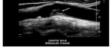


12 Smooth Carotid Plaque Plaque Features: Smooth and Homogeneous Surface Features: Irregular Plaque Irregular Plaque Surface Features: Crater/Ulcer Surface Features: Crater/Ulcer 12


13 Surface Features: Crater/Ulcer Plaque Features: Crater filled with thrombus Calcification and Shadowing Plaque Features: Calcification/Shadowing Plaque Features: Calcification/Shadowing Homgeneous Plaque 13


14 Plaque Features: Smooth; Heterogeneous Large hypoechoic ICA plaque CCA ICA Residual lumen BIF Plaque Features: Hypoechoic (Lusby I) Plaque Features: Hypoechoic region/? IPH Plaque Features: Complex Plaque Thrombosis/Occlusion of ICA 14


 Johnson (depth of crater predicts risk) Johnson (depth of")
 Crater/ulcer less critical independent risk Role in stroke risk, esp")
15 Thrombus in Wall (Dissection) ICA Dissection Transverse View Thrombus in CCA Mobile Component Thrombus in CCA Post-Angio Study Plaque Characteristics Suffered from lack of standardized nomenclature and scheme Many suggested systems, but pathologic correlations mixed More emphasis on hemodynamics, color flow, technical challenges, and time Plaque Features Clinical Implications Lusby (%/location of hypoechoic regions predict risk) Johnson (depth of crater predicts risk) Johnson (depth of crater predicts risk) Leahy (heterogeneity predicts risk) NASCET (ulcer high risk in nonsurgical pt) Crater/ulcer less critical independent risk Role in stroke risk, esp in less % stenosis 15


16 Plaque Features Lusby Criteria Carotid Protocol & Techniques B-mode suggestions Transverse image if unclear or large plaque ICA usually post/lat; ECA ant/medial Quick interrogation of internal jugular vein with B-mode and/or color flow imaging Note appearance of thyroid on transverse view, and report cyst/lesions > 1 cm dia. Carotid Protocol & Techniques Suggestions/Biases Adjust monitors for light in the room Adjust instrument settings; don t just cookbook the study with high contrast B/W images Learn to recognize artifacts Look for color voids to suggest unsuspected hypoechoic plaque Prior CEA Suture Line Carotid Bypass Graft Vein Wall Harmonic Artifact 16


 ID")

17 Carotid Duplex Sonography Duplex Doppler ICA Tight Stenosis Combines PW Doppler & B-mode imaging Image guided placement of sample gate Angle correction Option for color flow imaging Overcomes pitfalls of stand alone tests Expect 90% sens/spec for tight stenosis Color Flow Imaging CCA Color Flow Imaging Quick ID presence/direction of flow Road map for spectral Doppler More accurate angle of insonation Improved data on surface features ID of hypoechoic plaque (color void) ID string sign/near occlusions Speed up examination Color Flow: Tortuosity ICA ICA Tortuosity 360 degree loop/coil 17


18 Color Changes Direction of Flow Relative to Transducer Color Changes Direction vs Aliasing Color Change Distal to Plaque Color Flow ICA Stenosis Color Duplex of ICA Stenosis Color Imaging ID of hypoechoic plaque: color void 18


19 Color Flow Imaging Color void of hypoechoic plaque Color Flow Imaging Surface features Color Flow Suggests Occlusion Duplex Doppler in ICA Stump Doppler Signal Proximal to Occlusion High resistance waveform Color Flow Imaging ICA Occlusion 19


20 Low Velocity, High Resistance Doppler Signals ICA Subtotal Occlusion Low Velocity, High Resistance Doppler Signals ICA Subtotal Occlusion ICA Subtotal Occlusion Color flashes suggest patency Indirect Changes High Resistance Pattern Distal Occlusion Indirect Changes Distal Occlusion Color Flow ICA Occlusion 20


21 Indirect Changes Intracranial Effect ACA Reversal Color Flow Imaging Distal flow confirms patency Angiogram: Near occlusion ICA Carotid Dissection B-mode appearance Carotid Dissection Color Flow shows double lumen Internal Jugular Spontaneous Echo Contrast 21


22 Internal Jugular Spontaneous Echo Contrast Internal Jugular Transverse Spontaneous Echo Contrast Internal Jugular Thrombosis Internal Jugular Thombosis Internal Jugular Thrombosis B-mode Imaging Internal Jugular Thrombus 22


23 Color Flow of IJV Thrombus Power Doppler Imaging Power Imaging ICA Stenosis and Shadowing B-Flow Imaging Cerebrovascular Anatomy Color Duplex Subclavian Artery 23


24 Color Flow Vertebral Origin Vertebral Artery Interosseous/Intertransverse Segment Duplex Interosseous Vertebral Vertebral/Subclavian Steal Carotid Protocol & Techniques Ancillary Methods Cuff Test If vertebral waveform abnormal (from systolic decceleration to frank reversal), do cuff test to check for latent steal BP cuff to 5-10 mm over systolic on ipsilateral arm for 3 minutes to provoke increased metabolic demand in arm Causes flow reversal or worsening if SSS 24


25 Vertebral Duplex Complete reversal after cuff test Vertebral Duplex Cuff test shows latent SSS Carotid Protocol & Techniques Ancillary Methods Positional Testing Occasionally symptoms with certain positional changes (as with kinks, arthritis) Monitoring of carotid or vertebrals through range of neck flexion/extension, turning can show functional blockages only present with positional changes Carotid Protocol & Techniques Ancillary Methods Volume Flow Velocities alone can be deceiving Volume flow key to hemodynamic view Extensive experience with Color Velocity Imaging Quantification, Philips Ultrasound Time domain processing, m-mode display of velocities across vessel, and flow lumen, over time. Doppler now available on most instruments CCA VFR with CVI-Q Carotid Protocol & Techniques Ancillary Methods Volume Flow Best access and accuracy for CCA, less for ICA, and even less for vertebral Predictable changes with both ipsilateral and contralateral stenosis/occlusion. VFR drops with severe ipsilateral distal disease, increases contralateral (if disease not bilateral), if intracranial collaterals normal 25


26 Carotid Stenosis & VFR Volume Flow Measurments Use at WFUSM CCA VFR if 75% or greater stenosis If spectral changes of distal/prox sten/occl Bilateral high or low velocities Waveform suggesting AVM Assess collateral function; avoid error contralaterally Follow progression of stenosis Carotid Protocol & Techniques Ancillary Methods- Ophthalmics Ophthalmic flow direction helps understand collateral flow; sources and adequacy Ophthalmics reversed when collateral from ECA through orbit to ICA siphon Suggests that carotid lesion tight Suggests relative inadequacy of A-Com and P-Com arteries Ophthalmic Collateral Flow Orbital Color Flow Helpful for Hemodynamic Picture 26


27 Orbital Color Flow Reversed Ophthalmic Artery Orbital Color Flow 27
Carotid Abnormalities Coils, Kinks and Tortuosity David Lorelli M.D., RVT, FACS Michigan Vascular Association Conference Saturday, October 20, 2012
 Carotid Abnormalities Coils, Kinks and Tortuosity David Lorelli M.D., RVT, FACS Michigan Vascular Association Conference Saturday, October 20, 2012 Page 1 Table of Contents Carotid Anatomy Carotid Duplex
Disclosure Statement:
 Marsha M. Neumyer, BS, RVT, FSVU, FSDMS, FAIUM International Director Vascular Diagnostic Educational Services Vascular Resource Associates Harrisburg, PA Disclosure Statement: CME Calendar QR Code Marsha
GUNDERSEN/LUTHERAN ULTRASOUND DEPARTMENT POLICY AND PROCEDURE MANUAL
 GUNDERSEN/LUTHERAN ULTRASOUND DEPARTMENT POLICY AND PROCEDURE MANUAL SUBJECT: Carotid Duplex Ultrasound SECTION: Vascular Ultrasound ORIGINATOR: Deborah L. Richert, BSVT, RDMS, RVT DATE: October 15, 2015
Pre-and Post Procedure Non-Invasive Evaluation of the Patient with Carotid Disease
 Pre-and Post Procedure Non-Invasive Evaluation of the Patient with Carotid Disease Michael R. Jaff, D.O., F.A.C.P., F.A.C.C. Assistant Professor of Medicine Harvard Medical School Director, Vascular Medicine
Carotid Ultrasound: Improving Ultrasound
 Carotid Ultrasound: Improving Ultrasound Edward I. Bluth, M.D., F.A.C.R. Chairman Emeritus, Department of Radiology, Ochsner Clinic Foundation, New Orleans, Louisiana Professor, Ochsner Clinical School,
Protokollanhang zur SPACE-2-Studie Neurology Quality Standards
 Protokollanhang zur SPACE-2-Studie Neurology Quality Standards 1. General remarks In contrast to SPACE-1, the neurological center participating in the SPACE-2 trial will also be involved in the treatment
Carotid Artery Doppler
 Carotid Artery Doppler Patient Position supine or semisupine head slightly hyper extended rotated 45 away from the side being examined. Higher frequency linear transducers (7 MHz) Vessels should be imaged
Carotid Duplex: Beyond Stenosis Ido Weinberg, MD Vascular Medicine Massachusetts General Hospital Assistant Professor of Medicine Harvard Medical
 Carotid Duplex: Beyond Stenosis Ido Weinberg, MD Vascular Medicine Massachusetts General Hospital Assistant Professor of Medicine Harvard Medical School Boston, Massachusetts Disclosures I do not have
Ultrasound Imaging of The Posterior Circulation
 Ultrasound Imaging of The Posterior Circulation Michigan Sonographers Society 2 Nd Annual Fall Vascular Conference Larry N. Raber RDMS-RVT Clinical Manager General Ultrasound/Neurovascular Laboratory Cleveland
Beyond Stenosis Severity: Top 5 Important Duplex Characteristics to Identify in a Patient with Carotid Disease
 Beyond Stenosis Severity: Top 5 Important Duplex Characteristics to Identify in a Patient with Carotid Disease Jan M. Sloves RVT, RCS, FASE Technical Director New York Cardiovascular Associates Disclosures
NON-ATHEROSCLEROTIC PATHOLOGY OF THE CAROTID ARTERIES
 NON-ATHEROSCLEROTIC PATHOLOGY OF THE CAROTID ARTERIES Leslie M. Scoutt, MD, FACR Professor of Diagnostic Radiology & Surgery Vice Chair, Dept of Radiology & Biomedical Imaging Chief, Ultrasound Section
DISCLOSURE TEST YOUR WAVEFORM IQ. Partial volume artifact. 86 yo female with right arm swelling, picc line. AVF on left? Dx?
 Deborah Rubens University of Rochester Rochester, NY DISCLOSURE Neither I nor my immediate family have a financial relationship with a commercial organization that may have a direct or indirect interest
Pitfalls in the evaluation of carotid artery stenosis. Serge Kownator «Centre Cardiologique et Vasculaire» Thionville, Fr
 Pitfalls in the evaluation of carotid artery stenosis Serge Kownator «Centre Cardiologique et Vasculaire» Thionville, Fr Disclosure Statement of Financial Interest I currently have, or have had over the
What effects will proximal or distal disease have on an waveform?
 Spectral Doppler Interpretation Director Director of of Ultrasound Ultrasound Education Education & & Quality Quality Assurance Assurance Baylor Baylor College College of of Medicine Medicine Division
What effects will proximal or distal disease have on a waveform?
 Spectral Doppler Interpretation Director of Ultrasound Education & Quality Assurance Baylor College of Medicine Division of Maternal-Fetal Medicine Maternal Fetal Center Imaging Manager Texas Children
Non-invasive examination
 Non-invasive examination Segmental pressure and Ankle-Brachial Index (ABI) The segmental blood pressure (SBP) examination is a simple, noninvasive method for diagnosing and localizing arterial disease.
Carotid Imaging IT S ABOUT MORE THAN JUST OBTAINING THE IMAGES
 Carotid Imaging IT S ABOUT MORE THAN JUST OBTAINING THE IMAGES No financial or commercial relationships to disclose Carotid artery disease: Stroke is one of the most serious causes of mortality and morbidity
Contemporary Carotid Imaging and Approach to Treatment: Course Notes Thursday, June 22, 2017 David M. Pelz, MD, FRCPC
 CNSF Meeting, Victoria, BC. June 2017 Contemporary Carotid Imaging and Approach to Treatment: Course Notes Thursday, June 22, 2017 David M. Pelz, MD, FRCPC A. Objectives 1. To understand the current imaging
Vascular Portfolio: Carotid Reflection. Paige Fabre
 Vascular Portfolio: Carotid Reflection Paige Fabre 13654584 14 Carotid Reflection For this portfolio I produced three pieces of work; a case study, a PowerPoint of study protocol and a poster of stenosis
TRANSCRANIAL DOPPLER ULTRASOUND INTRODUCTION TO TCD INTERPRETATION
 TRANSCRANIAL DOPPLER ULTRASOUND INTRODUCTION TO TCD INTERPRETATION ---Rune Aaslid First TCD Publication 1982 WHAT IS TCD? Uses 2 MHz pulsed Doppler ultrasound Passes through cranial windows Provides information
8/20/18. The Doppler Effect. Objectives. What is the Doppler Effect. Doppler principles. Spectral Waveform. Image recognition. Vascular Ultrasound
 Vascular Ultrasound: Physics and Haemodynamics Objectives Doppler principles Spectral Waveform Key factors Haemodynamics: Stenosis Waveforms Image recognition Vascular Ultrasound: A flawed paradigm What
HD Scanning: Velocities and Volume Flow
 HD Scanning: Velocities and Volume Flow Non-Invasive Lab Symposium West Orange, NJ April 27, 2018 Volume Flow Cindy Sturt, MD, FACS, RVT 500,000 Americans on dialysis 20-25% annual mortality 65% 5 year
Optimising your Doppler settings for an accurate PI. Alison McGuinness Mid Yorks Hospitals
 Optimising your Doppler settings for an accurate PI Alison McGuinness Mid Yorks Hospitals Applications Both maternal uterine and fetal circulations can be studied with doppler sonography Uterine arteries
Recommendations for documentation of neurosonographic examinations
 Recommendations for documentation of neurosonographic examinations The documentation of ultrasound examinations is subject to a dynamic development particularly as regards newer applications. The present
Vascular Sonography Examination
 Vascular Sonography Examination The purpose of The American Registry of Radiologic Technologists (ARRT ) Vascular Sonography Examination is to assess the knowledge and cognitive skills underlying the intelligent
Physician s Vascular Interpretation Examination Content Outline
 Physician s Vascular Interpretation Examination Content Outline (Outline Summary) # Domain Subdomain Percentage 1 2 3 4 5 6 Cerebrovascular Abdominal Peripheral Arterial - Duplex Imaging Peripheral Arterial
New Trials in Progress: ACT 1. Jon Matsumura, MD Cannes, France June 28, 2008
 New Trials in Progress: ACT 1 Jon Matsumura, MD Cannes, France June 28, 2008 Faculty Disclosure I disclose the following financial relationships: Consultant, CAS training director, and/or research grants
Policies and Statements D16. Intracranial Cerebrovascular Ultrasound
 Policies and Statements D16 Intracranial Cerebrovascular Ultrasound SECTION 1: INSTRUMENTATION Policies and Statements D16 Intracranial Cerebrovascular Ultrasound May 2006 (Reaffirmed July 2007) Essential
Duplex Doppler Sonography of the Carotid Artery: False-Positive Results in an Artery Contralateral to an Artery with Marked Stenosis
 049 Duplex Doppler Sonography of the Carotid Artery: False-Positive Results in an Artery Contralateral to an Artery with Marked Stenosis William W. Beckett, Jr. Patricia C. Davis James C. Hoffman, Jr.
Lezione 3 Tronchi Sovraortici
 CORSO DI CERTIFICAZIONE DI COMPETENZA in ECOGRAFIA VASCOLARE GENERALE Lezione 3 Tronchi Sovraortici Settore formazione 2007-2009: Direttore: Paolo G. Pino Marco Campana, Antonella Moreo, Fausto Rigo, Ketty
No financial or commercial relationships to disclose
 Deanna New, RVT No financial or commercial relationships to disclose IAC REQUIREMENTS: The main duty of a sonographer is to make the physician or radiologists job easier by capturing images and doing
STRUCTURED EDUCATION REQUIREMENTS IMPLEMENTATION DATE: JULY 1, 2016
 STRUCTURED EDUCATION REQUIREMENTS Vascular Sonography The purpose of structured education is to provide the opportunity for individuals to develop mastery of discipline-specific knowledge that, when coupled
Image Formation (10) 2 Evaluation and Selection of Representative Images (10)
 2 Evaluation and Selection of Representative Images (10)") STRUCTURED SELF ASSESSMENT CONTENT SPECIFICATIONS SSA LAUNCH DATE: JANUARY 1, 2018 Vascular Sonography The purpose of continuing qualifications requirements (CQR) is to assist registered technologists
STRUCTURED SELF ASSESSMENT CONTENT SPECIFICATIONS SSA LAUNCH DATE: JANUARY 1, 2018 Vascular Sonography The purpose of continuing qualifications requirements (CQR) is to assist registered technologists
Carotid US: More than just a chart on the wall
 Carotid US: More than just a chart on the wall Leslie M. Scoutt, MD, FACR Professor of Diagnostic Radiology & Surgery Vice Chair, Dept of Radiology & Biomedical Imaging Chief, Ultrasound Section Medical
Vascular Surgery Cases: Detours. Brian F. Stull, RDMS, RVT UNC REX Healthcare Vascular Specialists
 Vascular Surgery Cases: Detours Brian F. Stull, RDMS, RVT UNC REX Healthcare Vascular Specialists Brian.Stull@Unchealth.unc.edu Objectives Anatomy of a bypass graft Where does it connect, where does it
Essentials of Clinical MR, 2 nd edition. 99. MRA Principles and Carotid MRA
 99. MRA Principles and Carotid MRA As described in Chapter 12, time of flight (TOF) magnetic resonance angiography (MRA) is commonly utilized in the evaluation of the circle of Willis. TOF MRA allows depiction
COPYRIGHTED MATERIAL. I How to Perform Ultrasound Tests
 I How to Perform Ultrasound Tests COPYRIGHTED MATERIAL 1 2 1 Principles of Extracranial Ultrasound Examination Andrei V. Alexandrov 1, Alice Robinson-Vaughn 1, Clotilde Balucani 2 & Marsha M. Neumyer 3
Non-invasive Imaging of Carotid Artery Atherosclerosis
 Non-invasive Imaging of Carotid Artery Atherosclerosis 최연현 성균관의대삼성서울병원영상의학과 Noninvasive Techniques US with Doppler CT MRI Ultrasonography Techniques of Carotid US US Anatomy (ICA vs ECA) Gray scale and
Carotid Artery Revascularization: Current Strategies. Shonda Banegas, D.O. Vascular Surgery Carondelet Heart and Vascular Institute September 6, 2014
 Carotid Artery Revascularization: Current Strategies Shonda Banegas, D.O. Vascular Surgery Carondelet Heart and Vascular Institute September 6, 2014 Disclosures None 1 Stroke in 2014 Stroke kills almost
What Do We Know? Disclosure Statement: 3/11/2015. Deep abdominal imaging
 Marsha M. Neumyer, BS, RVT, FSVU, FSDMS, FAIUM International Director Vascular Diagnostic Educational Services Vascular Resource Associates Harrisburg, PA Disclosure Statement: CME Calendar QR Code Marsha
Goals. Access flow and renal artery stenosis evaluation by Doppler ultrasound. Reimbursement. WHY use of Doppler Ultrasound
 Access flow and renal artery stenosis evaluation by Doppler ultrasound Adina Voiculescu, MD Interventional Nephrology Brigham and Women s Hospital Boston Instructor at Harvard Medical School Understand
Duplex Ultrasound of the Renal Arteries. Duplex Ultrasound. In the Beginning
 Duplex Ultrasound of the Renal Arteries DIMENSIONS IN HEART AND VASCULAR CARE 2013 PENN STATE HEART AND VASCULAR INSTITUTE ROBERT G. ATNIP MD PROFESSOR OF SURGERY AND RADIOLOGY Duplex Ultrasound Developed
Radial Artery Assessment for Coronary Artery Bypass
 VASCULAR TECHNOLOGY PROFESSIONAL PERFORMANCE GUIDELINES Radial Artery Assessment for Coronary Artery Bypass This Guideline was prepared by the Professional Guidelines Subcommittee of the Society for Vascular
Neuro Quiz 29 Transcranial Doppler Monitoring
 Verghese Cherian, MD, FFARCSI Penn State Hershey Medical Center, Hershey Quiz Team Shobana Rajan, M.D Suneeta Gollapudy, M.D Angele Marie Theard, M.D Neuro Quiz 29 Transcranial Doppler Monitoring This
RPVI Exam Review ecourse
 RPVI Exam Review ecourse The RPVI Exam Review ecourse consists of ten Vascular Physics Modules and fourteen Vascular Specialty Modules. Detailed descriptions of module content are listed below. During
Vascular disease. Structural evaluation of vascular disease. Goo-Yeong Cho, MD, PhD Seoul National University Bundang Hospital
 Vascular disease. Structural evaluation of vascular disease Goo-Yeong Cho, MD, PhD Seoul National University Bundang Hospital resistance vessels : arteries
US of Neurovascular Occlusive Disease: Interpretive Pearls and Pitfalls 1
 EDUCATION EXHIBIT 1165 US of Neurovascular Occlusive Disease: Interpretive Pearls and Pitfalls 1 CME FEATURE See accompanying test at http:// www.rsna.org /education /rg_cme.html LEARNING OBJECTIVES FOR
Carotid Artery Stenting (CAS) Pathophysiology. Technical Considerations. Plaque characteristics: relevant concepts. CAS and CEA
 Pathophysiology. Technical Considerations. Plaque characteristics: relevant concepts. CAS and CEA") Carotid Artery Stenting (CAS) Carotid Artery Stenting for Stroke Risk Reduction Matthew A. Corriere MD, MS, RPVI Assistant Professor of Surgery Department of Vascular and Endovascular Surgery Rationale:
Carotid Artery Stenting (CAS) Carotid Artery Stenting for Stroke Risk Reduction Matthew A. Corriere MD, MS, RPVI Assistant Professor of Surgery Department of Vascular and Endovascular Surgery Rationale:
Antegrade and retrograde flow of carotid
 Antegrade and retrograde flow of carotid The ECA waveform is high resistance and may have retrograde flow in diastole.. They should always demonstrate antegrade flow (toward the brain) and be. external
Transcranial Doppler (Basic Step) Dae-il Chang, M.D., Sung Sang Yoon, M.D. Department of Neurology, College of Medicine, Kyunghee university
 Dae-il Chang, M.D., Sung Sang Yoon, M.D. Department of Neurology, College of Medicine, Kyunghee university") Transcranial Doppler (Basic Step) Dae-il Chang, M.D., Sung Sang Yoon, M.D. Department of Neurology, College of Medicine, Kyunghee university Principles of Doppler Ultrasonography Major target Speed & direction
Transcranial Doppler (Basic Step) Dae-il Chang, M.D., Sung Sang Yoon, M.D. Department of Neurology, College of Medicine, Kyunghee university Principles of Doppler Ultrasonography Major target Speed & direction
[(PHY-3a) Initials of MD reviewing films] [(PHY-3b) Initials of 2 nd opinion MD]
![[(PHY-3a) Initials of MD reviewing films] [(PHY-3b) Initials of 2 nd opinion MD]](/thumbs/89/98619893.jpg "[(PHY-3a) Initials of MD reviewing films] [(PHY-3b) Initials of 2 nd opinion MD]") 2015 PHYSICIAN SIGN-OFF (1) STUDY NO (PHY-1) CASE, PER PHYSICIAN REVIEW 1=yes 2=no [strictly meets case definition] (PHY-1a) CASE, IN PHYSICIAN S OPINION 1=yes 2=no (PHY-2) (PHY-3) [based on all available
2015 PHYSICIAN SIGN-OFF (1) STUDY NO (PHY-1) CASE, PER PHYSICIAN REVIEW 1=yes 2=no [strictly meets case definition] (PHY-1a) CASE, IN PHYSICIAN S OPINION 1=yes 2=no (PHY-2) (PHY-3) [based on all available
Carotid Artery Stenting
 Carotid Artery Stenting JESSICA MITCHELL, ACNP CENTRAL ILLINOIS RADIOLOGICAL ASSOCIATES External Carotid Artery (ECA) can easily be identified from Internal Carotid Artery (ICA) by noticing the branches.
ASDIN 7th Annual Scientific Meeting DISCLOSURES TECHNICAL CONSIDERATIONS TECHNICAL CONSIDERATIONS UTILITY OF ULTRASOUND IN EVALUATING ACCESS
 DISCLOSURES UTILITY OF ULTRASOUND IN EVALUATING ACCESS DYSFUNCTION None Vandana Dua Niyyar, MD Assistant Professor of Medicine, Division of Nephrology, Emory University UTILITY OF ULTRASOUND IN ACCESS
Radiologic Importance of a High- Resistive Vertebral Artery Doppler Waveform on Carotid Duplex Ultrasonography
 CME Article Radiologic Importance of a High- Resistive Vertebral Artery Doppler Waveform on Carotid Duplex Ultrasonography Esther S. H. Kim, MD, MPH, Megan Thompson, Kristine M. Nacion, BA, Carmel Celestin,
TO CATCH A THIEF: IMAGING OF SUBCLAVIAN STEAL
 October 2013 TO CATCH A THIEF: IMAGING OF SUBCLAVIAN STEAL Sumir Pandit, Harvard Medical School, Year III 1 AGENDA Introduction to our patient A.B. Anatomy review of aorta and branches CT imaging of our
VFI Technology to Change the Way You Work
 Analogic Ultrasound VFI Technology to Change the Way You Work Vascular Ultrasound Made Easier Vector Flow Imaging VFI VFI is a ground-breaking technology that can revolutionize the workflow for many Doppler
Diagnosis of Middle Cerebral Artery Occlusion with Transcranial Color-Coded Real-Time Sonography
 Diagnosis of Middle Cerebral Artery Occlusion with Transcranial Color-Coded Real-Time Sonography Kazumi Kimura, Yoichiro Hashimoto, Teruyuki Hirano, Makoto Uchino, and Masayuki Ando PURPOSE: To determine
Doppler ultrasound as noninvasive diagnosis of peripheral arterial disease
 Doppler ultrasound as noninvasive diagnosis of peripheral arterial disease Poster No.: C-0246 Congress: ECR 2012 Type: Scientific Exhibit Authors: C. Ballester Valles, F. Aparici-Robles; Valencia/ES Keywords:
Carotid artery occlusion: Positive predictive value of duplex sonography compared with arteriography
 Carotid artery occlusion: Positive predictive value of duplex sonography compared with arteriography Jonathan D. Kirsch, MD, Louis R. Wagner, MD, E. Meredith James, MD, J. William Charboneau, MD, Douglas
Quality ID #195 (NQF 0507): Radiology: Stenosis Measurement in Carotid Imaging Reports National Quality Strategy Domain: Effective Clinical Care
: Radiology: Stenosis Measurement in Carotid Imaging Reports National Quality Strategy Domain: Effective Clinical Care") Quality ID #195 (NQF 0507): Radiology: Stenosis Measurement in Carotid Imaging Reports National Quality Strategy Domain: Effective Clinical Care 2018 OPTIONS FOR INDIVIDUAL MEASURES: REGISTRY ONLY MEASURE
Quality ID #195 (NQF 0507): Radiology: Stenosis Measurement in Carotid Imaging Reports National Quality Strategy Domain: Effective Clinical Care 2018 OPTIONS FOR INDIVIDUAL MEASURES: REGISTRY ONLY MEASURE
Intracranial Cerebrovascular Evaluation Transcranial Doppler (Non-Imaging) and Transcranial Duplex Imaging (TCD-I)
 and Transcranial Duplex Imaging (TCD-I)") VASCULAR TECHNOLOGY PROFESSIONAL PERFORMANCE GUIDELINES Intracranial Cerebrovascular Evaluation Transcranial Doppler (Non-Imaging) and Transcranial Duplex Imaging (TCD-I) This Guideline was prepared by
VASCULAR TECHNOLOGY PROFESSIONAL PERFORMANCE GUIDELINES Intracranial Cerebrovascular Evaluation Transcranial Doppler (Non-Imaging) and Transcranial Duplex Imaging (TCD-I) This Guideline was prepared by
noninvasive, nonionizing, portable, inexpensive, safe for serial or prolonged studies
 TRANS CRANIAL DOPPLER Presented by : Anil Garg Transcranial Doppler 1982, Aaslid and colleagues introduced TCD as a non-invasive technique for monitoring blood flow velocity in basal cerebral arteries
FIRST COAST SERVICE OPTIONS FLORIDA MEDICARE PART B LOCAL COVERAGE DETERMINATION
 FIRST COAST SERVICE OPTIONS FLORIDA MEDICARE PART B LOCAL COVERAGE DETERMINATION CPT/HCPCS Codes 93875 Non-invasive physiologic studies of extracranial arteries, complete bilateral study (eg, periorbital
Screening for asymptomatic internal artery stenosis: Duplex criteria for discriminating 60% to 99% stenosis
 Screening for asymptomatic internal artery stenosis: Duplex criteria for discriminating 60% to 99% stenosis carotid Gregory L. Moneta, MD, James M. Edwards, MD, George Papanicolaou, MD, Thomas Hatsukami,
Carotid Endarterectomy for Symptomatic Complete Occlusion of the Internal Carotid Artery
 2011 65 4 239 245 Carotid Endarterectomy for Symptomatic Complete Occlusion of the Internal Carotid Artery a* a b a a a b 240 65 4 2011 241 9 1 60 10 2 62 17 3 67 2 4 64 7 5 69 5 6 71 1 7 55 13 8 73 1
NCVH. Ultrasongraphy: State of the Art Vein Forum 2015 A Multidisciplinary Approach to Otptimizing Venous Circulation From Wounds to WOW
 Ultrasongraphy: State of the Art 2015 NCVH New Cardiovascular Horizons Vein Forum 2015 A Multidisciplinary Approach to Otptimizing Venous Circulation From Wounds to WOW Anil K. Chagarlamudi, M.D. Cardiovascular
Upper Extremity Venous Duplex. Michigan Sonographers Society Fall Ultrasound Symposium October 15, 2016
 Upper Extremity Venous Duplex Michigan Sonographers Society Fall Ultrasound Symposium October 15, 2016 Patricia A. (Tish) Poe, BA RVT FSVU Director of Quality Assurance Navix Diagnostix Patricia A. Poe
Indications: following: embolization. artery that has diseases 5. The evaluation. of suspected. such entities. a cold hand. biopsy
 Peripheral Arterial Ultrasound Protocol Using Color and Spectral Doppler Reviewed by: Mark Yuhasz, MD Last Review Date: January 2015 Contact: (866) 761 4200, Option 1 Indications: The indications for peripheral
Case 8038 Renal allograft complicated with renal artery stenosis
 Case 8038 Renal allograft complicated with renal artery stenosis Santiago I, Canelas A, Pinto AP Section: Cardiovascular Published: 2009, Nov. 30 Patient: 61 year(s), male Clinical History A 61-year-old
(Department of Radiology, Beylikdüzü State Hospital, İstanbul, Turkey) Corresponding Author: Dr. Mete Özdikici
 Corresponding Author: Dr. Mete Özdikici") Quest Journals Journal of Medical and Dental Science Research Volume 5~ Issue 6 (2018) pp: 61-65 ISSN(Online) : 2394-076X ISSN (Print):2394-0751 www.questjournals.org Research Paper Quantitative Measurements
Quest Journals Journal of Medical and Dental Science Research Volume 5~ Issue 6 (2018) pp: 61-65 ISSN(Online) : 2394-076X ISSN (Print):2394-0751 www.questjournals.org Research Paper Quantitative Measurements
MESENTERIC ISCHEMIA. Phillip J Bendick, PhD
 MESENTERIC ISCHEMIA Phillip J Bendick, PhD Arterial Celiac - Hepatic - Splenic Superior Mesenteric Artery Inferior Mesenteric Artery Venous Mesenteric system Porto - hepatic system Inferior Vena Cava Acute
Transducer Selection. Renal Artery Duplex Exam. Renal Scan. Renal Scan Echogenicity. How to Perform an Optimal Renal Artery Doppler Examination
 How to Perform an Optimal Renal Artery Doppler Examination Director of Ultrasound Education & Quality Assurance Baylor College of Medicine Division of Maternal-Fetal Medicine Maternal Fetal Center Imaging
2015 ARDMS Physicians Vascular Interpretation Job Task Analysis Summary Report
 P a g e 1 2015 ARDMS Physicians Vascular Interpretation Job Task Analysis Summary Report American Registry for Diagnostic Medical Sonography (ARDMS) P a g e 2 Table of Contents ABOUT THE REPORT... 3 METHODOLOGY...
Measure #195 (NQF 0507): Radiology: Stenosis Measurement in Carotid Imaging Reports National Quality Strategy Domain: Effective Clinical Care
: Radiology: Stenosis Measurement in Carotid Imaging Reports National Quality Strategy Domain: Effective Clinical Care") Measure #195 (NQF 0507): Radiology: Stenosis Measurement in Carotid Imaging Reports National Quality Strategy Domain: Effective Clinical Care 2017 OPTIONS FOR INDIVIDUAL MEASURES: CLAIMS ONLY MEASURE TYPE:
Measure #195 (NQF 0507): Radiology: Stenosis Measurement in Carotid Imaging Reports National Quality Strategy Domain: Effective Clinical Care 2017 OPTIONS FOR INDIVIDUAL MEASURES: CLAIMS ONLY MEASURE TYPE:
Vivek R. Deshmukh, MD Director, Cerebrovascular and Endovascular Neurosurgery Chairman, Department of Neurosurgery Providence Brain and Spine
 Vivek R. Deshmukh, MD Director, Cerebrovascular and Endovascular Neurosurgery Chairman, Department of Neurosurgery Providence Brain and Spine Institute The Oregon Clinic Disclosure I declare that neither
Carotid Doppler: Doppler wave forms obtained from the common, external and internal carotid arteries. As well as the vertebral and subclavian
 Competency Carotid Doppler: Doppler wave forms obtained from the common, external and internal carotid arteries. As well as the vertebral and subclavian arteries. Preferred angle is 60 degrees or less.
Evaluation of Carotid Vessels and Vertebral Artery in Stroke Patients with Color Doppler Ultrasound and MR Angiography
 Evaluation of Carotid Vessels and Vertebral Artery in Stroke Patients with Color Doppler Ultrasound and MR Angiography Dr. Pramod Shaha 1, Dr. Vinay Raj R 2, Dr. (Brig) K. Sahoo 3 Abstract: Aim & Objectives:
Carotid Imaging. Dr Andrew Farrall. Consultant Neuroradiologist
 20121123 SSCA http://www.neuroimage.co.uk/network Andrew Farrall Carotid Imaging Dr Andrew Farrall Consultant Neuroradiologist SFC Brain Imaging Research Centre (www.sbirc.ed.ac.uk), SINAPSE Collaboration
Profile of Extracranial Cerebrovascular Disease in Kelantan: A Study by Continuous Wave Doppler Ultrasonography
 ---- ------ - ------------- Profile of Extracranial Cerebrovascular Disease in Kelantan: A Study by Continuous Wave Doppler Ultrasonography R.c. Pratap, MD M. Mafauzy, MRCP Department of Medicine, University
Disclosures. CREST Trial: Summary. Lecture Outline 4/16/2015. Cervical Atherosclerotic Disease
 Disclosures Your Patient Has Carotid Bulb Stenosis and a Tandem Intracranial Stenosis: How Do SAMMPRIS and Other Evidence Inform Your Treatment? UCSF Vascular Symposium 2015 Steven W. Hetts, MD Associate
Guidelines, Policies and Statements D20 Statement on Peripheral Venous Ultrasound
 Guidelines, Policies and Statements D20 Statement on Peripheral Venous Ultrasound Disclaimer and Copyright The ASUM Standards of Practice Board have made every effort to ensure that this Guideline/Policy/Statement
Treatment Considerations for Carotid Artery Stenosis. Danielle Zielinski, RN, MSN, ACNP Rush University Neurosurgery
 Treatment Considerations for Carotid Artery Stenosis Danielle Zielinski, RN, MSN, ACNP Rush University Neurosurgery 4.29.2016 There is no actual or potential conflict of interest in regards to this presentation
Upper Extremity Venous Duplex Evaluation
 VASCULARTECHNOLOGY PROFESSIONAL PERFORMANCE GUIDELINES Upper Extremity Venous Duplex Evaluation This Guideline was prepared by the Professional Guidelines Subcommittee of the Society for Vascular Ultrasound
TCD in Subclavian Steal Syndrome
 ISSN 2005-7881 Journal of Neurosonology 2(Suppl. 1):25-30, 2010 TCD in Subclavian Steal Syndrome Soon-Tae Lee, M.D., Ph.D. Department of Neurology, Seoul National University Hospital, Seoul, South Korea
Disclosures. State of the Art Management of Carotid Stenosis. NIH funding for clinical trials Consultant for Scientia Vascular and Medtronic
 State of the Art Management of Carotid Stenosis Mark R. Harrigan, MD UAB Stroke Center Professor of Neurosurgery, Neurology, and Radiology University of Alabama, Birmingham Disclosures NIH funding for
SCAI Fall Fellows Course Subclavian/Innominate Case Presentation
 SCAI Fall Fellows Course 2012 Subclavian/Innominate Case Presentation Daniel J. McCormick DO, FACC, FSCAI Director, Cardiovascular Interventional Therapy Pennsylvania Hospital University of Pennsylvania
Hemodynamically significant subclavian artery stenosis
 REVIEW ARTICLE Duplex Ultrasonography of Vertebral and Subclavian Arteries Vijay G. Kalaria, MD, FACC, FSCAI, Sony Jacob, MD, William Irwin, RVT, and Robert M. Schainfeld, DO, Indianapolis, Indiana, and
Carotid Ddisease, Carotid IMT and Risk of Stroke
 Carotid Ddisease, Carotid IMT and Risk of Stroke TATJANA RUNDEK, MD PhD Professor of Neurology, Epidemiology and Public Health Director, Clinical translational Division Department of Neurology, Miller
Doppler Basic & Hemodynamic Calculations
 Doppler Basic & Hemodynamic Calculations August 19, 2017 Smonporn Boonyaratavej MD Division of Cardiology, Department of Medicine Chulalongkorn University Cardiac Center, King Chulalongkorn Memorial Hospital
Guidelines for Ultrasound Surveillance
 Guidelines for Ultrasound Surveillance Carotid & Lower Extremity by Ian Hamilton, Jr, MD, MBA, RPVI, FACS Corporate Medical Director BlueCross BlueShield of Tennessee guidelines for ultrasound surveillance
MORTALITY AND MORBIDITY RISK FROM CAROTID ARTERY ATHEROSCLEROSIS. 73 year old NS right-handed male applicant for $1 Million life insurance
 MORTALITY AND MORBIDITY RISK FROM CAROTID ARTERY ATHEROSCLEROSIS October 17, 2012 AAIM Triennial Conference, San Diego Robert Lund, MD What Is The Risk? 73 year old NS right-handed male applicant for $1
Carotid intima media thickness as an usefull tool in predicting cerebrovaskular events
 Carotid intima media thickness as an usefull tool in predicting cerebrovaskular events Poster No.: C-0005 Congress: ECR 2015 Type: Authors: Keywords: DOI: Scientific Exhibit A. Rahimic - Catic; Sarajevo/BA
MORTALITY AND MORBIDITY RISK FROM CAROTID ARTERY ATHEROSCLEROSIS. 73 year old NS right-handed male applicant for $1 Million Life Insurance
 MORTALITY AND MORBIDITY RISK FROM CAROTID ARTERY ATHEROSCLEROSIS October 17, 2012 AAIM Triennial Conference, San Diego Robert Lund, MD What Is The Risk? 73 year old NS right-handed male applicant for $1
Update on Carotid Disease
 Update on Carotid Disease L. Nelson Hopkins, MD Elad Levy, MD Adnan Siddiqui, MD,PhD Ken Snyder, MD,PhD Gates Vascular Institute LN Hopkins, MD I disclose the following financial relationship(s): President,
Noninvasive transcranial Doppler ultrasound recording of flow velocity in basal cerebral arteries
 J Neurosurg 57:769-774, 1982 Noninvasive transcranial Doppler ultrasound recording of flow velocity in basal cerebral arteries RUNE AASLID, PH.D., THOMAS-MARC MARKWALDER, M.D., AND HEt,CE NORNES, M.D.
Duplex Carotid Sonography Peak Systolic Velocity in Quantifying Internal Carotid Artery Stenosis
 Duplex Carotid Sonography Peak Systolic Velocity in Quantifying Internal Carotid Artery Stenosis Cynthia E Withers, MD", Barbara B Gosink, MD", Alison M Keightley, MD", Giovanna Casola, MD", Arthur A Lee,
Transorbital blood flow sound recordings have the
 397 Noninvasive Detection of Intracranial Vascular Lesions by Recording Blood Flow Sounds Yasushi Kurokawa, MD; Seisho Abiko, MD; Kohsaku Watanabe, MD Background and Purpose Transorbital blood flow sound
Deb Coghlan AMS (Vascular and General ) Brisbane, Australia
 Brisbane, Australia") Deb Coghlan AMS (Vascular and General ) Brisbane, Australia ANEURYSMAL DIISEASE The infrarenal aorta enlarges with age, and is the commonest site for arterial aneurysms. An aneurysm is a permanent focal
Deb Coghlan AMS (Vascular and General ) Brisbane, Australia ANEURYSMAL DIISEASE The infrarenal aorta enlarges with age, and is the commonest site for arterial aneurysms. An aneurysm is a permanent focal
Carotid Stenosis 1/24/2019. Review of Primary Studies. NASCET- Moderate stenosis. ACAS (Asymptomatic Carotid Atherosclerosis Study) NASCET
 NASCET") Review of Primary Studies Carotid Stenosis NINDS National Institute of Neurological Disorders and Stroke 2 large studies to determine who would benefit from surgery NASCET North American Symptomatic Carotid
Review of Primary Studies Carotid Stenosis NINDS National Institute of Neurological Disorders and Stroke 2 large studies to determine who would benefit from surgery NASCET North American Symptomatic Carotid
Review Article Duplex Ultrasound Evaluation of Hemodialysis Access: A Detailed Protocol
 International Nephrology Volume 2012, Article ID 508956, 7 pages doi:10.1155/2012/508956 Review Article Duplex Ultrasound Evaluation of Hemodialysis Access: A Detailed Protocol Victoria Teodorescu, 1,