What effects will proximal or distal disease have on a waveform?
|
|
|
- Phebe Golden
- 5 years ago
- Views:
Transcription


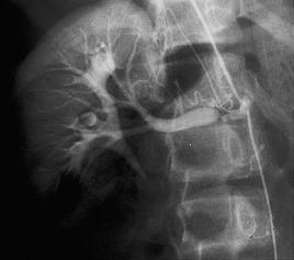
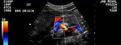
1 Spectral Doppler Interpretation Director of Ultrasound Education & Quality Assurance Baylor College of Medicine Division of Maternal-Fetal Medicine Maternal Fetal Center Imaging Manager Texas Children s Hospital, Pavilion for Women Houston Texas & Clinical Instructor Thomas Jefferson University Hospital - Radiology Department Philadelphia, Pennsylvania Spectral Analysis The final shape of the arterial Doppler waveform is dependent on numerous factors: Contraction by the heart Presence of stenosis in the vessel State of the downstream circulation OB Doppler How systolic and diastolic components of arterial waveforms appear in health and disease? What effects will proximal or distal disease have on a waveform? Distal disease Changes the resistance Distal Disease Changes the resistance Proximal disease Changes the strength of the signal Acute & chronic parenchymal disease Obstruction Renal vein thrombosis 1


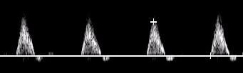
2 Distal Disease Changes the resistance Distal Disease Changes the resistance Distal Disease Changes the resistance Proximal Disease Changes the strength of the signal Tardus Parvus Waveform Tardus Slow & late Parvus Small & little Tardus Parvus Waveform Systolic acceleration diminished Acceleration time prolonged Waveform shape Diminished pulsatility 2


3 Caution! Increase Sweep Speed Proximal Disease Changes the strength of the signal Mesenteric Arteries Mesenteric Arteries Remember! It is more difficult to demonstrate tardus parvus in a stiff vessel Atherosclotic arteries & increased distal resistance masks the post-stenotic tardus parvus 3


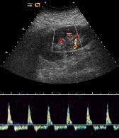
4 Qualitative Quantitative Doppler Analysis Doppler Analysis Qualitative The visual or acoustic evaluation of Doppler wave form Doppler Analysis Qualitative The visual or acoustic evaluation of Doppler wave form Quantitative Calculation of volume flow Calculation of indices Indirect method to evaluate blood perfusion Doppler Analysis Waveform is commonly described by pulsatility which can be measured Peak Systolic velocity PSV Resistance Index RI Pulsatility Index PI Systolic/Diastolic Ratio S/D Acceleration Index AI Acceleration Time AT How to Look at a Waveform? Spectral Doppler Where & how was signal obtained? Presence of flow Direction of flow Characterization of signal Quality of exam 4

5 Cursor is used for optimal alignment between vessel axis & Doppler scan line Angle of insonation Angle correction only used to measure velocity Spectral Doppler Sample Volume determines the location and area that the pulsed wave Doppler listens for a returning signal Cursor is used for optimal alignment between vessel axis & Doppler scan line Angle of insonation Angle correction only used to measure velocity Spectral Doppler Sample Volume determines the location and area that the pulsed wave Doppler listens for a returning signal Cursor is used for optimal alignment between vessel axis & Doppler scan line Angle of insonation Angle correction only used to measure velocity Spectral Doppler Sample Volume determines the location and area that the pulsed wave Doppler listens for a returning signal How to Look at a Waveform? Where & how was signal obtained? What is the angle of insonation Where is the sample volume What is the sample volume size 1 What is the Doppler Angle? What is the Doppler Angle? Angle is the result of Doppler line direction Cursor correction Angle affects velocity accuracy A V = 2. F t. cos Θ. F d C 5


6 What is the Doppler Angle? What is the Doppler Angle? What is the Doppler Angle? What is the Doppler Angle? What is the Doppler Angle? 6


7 In Straight Unbranched Vessels Blood Flows in Layers (or Laminar) 2 Where is the Sample Volume? 7


8 3 What is the Sample Volume Size? Size ranges from 0.7 to 15mm Larger gate to search for flow Smaller gate for precise information What is the Sample Volume Size? What is the Sample Volume Size? Spectral Doppler - Sample Volume What is the recommended size of the sample volume and why? The sample volume size should be no larger than 1/3 of the size of the vessel. If larger, the sample volume is capturing slower flow that occurs near the vessel walls. What is the Sample Volume Size? What is the Sample Volume Size? Too small a gate may give the false impression of reduced or even absent flow 8


 Increasing the scale smaller")
9 How to Look at a Waveform? Where & how was signal obtained? What is the angle of insonation Where is the sample volume What is the sample volume size Technical considerations Doppler Gain Velocity Scale Wall Filter Sweep Speed Spectral Doppler Gain Controls the amplification of the returning Doppler signals The Doppler gain should be adjusted to a level that fills in the gray scale of the spectral analysis waveform without creating noise Spectral Display effect of Doppler Gain Angle adjustments are not necessary since the shape of the waveform, rather than velocity, is used for interpretation Spectral Doppler - Velocity Scale Controls PRF (the rate at which the transducer is pulsed per second) Increasing the scale smaller waveform size Θ = 89 O Decreasing the scale bigger waveform size 9


10 Spectral Doppler - Velocity Scale Spectral Doppler - Velocity Scale Spectral Doppler - Velocity Scale Spectral Doppler - Velocity Scale Decreased PRF Increased PRF Spectral Doppler - Wall Filter Suppress velocities associated with tissue or wall motion Higher setting Reduce artifacts Can eliminate diagnostic information Spectral Doppler - Sweep Speed Controls how quickly the spectral information is updated Three sweep speeds Slow Moderate Fast OB Doppler 10


11 Spectral Doppler - Sweep Speed How to Look at a Waveform? Where & how was signal obtained? Flow direction OB Doppler Diagnostic Challenge How to Look at a Waveform? Where & how was signal obtained? Flow direction Characterization of signal Characterization of Signal Spectral Analysis Site of signal What is normal & abnormal Shape (edge) of spectrum Velocity of blood flow Pulsatility Structure of spectrum Distribution of blood velocities Spectral broadening Characterization of Signal Spectral Analysis Site of signal What is normal & abnormal Shape (edge) of spectrum Velocity of blood flow Pulsatility Structure of spectrum Distribution of blood velocities Spectral broadening Diagnosis 11


12 What does increased pulsatility in the hepatic veins suggest? What does loss of pulsatility in the hepatic veins suggest? What does loss of pulsatility in the portal veins suggest? 1. Cirrhosis 2. Compression from mass 3. Partial thrombosis What does pulsatile portal vein suggest? Portal Vein Gas Any communication between the systemic and portal veins, (portosystemic shunts, fistulae) may lead to a pulsatile portal vein Increased pulsatility of portal venous flow may also be seen with congestion of the liver, especially the passive congestion associated with right-sided cardiac failure and/or tricuspid regurgitation 12


13 Portal vein gas Characterization of Signal Edge of spectral envelope Waveform shape & pulsatility Peak velocities Ischemic, inflammatory, or infectious bowel diseases Pediatric age group Necrotizing enterocolitis Characterization of Signal Distribution of blood velocities Gray scale distribution of all RBC V189 cm/s Celiac Artery Important Signs of Stenosis Important Signs of Stenosis Proximal to stenosis Change in pulsitility At the stenosis Elevated velocities compared to pre-stenotic segment Laminar flow 13


14 Important Signs of Stenosis Important Signs of Stenosis Beyond the stenosis Post stenotic turbulence or disturb flow Spectral broadening Loss of well defined spectral edge Distal to stenosis Down stream Tardus-Parvus Velocity should drop off distal to stenosis Exceptions: long stenosis, near occlusive lesions Distribution of Doppler frequencies seen in spectrum filling of envelope Celiac Artery Celiac Artery 14


15 Characterization of Signal Characterization of Signal Aorta Lt Renal Artery Characterization of Signal Diastolic Flow Physiological and Pathological conditions: Cardiac and aortic factors Vessel compliance Downstream resistance Venous and arteriovenous connections Stenosis at, above or beyond vessel Increased Diastolic Flow Effect of Eating on Diastole Eating affects SMA Exercise affects muscles Neovascularity Inflammatory conditions Corpus luteum development Menstrual cycle on uterus Arteriovenous Shunting SMA Before Meal SMA After Meal 15


16 Nonspecificity of Neovascularity Inflammatory Conditions Ovarian Cancer Benign Hemorrhagic Cyst Orchitis Uterine Artery Flow Ovulatory cycles There is an increase in end diastolic flow velocities between proliferative & secretory phases Uterine Artery Persistent Notching Notch at 25 weeks implies incomplete trophoblastic invasion and is predictive of preeclampsia and/or delivering a growth restricted fetus Arteriovenous Shunting Small connections tumor vessels, arterioportal shunting in cirrhosis Large vessels AV Malformations vein of Galen aneurysm uterine AVM AV Fistulas traumatic AVF Decreased Diastolic Flow Change of resistance from lower to higher decreases diastolic flow Frequently seen in distal stenosis or occlusive disease Venous outflow obstruction 16


17 Distal Stenosis Occlusive Disease Sagittal Aorta Staccato Flow Thrombosed Distal Aorta & Common Iliac Arteries Occlusive Disease Vascular Destruction Capillary and vascular destruction obstructs flow decreasing diastole Common sites Renal disease Placental diseases Decreased Diastolic Flow Change of resistance from lower to higher decreases diastolic flow Frequently seen in distal stenosis or occlusive disease Venous outflow obstruction Venous Obstruction Venous outflow affects diastole Physiologic Erection Pathologic Renal vein thrombosis 17

18 Conclusion What effects will proximal or distal disease have on an waveform? How to look at a waveform? Doppler analysis Stenosis profiles Diastolic flow Spectral Doppler Interpretation Thank You 18
What effects will proximal or distal disease have on an waveform?
 Spectral Doppler Interpretation Director Director of of Ultrasound Ultrasound Education Education & & Quality Quality Assurance Assurance Baylor Baylor College College of of Medicine Medicine Division
Transducer Selection. Renal Artery Duplex Exam. Renal Scan. Renal Scan Echogenicity. How to Perform an Optimal Renal Artery Doppler Examination
 How to Perform an Optimal Renal Artery Doppler Examination Director of Ultrasound Education & Quality Assurance Baylor College of Medicine Division of Maternal-Fetal Medicine Maternal Fetal Center Imaging
DISCLOSURE TEST YOUR WAVEFORM IQ. Partial volume artifact. 86 yo female with right arm swelling, picc line. AVF on left? Dx?
 Deborah Rubens University of Rochester Rochester, NY DISCLOSURE Neither I nor my immediate family have a financial relationship with a commercial organization that may have a direct or indirect interest
HD Scanning: Velocities and Volume Flow
 HD Scanning: Velocities and Volume Flow Non-Invasive Lab Symposium West Orange, NJ April 27, 2018 Volume Flow Cindy Sturt, MD, FACS, RVT 500,000 Americans on dialysis 20-25% annual mortality 65% 5 year
Vascular Sonography Examination
 Vascular Sonography Examination The purpose of The American Registry of Radiologic Technologists (ARRT ) Vascular Sonography Examination is to assess the knowledge and cognitive skills underlying the intelligent
Visceral Vascular Ultrasound. Joel Thompson, MD, MPH Borg & Ide Imaging
 Visceral Vascular Ultrasound Joel Thompson, MD, MPH Borg & Ide Imaging Objectives: Review major abdominal vascular structures Identify normal peak systolic velocity (PSV) for major abdominal arteries.
Optimising your Doppler settings for an accurate PI. Alison McGuinness Mid Yorks Hospitals
 Optimising your Doppler settings for an accurate PI Alison McGuinness Mid Yorks Hospitals Applications Both maternal uterine and fetal circulations can be studied with doppler sonography Uterine arteries
Abdominal Doppler Mastering the next level of vascular anatomy in the belly. Cindy A. Owen, RDMS, RVT
 Abdominal Doppler Mastering the next level of vascular anatomy in the belly Cindy A. Owen, RDMS, RVT Introduction Abdominal Doppler is a tough exam Success is dependent on: Patient body habitus Patient
STRUCTURED EDUCATION REQUIREMENTS IMPLEMENTATION DATE: JULY 1, 2016
 STRUCTURED EDUCATION REQUIREMENTS Vascular Sonography The purpose of structured education is to provide the opportunity for individuals to develop mastery of discipline-specific knowledge that, when coupled
Image Formation (10) 2 Evaluation and Selection of Representative Images (10)
 2 Evaluation and Selection of Representative Images (10)") STRUCTURED SELF ASSESSMENT CONTENT SPECIFICATIONS SSA LAUNCH DATE: JANUARY 1, 2018 Vascular Sonography The purpose of continuing qualifications requirements (CQR) is to assist registered technologists
STRUCTURED SELF ASSESSMENT CONTENT SPECIFICATIONS SSA LAUNCH DATE: JANUARY 1, 2018 Vascular Sonography The purpose of continuing qualifications requirements (CQR) is to assist registered technologists
Vascular Imaging in the Pediatric Abdomen. Jonathan Swanson, MD
 Vascular Imaging in the Pediatric Abdomen Jonathan Swanson, MD Goals and Objectives To understand the imaging approach, appearance, and clinical manifestations of the common pediatric abdominal vascular
Goals. Access flow and renal artery stenosis evaluation by Doppler ultrasound. Reimbursement. WHY use of Doppler Ultrasound
 Access flow and renal artery stenosis evaluation by Doppler ultrasound Adina Voiculescu, MD Interventional Nephrology Brigham and Women s Hospital Boston Instructor at Harvard Medical School Understand
MESENTERIC ISCHEMIA. Phillip J Bendick, PhD
 MESENTERIC ISCHEMIA Phillip J Bendick, PhD Arterial Celiac - Hepatic - Splenic Superior Mesenteric Artery Inferior Mesenteric Artery Venous Mesenteric system Porto - hepatic system Inferior Vena Cava Acute
No financial or commercial relationships to disclose
 Deanna New, RVT No financial or commercial relationships to disclose IAC REQUIREMENTS: The main duty of a sonographer is to make the physician or radiologists job easier by capturing images and doing
What Do We Know? Disclosure Statement: 3/11/2015. Deep abdominal imaging
 Marsha M. Neumyer, BS, RVT, FSVU, FSDMS, FAIUM International Director Vascular Diagnostic Educational Services Vascular Resource Associates Harrisburg, PA Disclosure Statement: CME Calendar QR Code Marsha
Case 8038 Renal allograft complicated with renal artery stenosis
 Case 8038 Renal allograft complicated with renal artery stenosis Santiago I, Canelas A, Pinto AP Section: Cardiovascular Published: 2009, Nov. 30 Patient: 61 year(s), male Clinical History A 61-year-old
Abdomen Sonography Examination Content Outline
 Abdomen Sonography Examination Content Outline (Outline Summary) # Domain Subdomain Percentage 1 2 3 Anatomy, Perfusion, and Function Pathology, Vascular Abnormalities, Trauma, and Postoperative Anatomy
The Fetus: Five Top Do Not Miss Diagnoses. Doppler Ultrasound
 The Fetus: Five Top Do Not Miss Diagnoses Doppler Ultrasound Giancarlo Mari, MD, MBA Professor and Chair Department of Obstetrics and Gynecology University of Tennessee Health Science Center Memphis, TN
Postoperative AV Fistula Evaluation. Postoperative examination protocol. Postoperative AVF Protocol. Hemodialysis Access Surveillance
 Hemodialysis Access Surveillance Postoperative AV Fistula Evaluation Failure of maturation Stenosis Perigraft mass/fluid collection Joseph L. Mills, Sr., M.D. Professor of Surgery Chief, Division of Vascular
Adult Echocardiography Examination Content Outline
 Adult Echocardiography Examination Content Outline (Outline Summary) # Domain Subdomain Percentage 1 2 3 4 5 Anatomy and Physiology Pathology Clinical Care and Safety Measurement Techniques, Maneuvers,
US of Renovascular Hypertension. Jonathan R. Dillman, MD, MSc Associate Professor Director, Thoracoabdominal Imaging
 US of Renovascular Hypertension Jonathan R. Dillman, MD, MSc Associate Professor Director, Thoracoabdominal Imaging Disclosures Nothing Relevant Unrelated grant funding Siemens US Toshiba US Objectives
NON-ATHEROSCLEROTIC PATHOLOGY OF THE CAROTID ARTERIES
 NON-ATHEROSCLEROTIC PATHOLOGY OF THE CAROTID ARTERIES Leslie M. Scoutt, MD, FACR Professor of Diagnostic Radiology & Surgery Vice Chair, Dept of Radiology & Biomedical Imaging Chief, Ultrasound Section
Doppler ultrasound as noninvasive diagnosis of peripheral arterial disease
 Doppler ultrasound as noninvasive diagnosis of peripheral arterial disease Poster No.: C-0246 Congress: ECR 2012 Type: Scientific Exhibit Authors: C. Ballester Valles, F. Aparici-Robles; Valencia/ES Keywords:
8/20/18. The Doppler Effect. Objectives. What is the Doppler Effect. Doppler principles. Spectral Waveform. Image recognition. Vascular Ultrasound
 Vascular Ultrasound: Physics and Haemodynamics Objectives Doppler principles Spectral Waveform Key factors Haemodynamics: Stenosis Waveforms Image recognition Vascular Ultrasound: A flawed paradigm What
Carotid Abnormalities Coils, Kinks and Tortuosity David Lorelli M.D., RVT, FACS Michigan Vascular Association Conference Saturday, October 20, 2012
 Carotid Abnormalities Coils, Kinks and Tortuosity David Lorelli M.D., RVT, FACS Michigan Vascular Association Conference Saturday, October 20, 2012 Page 1 Table of Contents Carotid Anatomy Carotid Duplex
Doppler Ultrasonography of the Liver: What Every General Radiologist Should Know
 Doppler Ultrasonography of the Liver: What Every General Radiologist Should Know Poster No.: C-1658 Congress: ECR 2014 Type: Authors: Keywords: DOI: Educational Exhibit T. González de la Huebra Labrador,
Duplex Ultrasound of the Renal Arteries. Duplex Ultrasound. In the Beginning
 Duplex Ultrasound of the Renal Arteries DIMENSIONS IN HEART AND VASCULAR CARE 2013 PENN STATE HEART AND VASCULAR INSTITUTE ROBERT G. ATNIP MD PROFESSOR OF SURGERY AND RADIOLOGY Duplex Ultrasound Developed
Deb Coghlan AMS (Vascular and General ) Brisbane, Australia
 Brisbane, Australia") Deb Coghlan AMS (Vascular and General ) Brisbane, Australia ANEURYSMAL DIISEASE The infrarenal aorta enlarges with age, and is the commonest site for arterial aneurysms. An aneurysm is a permanent focal
Deb Coghlan AMS (Vascular and General ) Brisbane, Australia ANEURYSMAL DIISEASE The infrarenal aorta enlarges with age, and is the commonest site for arterial aneurysms. An aneurysm is a permanent focal
Doppler US of the Liver Made Simple 1
 Note: This copy is for your personal non-commercial use only. To order presentation-ready copies for distribution to your colleagues or clients, contact us at www.rsna.org/rsnarights. Gastrointestinal
Doppler Basic & Hemodynamic Calculations
 Doppler Basic & Hemodynamic Calculations August 19, 2017 Smonporn Boonyaratavej MD Division of Cardiology, Department of Medicine Chulalongkorn University Cardiac Center, King Chulalongkorn Memorial Hospital
Physician s Vascular Interpretation Examination Content Outline
 Physician s Vascular Interpretation Examination Content Outline (Outline Summary) # Domain Subdomain Percentage 1 2 3 4 5 6 Cerebrovascular Abdominal Peripheral Arterial - Duplex Imaging Peripheral Arterial
Appendix II: ECHOCARDIOGRAPHY ANALYSIS
 Appendix II: ECHOCARDIOGRAPHY ANALYSIS Two-Dimensional (2D) imaging was performed using the Vivid 7 Advantage cardiovascular ultrasound system (GE Medical Systems, Milwaukee) with a frame rate of 400 frames
Guide to Small Animal Vascular Imaging using the Vevo 770 Micro-Ultrasound System
 Guide to Small Animal Vascular Imaging using the Vevo 770 Micro-Ultrasound System January 2007 Objectives: After completion of this module, the participant will be able to accomplish the following: Understand
Volume Flow. Volume Flow
 Volume Flow Jonathan M. Rubin, M.D., Ph.D. Department of Radiology Volume Flow Technique initially described by Hottenger and Meindl in 1974 Describes method for measuring the total flux across a flow
Vascular Ultrasound: Current state, current needs, future directions
 Vascular Ultrasound: Current state, current needs, future directions Laurence Needleman, MD Thomas Jefferson University Hospitals Sidney Kimmel Medical College of Thomas Jefferson University Disclosures
Job Task Analysis for ARDMS Abdomen Data Collected: June 30, 2011
 Job Task Analysis for ARDMS Abdomen Data Collected: June 30, 2011 Reported: Analysis Summary for: Abdomen Examination Survey Dates 06/13/2011-06/26/2011 Invited Respondents 6,000 Surveys with Demographics
Assessment of fetal heart function and rhythm
 Assessment of fetal heart function and rhythm The fetal myocardium Early Gestation Myofibrils 30% of myocytes Less sarcoplasmic reticula Late Gestation Myofibrils 60% of myocytes Increased force per unit
Ultrasound of the Renal Arteries
 Ultrasound of the Renal Arteries Greg Curry Vascular Ultrasound Workshop Aug 2017 The Examination Technique Pathophysiology Role of US then and now Background Live Scanning Ultrasound Population: 20% Hypertensive
Indications: following: embolization. artery that has diseases 5. The evaluation. of suspected. such entities. a cold hand. biopsy
 Peripheral Arterial Ultrasound Protocol Using Color and Spectral Doppler Reviewed by: Mark Yuhasz, MD Last Review Date: January 2015 Contact: (866) 761 4200, Option 1 Indications: The indications for peripheral
RPVI Exam Review ecourse
 RPVI Exam Review ecourse The RPVI Exam Review ecourse consists of ten Vascular Physics Modules and fourteen Vascular Specialty Modules. Detailed descriptions of module content are listed below. During
4/19/2018. St. Cloud Hospital Perinatology Kristin Olson, RDMS, RVT
 St. Cloud Hospital Perinatology Kristin Olson, RDMS, RVT Review Fetal Circulation Provide Indications for Umbilical Artery, Middle Cerebral Artery, and Ductus Venosus Doppler studies. Demonstrate normal
Disclosure Statement:
 Marsha M. Neumyer, BS, RVT, FSVU, FSDMS, FAIUM International Director Vascular Diagnostic Educational Services Vascular Resource Associates Harrisburg, PA Disclosure Statement: CME Calendar QR Code Marsha
Hemodynamic Assessment. Assessment of Systolic Function Doppler Hemodynamics
 Hemodynamic Assessment Matt M. Umland, RDCS, FASE Aurora Medical Group Milwaukee, WI Assessment of Systolic Function Doppler Hemodynamics Stroke Volume Cardiac Output Cardiac Index Tei Index/Index of myocardial
ASDIN 7th Annual Scientific Meeting DISCLOSURES TECHNICAL CONSIDERATIONS TECHNICAL CONSIDERATIONS UTILITY OF ULTRASOUND IN EVALUATING ACCESS
 DISCLOSURES UTILITY OF ULTRASOUND IN EVALUATING ACCESS DYSFUNCTION None Vandana Dua Niyyar, MD Assistant Professor of Medicine, Division of Nephrology, Emory University UTILITY OF ULTRASOUND IN ACCESS
The Effect of Poststenotic Vessel Wall Compliance
 The Effect of Poststenotic Vessel Wall Compliance upon the Pulsus Tardus Phenomenon Ronald O. Bude, M.D. Jonathan M. Rubin, M.D., Ph.D. Joel F. Platt, M.D. and Ronald S. Adler, M.D., Ph.D. ANN ARBOR, MICHIGAN
Circulatory System MARE HEIFER. aorta cvc. iia eia ipa. aorta. dca. (uov) oa. uboa. uma. ubva va. bua. iia. uma. ubva. eia. ua bua. cvc.
 oa. uboa. uma. ubva va. bua. iia. uma. ubva. eia. ua bua. cvc.") Circulatory System 13 MARE aorta cvc ov (uov) oa dca uboa iia eia ipa ua uma bua ubva va HEIFER iia aorta cvc eia oa uma ua bua ubva va ov (uov) uboa 18 Chapter 1 Hemodynamics Plug flow Natural disturbed
Circulatory System 13 MARE aorta cvc ov (uov) oa dca uboa iia eia ipa ua uma bua ubva va HEIFER iia aorta cvc eia oa uma ua bua ubva va ov (uov) uboa 18 Chapter 1 Hemodynamics Plug flow Natural disturbed
11 TH ANNUAL VASCULAR NONINVASIVE TESTING SYMPOSIUM NOVEMBER 10, 2018
 11 TH ANNUAL VASCULAR NONINVASIVE TESTING SYMPOSIUM NOVEMBER 10, 2018 RENAL ARTERY DISEASE AND RENOVASCULAR HYPERTENSION 1 WHAT IS RENOVASCULAR HYPERTENSION? https://my.clevelandclinic.org/health/diseases/16459-renovascular-hypertension
Mesenteric/Splanchnic Artery Duplex Imaging
 VASCULAR TECHNOLOGY PROFESSIONAL PERFORMANCE GUIDELINES Mesenteric/Splanchnic Artery Duplex Imaging This Guideline was prepared by members of the Society for Vascular Ultrasound (SVU) as a template to
Treatment of choice for end stage renal disease Imaging to establish baseline and diagnosis of potential complications Review common surgical
 Treatment of choice for end stage renal disease Imaging to establish baseline and diagnosis of potential complications Review common surgical techniques Review normal appearance Discuss US diagnosis of
VFI Technology to Change the Way You Work
 Analogic Ultrasound VFI Technology to Change the Way You Work Vascular Ultrasound Made Easier Vector Flow Imaging VFI VFI is a ground-breaking technology that can revolutionize the workflow for many Doppler
Carotid Artery Doppler
 Carotid Artery Doppler Patient Position supine or semisupine head slightly hyper extended rotated 45 away from the side being examined. Higher frequency linear transducers (7 MHz) Vessels should be imaged
DICOM Correction Item
 Correction Number DICOM Correction Item CP-449 Log Summary: Type of Modification Correct Value Name of Standard PS 3.16-2004 Rationale for Correction: Clinical applications use arterial and venous branches
Review Article Duplex Ultrasound Evaluation of Hemodialysis Access: A Detailed Protocol
 International Nephrology Volume 2012, Article ID 508956, 7 pages doi:10.1155/2012/508956 Review Article Duplex Ultrasound Evaluation of Hemodialysis Access: A Detailed Protocol Victoria Teodorescu, 1,
2015 ARDMS Physicians Vascular Interpretation Job Task Analysis Summary Report
 P a g e 1 2015 ARDMS Physicians Vascular Interpretation Job Task Analysis Summary Report American Registry for Diagnostic Medical Sonography (ARDMS) P a g e 2 Table of Contents ABOUT THE REPORT... 3 METHODOLOGY...
(D) (E) (F) 6. The extrasystolic beat would produce (A) increased pulse pressure because contractility. is increased. increased
 (E) (F) 6. The extrasystolic beat would produce (A) increased pulse pressure because contractility. is increased. increased") Review Test 1. A 53-year-old woman is found, by arteriography, to have 5% narrowing of her left renal artery. What is the expected change in blood flow through the stenotic artery? Decrease to 1 2 Decrease
Review Test 1. A 53-year-old woman is found, by arteriography, to have 5% narrowing of her left renal artery. What is the expected change in blood flow through the stenotic artery? Decrease to 1 2 Decrease
Neuro Quiz 29 Transcranial Doppler Monitoring
 Verghese Cherian, MD, FFARCSI Penn State Hershey Medical Center, Hershey Quiz Team Shobana Rajan, M.D Suneeta Gollapudy, M.D Angele Marie Theard, M.D Neuro Quiz 29 Transcranial Doppler Monitoring This
Vascular Technology Examination Content Outline
 Vascular Technology Examination Content Outline (Outline Summary) # Domain Subdomain Percentage 1 Normal Anatomy, Perfusion, and Function Evaluate normal anatomy, perfusion, function 2 Pathology, Perfusion,
Non-invasive examination
 Non-invasive examination Segmental pressure and Ankle-Brachial Index (ABI) The segmental blood pressure (SBP) examination is a simple, noninvasive method for diagnosing and localizing arterial disease.
Assessment of Cardio- & Neurovascular Hemodynamics in the Human Circulatory System using 4D flow MRI
 Assessment of Cardio- & Neurovascular Hemodynamics in the Human Circulatory System using 4D flow MRI Michael Markl, Ph.D. Departments of Radiology & Biomedical Engineering Northwestern University, Chicago,
Diagnosis of Middle Cerebral Artery Occlusion with Transcranial Color-Coded Real-Time Sonography
 Diagnosis of Middle Cerebral Artery Occlusion with Transcranial Color-Coded Real-Time Sonography Kazumi Kimura, Yoichiro Hashimoto, Teruyuki Hirano, Makoto Uchino, and Masayuki Ando PURPOSE: To determine
found that some patients without stenotic lesions had blood velocity or pressure measurement across the
 Br Heart J 1985; 53: 640-4 Increased blood velocities in the heart and great vessels of patients with congenital heart disease An assessment of their significance in the absence of valvar stenosis STANLEY
Abdominal Doppler. Cases of Where, Why, and How
 Abdominal Doppler Cases of Where, Why, and How Jill D. Trotter, BS, RT(R), RDMS, RVT Director, Diagnostic Medical Sonography Program Vanderbilt University/Vanderbilt Medical Center Nashville, Tennessee
ViosWorks: A Paradigm Shift in Cardiac MR Imaging
 Figure 1. ViosWorks image of a patient with shunted pulmonary venous return. Image courtesy of Dr. Shreyas Vasanawala, Stanford University. ViosWorks: A Paradigm Shift in Cardiac MR Imaging The value of
Section II: Patient Interview Grade: 5
 Only written competency completed with this EXACT form will be accepted for grading. No modifications to the LAYOUT of the form will be accepted for a written competency. Failure to comply will result
Renal Artery Stenosis With Severe Hypertension: A Case Report
 CASE REPORT Renal Artery Stenosis With Severe Hypertension: A Case Report Suwaid MA ABSTRACT Background: Renal artery stenosis (RAS) is found in 77% of hypertensive patients and is responsible for 1-2%
PART II ECHOCARDIOGRAPHY LABORATORY OPERATIONS ADULT TRANSTHORACIC ECHOCARDIOGRAPHY TESTING
 PART II ECHOCARDIOGRAPHY LABORATORY OPERATIONS ADULT TRANSTHORACIC ECHOCARDIOGRAPHY TESTING STANDARD - Primary Instrumentation 1.1 Cardiac Ultrasound Systems SECTION 1 Instrumentation Ultrasound instruments
Back to Basics: Common Errors In Quantitation In Everyday Practice
 Back to Basics: Common Errors In Quantitation In Everyday Practice Deborah Agler, ACS, RDCS, FASE October 9, 2017 ASE: Echo Florida Rebecca T. Hahn, MD Director of Interventional Echocardiography Professor
CVS Hemodynamics. Change in blood pressure:
 CVS Hemodynamics -The distribution of blood inside the circulation: The major part of blood volume is found in the venous system 60% (2/3), that s why veins are called the capacitance vessels. -Arteries
Doppler Ultrasound Findings in the Hepatic Artery Shortly After Liver Transplantation
 Gastrointestinal Imaging Pictorial Essay García-riado et al. Hepatic rtery fter Liver Transplantation Gastrointestinal Imaging Pictorial Essay FOUS ON: Ángeles García-riado 1 Rosa Gilabert nnalisa erzigotti
Radial Artery Assessment for Coronary Artery Bypass
 VASCULAR TECHNOLOGY PROFESSIONAL PERFORMANCE GUIDELINES Radial Artery Assessment for Coronary Artery Bypass This Guideline was prepared by the Professional Guidelines Subcommittee of the Society for Vascular
Vikram Dogra, M.D. Professor of Radiology, Urology & BME Department of Imaging Sciences University Of Rochester Medical Center
 Ultrasound of the Scrotum Vikram Dogra, M.D. Professor of Radiology, Urology & BME Department of Imaging Sciences University Of Rochester Medical Center Etiologies of Acute Scrotal Pain Epididymitis/Orchitis
Lower Extremity Arterial Duplex Evaluation
 VASCULAR TECHNOLOGY PROFESSIONAL PERFORMANCE GUIDELINES Lower Extremity Arterial Duplex Evaluation This Guideline was prepared by the Professional Guidelines Subcommittee of the Society for Vascular Ultrasound
1. Long images of aorta (prox, mid, and dist) with AP measurements. 2. Trans images of aorta (prox, mid, and dist) with R/L measurements.
 with AP measurements. 2. Trans images of aorta (prox, mid, and dist) with R/L measurements.") Aorta 1. Long images of aorta (prox, mid, and dist) with AP measurements. 2. Trans images of aorta (prox, mid, and dist) with R/L measurements. 3. Long images of R/L common iliac arteries with AP measurements.
Aorta 1. Long images of aorta (prox, mid, and dist) with AP measurements. 2. Trans images of aorta (prox, mid, and dist) with R/L measurements. 3. Long images of R/L common iliac arteries with AP measurements.
NCVH. Ultrasongraphy: State of the Art Vein Forum 2015 A Multidisciplinary Approach to Otptimizing Venous Circulation From Wounds to WOW
 Ultrasongraphy: State of the Art 2015 NCVH New Cardiovascular Horizons Vein Forum 2015 A Multidisciplinary Approach to Otptimizing Venous Circulation From Wounds to WOW Anil K. Chagarlamudi, M.D. Cardiovascular
Pulsed Wave Doppler and Color Flow Doppler Evaluation in Healthy Dogs and Dogs with Cardiac Disease
 Cloud Publications International Journal of Advanced Veterinary Science and Technology 2016, Volume 5, Issue 2, pp. 256-265, Article ID Sci-446 ISSN 2320-3595 Research Article Open Access Pulsed Wave Doppler
Bits and Bobs secondary causes of heart problems. Dr Angela McBrien 9 th September 2017
 Bits and Bobs secondary causes of heart problems Dr Angela McBrien 9 th September 2017 Not the heart Dextroposition Heart in the right chest with the apex to the left Often caused by left sided chest mass
Imaging for Peripheral Vascular Disease
 Imaging for Peripheral Vascular Disease James G. Jollis, MD Director, Rex Hospital Cardiovascular Imaging Imaging for Peripheral Vascular Disease 54 year old male with exertional calf pain in his right
noninvasive, nonionizing, portable, inexpensive, safe for serial or prolonged studies
 TRANS CRANIAL DOPPLER Presented by : Anil Garg Transcranial Doppler 1982, Aaslid and colleagues introduced TCD as a non-invasive technique for monitoring blood flow velocity in basal cerebral arteries
Coronary Anomalies & Hemodynamic Identification
 Coronary Anomalies & Hemodynamic Identification David Stultz, MD Cardiology Fellow, PGY 6 May 2, 2006 Anomaly #1 Anomaly #2 Anomaly #3 Figure 18-27 Anomalous origin of the left circumflex artery.
Carotid Duplex: Beyond Stenosis Ido Weinberg, MD Vascular Medicine Massachusetts General Hospital Assistant Professor of Medicine Harvard Medical
 Carotid Duplex: Beyond Stenosis Ido Weinberg, MD Vascular Medicine Massachusetts General Hospital Assistant Professor of Medicine Harvard Medical School Boston, Massachusetts Disclosures I do not have
RENAL AND MESENTERIC ARTERY STENTS Are There Standard Velocity Criteria for Restenosis?
 RENAL AND MESENTERIC ARTERY STENTS Are There Standard Velocity Criteria for Restenosis? R. Eugene Zierler, M.D. The D. E. Strandness, Jr. Vascular Laboratory University of Washington Medical Center Division
Diastolic Function: What the Sonographer Needs to Know. Echocardiographic Assessment of Diastolic Function: Basic Concepts 2/8/2012
 Diastolic Function: What the Sonographer Needs to Know Pat Bailey, RDCS, FASE Technical Director Beaumont Health System Echocardiographic Assessment of Diastolic Function: Basic Concepts Practical Hints
TRANSCRANIAL DOPPLER ULTRASOUND INTRODUCTION TO TCD INTERPRETATION
 TRANSCRANIAL DOPPLER ULTRASOUND INTRODUCTION TO TCD INTERPRETATION ---Rune Aaslid First TCD Publication 1982 WHAT IS TCD? Uses 2 MHz pulsed Doppler ultrasound Passes through cranial windows Provides information
Renal Doppler Hospital Based Study and Review of Literature
 ORIGINAL RESEARCH www.ijcmr.com Hospital Based Study and Review of Literature Jahangeer Ahmad Bhat 1, Murassa Shamshad 2, Aresalan Malik 1, Iqbal Bhat 3, Iqbal Dar 3 ABSTRACT Introduction: Study was done
The Role of US in Chronic Mesenteric Ischemia. Sagar S. Gandhi, MD Vascular Health Alliance Greenville Health System
 The Role of US in Chronic Mesenteric Ischemia Sagar S. Gandhi, MD Vascular Health Alliance Greenville Health System No Disclosures Mesenteric Ischemia Anatomy Presentation Diagnostic tools Treatment Celiac
Ultrasound Physics & Terminology
 Ultrasound Physics & Terminology This module includes the following: Basic physics terms Basic principles of ultrasound Ultrasound terminology and terms Common artifacts seen Doppler principles Terms for
Enhancement of Cranial US: Utility of Supplementary Acoustic Windows and Doppler Harriet J. Paltiel, MD
 Enhancement of Cranial US: Utility of Supplementary Acoustic Windows and Doppler Harriet J. Paltiel, MD Boston Children s Hospital Harvard Medical School None Disclosures Conventional US Anterior fontanelle
PIAF study: Placental insufficiency and aortic isthmus flow Jean-Claude Fouron, MD
 Dear colleagues, I would like to thank you very sincerely for agreeing to participate in our multicentre study on the clinical significance of recording fetal aortic isthmus flow during placental circulatory
HISTORY. Question: What category of heart disease is suggested by the fact that a murmur was heard at birth?
 HISTORY 23-year-old man. CHIEF COMPLAINT: Decreasing exercise tolerance of several years duration. PRESENT ILLNESS: The patient is the product of an uncomplicated term pregnancy. A heart murmur was discovered
Duplex ultrasound is first-line imaging for all
 Our Protocol for Transabdominal Pelvic Vein Duplex Ultrasound A summary of s protocol for pelvic vein duplex ultrasonography, including equipment, patient positioning, ultrasound settings, and technique.
Fetal cardiac function: what to use and does it make a difference?
 17 th International Conference on Prenatal Diagnosis and Therapy Lisbon, June 2013 Fetal cardiac function: what to use and does it make a difference? Fàtima Crispi Department of Maternal-Fetal Medicine,
The Doppler Examination. Katie Twomley, MD Wake Forest Baptist Health - Lexington
 The Doppler Examination Katie Twomley, MD Wake Forest Baptist Health - Lexington OUTLINE Principles/Physics Use in valvular assessment Aortic stenosis (continuity equation) Aortic regurgitation (pressure
MR Advance Techniques. Vascular Imaging. Class II
 MR Advance Techniques Vascular Imaging Class II 1 Vascular Imaging There are several methods that can be used to evaluate the cardiovascular systems with the use of MRI. MRI will aloud to evaluate morphology
Uncommon Doppler Echocardiographic Findings of Severe Pulmonic Insufficiency
 Uncommon Doppler Echocardiographic Findings of Severe Pulmonic Insufficiency Rahul R. Jhaveri, MD, Muhamed Saric, MD, PhD, FASE, and Itzhak Kronzon, MD, FASE, New York, New York Background: Two-dimensional
Non- invasive vascular testing. Pros and Cons of ABIs and Alternative Physiologic Assessments
 Non- invasive vascular testing Pros and Cons of ABIs and Alternative Physiologic Assessments Non- Invasive Physiologic Arterial Studies Segmental Systolic Pressure Measurements ABIs, TBIs, and full segmentals
CARDIOVASCULAR SYSTEM
 CARDIOVASCULAR SYSTEM 1. Resting membrane potential of the ventricular myocardium is: A. -55 to-65mv B. --65 to-75mv C. -75 to-85mv D. -85 to-95 mv E. -95 to-105mv 2. Regarding myocardial contraction:
TAVR TTE INTERROGATION BY ALAN MATTHEWS
 TAVR TTE INTERROGATION BY ALAN MATTHEWS KEYS TO ACCURATE ASSESSMENT EDWARDS SAPIEN VALVE 3 PHASES OF TAVR TTE Evaluation (Qualifying) Placement (Intraoperative) Follow-up (Post-Op) GOALS High quality TTE
Ultrasound Imaging of The Posterior Circulation
 Ultrasound Imaging of The Posterior Circulation Michigan Sonographers Society 2 Nd Annual Fall Vascular Conference Larry N. Raber RDMS-RVT Clinical Manager General Ultrasound/Neurovascular Laboratory Cleveland
TEE Outside of the Cardiac OR
 TEE Outside of the Cardiac OR STEVE GIBSON MD PHD OU DEPARTMENT OF ANESTHEIOLOGY I have no financial relationships or conflicts of interest to disclose TRANSESOPHAGEAL ECHOCARDIOGRAPHY Basic principles
The role for contrast-enhanced ultrasonography outside of focal liver lesions
 The role for contrast-enhanced ultrasonography outside of focal liver lesions Paul S. Sidhu King s College Hospital, London, UK Introduction Contrast-enhanced ultrasonography (US) of focal liver lesions