What effects will proximal or distal disease have on an waveform?
|
|
|
- Alison May Gaines
- 6 years ago
- Views:
Transcription


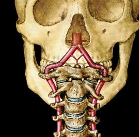
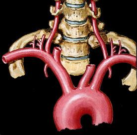
1 Spectral Doppler Interpretation Director Director of of Ultrasound Ultrasound Education Education & & Quality Quality Assurance Assurance Baylor Baylor College College of of Medicine Medicine Division Division of of Maternal-Fetal Maternal-Fetal Medicine Medicine Maternal Maternal Fetal Fetal Center Center Imaging Imaging Manager Manager Texas Texas Children s Children s Hospital, Hospital, Pavilion Pavilion for for Women Women Houston Houston Texas Texas & & Clinical Clinical Instructor Instructor Thomas Thomas Jefferson Jefferson University University Hospital Hospital -- Radiology Radiology Department Department Philadelphia, Philadelphia, Pennsylvania Pennsylvania What effects will proximal or distal disease have on an waveform? Distal Disease Changes the resistance Distal disease Changes the resistance Proximal disease Acute & chronic parenchymal disease Obstruction Renal vein thrombosis Changes the strength of the signal Distal Disease Distal Disease Changes the resistance Changes the resistance 1


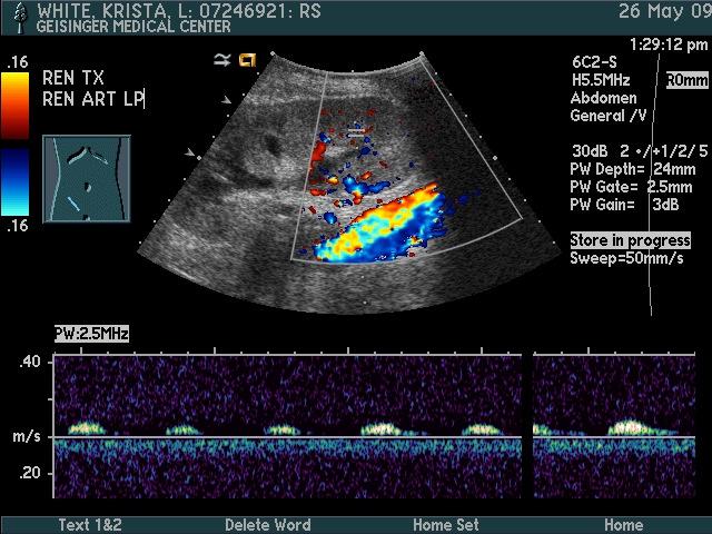
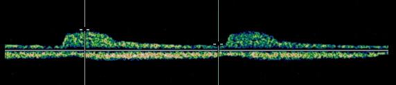
2 Distal Disease Proximal Disease Changes the resistance Changes the strength of the signal Caution! Increase Sweep Speed Tardus Parvus Waveform Tardus Slow & late Parvus Small & little Tardus Parvus Waveform Proximal Disease Changes the strength of the signal Systolic acceleration diminished Acceleration time prolonged Waveform shape Diminished pulsatility 2


3 Diagnostic Challenge Celiac Artery Stenosis Decreased or dampened waveform distal to stenosis Remember! It is more difficult to demonstrate tardus parvus in a stiff vessel Doppler Analysis Qualitative The visual or acoustic evaluation of Doppler wave form Atherosclotic arteries & increased distal resistance masks the post-stenotic tardus parvus 3


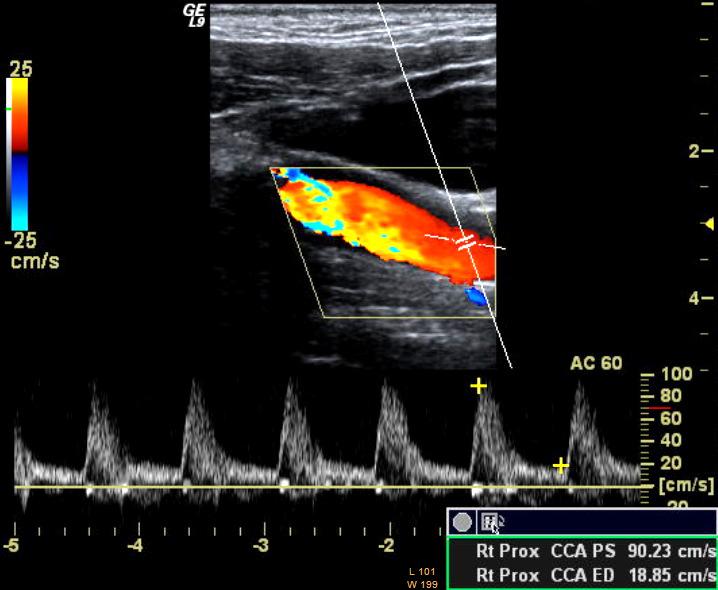
4 Doppler Analysis Qualitative The visual or acoustic evaluation of Doppler wave form Quantitative Calculation of volume flow Calculation of indices Indirect method to evaluate blood perfusion Doppler Waveform Waveform is commonly described by pulsatility which can be measured Peak Systolic velocity PSV Resistance Index RI Pulsatility Index PI Systolic/Diastolic Ratio S/D Acceleration Index AI Acceleration Time AT How to Look at a Waveform? Where & how was signal obtained? Presence of flow Direction of flow Characterization of signal Quality of exam How to Look at a Waveform? Where & how was signal obtained? 1 Where is the sample volume 2 What is the sample volume size 3 What is the Doppler angle 4 Technical considerations Where is the sample volume? What is the Sample Volume Size? 4


5 What is the Sample Volume Size? What is the Sample Volume Size? What is the Sample Volume Size? Too small a gate may give the false impression of reduced or even absent flow What is the Doppler Angle? Angle is the result of Doppler line direction Cursor correction Angle affects velocity accuracy V = 2. F t. cos Θ. F d C What is the Doppler Angle? What is the Doppler Angle? A 5


6 What is the Doppler Angle? What is the Doppler Angle? What is the Doppler Angle? What is the Doppler Angle? Caution Be Careful with Doppler Angle 6


7 Caution Angle to Flow Caution Angle to Flow Caution Spectral Display effect of Doppler Gain Caution Spectral Doppler - Velocity Scale Caution 7


8 Caution Spectral Doppler - Velocity Scale Spectral Doppler - Velocity Scale Decreased PRF Increased PRF How to Look at a Waveform? Where & how was signal obtained? Flow direction Diagnostic Challenge How to Look at a Waveform? Where & how was signal obtained? Flow direction Characterization of signal 8


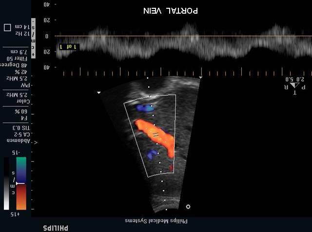
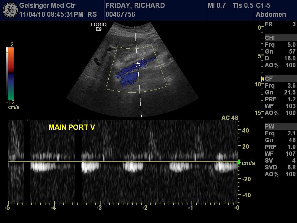
9 Characterization of Signal Spectral Analysis Site of signal What is normal & abnormal Shape (edge) of spectrum Velocity of blood flow Pulsatility Structure of spectrum Distribution of blood velocities Spectral broadening Characterization of Signal Spectral Analysis Site of signal What is normal & abnormal Shape (edge) of spectrum Velocity of blood flow Pulsatility Structure of spectrum Distribution of blood velocities Spectral broadening Diagnosis What does increased pulsatility in the hepatic veins suggest? What does loss of pulsatility in the hepatic veins suggest? 1. Cirrhosis 2. Compression from mass 3. Partial thrombosis 4. Occlusion or narrowing of the IVC above the level of the hepatic veins A What does loss of pulsatility in the portal veins suggest? B C D 9

 may")


10 What does pulsatile portal vein suggest? Portal Vein Gas Any communication between the systemic and portal veins, (portosystemic shunts, fistulae) may lead to a pulsatile portal vein Increased pulsatility of portal venous flow may also be seen with congestion of the liver, especially the passive congestion associated with right-sided cardiac failure and/or tricuspid regurgitation Portal vein gas Characterization of Signal Edge of spectral envelope Waveform shape & pulsatility Peak velocities Ischemic, inflammatory, or infectious bowel diseases Pediatric age group Necrotizing enterocolitis Characterization of Signal Celiac Artery Distribution of blood velocities Gray scale distribution of all RBC 10


11 Important Signs of Stenosis Important Signs of Stenosis Proximal to stenosis Change in pulsitility At the stenosis Elevated velocities compared to pre-stenotic segment Laminar flow Important Signs of Stenosis Important Signs of Stenosis Beyond the stenosis Post stenotic turbulence or disturb flow Spectral broadening Loss of well defined spectral edge Distal to stenosis Down stream Tardus-Parvus Velocity should drop off distal to stenosis Exceptions: long stenosis, near occlusive lesions Distribution of Doppler frequencies seen in spectrum filling of envelope Celiac Artery 11


12 Celiac Artery Characterization of Signal Characterization of Signal Aorta Lt Renal Artery Characterization of Signal Diastolic Flow Physiological and pathological conditions: Cardiac and aortic factors Vessel compliance Downstream resistance Venous and arteriovenous connections Stenosis at, above or beyond vessel 12


13 Increased Diastolic Flow Effect of Eating on Diastole Eating affects SMA Exercise affects muscles Neovascularity Inflammatory conditions Corpus luteum development Menstrual cycle on uterus Arteriovenous Shunting SMA Before Meal SMA After Meal Nonspecificity of Neovascularity Inflammatory Conditions Ovarian Cancer Benign Hemorrhagic Cyst Orchitis Uterine Artery Flow Ovulatory cycles There is an increase in end diastolic flow velocities between proliferative & secretory phases Uterine Artery Persistent Notching Notch at 25 weeks implies incomplete trophoblastic invasion and is predictive of preeclampsia and/or delivering a growth restricted fetus 13


14 Arteriovenous Shunting Arteriovenous Fistula Small connections tumor vessels, arterioportal shunting in cirrhosis Large vessels AV Malformations vein of Galen aneurysm uterine AVM AV Fistulas traumatic AVF HA R T E R I A L Fistula V E N O U S Flow Characterization-AVF the site of fistula Flow Characterization-AVF Low resistance arterial flow proximally Flow Characterization-AVF High resistance arterial flow distally Flow Characterization-AVF Arterialized venous flow 14


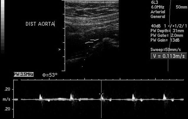
15 Decreased Diastolic Flow Change of resistance from lower to higher decreases diastolic flow Frequently seen in distal stenosis or occlusive disease Venous outflow obstruction Distal Stenosis Occlusive Disease Occlusive Disease Sagittal Aorta Staccato Flow Thrombosed Distal Aorta & Common Iliac Arteries Staccato Flow 15


16 Vascular Destruction Capillary and vascular destruction obstructs flow! decreasing diastole Common sites Renal disease Placental diseases Decreased Diastolic Flow Change of resistance from lower to higher decreases diastolic flow Frequently seen in distal stenosis or occlusive disease Venous outflow obstruction Venous Obstruction Venous outflow affects diastole Physiologic Erection Pathologic Renal vein thrombosis In systole the pressure in the artery is higher than in the pseudoaneurysm, blood flows in to pseudoaneurysm In diastole the pressure in the pseudoaneurysm is higher than in the artery, blood leaves the pseudoaneurysm Collateral Flow Flow Characterization In occlusive disease, a collateral may mimic the original vessel In ICA occlusive disease, the ECA may show increased diastolic flow In CCA occlusion, retrograde flow in external reconstitutes ICA and shows ICA type waveform 16
17 Conclusion Spectral Doppler Interpretation What effects will proximal or distal disease have on an waveform? How to look at a waveform? Doppler analysis Stenosis profiles Diastolic flow Thank You 17
What effects will proximal or distal disease have on a waveform?
 Spectral Doppler Interpretation Director of Ultrasound Education & Quality Assurance Baylor College of Medicine Division of Maternal-Fetal Medicine Maternal Fetal Center Imaging Manager Texas Children
Transducer Selection. Renal Artery Duplex Exam. Renal Scan. Renal Scan Echogenicity. How to Perform an Optimal Renal Artery Doppler Examination
 How to Perform an Optimal Renal Artery Doppler Examination Director of Ultrasound Education & Quality Assurance Baylor College of Medicine Division of Maternal-Fetal Medicine Maternal Fetal Center Imaging
DISCLOSURE TEST YOUR WAVEFORM IQ. Partial volume artifact. 86 yo female with right arm swelling, picc line. AVF on left? Dx?
 Deborah Rubens University of Rochester Rochester, NY DISCLOSURE Neither I nor my immediate family have a financial relationship with a commercial organization that may have a direct or indirect interest
Vascular Imaging in the Pediatric Abdomen. Jonathan Swanson, MD
 Vascular Imaging in the Pediatric Abdomen Jonathan Swanson, MD Goals and Objectives To understand the imaging approach, appearance, and clinical manifestations of the common pediatric abdominal vascular
HD Scanning: Velocities and Volume Flow
 HD Scanning: Velocities and Volume Flow Non-Invasive Lab Symposium West Orange, NJ April 27, 2018 Volume Flow Cindy Sturt, MD, FACS, RVT 500,000 Americans on dialysis 20-25% annual mortality 65% 5 year
Vascular Sonography Examination
 Vascular Sonography Examination The purpose of The American Registry of Radiologic Technologists (ARRT ) Vascular Sonography Examination is to assess the knowledge and cognitive skills underlying the intelligent
Abdominal Doppler Mastering the next level of vascular anatomy in the belly. Cindy A. Owen, RDMS, RVT
 Abdominal Doppler Mastering the next level of vascular anatomy in the belly Cindy A. Owen, RDMS, RVT Introduction Abdominal Doppler is a tough exam Success is dependent on: Patient body habitus Patient
STRUCTURED EDUCATION REQUIREMENTS IMPLEMENTATION DATE: JULY 1, 2016
 STRUCTURED EDUCATION REQUIREMENTS Vascular Sonography The purpose of structured education is to provide the opportunity for individuals to develop mastery of discipline-specific knowledge that, when coupled
Visceral Vascular Ultrasound. Joel Thompson, MD, MPH Borg & Ide Imaging
 Visceral Vascular Ultrasound Joel Thompson, MD, MPH Borg & Ide Imaging Objectives: Review major abdominal vascular structures Identify normal peak systolic velocity (PSV) for major abdominal arteries.
Image Formation (10) 2 Evaluation and Selection of Representative Images (10)
 2 Evaluation and Selection of Representative Images (10)") STRUCTURED SELF ASSESSMENT CONTENT SPECIFICATIONS SSA LAUNCH DATE: JANUARY 1, 2018 Vascular Sonography The purpose of continuing qualifications requirements (CQR) is to assist registered technologists
STRUCTURED SELF ASSESSMENT CONTENT SPECIFICATIONS SSA LAUNCH DATE: JANUARY 1, 2018 Vascular Sonography The purpose of continuing qualifications requirements (CQR) is to assist registered technologists
What Do We Know? Disclosure Statement: 3/11/2015. Deep abdominal imaging
 Marsha M. Neumyer, BS, RVT, FSVU, FSDMS, FAIUM International Director Vascular Diagnostic Educational Services Vascular Resource Associates Harrisburg, PA Disclosure Statement: CME Calendar QR Code Marsha
No financial or commercial relationships to disclose
 Deanna New, RVT No financial or commercial relationships to disclose IAC REQUIREMENTS: The main duty of a sonographer is to make the physician or radiologists job easier by capturing images and doing
Postoperative AV Fistula Evaluation. Postoperative examination protocol. Postoperative AVF Protocol. Hemodialysis Access Surveillance
 Hemodialysis Access Surveillance Postoperative AV Fistula Evaluation Failure of maturation Stenosis Perigraft mass/fluid collection Joseph L. Mills, Sr., M.D. Professor of Surgery Chief, Division of Vascular
Goals. Access flow and renal artery stenosis evaluation by Doppler ultrasound. Reimbursement. WHY use of Doppler Ultrasound
 Access flow and renal artery stenosis evaluation by Doppler ultrasound Adina Voiculescu, MD Interventional Nephrology Brigham and Women s Hospital Boston Instructor at Harvard Medical School Understand
NON-ATHEROSCLEROTIC PATHOLOGY OF THE CAROTID ARTERIES
 NON-ATHEROSCLEROTIC PATHOLOGY OF THE CAROTID ARTERIES Leslie M. Scoutt, MD, FACR Professor of Diagnostic Radiology & Surgery Vice Chair, Dept of Radiology & Biomedical Imaging Chief, Ultrasound Section
Optimising your Doppler settings for an accurate PI. Alison McGuinness Mid Yorks Hospitals
 Optimising your Doppler settings for an accurate PI Alison McGuinness Mid Yorks Hospitals Applications Both maternal uterine and fetal circulations can be studied with doppler sonography Uterine arteries
Case 8038 Renal allograft complicated with renal artery stenosis
 Case 8038 Renal allograft complicated with renal artery stenosis Santiago I, Canelas A, Pinto AP Section: Cardiovascular Published: 2009, Nov. 30 Patient: 61 year(s), male Clinical History A 61-year-old
Carotid Artery Doppler
 Carotid Artery Doppler Patient Position supine or semisupine head slightly hyper extended rotated 45 away from the side being examined. Higher frequency linear transducers (7 MHz) Vessels should be imaged
US of Renovascular Hypertension. Jonathan R. Dillman, MD, MSc Associate Professor Director, Thoracoabdominal Imaging
 US of Renovascular Hypertension Jonathan R. Dillman, MD, MSc Associate Professor Director, Thoracoabdominal Imaging Disclosures Nothing Relevant Unrelated grant funding Siemens US Toshiba US Objectives
8/20/18. The Doppler Effect. Objectives. What is the Doppler Effect. Doppler principles. Spectral Waveform. Image recognition. Vascular Ultrasound
 Vascular Ultrasound: Physics and Haemodynamics Objectives Doppler principles Spectral Waveform Key factors Haemodynamics: Stenosis Waveforms Image recognition Vascular Ultrasound: A flawed paradigm What
Carotid Abnormalities Coils, Kinks and Tortuosity David Lorelli M.D., RVT, FACS Michigan Vascular Association Conference Saturday, October 20, 2012
 Carotid Abnormalities Coils, Kinks and Tortuosity David Lorelli M.D., RVT, FACS Michigan Vascular Association Conference Saturday, October 20, 2012 Page 1 Table of Contents Carotid Anatomy Carotid Duplex
Doppler ultrasound as noninvasive diagnosis of peripheral arterial disease
 Doppler ultrasound as noninvasive diagnosis of peripheral arterial disease Poster No.: C-0246 Congress: ECR 2012 Type: Scientific Exhibit Authors: C. Ballester Valles, F. Aparici-Robles; Valencia/ES Keywords:
Doppler Ultrasonography of the Liver: What Every General Radiologist Should Know
 Doppler Ultrasonography of the Liver: What Every General Radiologist Should Know Poster No.: C-1658 Congress: ECR 2014 Type: Authors: Keywords: DOI: Educational Exhibit T. González de la Huebra Labrador,
Treatment of choice for end stage renal disease Imaging to establish baseline and diagnosis of potential complications Review common surgical
 Treatment of choice for end stage renal disease Imaging to establish baseline and diagnosis of potential complications Review common surgical techniques Review normal appearance Discuss US diagnosis of
MESENTERIC ISCHEMIA. Phillip J Bendick, PhD
 MESENTERIC ISCHEMIA Phillip J Bendick, PhD Arterial Celiac - Hepatic - Splenic Superior Mesenteric Artery Inferior Mesenteric Artery Venous Mesenteric system Porto - hepatic system Inferior Vena Cava Acute
Doppler US of the Liver Made Simple 1
 Note: This copy is for your personal non-commercial use only. To order presentation-ready copies for distribution to your colleagues or clients, contact us at www.rsna.org/rsnarights. Gastrointestinal
Abdomen Sonography Examination Content Outline
 Abdomen Sonography Examination Content Outline (Outline Summary) # Domain Subdomain Percentage 1 2 3 Anatomy, Perfusion, and Function Pathology, Vascular Abnormalities, Trauma, and Postoperative Anatomy
Indications: following: embolization. artery that has diseases 5. The evaluation. of suspected. such entities. a cold hand. biopsy
 Peripheral Arterial Ultrasound Protocol Using Color and Spectral Doppler Reviewed by: Mark Yuhasz, MD Last Review Date: January 2015 Contact: (866) 761 4200, Option 1 Indications: The indications for peripheral
Disclosure Statement:
 Marsha M. Neumyer, BS, RVT, FSVU, FSDMS, FAIUM International Director Vascular Diagnostic Educational Services Vascular Resource Associates Harrisburg, PA Disclosure Statement: CME Calendar QR Code Marsha
Duplex Ultrasound of the Renal Arteries. Duplex Ultrasound. In the Beginning
 Duplex Ultrasound of the Renal Arteries DIMENSIONS IN HEART AND VASCULAR CARE 2013 PENN STATE HEART AND VASCULAR INSTITUTE ROBERT G. ATNIP MD PROFESSOR OF SURGERY AND RADIOLOGY Duplex Ultrasound Developed
Ultrasound Imaging of The Posterior Circulation
 Ultrasound Imaging of The Posterior Circulation Michigan Sonographers Society 2 Nd Annual Fall Vascular Conference Larry N. Raber RDMS-RVT Clinical Manager General Ultrasound/Neurovascular Laboratory Cleveland
ASDIN 7th Annual Scientific Meeting DISCLOSURES TECHNICAL CONSIDERATIONS TECHNICAL CONSIDERATIONS UTILITY OF ULTRASOUND IN EVALUATING ACCESS
 DISCLOSURES UTILITY OF ULTRASOUND IN EVALUATING ACCESS DYSFUNCTION None Vandana Dua Niyyar, MD Assistant Professor of Medicine, Division of Nephrology, Emory University UTILITY OF ULTRASOUND IN ACCESS
Assessment of fetal heart function and rhythm
 Assessment of fetal heart function and rhythm The fetal myocardium Early Gestation Myofibrils 30% of myocytes Less sarcoplasmic reticula Late Gestation Myofibrils 60% of myocytes Increased force per unit
Volume Flow. Volume Flow
 Volume Flow Jonathan M. Rubin, M.D., Ph.D. Department of Radiology Volume Flow Technique initially described by Hottenger and Meindl in 1974 Describes method for measuring the total flux across a flow
11 TH ANNUAL VASCULAR NONINVASIVE TESTING SYMPOSIUM NOVEMBER 10, 2018
 11 TH ANNUAL VASCULAR NONINVASIVE TESTING SYMPOSIUM NOVEMBER 10, 2018 RENAL ARTERY DISEASE AND RENOVASCULAR HYPERTENSION 1 WHAT IS RENOVASCULAR HYPERTENSION? https://my.clevelandclinic.org/health/diseases/16459-renovascular-hypertension
The Fetus: Five Top Do Not Miss Diagnoses. Doppler Ultrasound
 The Fetus: Five Top Do Not Miss Diagnoses Doppler Ultrasound Giancarlo Mari, MD, MBA Professor and Chair Department of Obstetrics and Gynecology University of Tennessee Health Science Center Memphis, TN
Deb Coghlan AMS (Vascular and General ) Brisbane, Australia
 Brisbane, Australia") Deb Coghlan AMS (Vascular and General ) Brisbane, Australia ANEURYSMAL DIISEASE The infrarenal aorta enlarges with age, and is the commonest site for arterial aneurysms. An aneurysm is a permanent focal
Deb Coghlan AMS (Vascular and General ) Brisbane, Australia ANEURYSMAL DIISEASE The infrarenal aorta enlarges with age, and is the commonest site for arterial aneurysms. An aneurysm is a permanent focal
4/19/2018. St. Cloud Hospital Perinatology Kristin Olson, RDMS, RVT
 St. Cloud Hospital Perinatology Kristin Olson, RDMS, RVT Review Fetal Circulation Provide Indications for Umbilical Artery, Middle Cerebral Artery, and Ductus Venosus Doppler studies. Demonstrate normal
Physician s Vascular Interpretation Examination Content Outline
 Physician s Vascular Interpretation Examination Content Outline (Outline Summary) # Domain Subdomain Percentage 1 2 3 4 5 6 Cerebrovascular Abdominal Peripheral Arterial - Duplex Imaging Peripheral Arterial
Non-invasive examination
 Non-invasive examination Segmental pressure and Ankle-Brachial Index (ABI) The segmental blood pressure (SBP) examination is a simple, noninvasive method for diagnosing and localizing arterial disease.
Ultrasound of the Renal Arteries
 Ultrasound of the Renal Arteries Greg Curry Vascular Ultrasound Workshop Aug 2017 The Examination Technique Pathophysiology Role of US then and now Background Live Scanning Ultrasound Population: 20% Hypertensive
Adult Echocardiography Examination Content Outline
 Adult Echocardiography Examination Content Outline (Outline Summary) # Domain Subdomain Percentage 1 2 3 4 5 Anatomy and Physiology Pathology Clinical Care and Safety Measurement Techniques, Maneuvers,
Vascular Ultrasound: Current state, current needs, future directions
 Vascular Ultrasound: Current state, current needs, future directions Laurence Needleman, MD Thomas Jefferson University Hospitals Sidney Kimmel Medical College of Thomas Jefferson University Disclosures
Job Task Analysis for ARDMS Abdomen Data Collected: June 30, 2011
 Job Task Analysis for ARDMS Abdomen Data Collected: June 30, 2011 Reported: Analysis Summary for: Abdomen Examination Survey Dates 06/13/2011-06/26/2011 Invited Respondents 6,000 Surveys with Demographics
GUNDERSEN/LUTHERAN ULTRASOUND DEPARTMENT POLICY AND PROCEDURE MANUAL
 GUNDERSEN/LUTHERAN ULTRASOUND DEPARTMENT POLICY AND PROCEDURE MANUAL SUBJECT: Carotid Duplex Ultrasound SECTION: Vascular Ultrasound ORIGINATOR: Deborah L. Richert, BSVT, RDMS, RVT DATE: October 15, 2015
Doppler Ultrasound Findings in the Hepatic Artery Shortly After Liver Transplantation
 Gastrointestinal Imaging Pictorial Essay García-riado et al. Hepatic rtery fter Liver Transplantation Gastrointestinal Imaging Pictorial Essay FOUS ON: Ángeles García-riado 1 Rosa Gilabert nnalisa erzigotti
Pitfalls in the evaluation of carotid artery stenosis. Serge Kownator «Centre Cardiologique et Vasculaire» Thionville, Fr
 Pitfalls in the evaluation of carotid artery stenosis Serge Kownator «Centre Cardiologique et Vasculaire» Thionville, Fr Disclosure Statement of Financial Interest I currently have, or have had over the
Review Article Duplex Ultrasound Evaluation of Hemodialysis Access: A Detailed Protocol
 International Nephrology Volume 2012, Article ID 508956, 7 pages doi:10.1155/2012/508956 Review Article Duplex Ultrasound Evaluation of Hemodialysis Access: A Detailed Protocol Victoria Teodorescu, 1,
Mesenteric/Splanchnic Artery Duplex Imaging
 VASCULAR TECHNOLOGY PROFESSIONAL PERFORMANCE GUIDELINES Mesenteric/Splanchnic Artery Duplex Imaging This Guideline was prepared by members of the Society for Vascular Ultrasound (SVU) as a template to
Renal Artery Stenosis With Severe Hypertension: A Case Report
 CASE REPORT Renal Artery Stenosis With Severe Hypertension: A Case Report Suwaid MA ABSTRACT Background: Renal artery stenosis (RAS) is found in 77% of hypertensive patients and is responsible for 1-2%
TRANSCRANIAL DOPPLER ULTRASOUND INTRODUCTION TO TCD INTERPRETATION
 TRANSCRANIAL DOPPLER ULTRASOUND INTRODUCTION TO TCD INTERPRETATION ---Rune Aaslid First TCD Publication 1982 WHAT IS TCD? Uses 2 MHz pulsed Doppler ultrasound Passes through cranial windows Provides information
The Effect of Poststenotic Vessel Wall Compliance
 The Effect of Poststenotic Vessel Wall Compliance upon the Pulsus Tardus Phenomenon Ronald O. Bude, M.D. Jonathan M. Rubin, M.D., Ph.D. Joel F. Platt, M.D. and Ronald S. Adler, M.D., Ph.D. ANN ARBOR, MICHIGAN
Neuro Quiz 29 Transcranial Doppler Monitoring
 Verghese Cherian, MD, FFARCSI Penn State Hershey Medical Center, Hershey Quiz Team Shobana Rajan, M.D Suneeta Gollapudy, M.D Angele Marie Theard, M.D Neuro Quiz 29 Transcranial Doppler Monitoring This
Carotid Duplex: Beyond Stenosis Ido Weinberg, MD Vascular Medicine Massachusetts General Hospital Assistant Professor of Medicine Harvard Medical
 Carotid Duplex: Beyond Stenosis Ido Weinberg, MD Vascular Medicine Massachusetts General Hospital Assistant Professor of Medicine Harvard Medical School Boston, Massachusetts Disclosures I do not have
Hemodynamic Assessment. Assessment of Systolic Function Doppler Hemodynamics
 Hemodynamic Assessment Matt M. Umland, RDCS, FASE Aurora Medical Group Milwaukee, WI Assessment of Systolic Function Doppler Hemodynamics Stroke Volume Cardiac Output Cardiac Index Tei Index/Index of myocardial
Carotid US: More than just a chart on the wall
 Carotid US: More than just a chart on the wall Leslie M. Scoutt, MD, FACR Professor of Diagnostic Radiology & Surgery Vice Chair, Dept of Radiology & Biomedical Imaging Chief, Ultrasound Section Medical
TCD IN THE NICU, PICU AND OTHER APPLICATIONS. Dorothy Bulas M.D. Professor of Pediatrics & Radiology Children s National Washington D.C.
 TCD IN THE NICU, PICU AND OTHER APPLICATIONS Dorothy Bulas M.D. Professor of Pediatrics & Radiology Children s National Washington D.C. Objectives Recognize normal and abnormal cranial blood flow patterns
The role for contrast-enhanced ultrasonography outside of focal liver lesions
 The role for contrast-enhanced ultrasonography outside of focal liver lesions Paul S. Sidhu King s College Hospital, London, UK Introduction Contrast-enhanced ultrasonography (US) of focal liver lesions
Section II: Patient Interview Grade: 5
 Only written competency completed with this EXACT form will be accepted for grading. No modifications to the LAYOUT of the form will be accepted for a written competency. Failure to comply will result
Carotid Ultrasound: Improving Ultrasound
 Carotid Ultrasound: Improving Ultrasound Edward I. Bluth, M.D., F.A.C.R. Chairman Emeritus, Department of Radiology, Ochsner Clinic Foundation, New Orleans, Louisiana Professor, Ochsner Clinical School,
Lower Extremity Arterial Doppler
 Lower Extremity Arterial Doppler 1. Spectral Doppler waveform should be taken in distal aorta and common iliac arteries. 2. R/L common femoral artery (CFA) color Doppler with velocity and B-mode. 3. R/L
2015 ARDMS Physicians Vascular Interpretation Job Task Analysis Summary Report
 P a g e 1 2015 ARDMS Physicians Vascular Interpretation Job Task Analysis Summary Report American Registry for Diagnostic Medical Sonography (ARDMS) P a g e 2 Table of Contents ABOUT THE REPORT... 3 METHODOLOGY...
The Role of US in Chronic Mesenteric Ischemia. Sagar S. Gandhi, MD Vascular Health Alliance Greenville Health System
 The Role of US in Chronic Mesenteric Ischemia Sagar S. Gandhi, MD Vascular Health Alliance Greenville Health System No Disclosures Mesenteric Ischemia Anatomy Presentation Diagnostic tools Treatment Celiac
Assessment of Cardio- & Neurovascular Hemodynamics in the Human Circulatory System using 4D flow MRI
 Assessment of Cardio- & Neurovascular Hemodynamics in the Human Circulatory System using 4D flow MRI Michael Markl, Ph.D. Departments of Radiology & Biomedical Engineering Northwestern University, Chicago,
Doppler Basic & Hemodynamic Calculations
 Doppler Basic & Hemodynamic Calculations August 19, 2017 Smonporn Boonyaratavej MD Division of Cardiology, Department of Medicine Chulalongkorn University Cardiac Center, King Chulalongkorn Memorial Hospital
Doppler Waveform Parvus and Tardus A Sign of Proximal Flow Obstruction
 Doppler Waveform Parvus and Tardus A Sign of Proximal Flow Obstruction Pesho S. Kotval, MD, PhD The Doppler linear flow velocity versus time spectrum obtained in an arterial flow system in which there
RPVI Exam Review ecourse
 RPVI Exam Review ecourse The RPVI Exam Review ecourse consists of ten Vascular Physics Modules and fourteen Vascular Specialty Modules. Detailed descriptions of module content are listed below. During
Vascular Technology Examination Content Outline
 Vascular Technology Examination Content Outline (Outline Summary) # Domain Subdomain Percentage 1 Normal Anatomy, Perfusion, and Function Evaluate normal anatomy, perfusion, function 2 Pathology, Perfusion,
CVS Hemodynamics. Change in blood pressure:
 CVS Hemodynamics -The distribution of blood inside the circulation: The major part of blood volume is found in the venous system 60% (2/3), that s why veins are called the capacitance vessels. -Arteries
Measure #195 (NQF 0507): Radiology: Stenosis Measurement in Carotid Imaging Reports National Quality Strategy Domain: Effective Clinical Care
: Radiology: Stenosis Measurement in Carotid Imaging Reports National Quality Strategy Domain: Effective Clinical Care") Measure #195 (NQF 0507): Radiology: Stenosis Measurement in Carotid Imaging Reports National Quality Strategy Domain: Effective Clinical Care 2017 OPTIONS FOR INDIVIDUAL MEASURES: CLAIMS ONLY MEASURE TYPE:
Measure #195 (NQF 0507): Radiology: Stenosis Measurement in Carotid Imaging Reports National Quality Strategy Domain: Effective Clinical Care 2017 OPTIONS FOR INDIVIDUAL MEASURES: CLAIMS ONLY MEASURE TYPE:
Duplex ultrasound is first-line imaging for all
 Our Protocol for Transabdominal Pelvic Vein Duplex Ultrasound A summary of s protocol for pelvic vein duplex ultrasonography, including equipment, patient positioning, ultrasound settings, and technique.
DICOM Correction Item
 Correction Number DICOM Correction Item CP-449 Log Summary: Type of Modification Correct Value Name of Standard PS 3.16-2004 Rationale for Correction: Clinical applications use arterial and venous branches
Vascular complications after adult liver transplantation: Evaluation with Doppler ultrasound
 Vascular complications after adult liver transplantation: Evaluation with Doppler ultrasound Poster No.: C-2588 Congress: ECR 2012 Type: Educational Exhibit Authors: M. S. C. Sousa, M. C. Cordeiro, R.
Quality ID #195 (NQF 0507): Radiology: Stenosis Measurement in Carotid Imaging Reports National Quality Strategy Domain: Effective Clinical Care
: Radiology: Stenosis Measurement in Carotid Imaging Reports National Quality Strategy Domain: Effective Clinical Care") Quality ID #195 (NQF 0507): Radiology: Stenosis Measurement in Carotid Imaging Reports National Quality Strategy Domain: Effective Clinical Care 2018 OPTIONS FOR INDIVIDUAL MEASURES: REGISTRY ONLY MEASURE
Quality ID #195 (NQF 0507): Radiology: Stenosis Measurement in Carotid Imaging Reports National Quality Strategy Domain: Effective Clinical Care 2018 OPTIONS FOR INDIVIDUAL MEASURES: REGISTRY ONLY MEASURE
Copy Here VASCULAR ACCESS COMPLICATIONS TYPICAL ARTERIAL COMPLICATION PROTOCOL ARTERIAL EXAMINATION: NORMAL FINDINGS X X
 VASCULAR ACCESS COMPLICATIONS X X Copy Here Natalia Fendrikova Mahlay, MD, RPVI ARTERIAL EXAMINATION: NORMAL FINDINGS TYPICAL ARTERIAL COMPLICATION PROTOCOL Indications Prior vascular access Pulsatile
Vascular Surgery Cases: Detours. Brian F. Stull, RDMS, RVT UNC REX Healthcare Vascular Specialists
 Vascular Surgery Cases: Detours Brian F. Stull, RDMS, RVT UNC REX Healthcare Vascular Specialists Brian.Stull@Unchealth.unc.edu Objectives Anatomy of a bypass graft Where does it connect, where does it
Case Report 1. CTA head. (c) Tele3D Advantage, LLC
 Tele3D Advantage, LLC") Case Report 1 CTA head 1 History 82 YEAR OLD woman with signs and symptoms of increased intra cranial pressure in setting of SAH. CT Brain was performed followed by CT Angiography of head. 2 CT brain Extensive
Case Report 1 CTA head 1 History 82 YEAR OLD woman with signs and symptoms of increased intra cranial pressure in setting of SAH. CT Brain was performed followed by CT Angiography of head. 2 CT brain Extensive
Neurovascular Ultrasound Course
 Neurovascular Ultrasound Course William M. McKinney (6/6/30-10/24/03) Father of Neurosonology Founder, Neurosonology Course, WFUSM Welcome to Winston-Salem, NC, Wake Forest School of Medicine, and the
Circulatory System MARE HEIFER. aorta cvc. iia eia ipa. aorta. dca. (uov) oa. uboa. uma. ubva va. bua. iia. uma. ubva. eia. ua bua. cvc.
 oa. uboa. uma. ubva va. bua. iia. uma. ubva. eia. ua bua. cvc.") Circulatory System 13 MARE aorta cvc ov (uov) oa dca uboa iia eia ipa ua uma bua ubva va HEIFER iia aorta cvc eia oa uma ua bua ubva va ov (uov) uboa 18 Chapter 1 Hemodynamics Plug flow Natural disturbed
Circulatory System 13 MARE aorta cvc ov (uov) oa dca uboa iia eia ipa ua uma bua ubva va HEIFER iia aorta cvc eia oa uma ua bua ubva va ov (uov) uboa 18 Chapter 1 Hemodynamics Plug flow Natural disturbed
Lower Extremity Arterial Duplex Evaluation
 VASCULAR TECHNOLOGY PROFESSIONAL PERFORMANCE GUIDELINES Lower Extremity Arterial Duplex Evaluation This Guideline was prepared by the Professional Guidelines Subcommittee of the Society for Vascular Ultrasound
Radial Artery Assessment for Coronary Artery Bypass
 VASCULAR TECHNOLOGY PROFESSIONAL PERFORMANCE GUIDELINES Radial Artery Assessment for Coronary Artery Bypass This Guideline was prepared by the Professional Guidelines Subcommittee of the Society for Vascular
Abdominal Doppler. Cases of Where, Why, and How
 Abdominal Doppler Cases of Where, Why, and How Jill D. Trotter, BS, RT(R), RDMS, RVT Director, Diagnostic Medical Sonography Program Vanderbilt University/Vanderbilt Medical Center Nashville, Tennessee
Transcranial Doppler (Basic Step) Dae-il Chang, M.D., Sung Sang Yoon, M.D. Department of Neurology, College of Medicine, Kyunghee university
 Dae-il Chang, M.D., Sung Sang Yoon, M.D. Department of Neurology, College of Medicine, Kyunghee university") Transcranial Doppler (Basic Step) Dae-il Chang, M.D., Sung Sang Yoon, M.D. Department of Neurology, College of Medicine, Kyunghee university Principles of Doppler Ultrasonography Major target Speed & direction
Transcranial Doppler (Basic Step) Dae-il Chang, M.D., Sung Sang Yoon, M.D. Department of Neurology, College of Medicine, Kyunghee university Principles of Doppler Ultrasonography Major target Speed & direction
Guide to Small Animal Vascular Imaging using the Vevo 770 Micro-Ultrasound System
 Guide to Small Animal Vascular Imaging using the Vevo 770 Micro-Ultrasound System January 2007 Objectives: After completion of this module, the participant will be able to accomplish the following: Understand
(D) (E) (F) 6. The extrasystolic beat would produce (A) increased pulse pressure because contractility. is increased. increased
 (E) (F) 6. The extrasystolic beat would produce (A) increased pulse pressure because contractility. is increased. increased") Review Test 1. A 53-year-old woman is found, by arteriography, to have 5% narrowing of her left renal artery. What is the expected change in blood flow through the stenotic artery? Decrease to 1 2 Decrease
Review Test 1. A 53-year-old woman is found, by arteriography, to have 5% narrowing of her left renal artery. What is the expected change in blood flow through the stenotic artery? Decrease to 1 2 Decrease
ADDITIONS. The following codes have been added.
 ADDITIONS The following codes have been added. 99446 Interprofessional telephone/internet assessment and management service provided by treating/requesting physician or other qualified health care professional;
noninvasive, nonionizing, portable, inexpensive, safe for serial or prolonged studies
 TRANS CRANIAL DOPPLER Presented by : Anil Garg Transcranial Doppler 1982, Aaslid and colleagues introduced TCD as a non-invasive technique for monitoring blood flow velocity in basal cerebral arteries
Pre-and Post Procedure Non-Invasive Evaluation of the Patient with Carotid Disease
 Pre-and Post Procedure Non-Invasive Evaluation of the Patient with Carotid Disease Michael R. Jaff, D.O., F.A.C.P., F.A.C.C. Assistant Professor of Medicine Harvard Medical School Director, Vascular Medicine
The Doppler Examination. Katie Twomley, MD Wake Forest Baptist Health - Lexington
 The Doppler Examination Katie Twomley, MD Wake Forest Baptist Health - Lexington OUTLINE Principles/Physics Use in valvular assessment Aortic stenosis (continuity equation) Aortic regurgitation (pressure
Case Endovascular management of non maturing dyalisis vascular access
 Case 10238 Endovascular management of non maturing dyalisis vascular access Guedes Pinto 1, Erique; Madeira 2, Célia; Sousa 3, Marta; Penha 1, Diana; Rosa 1, Luís; Germano 1, Ana; Baptista 1, Manuela 1
1. Long images of aorta (prox, mid, and dist) with AP measurements. 2. Trans images of aorta (prox, mid, and dist) with R/L measurements.
 with AP measurements. 2. Trans images of aorta (prox, mid, and dist) with R/L measurements.") Aorta 1. Long images of aorta (prox, mid, and dist) with AP measurements. 2. Trans images of aorta (prox, mid, and dist) with R/L measurements. 3. Long images of R/L common iliac arteries with AP measurements.
Aorta 1. Long images of aorta (prox, mid, and dist) with AP measurements. 2. Trans images of aorta (prox, mid, and dist) with R/L measurements. 3. Long images of R/L common iliac arteries with AP measurements.
Recommendations for Follow-up After Vascular Surgery Arterial Procedures SVS Practice Guidelines
 Recommendations for Follow-up After Vascular Surgery Arterial Procedures 2018 SVS Practice Guidelines vsweb.org/svsguidelines About the guidelines Published in the July 2018 issue of Journal of Vascular
Introduction to the Native Arteriovenous Fistula: A primer for medical students and radiology residents
 Introduction to the Native Arteriovenous Fistula: A primer for medical students and radiology residents Jesus Contreras, D.O. PGY-4 John Yasmer, D.O. Department of Radiology No Disclosures Objectives Introduce
RadRx Your Prescription for Accurate Coding & Reimbursement Copyright All Rights Reserved.
 Interventional Radiology Coding Case Studies Prepared by Stacie L. Buck, RHIA, CCS-P, RCC, CIRCC, AAPC Fellow President & Senior Consultant Week of October 29, 2018 Mesenteric Arteriogram & Thrombectomy/Thrombolysis
Uncommon Doppler Echocardiographic Findings of Severe Pulmonic Insufficiency
 Uncommon Doppler Echocardiographic Findings of Severe Pulmonic Insufficiency Rahul R. Jhaveri, MD, Muhamed Saric, MD, PhD, FASE, and Itzhak Kronzon, MD, FASE, New York, New York Background: Two-dimensional
Obliterative hepatocavopathy ultrasound and cavography findings
 doi:10.2478/v10019-008-0020-6 case report Obliterative hepatocavopathy ultrasound and cavography findings Ramazan Kutlu Department of Radiology, Inonu University School of Medicine, Malatya, Turkey ackgound.
Radiologic Importance of a High- Resistive Vertebral Artery Doppler Waveform on Carotid Duplex Ultrasonography
 CME Article Radiologic Importance of a High- Resistive Vertebral Artery Doppler Waveform on Carotid Duplex Ultrasonography Esther S. H. Kim, MD, MPH, Megan Thompson, Kristine M. Nacion, BA, Carmel Celestin,
IAC Standards and Guidelines for Vascular Testing Accreditation
 IAC Standards and Guidelines for Vascular Testing Accreditation Table of Contents All entries in Table of Contents are linked to the corresponding sections. Introduction... 4 Part A: Organization... 5
Radiologic Evaluation of Peripheral Arterial Disease
 January 2003 Radiologic Evaluation of Peripheral Arterial Disease Grace Tye, Harvard Medical School Year III Patient D.M. CC: 44 y/o male with pain in his buttocks Occurs after walking 2 blocks. Pain is
PART II ECHOCARDIOGRAPHY LABORATORY OPERATIONS ADULT TRANSTHORACIC ECHOCARDIOGRAPHY TESTING
 PART II ECHOCARDIOGRAPHY LABORATORY OPERATIONS ADULT TRANSTHORACIC ECHOCARDIOGRAPHY TESTING STANDARD - Primary Instrumentation 1.1 Cardiac Ultrasound Systems SECTION 1 Instrumentation Ultrasound instruments