Carotid Artery Doppler
|
|
|
- Roxanne O’Connor’
- 6 years ago
- Views:
Transcription
1 Carotid Artery Doppler
2 Patient Position supine or semisupine head slightly hyper extended rotated 45 away from the side being examined. Higher frequency linear transducers (7 MHz)
3 Vessels should be imaged as completely as possible Caudal angulation of the transducer in the supraclavicular region and cephalic angulation at the level of the mandible Assessed both in gray scale and colour doppler settings
4 Limitations»short muscular neck»a high carotid bifurcation»tortuous vessels»calcified shadowing plaques
5 Optimal Scanning Techniques and Doppler Settings
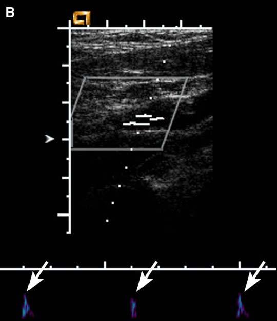
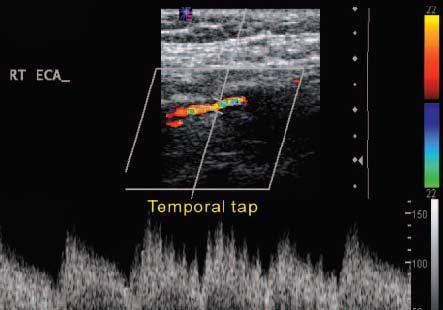
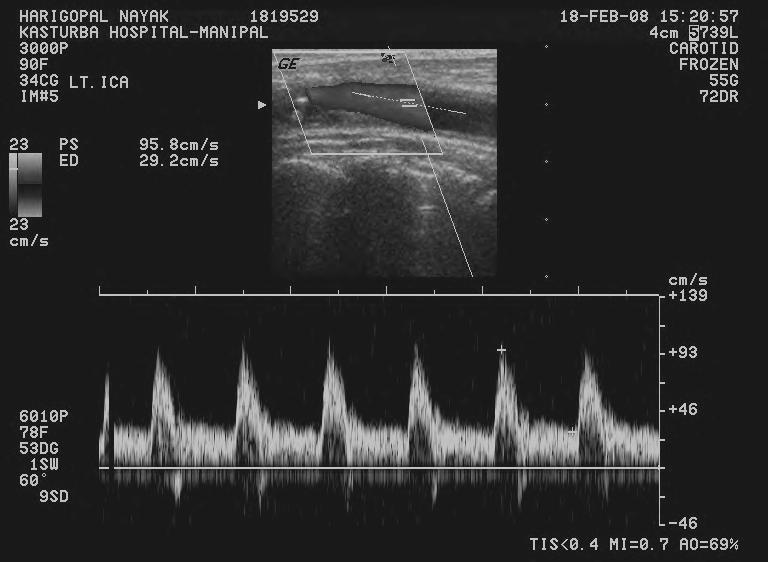
6 Scan both in transverse and longitudinal plane. Starting from proximal most CCA, bulb, ECA and ICA. Distal carotid 2 cm from the bulb ICA or ECA? Large in caliber, posterior and lateral low resistance wave form (not reliable) no branches no cluttering with temporal artery tapping,
7 Internal is not Internal
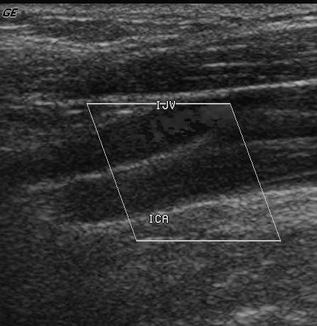
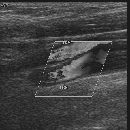
8 Color Doppler Sampling Window also known as the color box The size is adjusted to include all regions of interest. Adjustment of the angle by changing the box angles from left to center or right angling the transducer to ensure that the Doppler angle is less than 60 to the direction of blood flow
9

10 Proper steering
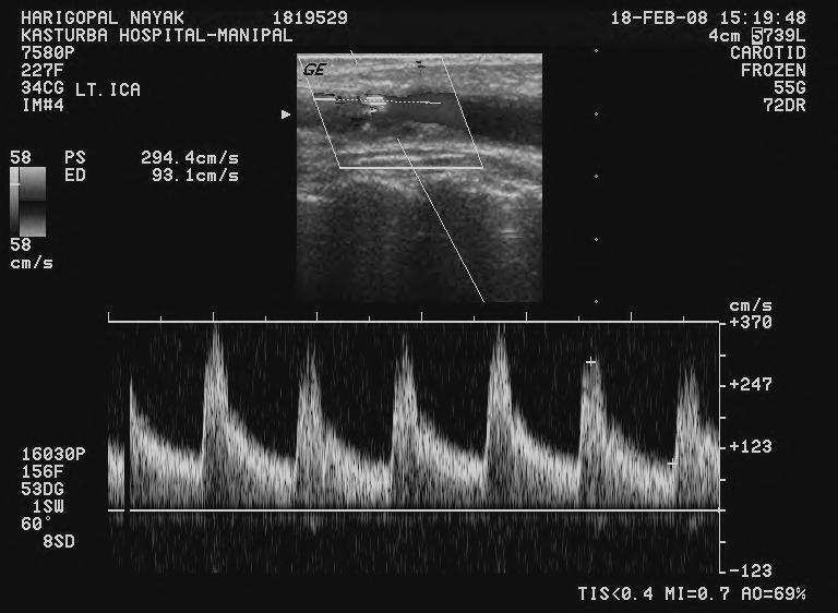
11 Sample Volume Gate and Angle Correction If the Doppler angle is small or more than 60 degree small error in the estimated velocity. preferred angle of incidence is 45 ± 4. The optimal position of the sample volume gate in a normal artery is in the mid lumen parallel to the vessel wall in a diseased vessel, parallel to the direction of blood flow should not be placed on the sharp curves of a tortuous artery falsely high velocity reading Should not be placed too close to the vessel wall spectral broadening.

12
13 Spectral Broadening Spectral broadening results from turbulence in the blood flow. Spurious spectral broadening a large Doppler angle a sample volume gate located close to the vessel wall a high Doppler gain setting The size of the gate is normally between 2 and 3 mm. too small (1.5 mm) the Doppler signal may be missed too large >3.5 spectral broadening
14 Color velocity scale If set below the mean velocity of blood flow, Aliasing throughout the vessel lumen set significantly higher than the mean velocity of blood flow, aliasing may disappear resulting in a missed stenosis In a normal carotid US examination, the color velocity scale should be set between 30 and 40 cm/sec (mean velocity).
15
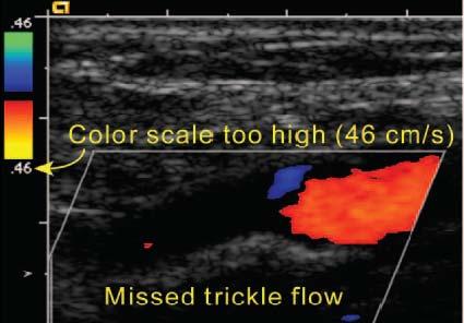
16 Color Gain Control The color gain should be set so that color just reaches the intimal surface of the vessel. If the color gain setting is too low, trickle flow may go undetected. If a color gain setting is high, bleeding of the color into the wall and surrounding tissues limit visualization of the plaque surface

17


18 Role of power doppler PDI may provide increased sensitivity to visualize the continuity of blood flow in arterial stenoses
19 Advantages of power doppler Angle independent No aliasing Very sensitive to low velocity and low amplitude flow Helps in differentiating critical stenosis from occlusion Disadvantages: motion sensitive does not give direction and velocity of flow
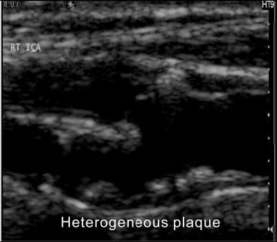
20 Carotid plaque Defined as a localized protrusion from the wall into the lumen with an area 50% greater than the intima media thickness of neighboring sites. low and high echogenic plaque. heterogeneous or homogeneous. regular (smooth) or irregular.
21 If more than 20% of the plaque echogenicity differed from the echogenicity of the rest of the plaque by two or more echogenicity grades is heterogenous. When height variations between 0.4 and 2 mm along the contour of the lesion is irregular Ulcerated plaques recesses in the contour of the lesion at least 2 mm in depth, with a well defined back wall at the base showing flow.
22


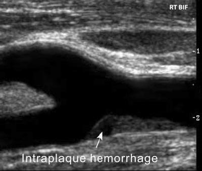
23 Heterogeneous plaques and ulcerated plaques are unstable or friable Potential for embolic TIA and cerebro vascular accidents

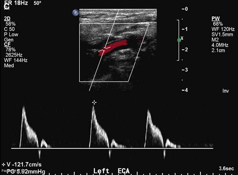
24 Fissuring or ulceration in the plaque
25


26 Plaque Classification Class I, homogeneous texture, uniformly hypoechoic Class II, heterogeneous texture, predominantly hypoechoic Class III, heterogeneous texture, predominantly hyperechoic Class IV, homogeneous texture, uniformly hyperechoic Class V, unclassified calcified plaques
27 After optimizing the setting **** Measure the velocity PSV and DV Proximal and distal CCA ICA and ECA Vertebral artery Wherever stenosis present at stenosis proximal to and distal to stenosis Compare bilateral carotid velocities symmetric or asymmetric
28 Waveform Analysis Normal Carotid Artery


29 CCA combination of ICA and ECA patterns, intermediate amount of continuous forward diastolic flow a sharp systolic upstroke and thin spectral envelope flow below the baseline or filling in of the spectral window normally should not be seen


30 ICA a low resistance waveform pattern systolic peak should be sharp and the spectral envelope thin continuous forward diastolic flow the systolic peak may be slightly blunter than the systolic peak of the ECA

 a normal")
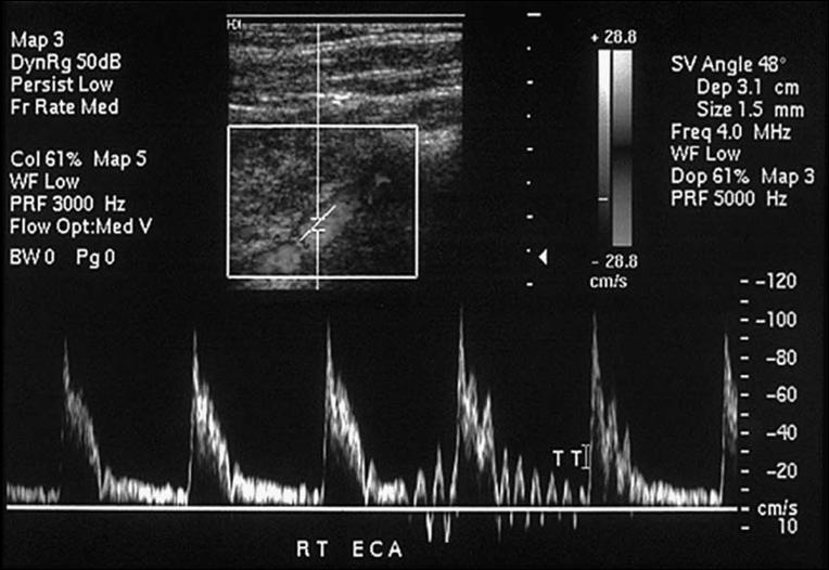
31 ECA the systolic upstroke is sharp the spectral envelope is thin. reduced to no diastolic flow diastolic flow should be symmetrical bilaterally Transient reversal in early diastole (characteristic early diastolic notch ) a normal finding

32
 no reversal of wave form")
33 VERTEBRAL ARTERY low resistance wave pattern forward diastolic flow no systolic or diastolic notch similar to carotid in flow (colour) no reversal of wave form
34 Look At Pattern Systolic contour Diastolic pattern PSV DV ICA PSVs / CCA PSVs ratio Compare Right and Left side
35 Abnormal CCA either low or high PSVs. abnormally high resistance waveform, an abnormally low resistance waveform,
36 Abnormally low PSVs A normal CCA PSV should be in the range of approximately Cm/s IF less than this, examine opposite side Symmetric Asymmetric (near normal) Low cardiac output Evaluate further A velocity difference of >20 cm/sec between the right and left is abnormal.
37 Causes for unilateral low PSVs Proximal stenosis (brachiocephalic) Parvus tardus waveform or normal pattern but asymmetrical PSVs. Distal stenosis (carotid bulb level) High resistance wave form
38 Innominate artery occlusion
39 High-resistance waveform in CCA High grade ICA stenosis or occlusion (externalization of the CCA) Distal waveforms should be assessed (support the diagnosis) EXCEPTION IF??? is bilateral and low PSVs indicates Aortic stenosis Severe cardiac failure

40 CCA ICA
41 Internalisation of ECA ICA
42 Focal stenosis of the CCA The ratio of the highest PSV at the CCA stenosis divided by the PSV 2 cm proximal to the stenosis should be calculated. PSVcca at stenosis/psvcca prox. If the ratio is 2 or more and less than 2.99 stenosis of 50% or more. If the ratio is 3 or more stenosis of 75% or more. also used if there are tandem stenosis.
43


44 CCA - mildly elevated resistance Luminal narrowing with color Aliasing High-velocity flow 627 cm/s, turbulence ICA- tardus-parvus waveform


45 Unusual finding in Case of CCA occlusion Reversal of flow in ECA and low resistance and low PSVs in ICA as it is fed by collaterals. This is to maintain the antegrade flow in ICA
46 ECA Reversal of flow ICA
47
48 Remember. If the stenosis is unilateral, there is marked asymmetry in the systolic contour of the waveforms of the right and the left CCAs. If the stenosis is central, such as with aortic stenosis, the waveforms are affected bilaterally.
49 ICA Normal is low resistance with high diastolic pattern. Most common site is ICA origin plaque extending from the bulb. High resistance pattern in the ICA Stenosis distally. PSVs raises Significant stenosis
50
51
52
53
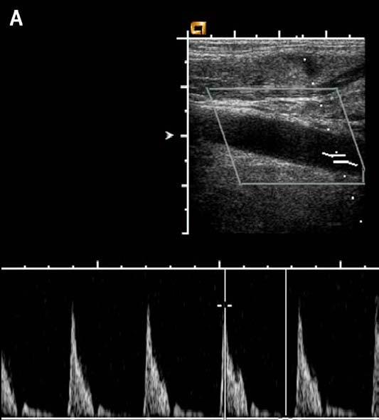
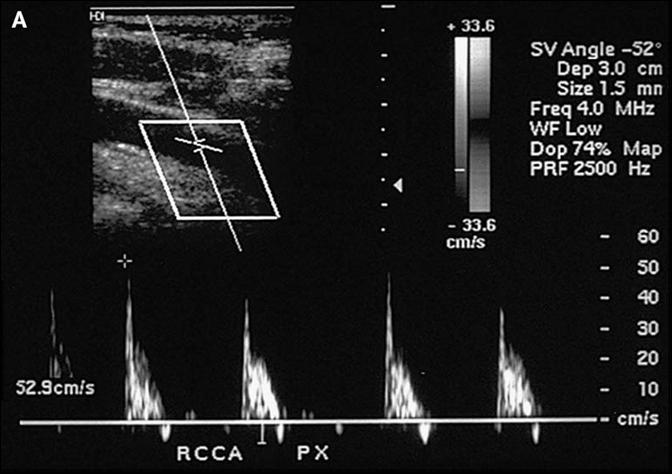
54

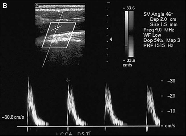
55 String sign - Near Total Occlusion
56 Total Occlusion
57 Near Total Occlusion or Total
58
59 Normal PSVs in ICA always normal??? As the stenotic grade increases PSVs start falling as flow through the tight stenosis reduces So measure EDV which raises Measure ratio ICA, PSVs / CCA PSVs Normal to <50% stenosis ratio will be < 2 As the stenosis increases ratio becomes > 4 or variable, internalization of ECA, opposite ICA PSVs increases Pitfalls Tortuous artery Plaque which is shadowing severe stenosis
60 So assess ICA In gray scale for amount of luminal narrowing Assess velocities in proper settings Should assess PSV, EDV and ratio of PSVs in ICA and CCA Assessed proximally, mid and distally If no color flow demonstrated in a tight stenotic segment even in power doppler confirm with other modality Assess opposite ICA for compensatory flow
61 ECA Confirm the ECA Is there any reversal of flow Is there any internalization
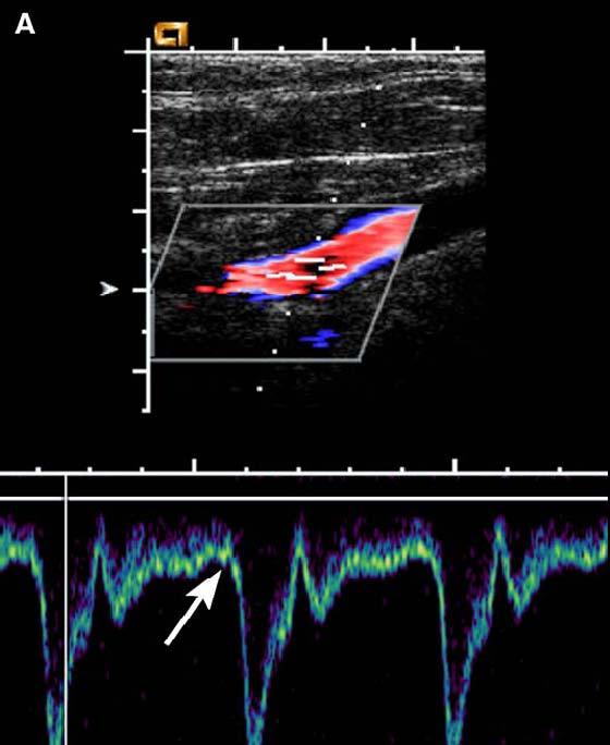
62 VERTEBRAL ARTERY LOOK AT Normal or hypoplastic or not seen Waveform pattern Direction of flow PSVs
63 Reversal of flow stenosis or occlusion at subclavian or brachiocephalic artery Transient systolic reversal in lesser digree stenosis High resistance wave pattern distal occlusion or stenosis Low resistance wave pattern more proximal stenosis

64 High resistance vertebral artery Distal vertebral stenosis


65 Parvus tardus Stenosis at vertebral origin- high PSV
66 Pitfall If not seen? Occlusion or small or congenitally absent Clinical correlation and other modality helps
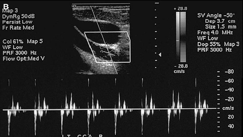
67 SUBCLAVIAN STEAL SYNDROME Subclavian artery steno occlusive disease proximal to the origin of the vertebral artery. Resulting in decreased blood pressure in the arm distal to the steno occlusive disease. Causes ipsilateral vertebral artery blood flow alteration Severe stenoses, flow reversal occurs in the ipsilateral vertebral artery as compensatory collateral to the vascular territory beyond the subclavian steno occlusive lesion.
68 Subclavian artery stenosis Innominate artery stenosis
69 Classification Based On Vertebral Artery Hemodynamics reduced antegrade vertebral flow (stage I) reversal of flow during reactive hyperemia testing of the arm (stage II) permanent retrograde vertebral flow (stage III).
70
71 at rest After inflation of a blood Pressure cuff on the left arm and rapid deflation Conversion of a presteal waveform to a complete steal following provocative maneuvers.


72 at rest After inflation of a blood Pressure cuff on the left arm and rapid deflation
73 The blood pressure cuff maneuver induces reactive hyperemia in the distal arm and increases blood flow across the subclavian stenosis, resulting in a complementary pressure drop and change in direction of blood flow in the ipsilateral vertebral artery towards the lower pressure subclavian origin.
74 Other wave patterns

75 Mid Systolic Retraction Pulsus Bisferience
76 PULSUS BISFERIENCE beat twice, Characterized by two systolic peaks with an interposed midsystolic retraction Seen in AR with or without concomitant AS Hypertophic obstructive cardiomyopathy Occasionally, may be seen in healthy, athletic, young individuals or in older patients.

77 Alternating systolic peak - Pulsus Alternans
78 PULSUS ALTERNANS Alternating peak systolic heights on sequential beats in a regular normal sinus rhythm Clinical conditions Intrinsic myocardial disease Ischemia Cardiomyopathies Valvular heart disease
79
80 water hammer pulse In aortic regurgitation reversed early diastolic flow in both CCAs with elevation of PSV and a sharp systolic upstroke Depending on the severity, the reversal of flow may be limited to early diastole with normalization of forward flow in end Diastole or may persist throughout diastole.
81 Appearance is Bilateral

82

83 Large cerebral infarct with uncal herniation
84 CAROTID DISSECTION


85 DISSECTION Trauma seat belt injury or repetitive trauma. Occasionally, spontaneous and isolated to the carotid arteries in Marfan syndrome, Ehlers Danlos syndrome, fibromuscular dysplasia, hypertension, or drug abuse Also direct extension of an aortic dissection. Rare but, dissection of the ICA is the most common cause of stroke in young patients. Most ICA dissections occur at the level of the carotid bifurcation.
86 Wave pattern is extremely bizarre in configuration: low PSV velocity with a highly irregular waveform contour with many spikes or fluttering with reversed or bidirectional of flow, such that it may be difficult to distinguish systole from diastole an intramural hematoma, causing a long segment tapering of the ICA without a break in the intima The residual lumen may be narrowed markedly, creating a string sign.


87 Thrombosis of the false lumen mimic stenosis The waveform may be indistinguishable from a stenosis except that typically it extends over a much longer segment and often no plaque is visualized.


88 The presence of early diastolic flow reversal in the ipsilateral CCA reduced peak systolic and diastolic velocities in the ipsilateral ICA are non specific, but warrant a search for a cause of increased peripheral vascular resistance.
89


90 Echogenic flap Hematoma with dissection


91 So What is this???


92 Thank you
Carotid Abnormalities Coils, Kinks and Tortuosity David Lorelli M.D., RVT, FACS Michigan Vascular Association Conference Saturday, October 20, 2012
 Carotid Abnormalities Coils, Kinks and Tortuosity David Lorelli M.D., RVT, FACS Michigan Vascular Association Conference Saturday, October 20, 2012 Page 1 Table of Contents Carotid Anatomy Carotid Duplex
GUNDERSEN/LUTHERAN ULTRASOUND DEPARTMENT POLICY AND PROCEDURE MANUAL
 GUNDERSEN/LUTHERAN ULTRASOUND DEPARTMENT POLICY AND PROCEDURE MANUAL SUBJECT: Carotid Duplex Ultrasound SECTION: Vascular Ultrasound ORIGINATOR: Deborah L. Richert, BSVT, RDMS, RVT DATE: October 15, 2015
DISCLOSURE TEST YOUR WAVEFORM IQ. Partial volume artifact. 86 yo female with right arm swelling, picc line. AVF on left? Dx?
 Deborah Rubens University of Rochester Rochester, NY DISCLOSURE Neither I nor my immediate family have a financial relationship with a commercial organization that may have a direct or indirect interest
Ultrasound Imaging of The Posterior Circulation
 Ultrasound Imaging of The Posterior Circulation Michigan Sonographers Society 2 Nd Annual Fall Vascular Conference Larry N. Raber RDMS-RVT Clinical Manager General Ultrasound/Neurovascular Laboratory Cleveland
Carotid Ultrasound: Improving Ultrasound
 Carotid Ultrasound: Improving Ultrasound Edward I. Bluth, M.D., F.A.C.R. Chairman Emeritus, Department of Radiology, Ochsner Clinic Foundation, New Orleans, Louisiana Professor, Ochsner Clinical School,
Beyond Stenosis Severity: Top 5 Important Duplex Characteristics to Identify in a Patient with Carotid Disease
 Beyond Stenosis Severity: Top 5 Important Duplex Characteristics to Identify in a Patient with Carotid Disease Jan M. Sloves RVT, RCS, FASE Technical Director New York Cardiovascular Associates Disclosures
NON-ATHEROSCLEROTIC PATHOLOGY OF THE CAROTID ARTERIES
 NON-ATHEROSCLEROTIC PATHOLOGY OF THE CAROTID ARTERIES Leslie M. Scoutt, MD, FACR Professor of Diagnostic Radiology & Surgery Vice Chair, Dept of Radiology & Biomedical Imaging Chief, Ultrasound Section
No financial or commercial relationships to disclose
 Deanna New, RVT No financial or commercial relationships to disclose IAC REQUIREMENTS: The main duty of a sonographer is to make the physician or radiologists job easier by capturing images and doing
Disclosure Statement:
 Marsha M. Neumyer, BS, RVT, FSVU, FSDMS, FAIUM International Director Vascular Diagnostic Educational Services Vascular Resource Associates Harrisburg, PA Disclosure Statement: CME Calendar QR Code Marsha
What effects will proximal or distal disease have on an waveform?
 Spectral Doppler Interpretation Director Director of of Ultrasound Ultrasound Education Education & & Quality Quality Assurance Assurance Baylor Baylor College College of of Medicine Medicine Division
Carotid US: More than just a chart on the wall
 Carotid US: More than just a chart on the wall Leslie M. Scoutt, MD, FACR Professor of Diagnostic Radiology & Surgery Vice Chair, Dept of Radiology & Biomedical Imaging Chief, Ultrasound Section Medical
Carotid Imaging IT S ABOUT MORE THAN JUST OBTAINING THE IMAGES
 Carotid Imaging IT S ABOUT MORE THAN JUST OBTAINING THE IMAGES No financial or commercial relationships to disclose Carotid artery disease: Stroke is one of the most serious causes of mortality and morbidity
What Do We Know? Disclosure Statement: 3/11/2015. Deep abdominal imaging
 Marsha M. Neumyer, BS, RVT, FSVU, FSDMS, FAIUM International Director Vascular Diagnostic Educational Services Vascular Resource Associates Harrisburg, PA Disclosure Statement: CME Calendar QR Code Marsha
Neurovascular Ultrasound Course
 Neurovascular Ultrasound Course William M. McKinney (6/6/30-10/24/03) Father of Neurosonology Founder, Neurosonology Course, WFUSM Welcome to Winston-Salem, NC, Wake Forest School of Medicine, and the
Case 8038 Renal allograft complicated with renal artery stenosis
 Case 8038 Renal allograft complicated with renal artery stenosis Santiago I, Canelas A, Pinto AP Section: Cardiovascular Published: 2009, Nov. 30 Patient: 61 year(s), male Clinical History A 61-year-old
Vertebral Artery Doppler Waveform Changes Indicating Subclavian Steal Physiology
 Downloaded from www.ajronline.org by 7.44.00.5 on 0/09/8 from IP address 7.44.00.5. Copyright RRS. For personal use only; all rights reserved Mark. Kliewer arbara S. Hertzberg David H. Kim James D. owie
Doppler Waveform Parvus and Tardus A Sign of Proximal Flow Obstruction
 Doppler Waveform Parvus and Tardus A Sign of Proximal Flow Obstruction Pesho S. Kotval, MD, PhD The Doppler linear flow velocity versus time spectrum obtained in an arterial flow system in which there
Hemodynamically significant subclavian artery stenosis
 REVIEW ARTICLE Duplex Ultrasonography of Vertebral and Subclavian Arteries Vijay G. Kalaria, MD, FACC, FSCAI, Sony Jacob, MD, William Irwin, RVT, and Robert M. Schainfeld, DO, Indianapolis, Indiana, and
Non-invasive examination
 Non-invasive examination Segmental pressure and Ankle-Brachial Index (ABI) The segmental blood pressure (SBP) examination is a simple, noninvasive method for diagnosing and localizing arterial disease.
Antegrade and retrograde flow of carotid
 Antegrade and retrograde flow of carotid The ECA waveform is high resistance and may have retrograde flow in diastole.. They should always demonstrate antegrade flow (toward the brain) and be. external
Radiologic Importance of a High- Resistive Vertebral Artery Doppler Waveform on Carotid Duplex Ultrasonography
 CME Article Radiologic Importance of a High- Resistive Vertebral Artery Doppler Waveform on Carotid Duplex Ultrasonography Esther S. H. Kim, MD, MPH, Megan Thompson, Kristine M. Nacion, BA, Carmel Celestin,
Deb Coghlan AMS (Vascular and General ) Brisbane, Australia
 Brisbane, Australia") Deb Coghlan AMS (Vascular and General ) Brisbane, Australia ANEURYSMAL DIISEASE The infrarenal aorta enlarges with age, and is the commonest site for arterial aneurysms. An aneurysm is a permanent focal
Deb Coghlan AMS (Vascular and General ) Brisbane, Australia ANEURYSMAL DIISEASE The infrarenal aorta enlarges with age, and is the commonest site for arterial aneurysms. An aneurysm is a permanent focal
Essentials of Clinical MR, 2 nd edition. 99. MRA Principles and Carotid MRA
 99. MRA Principles and Carotid MRA As described in Chapter 12, time of flight (TOF) magnetic resonance angiography (MRA) is commonly utilized in the evaluation of the circle of Willis. TOF MRA allows depiction
What effects will proximal or distal disease have on a waveform?
 Spectral Doppler Interpretation Director of Ultrasound Education & Quality Assurance Baylor College of Medicine Division of Maternal-Fetal Medicine Maternal Fetal Center Imaging Manager Texas Children
Optimising your Doppler settings for an accurate PI. Alison McGuinness Mid Yorks Hospitals
 Optimising your Doppler settings for an accurate PI Alison McGuinness Mid Yorks Hospitals Applications Both maternal uterine and fetal circulations can be studied with doppler sonography Uterine arteries
Vascular Portfolio: Carotid Reflection. Paige Fabre
 Vascular Portfolio: Carotid Reflection Paige Fabre 13654584 14 Carotid Reflection For this portfolio I produced three pieces of work; a case study, a PowerPoint of study protocol and a poster of stenosis
Carotid Duplex: Beyond Stenosis Ido Weinberg, MD Vascular Medicine Massachusetts General Hospital Assistant Professor of Medicine Harvard Medical
 Carotid Duplex: Beyond Stenosis Ido Weinberg, MD Vascular Medicine Massachusetts General Hospital Assistant Professor of Medicine Harvard Medical School Boston, Massachusetts Disclosures I do not have
Vascular Sonography Examination
 Vascular Sonography Examination The purpose of The American Registry of Radiologic Technologists (ARRT ) Vascular Sonography Examination is to assess the knowledge and cognitive skills underlying the intelligent
US of Neurovascular Occlusive Disease: Interpretive Pearls and Pitfalls 1
 EDUCATION EXHIBIT 1165 US of Neurovascular Occlusive Disease: Interpretive Pearls and Pitfalls 1 CME FEATURE See accompanying test at http:// www.rsna.org /education /rg_cme.html LEARNING OBJECTIVES FOR
STRUCTURED EDUCATION REQUIREMENTS IMPLEMENTATION DATE: JULY 1, 2016
 STRUCTURED EDUCATION REQUIREMENTS Vascular Sonography The purpose of structured education is to provide the opportunity for individuals to develop mastery of discipline-specific knowledge that, when coupled
TRANSCRANIAL DOPPLER ULTRASOUND INTRODUCTION TO TCD INTERPRETATION
 TRANSCRANIAL DOPPLER ULTRASOUND INTRODUCTION TO TCD INTERPRETATION ---Rune Aaslid First TCD Publication 1982 WHAT IS TCD? Uses 2 MHz pulsed Doppler ultrasound Passes through cranial windows Provides information
Transducer Selection. Renal Artery Duplex Exam. Renal Scan. Renal Scan Echogenicity. How to Perform an Optimal Renal Artery Doppler Examination
 How to Perform an Optimal Renal Artery Doppler Examination Director of Ultrasound Education & Quality Assurance Baylor College of Medicine Division of Maternal-Fetal Medicine Maternal Fetal Center Imaging
Lezione 3 Tronchi Sovraortici
 CORSO DI CERTIFICAZIONE DI COMPETENZA in ECOGRAFIA VASCOLARE GENERALE Lezione 3 Tronchi Sovraortici Settore formazione 2007-2009: Direttore: Paolo G. Pino Marco Campana, Antonella Moreo, Fausto Rigo, Ketty
Pre-and Post Procedure Non-Invasive Evaluation of the Patient with Carotid Disease
 Pre-and Post Procedure Non-Invasive Evaluation of the Patient with Carotid Disease Michael R. Jaff, D.O., F.A.C.P., F.A.C.C. Assistant Professor of Medicine Harvard Medical School Director, Vascular Medicine
Indications: following: embolization. artery that has diseases 5. The evaluation. of suspected. such entities. a cold hand. biopsy
 Peripheral Arterial Ultrasound Protocol Using Color and Spectral Doppler Reviewed by: Mark Yuhasz, MD Last Review Date: January 2015 Contact: (866) 761 4200, Option 1 Indications: The indications for peripheral
Image Formation (10) 2 Evaluation and Selection of Representative Images (10)
 2 Evaluation and Selection of Representative Images (10)") STRUCTURED SELF ASSESSMENT CONTENT SPECIFICATIONS SSA LAUNCH DATE: JANUARY 1, 2018 Vascular Sonography The purpose of continuing qualifications requirements (CQR) is to assist registered technologists
STRUCTURED SELF ASSESSMENT CONTENT SPECIFICATIONS SSA LAUNCH DATE: JANUARY 1, 2018 Vascular Sonography The purpose of continuing qualifications requirements (CQR) is to assist registered technologists
Introduction History Preceded by Arterial Doppler and ABI Indications
 Elise Brady, RVT, RDMS Introduction History Preceded by Arterial Doppler and ABI Indications 1) Abnormal ABI (within 2weeks of duplex) 2) Abnormal Doppler waveforms 3) Claudication 4) History of PVD 5)
Diagnosis of Middle Cerebral Artery Occlusion with Transcranial Color-Coded Real-Time Sonography
 Diagnosis of Middle Cerebral Artery Occlusion with Transcranial Color-Coded Real-Time Sonography Kazumi Kimura, Yoichiro Hashimoto, Teruyuki Hirano, Makoto Uchino, and Masayuki Ando PURPOSE: To determine
HD Scanning: Velocities and Volume Flow
 HD Scanning: Velocities and Volume Flow Non-Invasive Lab Symposium West Orange, NJ April 27, 2018 Volume Flow Cindy Sturt, MD, FACS, RVT 500,000 Americans on dialysis 20-25% annual mortality 65% 5 year
Echocardiography as a diagnostic and management tool in medical emergencies
 Echocardiography as a diagnostic and management tool in medical emergencies Frank van der Heusen MD Department of Anesthesia and perioperative Care UCSF Medical Center Objective of this presentation Indications
Neuro Quiz 29 Transcranial Doppler Monitoring
 Verghese Cherian, MD, FFARCSI Penn State Hershey Medical Center, Hershey Quiz Team Shobana Rajan, M.D Suneeta Gollapudy, M.D Angele Marie Theard, M.D Neuro Quiz 29 Transcranial Doppler Monitoring This
Duplex Doppler Sonography of the Carotid Artery: False-Positive Results in an Artery Contralateral to an Artery with Marked Stenosis
 049 Duplex Doppler Sonography of the Carotid Artery: False-Positive Results in an Artery Contralateral to an Artery with Marked Stenosis William W. Beckett, Jr. Patricia C. Davis James C. Hoffman, Jr.
Protokollanhang zur SPACE-2-Studie Neurology Quality Standards
 Protokollanhang zur SPACE-2-Studie Neurology Quality Standards 1. General remarks In contrast to SPACE-1, the neurological center participating in the SPACE-2 trial will also be involved in the treatment
Carotid Doppler: Doppler wave forms obtained from the common, external and internal carotid arteries. As well as the vertebral and subclavian
 Competency Carotid Doppler: Doppler wave forms obtained from the common, external and internal carotid arteries. As well as the vertebral and subclavian arteries. Preferred angle is 60 degrees or less.
Adult Echocardiography Examination Content Outline
 Adult Echocardiography Examination Content Outline (Outline Summary) # Domain Subdomain Percentage 1 2 3 4 5 Anatomy and Physiology Pathology Clinical Care and Safety Measurement Techniques, Maneuvers,
TO CATCH A THIEF: IMAGING OF SUBCLAVIAN STEAL
 October 2013 TO CATCH A THIEF: IMAGING OF SUBCLAVIAN STEAL Sumir Pandit, Harvard Medical School, Year III 1 AGENDA Introduction to our patient A.B. Anatomy review of aorta and branches CT imaging of our
Visceral Vascular Ultrasound. Joel Thompson, MD, MPH Borg & Ide Imaging
 Visceral Vascular Ultrasound Joel Thompson, MD, MPH Borg & Ide Imaging Objectives: Review major abdominal vascular structures Identify normal peak systolic velocity (PSV) for major abdominal arteries.
Background & Indications Probe Selection
 Teresa S. Wu, MD, FACEP Director, EM Ultrasound Program & Fellowship Co-Director, Simulation Based Training Program & Fellowship Associate Program Director, EM Residency Program Maricopa Medical Center
Goals. Access flow and renal artery stenosis evaluation by Doppler ultrasound. Reimbursement. WHY use of Doppler Ultrasound
 Access flow and renal artery stenosis evaluation by Doppler ultrasound Adina Voiculescu, MD Interventional Nephrology Brigham and Women s Hospital Boston Instructor at Harvard Medical School Understand
Intracranial Cerebrovascular Evaluation Transcranial Doppler (Non-Imaging) and Transcranial Duplex Imaging (TCD-I)
 and Transcranial Duplex Imaging (TCD-I)") VASCULAR TECHNOLOGY PROFESSIONAL PERFORMANCE GUIDELINES Intracranial Cerebrovascular Evaluation Transcranial Doppler (Non-Imaging) and Transcranial Duplex Imaging (TCD-I) This Guideline was prepared by
VASCULAR TECHNOLOGY PROFESSIONAL PERFORMANCE GUIDELINES Intracranial Cerebrovascular Evaluation Transcranial Doppler (Non-Imaging) and Transcranial Duplex Imaging (TCD-I) This Guideline was prepared by
Internal Carotid Artery Dissection
 May 2011 Internal Carotid Artery Dissection Carolyn April, HMS IV Agenda Presentation of a clinical case Discussion of the clinical features of ICA dissection Discussion of the imaging modalities used
Recommendations for documentation of neurosonographic examinations
 Recommendations for documentation of neurosonographic examinations The documentation of ultrasound examinations is subject to a dynamic development particularly as regards newer applications. The present
(Department of Radiology, Beylikdüzü State Hospital, İstanbul, Turkey) Corresponding Author: Dr. Mete Özdikici
 Corresponding Author: Dr. Mete Özdikici") Quest Journals Journal of Medical and Dental Science Research Volume 5~ Issue 6 (2018) pp: 61-65 ISSN(Online) : 2394-076X ISSN (Print):2394-0751 www.questjournals.org Research Paper Quantitative Measurements
Quest Journals Journal of Medical and Dental Science Research Volume 5~ Issue 6 (2018) pp: 61-65 ISSN(Online) : 2394-076X ISSN (Print):2394-0751 www.questjournals.org Research Paper Quantitative Measurements
Duplex Ultrasound of the Renal Arteries. Duplex Ultrasound. In the Beginning
 Duplex Ultrasound of the Renal Arteries DIMENSIONS IN HEART AND VASCULAR CARE 2013 PENN STATE HEART AND VASCULAR INSTITUTE ROBERT G. ATNIP MD PROFESSOR OF SURGERY AND RADIOLOGY Duplex Ultrasound Developed
COLOUR DOPPLER EVALUATION OF DEGREE OF STENOSIS AND PLAQUE MORPHOLOGY IN EXTRACRANIAL CAROTID ARTERIES IN PATIENTS OF STROKE
 wjpmr, 2019, 5(1), 122-128 SJIF Impact Factor: 4.639 Hassan et al. Research Article WORLD JOURNAL OF PHARMACEUTICAL AND MEDICAL RESEARCH ISSN 2455-3301 www.wjpmr.com WJPMR COLOUR DOPPLER EVALUATION OF
8/20/18. The Doppler Effect. Objectives. What is the Doppler Effect. Doppler principles. Spectral Waveform. Image recognition. Vascular Ultrasound
 Vascular Ultrasound: Physics and Haemodynamics Objectives Doppler principles Spectral Waveform Key factors Haemodynamics: Stenosis Waveforms Image recognition Vascular Ultrasound: A flawed paradigm What
Pitfalls in the evaluation of carotid artery stenosis. Serge Kownator «Centre Cardiologique et Vasculaire» Thionville, Fr
 Pitfalls in the evaluation of carotid artery stenosis Serge Kownator «Centre Cardiologique et Vasculaire» Thionville, Fr Disclosure Statement of Financial Interest I currently have, or have had over the
Review Article Duplex Ultrasound Evaluation of Hemodialysis Access: A Detailed Protocol
 International Nephrology Volume 2012, Article ID 508956, 7 pages doi:10.1155/2012/508956 Review Article Duplex Ultrasound Evaluation of Hemodialysis Access: A Detailed Protocol Victoria Teodorescu, 1,
NCVH. Ultrasongraphy: State of the Art Vein Forum 2015 A Multidisciplinary Approach to Otptimizing Venous Circulation From Wounds to WOW
 Ultrasongraphy: State of the Art 2015 NCVH New Cardiovascular Horizons Vein Forum 2015 A Multidisciplinary Approach to Otptimizing Venous Circulation From Wounds to WOW Anil K. Chagarlamudi, M.D. Cardiovascular
COPYRIGHTED MATERIAL. I How to Perform Ultrasound Tests
 I How to Perform Ultrasound Tests COPYRIGHTED MATERIAL 1 2 1 Principles of Extracranial Ultrasound Examination Andrei V. Alexandrov 1, Alice Robinson-Vaughn 1, Clotilde Balucani 2 & Marsha M. Neumyer 3
Fibromuscular Dysplasia. Miranda Forrest Baker College
 Fibromuscular Dysplasia Miranda Forrest Baker College Overview Case Study Patient Information Exam Images Findings FMD Types Signs and Symptoms Treatment Case Study Patient Information Female 57 years
Problems of Carotid Doppler Scanning Which Can Be Overcome by Using Frequency Analysis
 Problems of Carotid Doppler Scanning Which Can Be Overcome by Using Frequency Analysis K. W. JOHNSTON, M.D., F.R.C.S.(C), F.A.C.S., P. M. BROWN, M.D., F.R.C.S.(C), AND M. KASSAM, M.A.SC. SUMMARY The value
Transcranial Doppler (Basic Step) Dae-il Chang, M.D., Sung Sang Yoon, M.D. Department of Neurology, College of Medicine, Kyunghee university
 Dae-il Chang, M.D., Sung Sang Yoon, M.D. Department of Neurology, College of Medicine, Kyunghee university") Transcranial Doppler (Basic Step) Dae-il Chang, M.D., Sung Sang Yoon, M.D. Department of Neurology, College of Medicine, Kyunghee university Principles of Doppler Ultrasonography Major target Speed & direction
Transcranial Doppler (Basic Step) Dae-il Chang, M.D., Sung Sang Yoon, M.D. Department of Neurology, College of Medicine, Kyunghee university Principles of Doppler Ultrasonography Major target Speed & direction
Evaluation of Carotid Vessels and Vertebral Artery in Stroke Patients with Color Doppler Ultrasound and MR Angiography
 Evaluation of Carotid Vessels and Vertebral Artery in Stroke Patients with Color Doppler Ultrasound and MR Angiography Dr. Pramod Shaha 1, Dr. Vinay Raj R 2, Dr. (Brig) K. Sahoo 3 Abstract: Aim & Objectives:
Radial Artery Assessment for Coronary Artery Bypass
 VASCULAR TECHNOLOGY PROFESSIONAL PERFORMANCE GUIDELINES Radial Artery Assessment for Coronary Artery Bypass This Guideline was prepared by the Professional Guidelines Subcommittee of the Society for Vascular
Section II: Patient Interview Grade: 5
 Only written competency completed with this EXACT form will be accepted for grading. No modifications to the LAYOUT of the form will be accepted for a written competency. Failure to comply will result
Vikram Dogra, M.D. Professor of Radiology, Urology & BME Department of Imaging Sciences University Of Rochester Medical Center
 Ultrasound of the Scrotum Vikram Dogra, M.D. Professor of Radiology, Urology & BME Department of Imaging Sciences University Of Rochester Medical Center Etiologies of Acute Scrotal Pain Epididymitis/Orchitis
Management of cervicocephalic arterial dissection. Ciro G. Randazzo, MD, MPH Thomas Jefferson University Hospital, Department of Neurosurgery
 Management of cervicocephalic arterial dissection Ciro G. Randazzo, MD, MPH Thomas Jefferson University Hospital, Department of Neurosurgery Definition Disruption of arterial wall, either at level of intima-media
Abdominal Doppler Mastering the next level of vascular anatomy in the belly. Cindy A. Owen, RDMS, RVT
 Abdominal Doppler Mastering the next level of vascular anatomy in the belly Cindy A. Owen, RDMS, RVT Introduction Abdominal Doppler is a tough exam Success is dependent on: Patient body habitus Patient
Acute Aortic Syndromes
 Acute Aortic Syndromes Carole J. Dennie, MD Acute Thoracic Aortic Syndromes Background Non-Traumatic Acute Thoracic Aortic Syndromes Carole Dennie MD FRCPC Associate Professor of Radiology and Cardiology
Enhancement of Cranial US: Utility of Supplementary Acoustic Windows and Doppler Harriet J. Paltiel, MD
 Enhancement of Cranial US: Utility of Supplementary Acoustic Windows and Doppler Harriet J. Paltiel, MD Boston Children s Hospital Harvard Medical School None Disclosures Conventional US Anterior fontanelle
PART II ECHOCARDIOGRAPHY LABORATORY OPERATIONS ADULT TRANSTHORACIC ECHOCARDIOGRAPHY TESTING
 PART II ECHOCARDIOGRAPHY LABORATORY OPERATIONS ADULT TRANSTHORACIC ECHOCARDIOGRAPHY TESTING STANDARD - Primary Instrumentation 1.1 Cardiac Ultrasound Systems SECTION 1 Instrumentation Ultrasound instruments
Case 9799 Stanford type A aortic dissection: US and CT findings
 Case 9799 Stanford type A aortic dissection: US and CT findings Accogli S, Aringhieri G, Scalise P, Angelini G, Pancrazi F, Bemi P, Bartolozzi C Department of Diagnostic and Interventional Radiology, University
EXPLORING ULTRASOUND IMAGES OF THE CAROTID ARTERIES USING NEURAL NETWORK TOOLS
 Proceedings of the 6th International Conference on Mechanics and Materials in Design, Editors: J.F. Silva Gomes & S.A. Meguid, P.Delgada/Azores, 26-30 July 2015 PAPER REF: 5430 EXPLORING ULTRASOUND IMAGES
Shadow because the air
 Thyroid Ultrasound Thyroid US examination needs: 1. high frequency transducer 2. extended patient's neck 3. check all the neck area because the swelling could be in areas other than the thyroid such as
Vascular Imaging in the Pediatric Abdomen. Jonathan Swanson, MD
 Vascular Imaging in the Pediatric Abdomen Jonathan Swanson, MD Goals and Objectives To understand the imaging approach, appearance, and clinical manifestations of the common pediatric abdominal vascular
US of Renovascular Hypertension. Jonathan R. Dillman, MD, MSc Associate Professor Director, Thoracoabdominal Imaging
 US of Renovascular Hypertension Jonathan R. Dillman, MD, MSc Associate Professor Director, Thoracoabdominal Imaging Disclosures Nothing Relevant Unrelated grant funding Siemens US Toshiba US Objectives
Policies and Statements D16. Intracranial Cerebrovascular Ultrasound
 Policies and Statements D16 Intracranial Cerebrovascular Ultrasound SECTION 1: INSTRUMENTATION Policies and Statements D16 Intracranial Cerebrovascular Ultrasound May 2006 (Reaffirmed July 2007) Essential
Vascular Surgery Cases: Detours. Brian F. Stull, RDMS, RVT UNC REX Healthcare Vascular Specialists
 Vascular Surgery Cases: Detours Brian F. Stull, RDMS, RVT UNC REX Healthcare Vascular Specialists Brian.Stull@Unchealth.unc.edu Objectives Anatomy of a bypass graft Where does it connect, where does it
The production of murmurs is due to 3 main factors:
 Heart murmurs The production of murmurs is due to 3 main factors: high blood flow rate through normal or abnormal orifices forward flow through a narrowed or irregular orifice into a dilated vessel or
Aortic arch pathology. Cerebral ischemia following carotid artery stenosis.
 Important: -Subclavian Steal Syndrome -Cerebral ischemia Aortic arch pathology. Cerebral ischemia following carotid artery stenosis. Mina Aubeed & Alba Hernández Pinilla Aortic arch pathology Common arch
11 TH ANNUAL VASCULAR NONINVASIVE TESTING SYMPOSIUM NOVEMBER 10, 2018
 11 TH ANNUAL VASCULAR NONINVASIVE TESTING SYMPOSIUM NOVEMBER 10, 2018 RENAL ARTERY DISEASE AND RENOVASCULAR HYPERTENSION 1 WHAT IS RENOVASCULAR HYPERTENSION? https://my.clevelandclinic.org/health/diseases/16459-renovascular-hypertension
Hemodynamic Assessment. Assessment of Systolic Function Doppler Hemodynamics
 Hemodynamic Assessment Matt M. Umland, RDCS, FASE Aurora Medical Group Milwaukee, WI Assessment of Systolic Function Doppler Hemodynamics Stroke Volume Cardiac Output Cardiac Index Tei Index/Index of myocardial
Doppler Ultrasonography of the Liver: What Every General Radiologist Should Know
 Doppler Ultrasonography of the Liver: What Every General Radiologist Should Know Poster No.: C-1658 Congress: ECR 2014 Type: Authors: Keywords: DOI: Educational Exhibit T. González de la Huebra Labrador,
AN INTRODUCTION TO DOPPLER. Sarah Gardner, Clinical lead, Tissue viability service. Oxford Health NHS Foundation Trust.
 AN INTRODUCTION TO DOPPLER Sarah Gardner, Clinical lead, Tissue viability service. Oxford Health NHS Foundation Trust. THE DOPPLER EFFECT The Doppler Principle was described by Physicist and mathematician
MESENTERIC ISCHEMIA. Phillip J Bendick, PhD
 MESENTERIC ISCHEMIA Phillip J Bendick, PhD Arterial Celiac - Hepatic - Splenic Superior Mesenteric Artery Inferior Mesenteric Artery Venous Mesenteric system Porto - hepatic system Inferior Vena Cava Acute
PTA 106 Unit 1 Lecture 3
 PTA 106 Unit 1 Lecture 3 The Basics Arteries: Carry blood away from the heart toward tissues. They typically have thicker vessels walls to handle increased pressure. Contain internal and external elastic
FFR: Tips and Tricks. A/Prof (Adj) Yeo Khung Keong, MBBS, FAMS, FACC, FSCAI National Heart Centre Singapore
 Yeo Khung Keong, MBBS, FAMS, FACC, FSCAI National Heart Centre Singapore") FFR: Tips and Tricks A/Prof (Adj) Yeo Khung Keong, MBBS, FAMS, FACC, FSCAI National Heart Centre Singapore Disclosures Abbott Vascular: Speaker, Proctor (MitraClip) Boston Scientific: Consultant, honorarium
FFR: Tips and Tricks A/Prof (Adj) Yeo Khung Keong, MBBS, FAMS, FACC, FSCAI National Heart Centre Singapore Disclosures Abbott Vascular: Speaker, Proctor (MitraClip) Boston Scientific: Consultant, honorarium
BEDSIDE ULTRASOUND BEDSIDE ULTRASOUND. Deep Vein Thrombosis. Probe used
 BEDSIDE ULTRASOUND Part 2 Diagnosis of deep vein thrombosis Kishore Kumar Pichamuthu, Professor, Department of Critical Care, CMC, Vellore Summary: Deep vein thrombosis (DVT) is a problem encountered in
Subclavian artery Stenting
 Subclavian artery Stenting Etiology Atherosclerosis Takayasu s arteritis Fibromuscular dysplasia Giant Cell Arteritis Radiation-induced Vascular Injury Thoracic Outlet Syndrome Neurofibromatosis Incidence
noninvasive, nonionizing, portable, inexpensive, safe for serial or prolonged studies
 TRANS CRANIAL DOPPLER Presented by : Anil Garg Transcranial Doppler 1982, Aaslid and colleagues introduced TCD as a non-invasive technique for monitoring blood flow velocity in basal cerebral arteries
Michigan Vascular Association 2012 Conference Case studies from Massachusetts General Hospital. Our lab
 Michigan Vascular Association 2012 Conference Case studies from Massachusetts General Hospital Kathleen Hannon, MS, RVT, RDMS khannon@partners.org Our lab #1 in the nation! 15 full time RVT s 11 MD s IAC
Guidelines, Policies and Statements D5 Statement on Abdominal Scanning
 Guidelines, Policies and Statements D5 Statement on Abdominal Scanning Disclaimer and Copyright The ASUM Standards of Practice Board have made every effort to ensure that this Guideline/Policy/Statement
Michael Horowitz, MD Pittsburgh, PA
 Michael Horowitz, MD Pittsburgh, PA Introduction Cervical Artery Dissection occurs by a rupture within the arterial wall leading to an intra-mural Hematoma. A possible consequence is an acute occlusion
PROSTHETIC VALVE BOARD REVIEW
 PROSTHETIC VALVE BOARD REVIEW The correct answer D This two chamber view shows a porcine mitral prosthesis with the typical appearance of the struts although the leaflets are not well seen. The valve
Sonographic Characterization of Carotid Plaque: Detection of Hemorrhage
 311 Sonographic Characterization of Carotid Plaque: Detection of Hemorrhage E. I. Bluth' D.Kai C. R. B. Merritt' M. Sullivan' G. Farr2 N. L. Mills 3 M. Foreman' K. Sloan' M. Schlater' J. Stewart 3 By careful
CT Imaging of Blunt and Penetrating Vascular Trauma DENNIS FOLEY MEDICAL COLLEGE WISCONSIN
 CT Imaging of Blunt and Penetrating Vascular Trauma DENNIS FOLEY MEDICAL COLLEGE WISCONSIN THORACO ABDOMINAL TRAUMA 0 10 20 30 40 50 60 5 cc/sec 30 secs 1.25 mm/ 55 mm Z1.375 2.5 mm/ 55 mm Z 1.375 Grade
Noninvasive transcranial Doppler ultrasound recording of flow velocity in basal cerebral arteries
 J Neurosurg 57:769-774, 1982 Noninvasive transcranial Doppler ultrasound recording of flow velocity in basal cerebral arteries RUNE AASLID, PH.D., THOMAS-MARC MARKWALDER, M.D., AND HEt,CE NORNES, M.D.
Duplex Criteria for Determination of 50% or Greater Carotid Stenosis
 Article Duplex Criteria for Determination of 50% or Greater Carotid Stenosis David G. Neschis, MD, Frank J. Lexa, MD, Julia T. Davis, RN, RVT, Jeffrey P. Carpenter, MD, RVT Recently the North American
Reduction of flow velocities in patients with ischemic events in the middle cerebral artery long-term follow-up with ultrasound
 Acta Neurol. Belg., 20,, -5 Original articles Reduction of flow velocities in patients with ischemic events in the middle cerebral artery long-term follow-up with ultrasound Christine Kremer and Kasim