MRI Assessment of the Right Ventricle and Pulmonary Blood Flow, Perfusion and Ventilation
|
|
|
- Roxanne Hopkins
- 5 years ago
- Views:
Transcription


1 MRI Assessment of the Right Ventricle and Pulmonary Blood Flow, Perfusion and Ventilation Dr. Richard Thompson Department of Biomedical Engineering University of Alberta Heart and Lung Imaging Many Constantly Evolving Modalities: Echocardiography Computed Tomography (CT) SPECT Magnetic Resonance Imaging (MRI) Goals Understand the Present and Future Capabilities of MRI in Childhood Heart and Lung Imaging
 Requirement for Breath holding / Breathing Artifacts Heart and Lung Imaging MRI Strengths Non Invasive and Non Ionizing Radiation")
 MRI offers much more than RV asessment pulmonary artery anatomy and blood flow, perfusion, ventilation (Comprehensive")
2 Heart and Lung Imaging MRI Weaknesses Limited Availability / Long Wait Time for Studies (expensive) Long Scan Times (sedation of younger children/newborns) Requirement for Breath holding / Breathing Artifacts Heart and Lung Imaging MRI Strengths Non Invasive and Non Ionizing Radiation (safe) Arbitrary Slice Orientations (good views of complex anatomy) Excellent Soft Tissue Contrast (many contrast mechanisms) Accurate and Reproducible (RV mass / Volumes) MRI offers much more than RV asessment pulmonary artery anatomy and blood flow, perfusion, ventilation (Comprehensive and Quantitative - Endpoint)
 correlation to MRI RV volumes and systematically underestimate volumes 3D echocardiography: evaluation of the right ventricle. Curr Opin Cardiol.")

3 MRI for RV Imaging Is it still the Best? Evaluation of right ventricular volume and function by 2D and 3D echocardiography compared to MRI. Eur J Echocardiogr Dec;7(6): Epub 2005 Dec 9. 2D and 3D Echo have similar (moderate) correlation to MRI RV volumes and systematically underestimate volumes 3D echocardiography: evaluation of the right ventricle. Curr Opin Cardiol Sep;24(5): D Echo underestimates volumes, and has technical limitations, should still be used in conjunction with conventional 2D methods Multimodality comparison of quantitative volumetric analysis of the right ventricle. JACC Cardiovasc Imaging Jan;3(1):10 8. MRI, CT and 3D Echo are all similar for RV volume analysis Several Additional Studies Indicate that Echocardiography is similar to MRI for RV Volume Assessment in Children Dependent on the Institution, hardware and software RV Imaging Depends on Your Tools! Reproducibility is strongly dependent on volume assessment protocol 2 3% coefficient of variation in RV volumes

")
4 RV Imaging Summary RV imaging alone is not sufficient impetus to perform MRI, typically acceptable with echocardiography new hardware and software improving the robustness of Echo. Reproducibility can still be highly variable depending on hardware and software (MRI, Echo, CT) should be determined at each site for the purpose of clinical research Ventricular Tissue Characterization Delayed Enhancement of myocardium following injection of contrast (Gd DTPA) identification of fibrosis and cell death (viability) Dark Myocardium (normal) Bright Myocardium (fibrosis)


 45")
5 Ventricular Tissue Characterization T 1 Mapping Imaging of Diffuse Fibrosis Ventricular Tissue Characterization T 2 Mapping Imaging Edema Delayed Enhancement Image - MI Quantitative T2 Values (Normal ms) 70 T 2 (ms) 45
6 Ventricular Tissue Characterization T 2 Mapping Imaging Edema base 70 Regional or Global Diffuse Changes Any change in local tissue environment that effects the mobility of water (fibrosis, ischemia) apex T2 (ms) Early marker of transplanted heart rejection 45 Ventricular Tissue Characterization Summary MRI offers the ability to image regional or diffuse fibrosis and edema as part of a standard clinical exam. Delayed Enhancement imaging (regional fibrosis) is available on all modern MRI scanners. Quantitative T 1 and T 2 imaging (diffuse fibrosis and edema) is increasingly available will be on all next generation scanners. Studies in younger children still needed to determine normal/abnormal values.


7 MR Imaging of Blood Velocity and Flow Phase Contrast MRI Measures Blood Velocity in all Directions Velocity Image MR Imaging of Blood Velocity and Flow 5 yrs of age Hypoplastic Left Heart Syndrome Post Fontan Operation


")
8 MR Imaging of Blood Velocity and Flow MR Imaging of Blood Velocity and Flow Calculation of timing parameters (acceleration time) 115 ms MRI Volume measurements known to have systematic errors (10%)

9 MR Imaging of Blood Velocity and Flow Other Applications: Visualization of Blood Flow MR Imaging of Blood Velocity and Flow Other Applications: Visualization of Blood Flow
. Offers unique 3 Dimensional flow imaging.")
10 MR Imaging of Blood Velocity and Flow Other Applications: Visualization of Blood Flow PAH Patient 5 cm MR Imaging of Blood Velocity and Flow Summary Phase Contrast blood velocity imaging is available on all modern MRI scanners Similar to Color Doppler with Echocardiography (standard metrics, e.g. acceleration time). Offers unique 3 Dimensional flow imaging. Data processing tools do exist but are not generally available (research)
 is well")

 *Courtesy of Dr.")
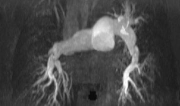
11 Magnetic Resonance Angiography MR Pulmonary Angiography (contrast enhanced) is well established in the clinic with significant improvements in the last decade Magnetic Resonance Angiography Lumenography (1 2 mm spatial resolution) *Courtesy of Dr. Scott Reeder, University of Wisconsin

 for the detection of thrombus.")
12 Magnetic Resonance Angiography Qualitative assessment of perfusion *Courtesy of Dr. Scott Reeder, University of Wisconsin Magnetic Resonance Angiography Summary Pulmonary angiography with MRI has improved significantly in recent years (faster and higher resolution) similar sensitivity and specificity to computed tomography (CT) for the detection of thrombus. Excellent for visualization of characteristic patterns (pruned arteries in PAH) RA PA
13 Lung Perfusion and Ventilation MRI 1) Regional Perfusion (Delivery of Blood to Capillaries/Tissue) Contrast Enhanced Non Contrast (Arterial Spin Labeling) 2) Spatially Matched Regional Ventilation Contrast Enhanced Lung Perfusion Pulmonary Blood Flow (PBF) Estimated using the rate and extent of MRI signal enhancement immediately following an injected contrast agent (indicator dilution theory) C Tissue t ( t) = PBF C ( t) R( t τ ) dτ 0 Artery PBV t 0 = t 0 C C tissue artery ( t) dt ( t) dt Blood Flow (PBF) calculated from arterial(input) and tissue contrast agent concentrations MTT = PBV / PBF
 Images acquired every 600 ms following")
 and C Artery (t) Contrast Enhanced Lung Perfusion")

14 Contrast Enhanced Lung Perfusion Sample Perfusion Time Intensity Curves (t) C Artery Patient with ipah (axial slices) Images acquired every 600 ms following injection of standard contrast agent (Gd DTPA) C Tissue (t) Calculate perfusion using measured C Tissue (t) and C Artery (t) Contrast Enhanced Lung Perfusion Pulmonary Blood Flow (PBF) images are calculated using the C Tissue (t) time curves in each pixel. MR Angiogram Low Flow Region MR Perfusion Image (PBF) RA PA High Flow Region


15 Case Study: Pre-Thromboarterectomy MR Angiogram Coronal MIP ? Upper Right Lung No Visible Arteries PBF Case Study: 1 Week Post-Thromboarterectomy MR Angiogram Coronal MIP Pre Post


")


16 Case Study: 3 Months Post-Thromboarterectomy Pre 1 Week Post 3 Months Post Lung Perfusion without Contrast Agents (Arterial Spin Labeling) Pulmonary Blood Flow (PBF) Estimated using the reduction in MRI signal intensity with labeling (saturation) of MRI signal in the arterial blood. Labeled Baseline Blood Labeling Chest Liver Perfusion Image PBF (ml/min/cm 3 ) 0
 Direct quantification of lung deformation during breathing Free Breathing Post-Correction Image morphing is used to correct for breathing")
17 Imaging Ventilation with MRI 1) Direct quantification of lung deformation during breathing 2) Changes in MRI signal intensity with breathing Imaging Ventilation indirectly by measuring regional changes in water density Imaging Ventilation with MRI 1) Direct quantification of lung deformation during breathing Free Breathing Post-Correction Image morphing is used to correct for breathing deformation of the lungs (reflects regional changes in volume ventilation) Validation: Comparison with Spirometer

 0 Ventilation and")
 Ventilation is ~2 times")
18 Imaging Ventilation with MRI 2) Changes in MRI signal intensity with breathing Imaging Ventilation indirectly by measuring regional changes in water density Imaging Ventilation with MRI Example Ventilation and Non Contrast Perfusion in a patient with ipah MR Angiogram Sagittal Slice Perfusion Image PBF (ml/min/cm 3 ) 0 Ventilation and perfusion images acquired in ~30 seconds without the need for breath holding or contrast Chest Liver Ventilation Image Ventilation (ml/min/cm 3 ) Ventilation is ~2 times larger than perfusion
19 Summary In a 45 minute study, MRI offers: RV anatomy and function (standard) RV tissue characterization (standard/emerging) Pulmonary Angiography (standard) Doppler like blood velocity imaging (standard) 3D blood flow visualization and analysis (emerging) Contrast enhanced perfusion (emerging) Non Contrast perfusion and ventilation (emerging) Clinicians and Imaging Scientists/Radiologists Need to Work Closely Together: Emerging >Standard MRI Offers Single Stop Diagnostics and Novel Endpoints Based on both RV and Pulmonary Function Acknowledgements Students Kelvin Chow June Cheng Baron Collaborators Dr. Evangelos Michelakis Linda Webster Dr. Ian Paterson Dr. Edythe Tham Dr. Bernard Thebaud Dr. Mike Stickland Funding and Salary Support Alberta Innovates Health Solutions Natural Sciences and Engineering Research Council of Canada University of Alberta Hospital Foundation
Introduction. Cardiac Imaging Modalities MRI. Overview. MRI (Continued) MRI (Continued) Arnaud Bistoquet 12/19/03
 MRI (Continued) Arnaud Bistoquet 12/19/03") Introduction Cardiac Imaging Modalities Arnaud Bistoquet 12/19/03 Coronary heart disease: the vessels that supply oxygen-carrying blood to the heart, become narrowed and unable to carry a normal amount
Introduction Cardiac Imaging Modalities Arnaud Bistoquet 12/19/03 Coronary heart disease: the vessels that supply oxygen-carrying blood to the heart, become narrowed and unable to carry a normal amount
General Cardiovascular Magnetic Resonance Imaging
 2 General Cardiovascular Magnetic Resonance Imaging 19 Peter G. Danias, Cardiovascular MRI: 150 Multiple-Choice Questions and Answers Humana Press 2008 20 Cardiovascular MRI: 150 Multiple-Choice Questions
Sung A Chang Department of Internal Medicine, Division of Cardiology, Sungkyunkwan University School of Medicine, Samsung Medical Center
 CMR Perfusion and Viability A STICH Out of Time? Sung A Chang Department of Internal Medicine, Division of Cardiology, Sungkyunkwan University School of Medicine, Samsung Medical Center Can Imaging Improve
Cardiology for the Practitioner Advanced Cardiac Imaging: Worth the pretty pictures?
 Keenan Research Centre Li Ka Shing Knowledge Institute Cardiology for the Practitioner Advanced Cardiac Imaging: Worth the pretty pictures? Howard Leong-Poi, MD, FRCPC Associate Professor of Medicine St.
Qualitative and Quantitative Assessment of Perfusion
 APCDE 2011 Qualitative and Quantitative Assessment of Perfusion Hyun Ju Yoon Chonnam National University Hospital Gwangju, Korea ISCHEMIC CASCADE Blood flow mismatch Perfusion defects on nuclear imaging,
Cardiac Imaging Tests
 Cardiac Imaging Tests http://www.medpagetoday.com/upload/2010/11/15/23347.jpg Standard imaging tests include echocardiography, chest x-ray, CT, MRI, and various radionuclide techniques. Standard CT and
Current Indications for Cardiac MRI: What You See is What You Get?
 Current Indications for Cardiac MRI: What You See is What You Get? Javier Ganame, MD, PhD, FASE No disclosures Cardiology Update, Niagara, Sept 24th, 2016 The Ideal Diagnostic Technique Easy to apply Accurate
A Light in the Dark: Cardiac MRI and Risk Mitigation. J. Ronald Mikolich MD Professor of Internal Medicine Northeast Ohio Medical University (NEOMED)
") A Light in the Dark: Cardiac MRI and Risk Mitigation J. Ronald Mikolich MD Professor of Internal Medicine Northeast Ohio Medical University (NEOMED) Dr. Mikolich has NO financial disclosures relative to
A Light in the Dark: Cardiac MRI and Risk Mitigation J. Ronald Mikolich MD Professor of Internal Medicine Northeast Ohio Medical University (NEOMED) Dr. Mikolich has NO financial disclosures relative to
Horizon Scanning Technology Summary. Magnetic resonance angiography (MRA) imaging for the detection of coronary artery disease
 imaging for the detection of coronary artery disease") Horizon Scanning Technology Summary National Horizon Scanning Centre Magnetic resonance angiography (MRA) imaging for the detection of coronary artery disease April 2007 This technology summary is based
Horizon Scanning Technology Summary National Horizon Scanning Centre Magnetic resonance angiography (MRA) imaging for the detection of coronary artery disease April 2007 This technology summary is based
Perfusion, Viability, Edema and Hemorrhage: How it Can (and Should) Change Clinical Practice. Rohan Dharmakumar, Ph.D.
 Change Clinical Practice. Rohan Dharmakumar, Ph.D.") Perfusion, Viability, Edema and Hemorrhage: How it Can (and Should) Change Clinical Practice Rohan Dharmakumar, Ph.D. Director, Translational Cardiac Imaging Research Associate Director, Biomedical Imaging
Perfusion, Viability, Edema and Hemorrhage: How it Can (and Should) Change Clinical Practice Rohan Dharmakumar, Ph.D. Director, Translational Cardiac Imaging Research Associate Director, Biomedical Imaging
3D-stress echocardiography Bernard Cosyns, MD, PhD
 3D-stress echocardiography Bernard Cosyns, MD, PhD No Disclosure The Pro-Technology bias Sicari et al. Cardiovascular Ultrasound 2006, 4:11 Overview 2D stress echocardiography: main limitations 3D echocardiography:
Perfusion MRI. Youngkyoo Jung, PhD Associate Professor Radiology, Biomedical Engineering, and Clinical & Translational Science Institute
 Perfusion MRI Youngkyoo Jung, PhD Associate Professor Radiology, Biomedical Engineering, and Clinical & Translational Science Institute Perfusion The delivery of blood to a capillary bed in tissue Perfusion
Fulfilling the Promise
 Fulfilling the Promise of Cardiac MR Non-contrast, free-breathing technique generates comprehensive evaluation of the coronary arteries By Maggie Fung, MR Cardiovascular Clinical Development Manager; Wei
Cardiac magnetic resonance imaging in rheumatoid arthritis: promising or misleading? Sophie Mavrogeni MD FESC
 Cardiac magnetic resonance imaging in rheumatoid arthritis: promising or misleading? Sophie Mavrogeni MD FESC Onassis Cardiac Surgery Center Athens Greece Nothing to disclose Financial disclosure Cardiac
Tissue Doppler Imaging in Congenital Heart Disease
 Tissue Doppler Imaging in Congenital Heart Disease L. Youngmin Eun, M.D. Department of Pediatrics, Division of Pediatric Cardiology, Kwandong University College of Medicine The potential advantage of ultrasound
Cardiac Viability Testing A Clinical Perspective Annual Cardiac Imaging Symposium. Lisa M Mielniczuk MD FRCPC University of Ottawa Heart Institute
 Cardiac Viability Testing A Clinical Perspective Annual Cardiac Imaging Symposium Lisa M Mielniczuk MD FRCPC University of Ottawa Heart Institute 62 year old male Anterior STEMI late presentation, occluded
PERFUSION MRI CONTRAST BASED TECHNIQUES
 PERFUSION MRI CONTRAST BASED TECHNIQUES by Kenny K Israni Mar 28, 2006 PERFUSION - MRI Dynamic Susceptibility contrast Dynamic Relaxivity contrast STEADY-STATE STATE TECHNIQUES Steady-state Susceptibility
Cardiac MRI in ACHD What We. ACHD Patients
 Cardiac MRI in ACHD What We Have Learned to Apply to ACHD Patients Faris Al Mousily, MBChB, FAAC, FACC Consultant, Pediatric Cardiology, KFSH&RC/Jeddah Adjunct Faculty, Division of Pediatric Cardiology
AMERICAN IMAGING MANAGEMENT
 2012 CPT Codes Computerized Tomography (CT) CPT Description Abdomen 74150 CT abdomen; w/o 74160 CT abdomen; with 74170 CT abdomen; w/o followed by Chest 71250 CT thorax; w/o 71260 CT thorax; with 71270
AMERICAN IMAGING MANAGEMENT
 2010 BCBS of Georgia CPT Codes With Grouper Numbers Computerized Tomography (CT) CPT Description Abdomen 74150 CT abdomen; w/o contrast 6 74160 CT abdomen; with contrast 74170 CT abdomen; w/o contrast
Imaging of the Heart Todd Tessendorf MD FACC
 Imaging of the Heart Todd Tessendorf MD FACC Outline Imaging Modalities for Structural Heart Disease ECHO, MRI Imaging Modalities for Ischemic Heart Disease SPECT, PET, CCTA Show lots of pretty pictures
2014 CPT Radiology Codes Requiring Review
 CT Head 1 70480 CT orbit, sella or posterior fossa; w/o contrast 1 CT Head 1 70481 CT orbit, sella or posterior fossa; with CT orbit, sella or posterior fossa; w/o contrast CT Head 1 70482 followed by
Diagnostic Imaging Utilization Management and Consultation Management Programs Imaging Code Listing for Connecticut, Maine and New Hampshire
 Diagnostic Imaging Utilization Management and Consultation Management Programs Imaging Code Listing for Connecticut, Maine and New Hampshire The grid below contains the CPT * codes that are subject to
Functional Chest MRI in Children Hyun Woo Goo
 Functional Chest MRI in Children Hyun Woo Goo Department of Radiology and Research Institute of Radiology Asan Medical Center, University of Ulsan College of Medicine, Seoul, Korea No ionizing radiation
Review of Cardiac Imaging Modalities in the Renal Patient. George Youssef
 Review of Cardiac Imaging Modalities in the Renal Patient George Youssef ECHO Left ventricular hypertrophy (LVH) assessment Diastolic dysfunction Stress ECHO Cardiac CT angiography Echocardiography - positives
CARDIAC MRI. Cardiovascular Disease. Cardiovascular Disease. Cardiovascular Disease. Overview
 CARDIAC MRI Dr Yang Faridah A. Aziz Department of Biomedical Imaging University of Malaya Medical Centre Cardiovascular Disease Diseases of the circulatory system, also called cardiovascular disease (CVD),
CT Versus MR for the Runoff
 CT Versus MR for the Runoff Robert R. Edelman, M.D. Dept. of Radiology NorthShore University HealthSystem Feinberg School of Medicine, Northwestern University Magnetic Resonance Computed Tomography Radio
Multiple Gated Acquisition (MUGA) Scanning
 Scanning") Multiple Gated Acquisition (MUGA) Scanning Dmitry Beyder MPA, CNMT Nuclear Medicine, Radiology Barnes-Jewish Hospital / Washington University St. Louis, MO Disclaimers/Relationships Standard of care research
Multiple Gated Acquisition (MUGA) Scanning Dmitry Beyder MPA, CNMT Nuclear Medicine, Radiology Barnes-Jewish Hospital / Washington University St. Louis, MO Disclaimers/Relationships Standard of care research
Policy #: 222 Latest Review Date: March 2009
 Name of Policy: MRI Phase-Contrast Flow Measurement Policy #: 222 Latest Review Date: March 2009 Category: Radiology Policy Grade: Active Policy but no longer scheduled for regular literature reviews and
Rotation: Imaging 2. Nuclear Cardiology (in Imaging 1 and 2)
") Rotation: Imaging 2 Imaging 2 provides addition nuclear cardiology experience and COCATS Level 1 cardiac MRI experience. Fellows administer, process, and read VHVI cardiac nuclear studies with cardiology
Rotation: Imaging 2 Imaging 2 provides addition nuclear cardiology experience and COCATS Level 1 cardiac MRI experience. Fellows administer, process, and read VHVI cardiac nuclear studies with cardiology
MR Advance Techniques. Vascular Imaging. Class II
 MR Advance Techniques Vascular Imaging Class II 1 Vascular Imaging There are several methods that can be used to evaluate the cardiovascular systems with the use of MRI. MRI will aloud to evaluate morphology
HIGHLIGHT SESSION. Imaging. J. L. Zamorano Gomez (Madrid, ES) Disclosures: Speaker Philips
 Disclosures: Speaker Philips") Imaging. J. L. Zamorano Gomez (Madrid, ES) Disclosures: Speaker Philips Agenda ECHO Diagnosis & Prognosis : Functional MR Severity Aortic Stenosis CT How to select pts for TAVI Adding prognostic info to
Imaging. J. L. Zamorano Gomez (Madrid, ES) Disclosures: Speaker Philips Agenda ECHO Diagnosis & Prognosis : Functional MR Severity Aortic Stenosis CT How to select pts for TAVI Adding prognostic info to
Multimodality Imaging in Cardiac Stem Cell Research
 Multimodality Imaging in Cardiac Stem Cell Research IL SUK SOHN, MD, PhD Department of Cardiology Kyung Hee University Hospital at Gangdong Kyung Hee University School of Medicine, Seoul, Korea Stem Cell
Cardial MRI; Approaching the Level of Gold Standard for Viability Assessment
 Cardial MRI; Approaching the Level of Gold Standard for Viability Assessment 용환석 고려대학교구로병원영상의학과 Viability Hibernating myocardium a state of myocardial hypocontractility during chronic hypoperfusion, in
Cardiovascular Imaging Endpoints in Oncology Clinical Trials
 Cardiovascular Imaging Endpoints in Oncology Clinical Trials Bonnie Ky, MD, MSCE Assistant Professor of Medicine and Epidemiology Director, Penn Cardio-Oncology Center of Excellence Director, Penn Center
Clinical Applications
 C H A P T E R 16 Clinical Applications In selecting pulse sequences and measurement parameters for a specific application, MRI allows the user tremendous flexibility to produce variations in contrast between
Cardiac Stress MRI: Detection of Ischemia. Disclosures: Dobutamine Stress MR. April 28, 2018
 Cardiac MRI: Detection of Ischemia Cardiac MRI in Today s Clinical Practice Foundations of Cardiovascular Magnetic Resonance Daniel C. Lee, MD, MSc Assistant Professor of Medicine and Radiology Co-Director,
A Light in the Dark: Cardiac MRI and Risk Mitigation. J. Ronald Mikolich MD Professor of Internal Medicine Northeast Ohio Medical University (NEOMED)
") A Light in the Dark: Cardiac MRI and Risk Mitigation J. Ronald Mikolich MD Professor of Internal Medicine Northeast Ohio Medical University (NEOMED) Dr. Mikolich has NO financial disclosures relative to
A Light in the Dark: Cardiac MRI and Risk Mitigation J. Ronald Mikolich MD Professor of Internal Medicine Northeast Ohio Medical University (NEOMED) Dr. Mikolich has NO financial disclosures relative to
Why Cardiac MRI? Presented by:
 Why Cardiac MRI? Presented by: Lisa G. Carkner, MD, FACC 1 Disclosures I have no financial disclosures Objectives Review basic principles of Cardiac MRI. What patient characteristics do I need to consider
Current Guidelines for Diagnosis of AMI Chest pain ST change on EKG Cardiac Enzymes
 Noninvasive Cardiac Imaging in Myocardial Infarction Sangchol Lee Sungkyunkwan University Samsung Medical Center Current Guidelines for Diagnosis of AMI Chest pain ST change on EKG Cardiac Enzymes Do We
Anatomical and Functional MRI of the Pancreas
 Anatomical and Functional MRI of the Pancreas MA Bali, MD, T Metens, PhD Erasme Hospital Free University of Brussels Belgium mbali@ulb.ac.be Introduction The use of MRI to investigate the pancreas has
General Imaging. Imaging modalities. Incremental CT. Multislice CT Multislice CT [ MDCT ]
![General Imaging. Imaging modalities. Incremental CT. Multislice CT Multislice CT [ MDCT ]](/thumbs/76/74079340.jpg "General Imaging. Imaging modalities. Incremental CT. Multislice CT Multislice CT [ MDCT ]") General Imaging Imaging modalities Conventional X-rays Ultrasonography [ US ] Computed tomography [ CT ] Radionuclide imaging Magnetic resonance imaging [ MRI ] Angiography conventional, CT,MRI Interventional
General Imaging Imaging modalities Conventional X-rays Ultrasonography [ US ] Computed tomography [ CT ] Radionuclide imaging Magnetic resonance imaging [ MRI ] Angiography conventional, CT,MRI Interventional
Press Presse Press Presse
 Press Presse Press Presse Barcelona, Spain, August 28, 2009 Siemens exhibits innovative solutions for cardiovascular medicine at ESC 2009 Siemens Healthcare exhibits its innovations for cardiology at the
1Pulse sequences for non CE MRA
 MRI: Principles and Applications, Friday, 8.30 9.20 am Pulse sequences for non CE MRA S. I. Gonçalves, PhD Radiology Department University Hospital Coimbra Autumn Semester, 2011 1 Magnetic resonance angiography
MRI protocol for post-repaired TOF
 2012 NASCI MRI protocol for post-repaired TOF Taylor Chung, M.D. Associate Director, Body and Cardiovascular Imaging Department of Diagnostic Imaging Children s Hospital & Research Center Oakland Oakland,
Use of Nuclear Cardiology in Myocardial Viability Assessment and Introduction to PET and PET/CT for Advanced Users
 Use of Nuclear Cardiology in Myocardial Viability Assessment and Introduction to PET and PET/CT for Advanced Users February 1 5, 2011 University of Santo Tomas Hospital Angelo King A-V Auditorium Manila,
THE first objective of this thesis was to explore possible shape parameterizations
 8 SUMMARY Columbus is not the only person who has discovered a new continent. So too have I. Anak Semua Bangsa (Child of All Nations) PRAMOEDYA ANANTA TOER 8.1 Myocardial wall motion modeling THE first
Cardiovascular Imaging
 Cardiovascular Imaging Cardiovascular Imaging Cardio and Vascular Imaging Vascularization / Angiogenesis Cardiovascular Imaging metabolic imaging of the heart myocardial perfusion imaging Cardiac CT Vascularization
The use of Cardiac CT and MRI in Clinical Practice
 The use of Cardiac CT and MRI in Clinical Practice Matthew W. Martinez, MD Assistant Professor of Medicine LVPG - Lehigh Valley Heart Specialists Lehigh Valley Health Network Oct. 3, 2009 DISCLOSURE Relevant
Cigna - Prior Authorization Procedure List: Radiology & Cardiology
 Cigna - Prior Authorization Procedure List: Radiology & Cardiology Product Category CPT Code CPT Code Description Radiology MR 70336 MRI Temporomandibular Joint(s), (TMJ) Radiology CT 70450 CT Head or
05/02/ CPT Preauthorization Groupings Effective May 2, Computerized Tomography (CT) Abdomen 6. CPT Description SEGR CT01
 Abdomen 6. CPT Description SEGR CT01") Computerized Tomography (CT) 6 & 101 5 Upper Extremity 11 Lower Extremity 12 Head 3 Orbit 1 Sinus 2 Neck 4 7 Cervical Spine 8 Thoracic Spine 9 Lumbar Spine 10 Colon 13 CPT Preauthorization Groupings CPT
Computerized Tomography (CT) 6 & 101 5 Upper Extremity 11 Lower Extremity 12 Head 3 Orbit 1 Sinus 2 Neck 4 7 Cervical Spine 8 Thoracic Spine 9 Lumbar Spine 10 Colon 13 CPT Preauthorization Groupings CPT
What the Cardiologist needs to know from Medical Images
 What the Cardiologist needs to know from Medical Images Gerald Maurer Department of Cardiology Medical University of Vienna What kinds of Cardiologists Plumbers Electricians Photographers And then there
Regional Comparisons of CT Air Trapping and MRI Ventilation Defect Percent in Asthma
 Regional Comparisons of CT Air Trapping and MRI Ventilation Defect Percent in Asthma V. A. Zavaletta 1, D.G. Mummy 4, T. Lampkins 4, W. Zha 4, M. L. Schiebler 1, N. Jarjour 3, L. Denlinger 3, and S.B.
After the Chest X-Ray:
 After the Chest X-Ray: What To Do Next Alan S. Brody Professor of Radiology and Pediatrics Chief of Thoracic Imaging Cincinnati Children s Hospital Cincinnati, Ohio USA What Should We Do Next? CT scan?
Multimodality Imaging of Septal Defects
 Multimodality Imaging of Septal Defects Ohio-ACC 2018 Annual Meeting October 27, 2018 Kan N. Hor, MD Director, Cardiac Magnetic Resonance Imaging Associate Professor of Pediatrics The Heart Center, Nationwide
The role of Magnetic Resonance Imaging in the diagnosis of viability & Coronary Artery Disease
 The role of Magnetic Resonance Imaging in the diagnosis of viability & Coronary Artery Disease G.P. Spanos, MSc, Phd Head of CardioVascular Imaging Tomographia Diagnostic Center Cardiovascular magnetic
The Value of Stress MRI in Evaluation of Myocardial Ischemia
 The Value of Stress MRI in Evaluation of Myocardial Ischemia Dr. Saeed Al Sayari, MBBS, EBCR, MBA Department of Radiology and Nuclear Medicine Mafraq Hospital, Abu Dhabi United Arab Emirates Introduction
Research Presentation June 23, Nimish Muni Resident Internal Medicine
 Research Presentation June 23, 2009 Nimish Muni Resident Internal Medicine Research Question In adult patients with repaired Tetralogy of Fallot, how does Echocardiography compare to MRI in evaluating
Disclosures. GETTING TO THE HEART OF THE MATTER WITH MULTIMODALITY CARDIAC IMAGING Organ Review Meeting 25 September. Overview
 GETTING TO THE HEART OF THE MATTER WITH MULTIMODALITY CARDIAC IMAGING Organ Review Meeting 25 September Disclosures None relevant to this presentation Mini Pakkal Assistant Professor of Radiology University
Cardiac MR -Complimentary -Competitor -Conqueror?
 Cardiac MR -Complimentary -Competitor -Conqueror? Dr Girish Dwivedi MRCP (UK), PhD (UK), FASE Staff Cardiologist, Assistant Professor in Medicine University of Ottawa Heart Institute University of Ottawa,
I have no financial disclosures
 Manpreet Singh MD I have no financial disclosures Exercise Treadmill Bicycle Functional capacity assessment Well validated prognostic value Ischemic assessment ECG changes ST segments Arrhythmias Hemodynamic
Conflict of Interests
 The Left Ventricle: How Should We Quantify Its Size and Function; Is It Time for 3D in Everyone? Roberto M Lang, MD Conflict of Interests Philips Medical Imaging Research Grants Speakers bureau Advisory
Cardiac Imaging. Kimberly Delcour, DO, FACC. Mahi Ashwath, MD, FACC, FASE. Director, Cardiac CT. Director, Cardiac MRI
 Cardiac Imaging Kimberly Delcour, DO, FACC Director, Cardiac CT Mahi Ashwath, MD, FACC, FASE Director, Cardiac MRI Cardiac Imaging Discuss the clinical applications of and indications for: Cardiac CT Nuclear
CMR stress Perfusion: what's new?
 CMR stress Perfusion: what's new? John P. Greenwood Professor of Cardiology, Leeds University, UK Consultant Cardiologist Leeds Teaching Hospitals NHS Trust, UK CMR: multi-parametric CMR: multi-parametric
Description MRI, TMJ C T Head Without Contrast C T Head With Contrast C T Head Without & With Contrast
 s Requiring Prior Authorization for the Advanced Imaging 70336 MRI, TMJ 70450 C T Head Without Contrast 70460 C T Head With Contrast 70470 C T Head Without & With Contrast 70480 C T Orbit Without Contrast
Functional aspects of anatomical imaging techniques
 Functional aspects of anatomical imaging techniques Nilendu Purandare Associate Professor & Consultant Radiologist Tata Memorial Centre Functional/metabolic/molecular imaging (radioisotope scanning) PET
Advanced imaging of the left atrium - strain, CT, 3D, MRI -
 Advanced imaging of the left atrium - strain, CT, 3D, MRI - Monica Rosca, MD Carol Davila University of Medicine and Pharmacy, Bucharest, Romania Declaration of interest: I have nothing to declare Case
Case Report Computed Tomography Angiography Successfully Used to Diagnose Postoperative Systemic-Pulmonary Artery Shunt Narrowing
 Case Reports in Cardiology Volume 2011, Article ID 802643, 4 pages doi:10.1155/2011/802643 Case Report Computed Tomography Angiography Successfully Used to Diagnose Postoperative Systemic-Pulmonary Artery
MRI ACS-ben. Tamás Simor MD, PhD, Med Hab. University of Pécs, Heart Institute
 MRI ACS-ben Tamás Simor MD, PhD, Med Hab Time Course of Changes in Infarct Size, Viable Myocardium, and LV Mass After Reperfused and Nonreperfused MI Blue lines denote reperfused myocardial infarction
Diagnostic Imaging Utilization Management and Consultation Management Programs Imaging Code Listing for Connecticut, Maine and New Hampshire
 Diagnostic Imaging Utilization Management and Consultation Management Programs Imaging Code Listing for Connecticut, Maine and New Hampshire The grid below contains the CPT * codes that are subject to
6/23/2009. Inversion Recovery (IR) Techniques and Applications. Variations of IR Technique. STIR, FLAIR, TI and TI Null. Applications of IR
 Techniques and Applications. Variations of IR Technique. STIR, FLAIR, TI and TI Null. Applications of IR") The Anatomy of Basic R Pulse Sequences Inversion Recovery () Techniques and Applications Chen Lin, PhD Indiana University School of edicine & Clarian Health Partners agnetization Preparation Section Chemical
The Anatomy of Basic R Pulse Sequences Inversion Recovery () Techniques and Applications Chen Lin, PhD Indiana University School of edicine & Clarian Health Partners agnetization Preparation Section Chemical
Evidence-Based Management of CAD: Last Decade Trials and Updated Guidelines
 Evidence-Based Management of CAD: Last Decade Trials and Updated Guidelines Enrico Ferrari, MD Cardiac Surgery Unit Cardiocentro Ticino Foundation Lugano, Switzerland Conflict of Interests No conflict
Chapter 5 Section 1.1. Diagnostic Radiology (Diagnostic Imaging)
") Radiology Chapter 5 Section 1.1 Issue Date: March 7, 1986 Authority: 32 CFR 199.4(a), (b)(2)(x), (c)(2)(viii), (e)(14) and 32 CFR 199.6(d)(2) 1.0 CPT 1 PROCEDURE CODES 70010-72292, 73000-76499, 77071-77084,
Radiology Chapter 5 Section 1.1 Issue Date: March 7, 1986 Authority: 32 CFR 199.4(a), (b)(2)(x), (c)(2)(viii), (e)(14) and 32 CFR 199.6(d)(2) 1.0 CPT 1 PROCEDURE CODES 70010-72292, 73000-76499, 77071-77084,
가천의대길병원소아심장과최덕영 PA C IVS THE EVALUATION AND PRINCIPLES OF TREATMENT STRATEGY
 가천의대길병원소아심장과최덕영 PA C IVS THE EVALUATION AND PRINCIPLES OF TREATMENT STRATEGY PA c IVS (not only pulmonary valve disease) Edwards JE. Pathologic Alteration of the right heart. In: Konstam MA, Isner M, eds.
Magnetic Resonance Angiography
 Magnetic Resonance Angiography 1 Magnetic Resonance Angiography exploits flow enhancement of GR sequences saturation of venous flow allows arterial visualization saturation of arterial flow allows venous
DECLARATION OF CONFLICT OF INTEREST. Grant support: Philips Healthcare Bayer Schering
 DECLARATION OF CONFLICT OF INTEREST Grant support: Philips Healthcare Bayer Schering Cardiac magnetic resonance imaging has replaced nuclear: pro Prof. Eike Nagel King s College London Which is the optimal
Aquilion ONE: Pediatric Imaging. Richard Mather, PhD. Senior Manager, CT Clinical Science Toshiba America Medical Systems, Inc.
 Aquilion ONE: Pediatric Imaging Richard Mather, PhD Senior Manager, CT Clinical Science Toshiba America Medical Systems, Inc. The use of CT in pediatric diagnostic procedures has increased significantly
Cardiac MRI in Small Rodents
 Cardiac MRI in Small Rodents Andreas Pohlmann, PhD Berlin Ultrahigh Field Facility, Max Delbrück Center for Molecular Medicine (MDC), Berlin, Germany Introduction The art of producing animal models has
Biomarkers and the Future of. John R. Votaw CBIS 5 th Year Anniversary Celebration/Look to the future February 8, 2013
 Biomarkers and the Future of Radiology John R. Votaw CBIS 5 th Year Anniversary Celebration/Look to the future February 8, 2013 Statistics/Radiology Collaboration The utility of Radiologic procedures
Objectives 8/17/2011. Challenges in Cardiac Imaging. Challenges in Cardiac Imaging. Basic Cardiac MRI Sequences
 8/17/2011 Traditional Protocol Model for Tomographic Imaging Cardiac MRI Sequences and Protocols Frandics Chan, M.D., Ph.D. Stanford University Medical Center Interpretation Lucile Packard Children s Hospital
ECHO HAWAII. My home. Pulmonary Hypertension and Pulmonary Embolism: Role of Echo U.S.A. Japan. Hawaii Island 1/9/2018
 Pulmonary Hypertension and Pulmonary Embolism: Role of Echo ECHO HAWAII January 15 19, 2018 Kenya Kusunose, MD, PhD, FASE Tokushima University Hospital Japan My home Japan U.S.A Hawaii Island 1 Economy
THE RIGHT VENTRICLE IN PULMONARY HYPERTENSION R. DRAGU
 THE RIGHT VENTRICLE IN PULMONARY HYPERTENSION R. DRAGU Cardiology Dept. Rambam Health Care Campus Rappaport Faculty of Medicine Technion, Israel Why the Right Ventricle? Pulmonary hypertension (PH) Right
ADI Procedure Codes. August 2016 Revised April 2017 Page 1 of 7 ADI Procedure Codes
 Code Description 70450 CT Head without contrast 70460 CT Head with contrast 70470 CT Head with & without contrast 70480 CT Orbit, et al without contrast 70481 CT Orbit, et al with contrast 70482 CT Orbit,
The bright Future of Diagnostic Imaging. Paul Smit Senior Vice President Medical Systems
 The bright Future of Diagnostic Imaging Paul Smit Senior Vice President Overview The role of Imaging in healthcare State of the art Philips position New developments 2 Role of imaging in healthcare Treatment
Dosimetric Analysis Report
 RT-safe 48, Artotinis str 116 33, Athens Greece RT-safe +30 2107563691 info@rt-safe.com Dosimetric Analysis Report SAMPLE, for demonstration purposes only Date of report: ------ Irradiation system: ------
DISCLOSURE OBJECTIVES PULMONARY VEIN STENOSIS DIAGNOSTIC TOOLS. Echo with Doppler Catheterization with angiography CT angiography MRI
 1 2 ND INTERNATIONAL CONFERENCE: NEONATAL AND CHILDHOOD PULMONARY VASCULAR DISEASE, MARCH 13-14, 2009, SAN FRANCISCO, USA PATHOPHYSIOLOGY OF PULMONARY VEIN FLOW: IMAGING NORMAL AND ABNORMAL PULMONARY VEIN
Multimodality Imaging of Anomalous Left Coronary Artery from the Pulmonary
 1 IMAGES IN CARDIOVASCULAR ULTRASOUND 2 3 4 Multimodality Imaging of Anomalous Left Coronary Artery from the Pulmonary Artery 5 6 7 Byung Gyu Kim, MD 1, Sung Woo Cho, MD 1, Dae Hyun Hwang, MD 2 and Jong
The best from Euro-Echo Ischemic heart disease. Fausto Rigo,FESC Department of Cardiology Mestre-Venezia Hospital,Italy
 The best from Euro-Echo 2011 Ischemic heart disease Fausto Rigo,FESC Department of Cardiology Mestre-Venezia Hospital,Italy faustorigo@alice.it DECLARATION OF CONFLICT OF INTEREST No conflict of interest
Cardiac Computed Tomography
 Cardiac Computed Tomography Authored and approved by Koen Nieman Stephan Achenbach Francesca Pugliese Bernard Cosyns Patrizio Lancellotti Anastasia Kitsiou Contents CARDIAC COMPUTED TOMOGRAPHY Page 1.
CT for Myocardial Characterization of Cardiomyopathy. Byoung Wook Choi, Yonsei University Severance Hospital, Seoul, Korea
 CT for Myocardial Characterization of Cardiomyopathy Byoung Wook Choi, Yonsei University Severance Hospital, Seoul, Korea Cardiomyopathy Elliott P et al. Eur Heart J 2008;29:270-276 The European Society
Advanced Imaging MRI and CTA
 Advanced Imaging MRI and CTA Who and why may benefit. Matthew W. Martinez, M.D. FACC Lehigh Valley Health Network Director, Cardiovascular Imaging Learning Objectives Review basics of CMR and CTA Review
Can SCMR CMR protocol recommendations
 Can SCMR CMR protocol recommendations V1.3 - April 2009 CanSCMR CMR Protocol and SOP Recommendation 2009 (15 minutes) 2 Planning of LV fct. real time multiple axes Realtime 3 cine long axis 6 long axes
Mechanisms and role of contrast echocardiography
 Mechanisms and role of contrast echocardiography Seol Sang-Hoon Inje University College of Medicine, Haeundae Paik Hospital, Busan, Korea Physical Principles of Contrast Ultrasound Contrast echocardiography
ROLE OF MULTISLICE COMPUTED TOMOGRAPHY IN CARDIAC IMAGING
 ROLE OF MULTISLICE COMPUTED TOMOGRAPHY IN CARDIAC IMAGING Non-invasive coronary angiography along with multidetector computed tomography or magnetic resonance imaging is attracting increasing interest
BlueAdvantage SM. & BlueChoice SM Radiology Prior Authorization Program Code List CPT /HCPS
 BlueAdvantage SM & BlueChoice SM Radiology Prior Authorization Program Code List CPT /HCPS 70336 MRI TMJ 70450 CT Head Without Contrast 70460 CT Head With Contrast 70470 CT Head Without & With Contrast
Diagnostic Imaging
 www.fisiokinesiterapia.biz Diagnostic Imaging Diagnostic Imaging is no longer limited to radiography. Major technological advancements have lead to the use of new and improved imaging technologies. The
A Case of Impending Cardiac Tamponade Caused by Effusive Constrictive Pericarditis
 Archives of Clinical and Medical Case Reports doi: 10.26502/acmcr.96550038 Volume 2, Issue 5 Case Report A Case of Impending Cardiac Tamponade Caused by Effusive Constrictive Pericarditis Catalina Sanchez-Alvarez
8/3/2016. Background. CT-Ventilation. Validation, Clinical Endpoints and Opportunities for CT Ventilation. 4DCT-Ventilation Imaging.
 Validation, Clinical Endpoints and Opportunities for CT Ventilation Yevgeniy Vinogradskiy PhD University of Colorado School of Medicine Department Of Radiation Oncology 4DCT-Ventilation Imaging Background
Non-Invasive Techniques
 Non-Invasive Techniques Key: Does not hurt the organism Psychology 372 Physiological Psychology Steven E. Meier, Ph.D. Listen to the audio lecture while viewing these slides or view the video presentation
Non-Invasive Techniques
 Many Procedures Non-Invasive Techniques Key: Does not hurt the organism Psychology 372 Physiological Psychology Steven E. Meier, Ph.D. Listen to the audio lecture while viewing these slides or view the