Standardising echocardiography and images. Version 2, 13/04/15
|
|
|
- Hester Griffith
- 5 years ago
- Views:
Transcription
1 Standardising echocardiography and images
2 1. Review of ECHO eligibility criteria - trial entry - rescue treatment 2. Assessments - personnel - timing 3. Technical aspects of ECHO examination
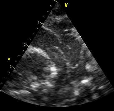
3 1. Trial entry What assessments need to be performed? Assessing eligibility for inclusion: Obtain high left parasternal ( ductal cut ) view Assess ductal patency and ductal flow characteristics Measure ductal dimension
4 What assessments need to be performed? (1) 2. Subsequent examinations - 3 weeks - Discharge ECHO - Rescue treatment In addition to (1) the following assessments should be performed: Assess presence of a hyperdynamic circulation Assess presence of ductal steal
5 ECHO criteria for Baby-OSCAR trial Trial entry 1. PDA dimension of 1.5 mm and 2. Unrestrictive pulsatile left to right flow in PDA Rescue treatment 1. Presence of a large PDA 2.0 mm and 2. Unrestrictive pulsatile left to right flow in PDA and 3. Presence of a hyperdynamic circulation or ductal steal
6 Who can perform ECHO assessments? Neonatologists or cardiologists (consultants/trainees) who have expertise in neonatal echocardiography and are able to visualise and assess the ductus arteriosus using conventional views Nominated echocardiographers should appear in Delegation Log
7 Cardiac structure At the first scan, it is desirable that the echocardiographer screens for normal cardiac anatomy. If any structural heart disease is suspected clinically or echocardiographically, appropriate action should follow according to local policy


8 WARNING You are entering an Evidence Free Zone
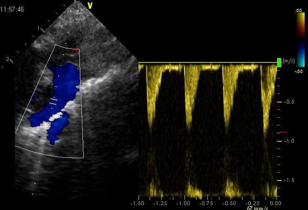
9 ECHO assessment Assessment of ductal patency and flow characteristics Assessment of ductal dimension Assessment of a hyperdynamic circulation Assessment of ductal steal


10 1. Assessment of ductal patency and flow characteristics


11 Right to left shunt > 30% of cardiac cycle should be considered abnormal
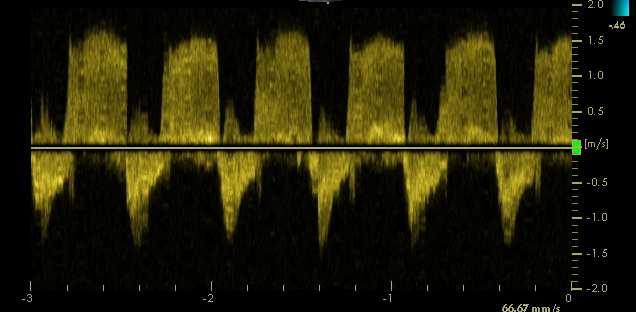
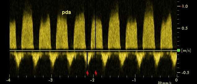
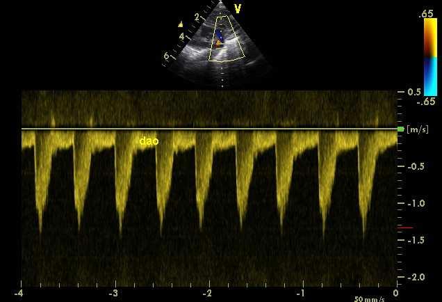
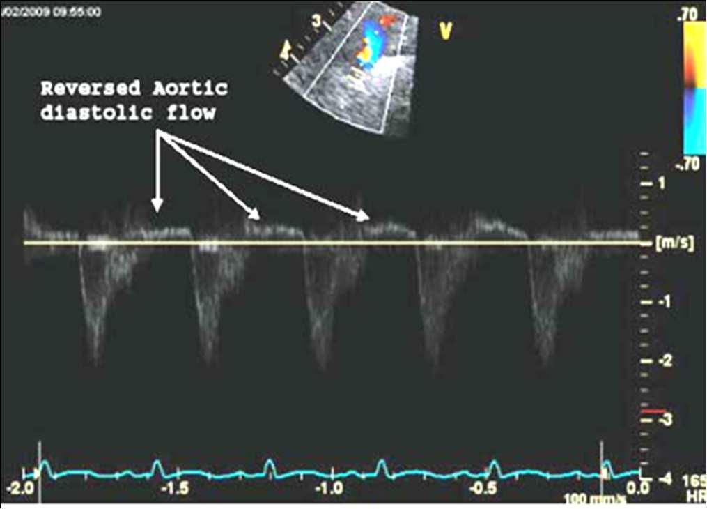
12 Bidirectional pattern Growing pattern

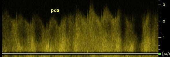
13 Closing pattern Pulsatile pattern


14 Pulsatility ratio = 3.13/2.03 = 1.54 Pulsatility ratio > 2 considered to represent pulsatile flow pattern Pulsatility ratio = 2.55/0.47 = 5.43
15 2. Assessment of ductal dimension Obtain high left parasternal ( ductal cut ) view. Optimise colour flow gain settings by: (1) adjusting colour gain scale to obtain optimal colour flow within the course of the ductus (2) adjusting colour gain to eliminate any peripheral colour interference by reducing gain until colour flow cannot be seen outside blood vessels Measure colour flow dimension at narrowest point by frame-to-frame analysis of the video loop selecting frames with the clearest discrete appearance of the ductus. Use 2D imaging to guide the point at which colour dimension should be measured. Calculate the mean of at least three measurements.

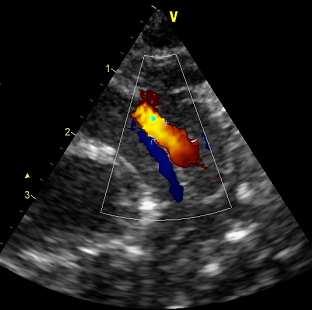
16 Pulmonary end of ductus
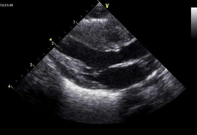
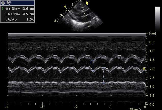
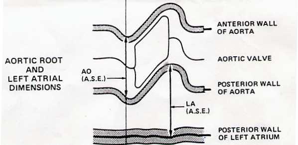
17 3. Assessment of a hyperdynamic circulation Measure left atrial: aortic root ratio using M-mode, ensuring the cursor is at right angles to the aorta and the posterior wall of the left atrium. Repeat (b) and calculate the mean of at least three measurements A LA/Ao ratio of > 2.0 is considered to represent significant left atrial dilatation secondary to volume overload of the left heart
18
19 4. Assessment of ductal steal Obtain view of descending aorta using a high parasternal or arch view Sample post-ductal aortic flow using pulse wave Doppler using angle-correction if necessary Record the presence of retrograde post-ductal aortic flow. Coeliac or superior mesenteric artery flows are acceptable alternatives
20


21 Retrograde diastolic flow is considered abnormal
22 ECHO assessment: summary Screening echo to detect structural heart disease, followed by: 1. Assessment of ductal patency and flow 2. Assessment of ductal dimension 3. Assessment of a hyperdynamic circulation 4. Assessment of ductal steal
23 ECHO criteria for Baby-OSCAR trial Trial entry 1. PDA dimension of 1.5 mm and 2. Unrestrictive pulsatile left to right flow in PDA Rescue treatment 1. Presence of a large PDA 2.0 mm and 2. Unrestrictive pulsatile left to right flow in PDA and 3. Presence of a hyperdynamic circulation or ductal steal

24 Contact details
Appendix II: ECHOCARDIOGRAPHY ANALYSIS
 Appendix II: ECHOCARDIOGRAPHY ANALYSIS Two-Dimensional (2D) imaging was performed using the Vivid 7 Advantage cardiovascular ultrasound system (GE Medical Systems, Milwaukee) with a frame rate of 400 frames
A preliminary study of the application of the transductal velocity ratio for assessing persistent ductus arteriosus
 Arch Dis Child Fetal Neonatal Ed 2000;82:F195 F199 F195 Division of Neonatal Services, Royal Women s Hospital, Melbourne, Australia M W Davies F R Betheras M Swaminathan Correspondence to: Dr Davies, Perinatal
Adult Echocardiography Examination Content Outline
 Adult Echocardiography Examination Content Outline (Outline Summary) # Domain Subdomain Percentage 1 2 3 4 5 Anatomy and Physiology Pathology Clinical Care and Safety Measurement Techniques, Maneuvers,
PART II ECHOCARDIOGRAPHY LABORATORY OPERATIONS ADULT TRANSTHORACIC ECHOCARDIOGRAPHY TESTING
 PART II ECHOCARDIOGRAPHY LABORATORY OPERATIONS ADULT TRANSTHORACIC ECHOCARDIOGRAPHY TESTING STANDARD - Primary Instrumentation 1.1 Cardiac Ultrasound Systems SECTION 1 Instrumentation Ultrasound instruments
Certificate in Clinician Performed Ultrasound (CCPU) Syllabus. Rapid Cardiac Echo (RCE)
 Syllabus. Rapid Cardiac Echo (RCE)") Certificate in Clinician Performed Ultrasound (CCPU) Syllabus Rapid Cardiac Echo (RCE) Purpose: Rapid Cardiac Echocardiography (RCE) This unit is designed to cover the theoretical and practical curriculum
Certificate in Clinician Performed Ultrasound (CCPU) Syllabus Rapid Cardiac Echo (RCE) Purpose: Rapid Cardiac Echocardiography (RCE) This unit is designed to cover the theoretical and practical curriculum
Diagnostic approach to heart disease
 Diagnostic approach to heart disease Initial work up History Physical exam Chest radiographs ECG Special studies Echocardiography Cardiac catheterization Echocardiography principles Technique of producing
Hypoplastic Left Heart Syndrome: Echocardiographic Assessment
 Hypoplastic Left Heart Syndrome: Echocardiographic Assessment Craig E Fleishman, MD, FACC, FASE Director, Non-invasive Cardiac Imaging The Hear Center at Arnold Palmer Hospital for Children, Orlando SCAI
Certificate in Allied Health Performed Ultrasound (CAHPU)
") Certificate in Allied Health Performed Ultrasound (CAHPU) Syllabus Advanced Allied Health Performed Neonatal Ultrasound Advanced Allied Health Performed Neonatal Ultrasound Purpose: Prerequisites: Training:
Certificate in Allied Health Performed Ultrasound (CAHPU) Syllabus Advanced Allied Health Performed Neonatal Ultrasound Advanced Allied Health Performed Neonatal Ultrasound Purpose: Prerequisites: Training:
Cardiac ultrasound protocols
 Cardiac ultrasound protocols IDEXX Telemedicine Consultants Two-dimensional and M-mode imaging planes Right parasternal long axis four chamber Obtained from the right side Displays the relative proportions
Uncommon Doppler Echocardiographic Findings of Severe Pulmonic Insufficiency
 Uncommon Doppler Echocardiographic Findings of Severe Pulmonic Insufficiency Rahul R. Jhaveri, MD, Muhamed Saric, MD, PhD, FASE, and Itzhak Kronzon, MD, FASE, New York, New York Background: Two-dimensional
Atrial Septal Defects
 Supplementary ACHD Echo Acquisition Protocol for Atrial Septal Defects The following protocol for echo in adult patients with atrial septal defects (ASDs) is a guide for performing a comprehensive assessment
COMPREHENSIVE EVALUATION OF FETAL HEART R. GOWDAMARAJAN MD
 COMPREHENSIVE EVALUATION OF FETAL HEART R. GOWDAMARAJAN MD Disclosure No Relevant Financial Relationships with Commercial Interests Fetal Echo: How to do it? Timing of Study -optimally between 22-24 weeks
Hybrid Stage I Palliation / Bilateral PAB
 Hybrid Stage I Palliation / Bilateral PAB Jeong-Jun Park Dept. of Thoracic & Cardiovascular Surgery Asan Medical Center, University of Ulsan CASE 1 week old neonate with HLHS GA 38 weeks Birth weight 3.0Kg
Fetal Ductus Arteriosus: The Good, Bad, and the Ugly. Tet Abs Valve syndrome With PDA. Fetal Ductus Arteriosus. James C. Huhta, M.D.
 Fetal Ductus Arteriosus: The Good, Bad, and the Ugly Tet Abs Valve syndrome With PDA James C. Huhta, M.D. Perinatal Cardiology Tampa, Florida 7 th Annual Fetal Echocardiography Course Friday, November
List of Videos. Video 1.1
 Video 1.1 Video 1.2 Video 1.3 Video 1.4 Video 1.5 Video 1.6 Video 1.7 Video 1.8 The parasternal long-axis view of the left ventricle shows the left ventricular inflow and outflow tract. The left atrium
Heart and Lungs. LUNG Coronal section demonstrates relationship of pulmonary parenchyma to heart and chest wall.
 Heart and Lungs Normal Sonographic Anatomy THORAX Axial and coronal sections demonstrate integrity of thorax, fetal breathing movements, and overall size and shape. LUNG Coronal section demonstrates relationship
Ductus Arteriosus: Dilemmas of Treatment vs. No Treatment in the Premature Neonate
 Ductus Arteriosus: Dilemmas of Treatment vs. No Treatment in the Premature Neonate Time allotted: 30 Minutes. No Disclosures. Norman H Silverman MD. D Sc (Med), FACC, FASE, FAHA. Professor of Pediatrics
Cardiac MRI in ACHD What We. ACHD Patients
 Cardiac MRI in ACHD What We Have Learned to Apply to ACHD Patients Faris Al Mousily, MBChB, FAAC, FACC Consultant, Pediatric Cardiology, KFSH&RC/Jeddah Adjunct Faculty, Division of Pediatric Cardiology
Index. Note: Page numbers of article titles are in boldface type.
 Index Note: Page numbers of article titles are in boldface type. A Acute coronary syndrome(s), anticoagulant therapy in, 706, 707 antiplatelet therapy in, 702 ß-blockers in, 703 cardiac biomarkers in,
British Society of Echocardiography
 British Society of Echocardiography Affiliated to the British Cardiac Society A Minimum Dataset for a Standard Adult Transthoracic Echocardiogram From the British Society of Echocardiography Education
Re-evaluation of the left atrial to aortic root ratio as a marker of patent ductus arteriosus
 Fl112 Department of Perinatal Medicine, King George V Hospital for Mothers and Babies, Part of Royal Prince Alfred Hospital, Sydney, New South Wales, Australia Parvathi Iyer Nick Evans Correspondence to:
Natural history of innocent heart murmurs in newborn babies: controlled echocardiographic study
 F166 Arch Dis Child Fetal Neonatal Ed 1998;78:F166 F170 ORIGINAL ARTICLES Natural history of innocent heart murmurs in newborn babies: controlled echocardiographic study Romaine Arlettaz, Nicholas Archer,
Transcatheter closure of the patent ductus arteriosus using the new Amplatzer duct occluder: Initial clinical applications in children
 Transcatheter closure of the patent ductus arteriosus using the new Amplatzer duct occluder: Initial clinical applications in children Basil Thanopoulos, MD, PhD, a Nikolaos Eleftherakis, MD, a Konstantinos
The sinus venosus represent the venous end of the heart It receives 3 veins: 1- Common cardinal vein body wall 2- Umbilical vein from placenta 3-
 1 2 The sinus venosus represent the venous end of the heart It receives 3 veins: 1- Common cardinal vein body wall 2- Umbilical vein from placenta 3- Vitelline vein from yolk sac 3 However!!!!! The left
Journal of American Science 2014;10(9) Congenital Heart Disease in Pediatric with Down's Syndrome
 Congenital Heart Disease in Pediatric with Down's Syndrome") Journal of American Science 2014;10(9) http://www.jofamericanscience.org Congenital Heart Disease in Pediatric with Down's Syndrome Jawaher Khalid Almaimani; Maryam Faisal Zafir; Hanan Yousif Abbas and
Journal of American Science 2014;10(9) http://www.jofamericanscience.org Congenital Heart Disease in Pediatric with Down's Syndrome Jawaher Khalid Almaimani; Maryam Faisal Zafir; Hanan Yousif Abbas and
Transposition of the Great Arteries Preoperative Diagnostic Considerations. John Simpson Evelina Children s Hospital London, UK
 Transposition of the Great Arteries Preoperative Diagnostic Considerations John Simpson Evelina Children s Hospital London, UK Areas to be covered Definitions Scope of occurrence of transposition of the
Large veins of the thorax Brachiocephalic veins
 Large veins of the thorax Brachiocephalic veins Right brachiocephalic vein: formed at the root of the neck by the union of the right subclavian & the right internal jugular veins. Left brachiocephalic
Pediatric Echocardiography Examination Content Outline
 Pediatric Echocardiography Examination Content Outline (Outline Summary) # Domain Subdomain Percentage 1 Anatomy and Physiology Normal Anatomy and Physiology 10% 2 Abnormal Pathology and Pathophysiology
Pathophysiology: Left To Right Shunts
 Pathophysiology: Left To Right Shunts Daphne T. Hsu, MD dh17@columbia.edu Learning Objectives Learn the relationships between pressure, blood flow, and resistance Review the transition from fetal to mature
Assessment of fetal heart function and rhythm
 Assessment of fetal heart function and rhythm The fetal myocardium Early Gestation Myofibrils 30% of myocytes Less sarcoplasmic reticula Late Gestation Myofibrils 60% of myocytes Increased force per unit
Data Collected: June 17, Reported: June 30, Survey Dates 05/24/ /07/2010
 Job Task Analysis for ARDMS Pediatric Echocardiography Data Collected: June 17, 2010 Reported: Analysis Summary For: Pediatric Echocardiography Exam Survey Dates 05/24/2010-06/07/2010 Invited Respondents
NEONATAL CLINICAL PRACTICE GUIDELINE
 NEONATAL CLINICAL PRACTICE GUIDELINE Approval Date: January 2015 Approved by: Neonatal Patient Care Teams, HSC & SBH Child Health Standards Committee Pages: 1 of 6 Supercedes: N/A 1.0 PURPOSE and INTENT
NOT YET!! PDA - Pathological or innocent physiologic bystander? PDA From Physiology to Treatment 9/8/2014
 PDA - Pathological or innocent physiologic bystander? PDA From Physiology to Treatment Martin Kluckow MBBS FRACP PhD CCPU Associate Professor Royal North Shore Hospital & University of Sydney, Australia
September 28-30, 2018
 September 28-30, 2018 Course Director Optimizing Detection of Congenital Heart Disease: Important Anatomic Cardiac Regions The Top 5 Critical Anatomic Regions in Fetal Cardiac Imaging Alfred Abuhamad,
Adel Hasanin Ahmed 1
 Adel Hasanin Ahmed 1 PERICARDIAL DISEASE The pericardial effusion ends anteriorly to the descending aorta and is best visualised in the PLAX. PSAX is actually very useful sometimes for looking at posterior
Large Arteries of Heart
 Cardiovascular System (Part A-2) Module 5 -Chapter 8 Overview Arteries Capillaries Veins Heart Anatomy Conduction System Blood pressure Fetal circulation Susie Turner, M.D. 1/5/13 Large Arteries of Heart
Systematic approach to Fetal Echocardiography. Objectives. Introduction 11/2/2015
 Systematic approach to Fetal Echocardiography. Pediatric Echocardiography Conference, JCMCH November 7, 2015 Rajani Anand Objectives Fetal cardiology pre-test Introduction Embryology and Physiology of
Pathophysiology: Left To Right Shunts
 Pathophysiology: Left To Right Shunts Daphne T. Hsu, MD dh17@columbia.edu Learning Objectives Learn the relationships between pressure, blood flow, and resistance Review the transition from fetal to mature
The Fontan circulation. Folkert Meijboom
 The Fontan circulation Folkert Meijboom What to expect? Why a Fontan-circulation Indications How does it work Types of Fontan circulation Historical overview Role of echocardiography What to expect? Why
CONGENITAL HEART DISEASE (CHD)
") CONGENITAL HEART DISEASE (CHD) DEFINITION It is the result of a structural or functional abnormality of the cardiovascular system at birth GENERAL FEATURES OF CHD Structural defects due to specific disturbance
CONGENITAL HEART DISEASE (CHD) DEFINITION It is the result of a structural or functional abnormality of the cardiovascular system at birth GENERAL FEATURES OF CHD Structural defects due to specific disturbance
Practical Echocardiography: ECHOES in the REAL WORLD Know When to Hold Em and When to Fold Em
 Practical Echocardiography: ECHOES in the REAL WORLD Know When to Hold Em and When to Fold Em Introduction The use of ultrasound in private veterinary practice is continuing to grow. The popularity of
SWISS SOCIETY OF NEONATOLOGY. Prenatal closure of the ductus arteriosus
 SWISS SOCIETY OF NEONATOLOGY Prenatal closure of the ductus arteriosus March 2007 Leone A, Fasnacht M, Beinder E, Arlettaz R, Neonatal Intensive Care Unit (LA, AR), University Hospital Zurich, Cardiology
5.8 Congenital Heart Disease
 5.8 Congenital Heart Disease Congenital heart diseases (CHD) refer to structural or functional heart diseases, which are present at birth. Some of these lesions may be discovered later. prevalence of Chd
Paediatrics Revision Session Cardiology. Emma Walker 7 th May 2016
 Paediatrics Revision Session Cardiology Emma Walker 7 th May 2016 Cardiovascular Examination! General:! Make it fun!! Change how you act depending on their age! Introduction! Introduce yourself & check
Teaching Manual. Afif EL-Khuffash The Rotunda Hospital, Dublin, Ireland. Patrick McNamara Hospital for Sick Children, Toronto
 Teaching Manual Afif EL-Khuffash The Rotunda Hospital, Dublin, Ireland Patrick McNamara Hospital for Sick Children, Toronto Second Edition 2014 INDEX INTRODUCTION 4 TNECHO TRAINING IN THE ROTUNDA HOSPITAL
A2b. PDA Management--A Reflection on the Evidence: Does it Help with Management? Session Summary. Session Objectives. References.
 FANNP 23RD NATIONAL NNP SYMPOSIUM: CLINICAL UTE AND REVIEW A2b Management--A Reflection on the Evidence: Does it Help with Management? Alfonso Vargas, MD Neonatologist Pediatrix Medical Group, Tampa, FL
Coarctation of the aorta: difficulties in prenatal
 7 Department of Fetal Cardiology, Guy's Hospital, London G K Sharland K-Y Chan L D Allan Correspondence to: Dr G Sharland, Department of Paediatric Cardiology, 1 lth Floor, Guy's Tower, Guy's Hospital,
PDA: Closure Using Coils and Devices: Indications, Technique & Outcome
 PDA: Closure Using Coils and Devices: Indications, Technique & Outcome Daniel Levi, MD, FSCAI Associate Professor of Pediatrics UCLA Biomedical Engineering Mattel Children s Hospital at UCLA, Division
Introduction to Fetal Doppler Echocardiography
 Chapter 32 Introduction to Fetal Doppler Echocardiography Dev Maulik Introduction Evaluation of the fetal heart constitutes one of the critical areas of prenatal diagnosis. Advances in diagnostic medical
Optimising your Doppler settings for an accurate PI. Alison McGuinness Mid Yorks Hospitals
 Optimising your Doppler settings for an accurate PI Alison McGuinness Mid Yorks Hospitals Applications Both maternal uterine and fetal circulations can be studied with doppler sonography Uterine arteries
PROSTHETIC VALVE BOARD REVIEW
 PROSTHETIC VALVE BOARD REVIEW The correct answer D This two chamber view shows a porcine mitral prosthesis with the typical appearance of the struts although the leaflets are not well seen. The valve
Case 9799 Stanford type A aortic dissection: US and CT findings
 Case 9799 Stanford type A aortic dissection: US and CT findings Accogli S, Aringhieri G, Scalise P, Angelini G, Pancrazi F, Bemi P, Bartolozzi C Department of Diagnostic and Interventional Radiology, University
Certificate in Clinician Performed Ultrasound (CCPU) Syllabus. Basic Echocardiography in Life Support
 Syllabus. Basic Echocardiography in Life Support") Certificate in Clinician Performed Ultrasound (CCPU) Syllabus Basic Echocardiography in Life Support Page 1 of 7 05/18 ACN 001 679 161 ABN 64 001 679 Basic Echocardiography in Life Support (BELS) Syllabus
Certificate in Clinician Performed Ultrasound (CCPU) Syllabus Basic Echocardiography in Life Support Page 1 of 7 05/18 ACN 001 679 161 ABN 64 001 679 Basic Echocardiography in Life Support (BELS) Syllabus
Rotation: Echocardiography: Transthoracic Echocardiography (TTE)
") Rotation: Echocardiography: Transthoracic Echocardiography (TTE) Rotation Format and Responsibilities: Fellows rotate in the echocardiography laboratory in each clinical year. Rotations during the first
Rotation: Echocardiography: Transthoracic Echocardiography (TTE) Rotation Format and Responsibilities: Fellows rotate in the echocardiography laboratory in each clinical year. Rotations during the first
Echocardiography in adult congenital heart disease
 S12 Department of Cardiology, Royal Hospital for Sick Children, Glasgow G3 8SJ, UK A Houston S Lilley T Richens University Department of Medicine and Therapeutics, Western Infirmary, Glasgow G11 6NT, UK
Adult Congenital Heart Disease: What All Echocardiographers Should Know Sharon L. Roble, MD, FACC Echo Hawaii 2016
 1 Adult Congenital Heart Disease: What All Echocardiographers Should Know Sharon L. Roble, MD, FACC Echo Hawaii 2016 DISCLOSURES I have no disclosures relevant to today s talk 2 Why should all echocardiographers
1. Definition/Overview
 VCAwestlaspecialty.com VCA West Los Angeles recently performed closure of a patent ductus arteriosus via percutaneous transarterial embolization with an Amplatz canine ductal occluder on a female, four-month-old
Total Anomalous Pulmonary Venous Connections: Anatomy and Diagnostic Imaging
 Total Anomalous Pulmonary Venous Connections: Anatomy and Diagnostic Imaging Timothy Slesnick, MD March 12, 2015 Congenital Cardiac Anesthesia Society Annual Meeting Disclosures I will discuss the use
Update on mangement of patent ductus arteriosus in preterm infants. Dr. Trinh Thi Thu Ha
 Update on mangement of patent ductus arteriosus in preterm infants Dr. Trinh Thi Thu Ha Outline 1. Overview of PDA 2. Timing of screening PDA? 3. When to treat PDA? Timing of ductal closure Prenatal
Debate in Management of native COA; Balloon Versus Surgery
 Debate in Management of native COA; Balloon Versus Surgery Dr. Amira Esmat, El Tantawy, MD Professor of Pediatrics Consultant Pediatric Cardiac Interventionist Faculty of Medicine Cairo University 23/2/2017
T wo dimensional and Doppler echocardiography is being
 F287 ORIGINAL ARTICLE Evaluation of echocardiography on the neonatal unit S Moss, D J Kitchiner, C W Yoxall, N V Subhedar... See end of article for authors affiliations... Correspondence to: Dr Subhedar,
Longitudinal changes in the diameter of the ductus arteriosus in ventilated preterm infants:
 F156 Department of Perinatal Medicine, King George V Hospital for Mothers and Babies, Part of Royal Prince Alfred Hospital, Camperdown, Sydney, NSW 2050, Australia N Evans P Iyer Correspondence to: Dr
TGA atrial vs arterial switch what do we need to look for and how to react
 TGA atrial vs arterial switch what do we need to look for and how to react Folkert Meijboom, MD, PhD, FES Dept ardiology University Medical entre Utrecht The Netherlands TGA + atrial switch: Follow-up
Echocardiography in Adult Congenital Heart Disease
 Echocardiography in Adult Congenital Heart Disease Michael Vogel Kinderherz-Praxis München CHD missed in childhood Subsequent lesions after repaired CHD Follow-up of cyanotic heart disease CHD missed in
AORTIC COARCTATION. Synonyms: - Coarctation of the aorta
 AORTIC COARCTATION Synonyms: - Coarctation of the aorta Definition: Aortic coarctation is a congenital narrowing of the aorta, usually located after the left subclavian artery, near the ductus or the ligamentum
Echocardiography Volume assessment. Justin Mandeville 2014
 Echocardiography Volume assessment Justin Mandeville 2014 Volume assessment and the intensivist Hypovolaemic shock Fluid tolerance Optimising cardiac output Avoiding overloading Guided fluid removal Add
IMAGES. in PAEDIATRIC CARDIOLOGY. Abstract
 IMAGES in PAEDIATRIC CARDIOLOGY Images Paediatr Cardiol. 2008 Apr-Jun; 10(2): 11 17. PMCID: PMC3232589 Transcatheter closure of symptomatic aortopulmonary window in an infant F Pillekamp, 1 T Hannes, 1
Screening for Critical Congenital Heart Disease
 Screening for Critical Congenital Heart Disease Caroline K. Lee, MD Pediatric Cardiology Disclosures I have no relevant financial relationships or conflicts of interest 1 Most Common Birth Defect Most
The Chest X-ray for Cardiologists
 Mayo Clinic & British Cardiovascular Society at the Royal College of Physicians, London : 21-23-October 2013 Cases-Controversies-Updates 2013 The Chest X-ray for Cardiologists Michael Rubens Royal Brompton
ULTRASOUND OF THE FETAL HEART
 ULTRASOUND OF THE FETAL HEART Cameron A. Manbeian, MD Disclosure Statement Today s faculty: Cameron Manbeian, MD does not have any relevant financial relationships with commercial interests or affiliations
PRACTICAL ECHOCARDIOGRAPHY IN THE ADULT with Doppler and color-doppler flow imaging
 PRACTICAL ECHOCARDIOGRAPHY IN THE ADULT with Doppler and color-doppler flow imaging PRACTICAL ECHOCARDIOGRAPHY IN THE ADULT with Doppler and color-doppler flow imaging by J.P.M. HAMER Thoraxcentre, Department
Superior vena cava flow in newborn infants: a novel marker of systemic blood flow
 F182 Arch Dis Child Fetal Neonatal Ed 2000;82:F182 F187 ORIGINAL ARTICLES Superior vena cava flow in newborn infants: a novel marker of systemic blood flow Martin Kluckow, Nick Evans Abstract Background
Slide 1. Slide 2. Slide 3 CONGENITAL HEART DISEASE. Papworth Hospital NHS Trust INTRODUCTION. Jakub Kadlec/Catherine Sudarshan INTRODUCTION
 Slide 1 CONGENITAL HEART DISEASE Jakub Kadlec/Catherine Sudarshan NHS Trust Slide 2 INTRODUCTION Most common congenital illness in the newborn Affects about 4 9 / 1000 full-term live births in the UK 1.5
5 Working With Measurements
 5 Working With Measurements Measurement Overview Measurements accompanying ultrasound images supplement other clinical procedures available to the attending physician. Accuracy of the measurements is determined
Diagnosis of Congenital Cardiac Defects Between 11 and 14 Weeks Gestation in High-Risk Patients
 Article Diagnosis of Congenital Cardiac Defects Between 11 and 14 Weeks Gestation in High-Risk Patients Zeev Weiner, MD, Abraham Lorber, MD, Eliezer Shalev, MD Objective. To examine the feasibility of
ECHOCARDIOGRAPHY DATA REPORT FORM
 Patient ID Patient Study ID AVM - - Date of form completion / / 20 Initials of person completing the form mm dd yyyy Study period Preoperative Postoperative Operative 6-month f/u 1-year f/u 2-year f/u
Assessment of LV systolic function
 Tutorial 5 - Assessment of LV systolic function Assessment of LV systolic function A knowledge of the LV systolic function is crucial in the undertanding of and management of unstable hemodynamics or a
Hemodynamic Assessment. Assessment of Systolic Function Doppler Hemodynamics
 Hemodynamic Assessment Matt M. Umland, RDCS, FASE Aurora Medical Group Milwaukee, WI Assessment of Systolic Function Doppler Hemodynamics Stroke Volume Cardiac Output Cardiac Index Tei Index/Index of myocardial
How to Recognize a Suspected Cardiac Defect in the Neonate
 Neonatal Nursing Education Brief: How to Recognize a Suspected Cardiac Defect in the Neonate https://www.seattlechildrens.org/healthcareprofessionals/education/continuing-medical-nursing-education/neonatalnursing-education-briefs/
An update on technique of fetal echocardiography with emphasis on anomalies detectable in four chambered view.
 An update on technique of fetal echocardiography with emphasis on anomalies detectable in four chambered view. Dr. Ranjitha.G Specialist Radiologist NMC-SH Al ain, UAE Fetal echocardiography is an essential
Diagnosis of fetal ductus arteriosus aneurysm: importance of the three-vessel view
 Ultrasound Obstet Gynecol 2005; 26: 57 62 Published online in Wiley InterScience (www.interscience.wiley.com). DOI: 10.1002/uog.1927 Diagnosis of fetal ductus arteriosus aneurysm: importance of the three-vessel
2) VSD & PDA - Dr. Aso
 VSD & PDA - Dr. Aso") 2) VSD & PDA - Dr. Aso Ventricular Septal Defect (VSD) Most common cardiac malformation 25-30 % Types of VSD: According to position perimembranous, inlet, muscular. According to size small, medium, large.
2) VSD & PDA - Dr. Aso Ventricular Septal Defect (VSD) Most common cardiac malformation 25-30 % Types of VSD: According to position perimembranous, inlet, muscular. According to size small, medium, large.
Debate: Should Ductal Stent Implantation be Considered for All Newborn Infants with Reduced Pulmonary Blood Flow?_Pros
 Debate: Should Ductal Stent Implantation be Considered for All Newborn Infants with Reduced Pulmonary Blood Flow?_Pros Mazeni Alwi Institut Jantung Negara Kuala Lumpur, Malaysia 5 th Asia Pacific Congenital
UvA-DARE (Digital Academic Repository) Central blood flow measurements in newborn infants de Waal, K.A. Link to publication
 Central blood flow measurements in newborn infants de Waal, K.A. Link to publication") UvA-DARE (Digital Academic Repository) Central blood flow measurements in newborn infants de Waal, K.A. Link to publication Citation for published version (APA): de Waal, K. A. (2011). Central blood flow
UvA-DARE (Digital Academic Repository) Central blood flow measurements in newborn infants de Waal, K.A. Link to publication Citation for published version (APA): de Waal, K. A. (2011). Central blood flow
PDA Device Closure with and without Arterial Access
 Research Article imedpub Journals www.imedpub.com Interventional Cardiology Journal DOI: 10.21767/2471-8157.100061 Abstract PDA Device Closure with and without Arterial Access Background: PDA device closure
AIMI-HF PROCEDURE MANUAL TECHNICAL GUIDE FOR ECHOCARDIOGRAPHY. MHI Core Laboratory E. O Meara - J.C. Tardif J. Vincent, G. Grenier, C.
 AIMI-HF PROCEDURE MANUAL TECHNICAL GUIDE FOR ECHOCARDIOGRAPHY MHI Core Laboratory E. O Meara - J.C. Tardif J. Vincent, G. Grenier, C. Roy February 2016 Montreal Heart Institute HF Research Aude Turgeon,
Christoph E. Schwarz 1*, Antonio Preusche 1, Winfried Baden 2, Christian F. Poets 1 and Axel R. Franz 1,3
 Schwarz et al. BMC Pediatrics (2016) 16:18 DOI 10.1186/s12887-016-0552-7 RESEARCH ARTICLE Open Access Repeatability of echocardiographic parameters to evaluate the hemodynamic relevance of patent ductus
Foetal Cardiology: How to predict perinatal problems. Prof. I.Witters Prof.M.Gewillig UZ Leuven
 Foetal Cardiology: How to predict perinatal problems Prof. I.Witters Prof.M.Gewillig UZ Leuven Cardiopathies Incidence : 8-12 / 1000 births ( 1% ) Most frequent - Ventricle Septum Defect 20% - Atrium Septum
Echocardiography in Congenital Heart Disease
 Chapter 44 Echocardiography in Congenital Heart Disease John L. Cotton and G. William Henry Multiple-plane cardiac imaging by echocardiography can noninvasively define the anatomy of the heart and the
Aortic Stenosis: Spectrum of Disease, Low Flow/Low Gradient and Variants
 Aortic Stenosis: Spectrum of Disease, Low Flow/Low Gradient and Variants Martin G. Keane, MD, FASE Professor of Medicine Lewis Katz School of Medicine at Temple University Basic root structure Parasternal
PRACTICAL GUIDE TO FETAL ECHOCARDIOGRAPHY IC Huggon and LD Allan
 PRACTICAL GUIDE TO FETAL ECHOCARDIOGRAPHY IC Huggon and LD Allan Fetal Cardiology Unit, Harris Birthright Research Centre for Fetal Medicine, King's College Hospital, London, UK IMPORTANCE OF PRENATAL
Echocardiographic and anatomical correlates in the fetus*
 Br Heart J 1980; : 51 Echocardiographic and anatomical correlates in the fetus* LINDSEY D ALLAN, MICHAEL J TYNAN, STUART CAMPBELL, JAMES L WILKINSON, ROBERT H ANDERSON From King's College Hospital, and
Case 47 Clinical Presentation
 93 Case 47 C Clinical Presentation 45-year-old man presents with chest pain and new onset of a murmur. Echocardiography shows severe aortic insufficiency. 94 RadCases Cardiac Imaging Imaging Findings C
Ductus Arteriosus: Dilemmas of Treatment vs. No Treatment in the Premature Neonate
 Ductus Arteriosus: Dilemmas of Treatment vs. No Treatment in the Premature Neonate Time allotted: 30 Minutes. No Disclosures. Norman H Silverman MD. D Sc (Med), FACC, FASE, FAHA. Professor of Pediatrics
Identification of congenital cardiac malformations by echocardiography in midtrimester fetus*
 Br Heart J 1981; 46: 358-62 Identification of congenital cardiac malformations by echocardiography in midtrimester fetus* LINDSEY D ALLAN, MICHAEL TYNAN, STUART CAMPBELL, ROBERT H ANDERSON From Guy's Hospital;
Doppler Basic & Hemodynamic Calculations
 Doppler Basic & Hemodynamic Calculations August 19, 2017 Smonporn Boonyaratavej MD Division of Cardiology, Department of Medicine Chulalongkorn University Cardiac Center, King Chulalongkorn Memorial Hospital
Congenital Heart Defects
 Normal Heart Congenital Heart Defects 1. Patent Ductus Arteriosus The ductus arteriosus connects the main pulmonary artery to the aorta. In utero, it allows the blood leaving the right ventricle to bypass
Lab 16. The Cardiovascular System Heart and Blood Vessels. Laboratory Objectives
 Lab 16 The Cardiovascular System Heart and Blood Vessels Laboratory Objectives Describe the anatomical structures of the heart to include the pericardium, chambers, valves, and major vessels. Describe
Patent ductus arteriosus (PDA)
") Patent ductus arteriosus (PDA) Information for families Great Ormond Street Hospital for Children NHS Foundation Trust 2 This information sheet explains about the congenital (present at birth) heart condition
Patent ductus arteriosus (PDA) Information for families Great Ormond Street Hospital for Children NHS Foundation Trust 2 This information sheet explains about the congenital (present at birth) heart condition
Heart and Soul Evaluation of the Fetal Heart
 Heart and Soul Evaluation of the Fetal Heart Ivana M. Vettraino, M.D., M.B.A. Clinical Associate Professor, Michigan State University College of Human Medicine Objectives Review the embryology of the formation