Chapter 04. Lecture Outline. See separate PowerPoint slides for all figures and tables pre-inserted into PowerPoint without notes.
|
|
|
- Lora Thornton
- 5 years ago
- Views:
Transcription


1 Chapter 04 Lecture Outline See separate PowerPoint slides for all figures and tables pre-inserted into PowerPoint without notes. Copyright The McGraw-Hill Companies, Inc. Permission required for reproduction or display.
2 Chapter 4-Tissues What is a tissue? group of cells with similar structure and function plus extracellular substance (matrix) Histology: study of tissues 2
3 Types of Tissues 1. Epithelial 2. Connective 3. Muscular 4. Nervous 3
4 Epithelial Tissues Location: - cover body (internal and external) - Ex. Skin, kidney, trachea, glands, etc. 4
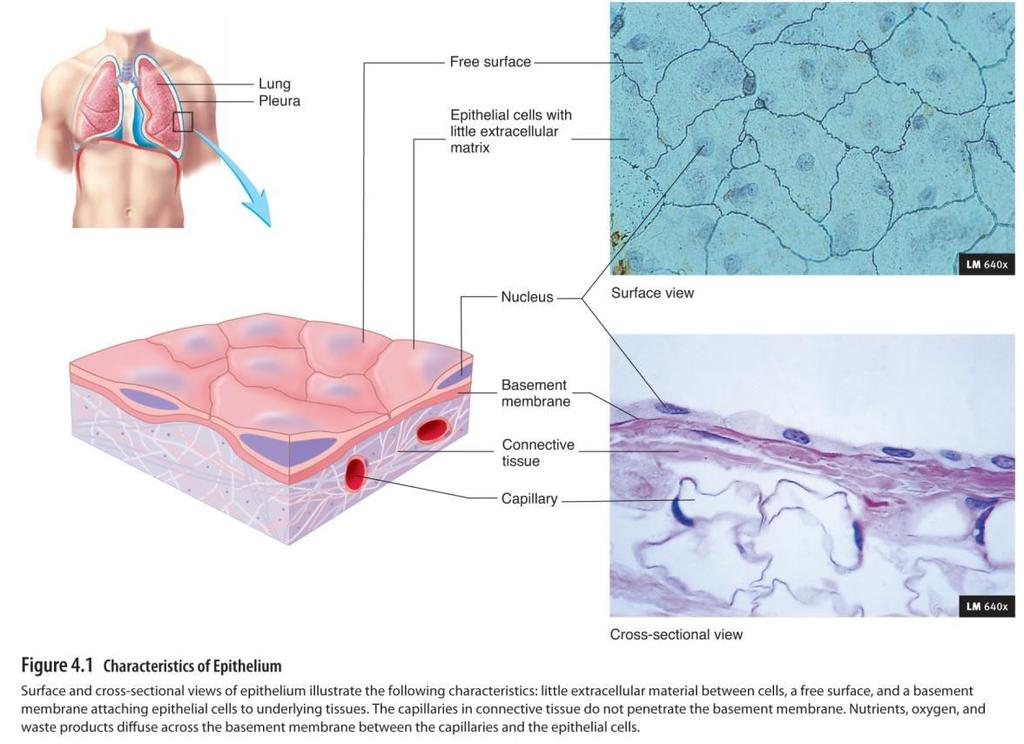
5 Characteristics: - cells close together (very little extracellular matrix) - form most glands - have free surface - Basal surface: attaches epithelial cells to underlying tissues 5
6
7 Functions of Epithelial Tissues 1. Protect: Ex. Skin 2. Act as a barrier: Ex. Skin keeps bacteria out 3. Diffusion and Filtration: Ex. Lungs and kidneys 4. Secretion: Ex. Sweat glands 5. Absorption: Ex. Small intestine 7
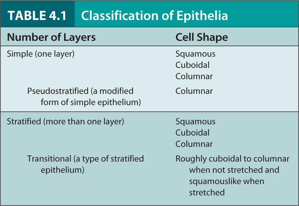
8 Classification of Epithelial Tissue Classified according to number of cell layers and cell shape Simple and stratified = number of cell layers Squamous, cuboidal, columnar, transitional= cell shape 8
9
10 Types of Epithelial Tissues Simple Epithelium Structure: 1 layer of cells Stratified Epithelium Structure: many layers of cells 10
11 Simple Squamous Structure: 1 layer of flat, tile-like cells Function: diffusion and filtration Location: blood vessels, lungs, heart, kidneys Simple Cuboidal Structure: 1 layer of square-shaped cells Function: secretion Location: glands, ovaries, kidneys 11
 Simple Squamous Epithelium Structure: Function: Location: Single layer of flat, often hexagonal cells; the nuclei appear as bumps when viewed in cross section")
 Lung alveoli Free surface Nucleus Basement membrane Simple squamous")
12 Copyright The McGraw-Hill Companies, Inc. Permission required for reproduction or display. TABLE 4.2 Simple Epithelium (a) Simple Squamous Epithelium Structure: Function: Location: Single layer of flat, often hexagonal cells; the nuclei appear as bumps when viewed in cross section because the cells are so flat Diffusion, filtration, some secretion, and some protection against friction Lining of blood vessels and the heart, lymphatic vessels, alveoli of the lungs, portions of the kidney tubules, lining of serous membranes of body cavities (pleural, pericardial, peritoneal) Lung alveoli Free surface Nucleus Basement membrane Simple squamous epithelial cell LM 640x a(2): McGraw-Hill Higher Education, Inc./Dennis Strete, photographer 12
13 Copyright The McGraw-Hill Companies, Inc. Permission required for reproduction or display. TABLE 4.2 continued (b) Simple Cuboidal Epithelium Structure: Single layer of cube-shaped cells; some cells have microvilli (kidney tubules) or cilia (terminal bronchioles of the lungs) Function: Location: Active transport and facilitated Kidney tubules, glands and their ducts, diffusion result in secretion and choroid plexuses of the brain, lining absorption by cells of the kidney of terminal bronchioles of the lungs, tubules; secretion by cells of glands and surfaces of the ovaries and choroid plexuses; movement of particles embedded in mucus out of the terminal bronchioles by ciliated cells Kidney Free surface Nucleus Simple cuboidal epithelial cell Basement membrane LM 640x b(2): Victor Eroschenko 13
14 Simple Columnar Structure: 1 layer of tall, narrow cells Function: secrete mucus and absorption Location: stomach, intestines, resp. tract Pseudostratified Columnar Structure: 1 layer of tall, narrow cells appears stratified but isn t Function: secrete mucus and propel debris out of resp. tract (cilia) Location: nasal cavity and trachea 14
 Simple Columnar Epithelium Structure: Function: Location: Single layer of tall, narrow cells; some cells have cilia (bronchioles of lungs, auditory tubes, uterine tubes, and uterus) or microvilli")

15 TABLE 4.2 continued Copyright The McGraw-Hill Companies, Inc. Permission required for reproduction or display. (c) Simple Columnar Epithelium Structure: Function: Location: Single layer of tall, narrow cells; some cells have cilia (bronchioles of lungs, auditory tubes, uterine tubes, and uterus) or microvilli (intestines) Movement of particles out of the bronchioles of the lungs by ciliated cells; partially responsible for the movement of oocytes through the uterine tubes by ciliated cells; secretion by cells of the glands, the stomach, and the intestine; absorption by cells of the intestine Glands and some ducts, bronchioles of lungs, auditory tubes, uterus, uterine tubes, stomach, intestines, gallbladder, bile ducts, and ventricles of the brain Lining of stomach and intestines Free surface Goblet cell containing mucus Nucleus Simple columnar epithelial cell Basement membrane LM 640x c(2): Victor Eroschenko 15
 Pseudostratified Columnar Epithelium Structure: Function: Location: Single layer of cells; some cells are tall and thin and reach the free surface, and others do not; the nuclei of")
16 Copyright The McGraw-Hill Companies, Inc. Permission required for reproduction or display. TABLE 4.2 continued (d) Pseudostratified Columnar Epithelium Structure: Function: Location: Single layer of cells; some cells are tall and thin and reach the free surface, and others do not; the nuclei of these cells are at different levels and appear stratified; the cells are almost always ciliated and are associated with goblet cells that secrete mucus on to the free surface Synthesize and secrete mucus on to the free surface and move mucus (or fluid) that contains foreign particles over the surface of the free surface and from passages Lining of nasal cavity, nasal sinuses, auditory tubes, pharynx, trachea, and bronchi of lungs Trachea Bronchus Cilia Free surface Goblet cell containing mucus Pseudostratified columnar epithelial cell Nucleus Basement membrane LM 413x d(2): Victor Eroschenko 16
17 Stratified Squamous Structure: many layers of flat, tile-like cells Function: protect and acts as a barrier Location: skin, mouth, throat, esophagus Transitional Structure: special type of stratified epi. changes shape (stretched squamous, not stretched cuboidal) Function: hold fluids Location: urinary bladder 17
 Stratified Squamous Epithelium Structure: Function: Location: Several layers of cells that are cuboidal Protects against abrasion, forms in the basal layer and progressively a")

18 Copyright The McGraw-Hill Companies, Inc. Permission required for reproduction or display. TABLE 4.3 Simple Epithelium (a) Stratified Squamous Epithelium Structure: Function: Location: Several layers of cells that are cuboidal Protects against abrasion, forms in the basal layer and progressively a barrier against infection, and flattened toward the surface; the reduces loss of water from the body epithelium can be nonkeratinized (moist) or keratinized; in nonkeratinized stratified squamous epithelium, the surface cells retain a nucleus and cytoplasm; in keratinized stratified epithelium, the cytoplasm of cells at the surface is replaced by a protein called keratin, and the cells are dead Keratinized outer layer of the skin; nonkeratinized mouth, throat, larynx, esophagus, anus, vagina, inferior urethra, and corneas Skin Cornea Mouth Esophagus Free surface Nonkeratinized stratified squamous epithelial cell Nuclei Basement membrane LM 286x a(2): Victor Eroschenko 18
 Transitional Epithelium Structure: Stratified cells that appear cuboidal when the organ or tube is not stretched and squamous when the organ or tube is stretched by fluid Function:")
19 Copyright The McGraw-Hill Companies, Inc. Permission required for reproduction or display. TABLE 4.3 continued (b) Transitional Epithelium Structure: Stratified cells that appear cuboidal when the organ or tube is not stretched and squamous when the organ or tube is stretched by fluid Function: Accommodates fluctuations in the volume of fluid in an organ or a tube; protects against the caustic effects of urine Location: Lining of urinary bladder, ureters, and superior urethra Free surface Transitional epithelial cell Ureter Urinary bladder Urethra Nucleus Basement membrane LM 413x Free surface Tissue not stretched Transitional epithelial cell Nucleus LM 413x Tissue stretched Basement membrane b(2, 3): Victor Eroschenko 19
20 Free Cell Surfaces Surface not in contact with other cells Smooth to reduce friction, Ex. Blood vessels Microvilli: - increase cell s surface area - Ex. Small intestine 20
21 Cilia: - move materials across cell s surface - Ex. Trachea Goblet cells: - produce mucus - Ex. Stomach 21
22 Cell Connections Tight junctions: - bind adjacent cells together - Ex. Intestines Desmosomes: mechanical links that bind cells 22
23 Hemidesmosomes: bind cells to basement membrane Gap junctions: - small channels that allow molecules to pass between cells - allow cells to communicate - most common 23

24 Figure 4.2
25 What are they? Glands structures that secrete substances onto a surface, into a cavity, or into blood Exocrine glands: - glands with ducts - Ex. Sweat or oil glands Endocrine glands: no ducts (directly into bloodstream) Ex. Thyroid, thymus, pituitary glands, etc. 25

26 Secretion in duct Copyright The McGraw-Hill Companies, Inc. Permission required for reproduction or display. Pinched-off portion of cell in the secretion Dying cell releases secretory products Vesicle releasing contents into duct Vesicle containing secretory products Secretory products stored in the cell Replacement cell Cell shed into the duct (a) Merocrine gland Cells of the gland produce secretions by active transport or produce vesicles that contain secretory products, and the vesicles empty their contents into the duct through exocytosis. (b) Apocrine gland Secretory products are stored in the cell near the lumen of the duct. A portion of the cell near the lumen containing secretory products is pinched off the cell and joins secretions produced by a merocine process. (c) Holocrine gland Secretory products are stored in the cells of the gland. Entire cells are shed by the gland and become part of the secretion. The lost cells are replaced by other cells deeper in the gland. 26
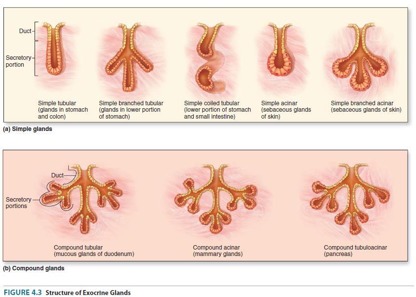
27 Types of Exocrine Glands Simple: no branches Compound: many branches Tubular: end of duct Alveolus: sac-like structure 27
28
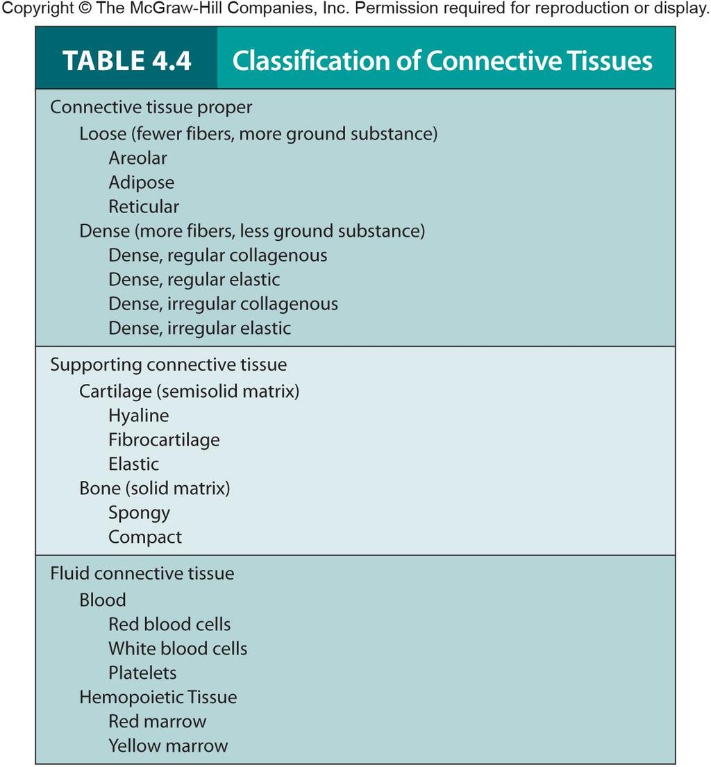
29 Connective Tissues Characteristics Cells far apart Contain large amounts of extracellular matrix Classified based on type of extracellular matrix and function Ex. Blast cells build, clast cells carve Extracellular matrix contains 3 components (in varying amounts): protein fibers, ground substance, fluid Ground substance: proteins and sugars 29
30 Types of Protein Fibers Collagen fibers: look like ropes and are flexible but resist stretching Reticular fibers: supporting network that fills spaces between organs and tissues Elastic fibers: recoil after being stretched 30
31 Functions of Connective Tissue 1. Enclose and separate: Ex. around organs and muscles 2. Connect tissues: Ex. Tendons: connect bone to muscle Ex. Ligaments: connect bone to bone 3. Support and Movement: Ex. bones 31
32 4. Storage: Ex. bones store calcium and adipose tissue stores fat 5. Cushion and insulate: Ex. adipose tissue protects organs and helps conserve heat 6. Transport: Ex. Blood 7. Protect: Ex. Immune cells 32
33
34 Types of Ordinary Connective Tissue Loose Location: between organs, muscles, glands, skin Structure: collagen fibers far apart Function: support and protect 34
 Areolar Connective Tissue Structure: Function: Location: A fine network of fibers (mostly collagen fibers with a few elastic fibers) with")

35 Copyright The McGraw-Hill Companies, Inc. Permission required for reproduction or display. TABLE 4.5 Connective Tissue Proper: Loose Connective Tissue (a) Areolar Connective Tissue Structure: Function: Location: A fine network of fibers (mostly collagen fibers with a few elastic fibers) with spaces between the fibers; fibroblasts, macrophages, and lymphocytes are located in the spaces Loose packing, support, and nourishment for the structures with which it is associated Widely distributed throughout the body; substance on which epithelial basement membranes rest; packing between glands, muscles, and nerves; attaches the skin to underlying tissues Elastic fiber Nucleus Collagen fiber Epidermis Dermis Skin Loose connective tissue with fat Muscle LM 400X (a): Ed Reschke 35
36 Dense Location: tendons, ligaments, skin Structure: collagen fibers packed close together Function: connect and can withstand pulling forces Adipose Location: under skin and around organs Structure: collagen and elastic fibers, cells filled with lipids Function: storage, insulate, cushion 36
 Dense Collagenous Connective Tissue Structure: Function: Location: Matrix composed of collagen fibers running in somewhat the same direction in")
37 Copyright The McGraw-Hill Companies, Inc. Permission required for reproduction or display. TABLE 4.6 Connective Tissue Proper: Dense Connective Tissue (a) Dense Collagenous Connective Tissue Structure: Function: Location: Matrix composed of collagen fibers running in somewhat the same direction in tendons and ligaments; collagen fibers run in several directions in the dermis of the skin and in organ capsules Withstand great pulling forces exerted in the direction of fiber orientation due to great tensile strength and stretch resistance Tendons (attach muscle to bone) and ligaments (attach bones to each other); also found in the dermis of the skin, organ capsules, and the outer layer of many blood vessels Nucleus of fibroblast Ligament Tendon Collagen fibers LM 165x (a): Victor Eroschenko 37

 and in the vocal cords; also found in elastic connective tissue of blood vessel walls Elastin fibers Nucleus of fibroblast Base of tongue Vocal folds (true vocal cords)")
38 TABLE 4.6 continued (b) Dense Elastic Connective Tissue Copyright The McGraw-Hill Companies, Inc. Permission required for reproduction or display. Structure: Matrix composed of collagen fibers and elastin fibers running in somewhat the same direction in elastic ligaments; elastic fibers run in connective tissue of blood vessel walls Function: Capable of stretching and recoiling like a rubber band with strength in the direction of fiber orientation Location: Elastic ligaments between the vertebrae and along the dorsal aspect of the neck (nucha) and in the vocal cords; also found in elastic connective tissue of blood vessel walls Elastin fibers Nucleus of fibroblast Base of tongue Vocal folds (true vocal cords) Vestibular fold (false vocal cord) LM 100x (b): Victor Eroschenko 38


39 TABLE 4.5 (b) Adipose Tissue continued Copyright The McGraw-Hill Companies, Inc. Permission required for reproduction or display. Structure: Function: Location: Little extracellular matrix surrounding cells; the adipocytes, or fat cells, are so full of lipid that the cytoplasm is pushed to the periphery of the cell Packing material, thermal insulator, energy storage, and protection of organs against injury from being bumped or jarred Predominantly in subcutaneous areas, mesenteries, renal pelves, around kidneys, attached to the surface of the colon, mammary glands, and in loose connective tissue that penetrates into spaces and crevices Adipose tissue Nucleus Mammary gland Adipocytes or fat cells LM 100x (b): Ed Reschke 39
40 Cartilage Type of connective tissue Composed of chondrocytes Contains collagen Withstands compressions Provides support, flexibility, strength 40
41 Types of Cartilage Hyaline cartilage Location: covers ends of bones Structure: some collagen fibers Function: reduces friction (cushion) Fibrocartilage Location: between vertebra Structure: lots of collagen fibers Function: can withstand compression 41
 Hyaline Cartilage Structure: Collagen fibers are small and evenly dispersed in the matrix, making the matrix appear transparent; the cartilage cells, or")
42 Copyright The McGraw-Hill Companies, Inc. Permission required for reproduction or display. TABLE 4.7 Supporting Connective Tissue: Cartilage (a) Hyaline Cartilage Structure: Collagen fibers are small and evenly dispersed in the matrix, making the matrix appear transparent; the cartilage cells, or chondrocytes, are found in spaces, or lacunae, within the firm but flexible matrix Function: Allows growth of long bones; provides rigidity with some flexibility in the trachea, bronchi, ribs, and nose; forms strong, smooth, yet somewhat flexible articulating surfaces; forms the embryonic skeleton Location: Growing long bones, cartilage rings of the respiratory system, costal cartilage of ribs, nasal cartilages, articulating surface of bones, and the embryonic skeleton Bone Chondrocyte in a lacuna Nucleus Matrix Hyaline cartilage LM 240x (a): Carolina Biological Supply/PhototakeUSA.com
43 Elastic cartilage Location: ear and tip of nose Structure: elastic fibers Function: can recoil 43
 Fibrocartilage Structure: Collagen fibers similar to those in hyaline cartilage; the fibers are more numerous than in other cartilages and are arranged in")

![, knees and temporomandibular [jaw] joints) Chondrocyte in lacuna Nucleus Intervertebral disk Collagen fibers in matrix LM 240x (c) Elastic Cartilage Structure: Similar to](/docs-images/82/87028960/images/44-4.jpg "hyaline cartilage, but matrix also contains elastin fibers Function: Provides rigidity with even more flexibility than hyaline cartilage because elastic fibers return to")
44 Copyright The McGraw-Hill Companies, Inc. Permission required for reproduction or display. TABLE 4.7 continued (b) Fibrocartilage Structure: Collagen fibers similar to those in hyaline cartilage; the fibers are more numerous than in other cartilages and are arranged in thick bundles Function: Somewhat flexible and capable of withstanding considerable pressure; connects structures subjected to great pressure Location: Intervertebral disks, pubic symphysis, and articular disks (e.g., knees and temporomandibular [jaw] joints) Chondrocyte in lacuna Nucleus Intervertebral disk Collagen fibers in matrix LM 240x (c) Elastic Cartilage Structure: Similar to hyaline cartilage, but matrix also contains elastin fibers Function: Provides rigidity with even more flexibility than hyaline cartilage because elastic fibers return to their original shape after being stretched Location: External ears, epiglottis, and auditory tubes Elastic fibers in matrix Chondrocytes in lacunae Nucleus LM 240x (b); Victor Eroschenko; (c): Victor Eroschenko
45 Hard connective tissue Bone 2 types: compact and spongy Composed of osteocytes 45
 are located within lacunae; the matrix is organized into layers")
46 TABLE 4.8 Copyright The McGraw-Hill Companies, Inc. Permission required for reproduction or display. Supporting Connective Tissue: Bone Structure: Hard, bony matrix predominates; many osteocytes (not seen in this bone preparation) are located within lacunae; the matrix is organized into layers called lamellae Function: Provides great strength and support and protects internal organs, such as the brain; bone also provides attachment sites for muscles and ligaments; the joints of bones allow movements Location: All bones of the body Lacuna Central canal Spongy bone LM 240x Matrix organized into lamellae Compact bone Trent Stephens
47 Blood Liquid connective tissue Erythrocytes, leukocytes, platelets Transport food, oxygen, waste, hormones 47

48 TABLE 4.9 Copyright The McGraw-Hill Companies, Inc. Permission required for reproduction or display. Fluid Connective Tissue: Blood Structure: Blood cells and a fluid matrix Function: Transports oxygen, carbon dioxide, hormones, nutrients, waste products, and other substances; protects the body from infections and is involved in temperature regulation Location: Within the blood vessels; white blood cells frequently leave the blood vessels and enter the interstitial spaces Red blood cells White blood cells LM 400x Ed Reschke
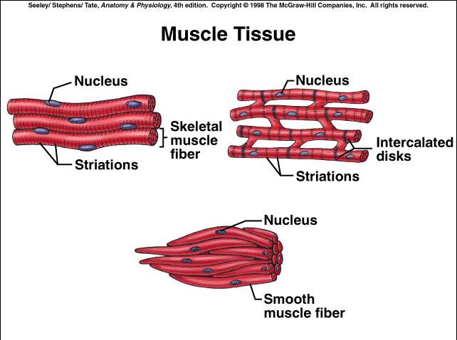
49 Muscular Tissue Muscle type Nucleus/i Nucleus/i location Striated Skeletal many peripheral Y (most muscle) Cardiac 1 centrally Y (heart) Smooth 1 centrally N (organs) 49
50 50
 Skeletal Muscle Structure: Skeletal muscle cells or fibers appear striated (banded); cells are large, long, and cylindrical, with many nuclei Function: Movement of the body;")
51 Copyright The McGraw-Hill Companies, Inc. Permission required for reproduction or display. TABLE 4.10 Muscle Tissue (a) Skeletal Muscle Structure: Skeletal muscle cells or fibers appear striated (banded); cells are large, long, and cylindrical, with many nuclei Function: Movement of the body; under voluntary control Location: Attached to bone or other connective tissue Muscle Nucleus (near periphery of cell) Skeletal muscle fiber Striations LM 800x a(2): Ed Reschke
 Cardiac Muscle Structure: Cardiac muscle cells are cylindrical and striated and have a single nucleus; they are branched and connected to one another by intercalated disks, which")
52 Copyright The McGraw-Hill Companies, Inc. Permission required for reproduction or display. TABLE 4.10 continued (b) Cardiac Muscle Structure: Cardiac muscle cells are cylindrical and striated and have a single nucleus; they are branched and connected to one another by intercalated disks, which contain gap junctions Function: Pumps the blood; under involuntary (unconscious) control Location: In the heart Nucleus Cardiac muscle cell Intercalated disks (special junctions between cells) Striations LM 800x b(2): Ed Reschke
 Smooth Muscle continued Structure: Smooth muscle cells are tapered at each end, are not striated, and have a single nucleus Function: Regulates the size of organs, forces fluid through tube,")
53 Copyright The McGraw-Hill Companies, Inc. Permission required for reproduction or display. TABLE 4.10 (c) Smooth Muscle continued Structure: Smooth muscle cells are tapered at each end, are not striated, and have a single nucleus Function: Regulates the size of organs, forces fluid through tube, controls the amount of light entering the eye, and produces goose bumps in the skin; under involuntary (unconscious) control Location: In hollow organs, such as the stomach and intestine; skin and eyes Wall of stomach Wall of colon Wall of small intestine Nucleus Smooth muscle cell LM 800x c(2): Victor Eroschenko
54 Nervous Tissue Consist of neurons or nerve cells Found in brain, spinal cord, and peripheral nerves Controls and coordinates body movements Includes axons, dendrites, cell bodies 54

55 Copyright The McGraw-Hill Companies, Inc. Permission required for reproduction or display. TABLE 4.11 Nervous Tissue Structure: A neuron consists of dendrites, a cell body, and a long axon; neuroglia, or support cells, surround the neurons Function: Neurons transmit information in the form of action potentials, store information, and integrate and evaluate data; neuroglia support, protect, and form specialized sheaths around axons Location: In the brain, spinal cord, and ganglia Brain Spinal cord Spinal nerves Dendrite Cell body of neuron Nucleus of neuron Nuclei of neuroglia Neuroglia Axon LM 240x Trent Stephens 55
56 What is it? Tissue Repair substitution of dead cells for viable cells Regeneration: cells of same type develop (no scar) Replacement: cells of a different type develop (scar) 56
57 Inflammation Occurs when tissues are damaged Signals the body s defenses (white blood cells) to destroy foreign materials and damaged cells so repair can occur. Chemical mediators: - released after injury - cause dilation of blood vessels 57
58 Symptoms of Inflammation 1. Redness: blood vessels dilate 2. Heat: due to increased blood flow 3. Swelling: from water and proteins 4. Pain: nerve endings are stimulated by damage and swelling 58

59 Copyright The McGraw-Hill Companies, Inc. Permission required for reproduction or display. Splinter Bacteria introduced 1 A splinter in the skin causes damage and introduces bacteria. Chemical mediators of inflammation are released or activated in injured tissues and adjacent blood vessels. Some blood vessels rupture, causing bleeding. 1 Epidermis 2 Chemical mediators cause capillaries to dilate and the skin to become red. Chemical mediators also increase capillary permeability, and fluid leaves the capillaries, producing swelling (arrows). 2 Dermis Blood vessel Bacteria proliferating 3 White blood cells (e.g., neutrophils) leave the dilated blood vessels and move to the site of bacterial infection, where they begin to phagocytize bacteria and other debris. 3 Neutrophil phagocytizing bacteria Neutrophil migrating through blood vessel wall
UNIT 4 T I S S U E S
 UNIT 4 T I S S U E S WHAT IS A TISSUE Group of cells that work together to do a function Cells are similar Extracellular fluid around them is similar Histology EPITHELIAL TISSUE Also called epithelium
Outline. Bio 105: Tissues Laboratory. Organization of the Human Body. Tissue - Epithelium. Tissues 3/2/ Copyright 2009 Pearson Education, Inc
 Outline Bio 105: Tissues Laboratory Laboratory 5 Reading: Chapter 4 I. Cell to cell contact II. Body Cavities III. Membranes IV. Homeostasis V. Integumentary System I. Includes skin, hair and nails 1 2
Chapter 5. Tissues. 4 Types of Body Tissues. Tissues
 Chapter 5 Tissues Tissues Tissues - groups of cells that are similar in structure & function RBC, WBC, & platelets are a group of cells working together to form BLOOD tissue Histology Pathohistology study
Hole s Human Anatomy and Physiology
 Hole s Human Anatomy and Physiology 1 Chapter 5 Tissues Four major tissue types 1. Epithelial 2. Connective 3. Muscle 4. Nervous 2 Epithelial Tissues General characteristics - cover organs and the body
Tissues 10/21/2016. Epithelial Tissue
 Tissues This is a generalized cell diagram. It shows the anatomy of a cell, but most cells do not actually look like this. Cells can have a wide variety of shapes and sizes, depending on their function.
Basic Histology. By Mrs. Bailey
 Basic Histology By Mrs. Bailey Primary Tissues 1. Epithelial Tissue 2. Connective Tissue 3. Muscle Tissue 4. Nervous Tissue Very cellular Supported by underlying connective tissue Epithelial & connective
Chapter 05. Review. Copyright The McGraw-Hill Companies, Inc. Permission required for reproduction or display.
 Chapter 05 Review 5.1: Introduction Similar cells with a common function are called tissues. The study of tissues is called histology. There are four (4) primary or major tissue types: 1. Epithelial Tissue
Histology. Study of body tissues
 Histology Study of body tissues 2 Introduction to Body Tissues 1. Composed of specialized cells of similar structure and perform a common function 2. Four major types (4 Cs) a. Epithelial - Cover b. Connective
Tissues, Glands, and Membranes. Chapter Five Mrs. Hornacek
 Tissues, Glands, and Membranes Chapter Five Mrs. Hornacek Objectives 1. Name the four main groups of tissues and give the location and general characteristics of each. 2. Differentiate between voluntary
Lab 1 ANIMAL TISSUES
 Lab 1 ANIMAL TISSUES Levels of Organization Animals are multicellular heterotrophs whose cells lack cell walls. Most animals exhibit a hierarchical level of organization: Cells are organized into tissues
Tissues. How do cells form tissues?
 Tissues How do cells form tissues? Using cell junctions Tissues Epithelial tissue Connective tissue Muscle tissue Nervous tissue Epithelial Tissue Closely packed cells in continuous sheets connected by
Body Tissues. Cells are specialized for particular functions Tissues - groups of cells with similar structure. and function Four primary tissue types:
 Chapter 3 Tissues Body Tissues Cells are specialized for particular functions Tissues - groups of cells with similar structure and function Four primary tissue types: Epithelium Connective tissue Nervous
Tissues Chapter 5...Tissue - a group or mass of similar cells working together to perform certain common functions
 Tissues Chapter 5...Tissue - a group or mass of similar cells working together to perform certain common functions There are 4 major types of tissue Epithelial Connective Muscle Nervous 1. Epithelial Tissue
Unit I Problem 9 Histology: Basic Tissues of The Body
 Unit I Problem 9 Histology: Basic Tissues of The Body - What is the difference between cytology and histology? Cytology: it is the study of the structure and functions of cells and their contents. Histology:
Anatomy and Physiology Tissue Review
 Anatomy and Physiology Tissue Review OVERVIEW Histology practicals can be rough, especially when access to slides is limited to the lab period. This resource provides an opportunity to learn or review
Study of different tissues Abnormal cells and tissues can be compared to normal tissues to identify disease, such as cancer Being able to know and
 CHAPTER 4 Study of different tissues Abnormal cells and tissues can be compared to normal tissues to identify disease, such as cancer Being able to know and recognize normal tissues under the microscope
Epithelial Tissue lining, covering, glandular tissue > Function protect, absorption, filtration, secretion, excretion
 Chapter 4: TISSUES IX. Tissues Intro Epithelial Tissue lining, covering, glandular tissue > Function protect, absorption, filtration, secretion, excretion Connective Tissue most widespread tissue type
Body Tissues Pearson Education, Inc.
 Body Tissues Tissues Groups of cells with similar structure and function Four primary types: Epithelial tissue (epithelium).1 Connective tissue.2 Muscle tissue.3 Nervous tissue.4 Epithelial Tissues Locations:
Epithelial Tissues. Types of Epithelial Tissues: Lining of Kidney
 Epithelial Tissues Covers the entire body surface and most of the body s inner cavities Outer epidermis (skin) protects from injury and drying out Inner epidermal tissue (on internal surfaces) often serves
Chapter 1: Cells and Tissues
 Chapter 1: Cells and Tissues Cells and Tissues Carry out all chemical activities needed to sustain life Cells are the building blocks of all living things Tissues are groups of cells that are similar in
Tissue Outline (chapter 4) Tissues group of cells that perform structural and roles. List the 4 types:
 Tissues group of cells that perform structural and roles. List the 4 types:") Tissue Outline (chapter 4) Tissues group of cells that perform structural and roles. List the 4 types: 1. 2. 3. 4. I. Epithelial Tissue covers all the surfaces, inside & out. Are the major tissues of,
Tissue Outline (chapter 4) Tissues group of cells that perform structural and roles. List the 4 types: 1. 2. 3. 4. I. Epithelial Tissue covers all the surfaces, inside & out. Are the major tissues of,
Tissues. Cells work together in functionally related groups called tissues Types of tissues: 1. Epithelial lining and covering. 2. Connective support
 Histology Tissues Cells work together in functionally related groups called tissues Types of tissues: 1. Epithelial lining and covering 2. Connective support 3. Muscle movement 4. Nervous control Epithelial
TISSUES. Dr. Gary Mumaugh
 TISSUES Dr. Gary Mumaugh Tissues Tissues - Groups of cells similar in structure and function and perform a common function Histology The study of tissues The four types of tissues Epithelial Connective
A. cells that perform related functions and are similar in structure. B. extracellular material - made by cells and secreted into interstitial space
 I. tissue components A. cells that perform related functions and are similar in structure B. extracellular material - made by cells and secreted into interstitial space II. tissue types A. epithelium (e.)
Basic Tissue Types and Functions
 Tissues Histology Basic Tissue Types and Functions 1) Epithelial tissue covering 2) Connective tissue support 3) Muscle tissue movement 4) Nervous tissue control Epithelial Tissue 1) Covers a body surface
Tissues. groups of cells similar in structure and function 4 types. epithelium connective muscle nervous
 Tissues groups of cells similar in structure and function 4 types epithelium connective muscle nervous Epithelial Tissue lining covering glandular Functions protection absorption filtration secretion Epithelium
THE TISSUE LEVEL OF ORGANIZATION PART I: EPITHELIAL TISSUE
 THE TISSUE LEVEL OF ORGANIZATION PART I: EPITHELIAL TISSUE 4 Main Tissue Types Epithelium Covers surfaces, lines cavities, forms glands Connective Tissue Support and protects body Muscular Tissue Movement
Air sacs of lungs and the lining of the heart, blood vessels, and lymphatic vessels
 Cells Location Function Simple squamous epithelium Air sacs of lungs and the lining of the heart, blood vessels, and lymphatic vessels Allows materials to pass through by diffusion and filtration, and
Epithelium. Four primary tissue types:
 Epithelium Four primary tissue types: Epithelial (covering) Connective (support) Nervous (control) Muscular (movement) Smooth muscle Cardiac muscle Skeletal muscle 1 Epithelial Tissue Features Epithelial
The Tissue Level of Organization
 Tissue The Tissue Level of Organization Chapter 3 Definition an aggregation of cells in which each cooperates with all others in the performance of a given function Examples of general functions Movement
Unit II: Tissues and Integumentary System
 Unit II: Tissues and Integumentary System 2.1 - Tissues Chapter 4 Written Response #1 1. What is a tissue? 2. What are four major types of tissues? Tissue Definition: a group or mass of similar cells working
Histology Notes -Part 1: Epithelial Tissues
 Introduction Group of cells w/ similar structure & function = TISSUE Four Basic Tissue Types 1. Epithelial-covers 2. Connective-supports 3. Muscular*-produces movement (will discuss in the muscular system
Histology 101! !! Name:! Block: Identify and describe the functions of major tissue types including their subclasses and varieties!
 Histology 101 Identify and describe the functions of major tissue types including their subclasses and varieties Name: Block: "1 Introduction to Tissues Histology Notes Tissue (living fabric) : groups
Epithelial Tissue. Simple Cuboidal Function: secretion and absorption. Simple Squamous
 Epithelial Tissue General Functions: Lines and covers organs Absorbs / secretes substances Gas exchange Protection Special Characteristics: - have an apical surface on top - have a basement membrane below
Tissue: The Living Fabric: Part A
 PowerPoint Lecture Slides prepared by Janice Meeking, Mount Royal College C H A P T E R 4 Tissue: The Living Fabric: Part A Tissues Groups of cells similar in structure and function Types of tissues Epithelial
Tissue = groups of cells that are similar in structure and function
 Tissue = groups of cells that are similar in structure and function Types Epithelial - covering Connective - support Muscle - movement Nervous - control Membranes line body cavities and hold organs together
ACTIVITY 2: HISTOLOGY AND INTEGUMENT
 ACTIVITY 2: HISTOLOGY AND INTEGUMENT Objectives: 1) How to get ready: Read Chapter 4 and 5, McKinley et al., Human Anatomy, 4e. All text references are for this textbook. 2) Identify each tissue (26 tissues)
Classification of Tissues
 6 R e v i e w S h e e t Exercise Classification of Tissues NAME LAB TIME/DATE Tissue Structure and Function General Review 1. Define tissue. A group of cells similar to one another in structure that perform
HOLE S ANATOMY CHAPTER 5, PART II Lecture notes
 HOLE S ANATOMY CHAPTER 5, PART II Lecture notes I. Connective Tissue A. Structure 1. have few cells that are spaced apart and can divide; two categories: a. fixed cells cells that are present in tissue
Cell and Tissue Types. Epithelial, Connective, Muscle, Nerve
 Cell and Tissue Types Epithelial, Connective, Muscle, Nerve Objectives Explain the major stages of the cell cycle and cellular division (mitosis). Describe specific events occurring in each of the phases
Tissues. Group of cells that are similar in structure and function. 4 primary types. Epithelium (covering) Connective (support) Nervous(control)
 Connective (support) Nervous(control)") Tissues Tissues Group of cells that are similar in structure and function 4 primary types Epithelium (covering) Connective (support) Nervous(control) Epithelial tissue (epithelium) Lining, covering, and
Tissues Tissues Group of cells that are similar in structure and function 4 primary types Epithelium (covering) Connective (support) Nervous(control) Epithelial tissue (epithelium) Lining, covering, and
Name: Test Date: Chapter 4- Tissues. Use the choices to identify the major tissue types found below:
 Name: Test Date: Chapter 4- Tissues Use the choices to identify the major tissue types found below: A. Connective B. Epithelium C. Muscle D. Nervous 1. B Lines body cavities and covers the body s external
B. Classification of epithelium: by number of cell layers present and by shape of the superficial cell layers.
 I. Introduction - tissue: group of cells that are closely associated, similar in structure and function, and perform a common or related function. - four primary tissues: epithelial tissue, connective
Histology= the study of tissues
 Unit 3-Histology Histology= the study of tissues A tissue is a group of cells that have a similar shape and function. Different types of tissues can be found in different organs. In humans, there are four
What is a tissue? Points to ponder. Tissues Connective Tissue. 1. Connective tissue 2/23/2019. Organization and Regulation of Body Systems
 Organization and Regulation of Body Systems Chapter 04 Lecture Outline See separate PowerPoint slides for all figures and tables preinserted into PowerPoint without notes. Copyright 2016 McGraw-Hill Education.
Tissues- of cells with similar and
 Tissues- of cells with similar and. Four types of tissues 1. 2. 3. 4. Characteristics of Epithelial Tissue -Highly Cellular -Special contacts -Polar (apical and basal surfaces) -Supported by connective
Use for reference if needed:
 A- 2.5 Describe how structure and function are related in terms of cell and tissue types. I can recognize different types of body tissue. I can explain how different tissue structures affect their functions.
HISTOLOGY. Simple squamal lungs
 HISTOLOGY Lab Objectives: Students should be able to... 1. Visually identify each class of tissue and examples within each class 2. Indicate the location (in the human body and/or organ) and function of
Tissues. tissue = many cells w/ same structure and function. cell shape aids its function tissue shape aids its function
 Tissues tissue = many cells w/ same structure and function cell shape aids its function tissue shape aids its function Histology = study of tissues 4 types of tissues Epithelial coverings contact openings
8/30/2017. Tissue: The Living Fabric. 4.3 Connective Tissue
 Chapter 4 Part B Tissue: The Living Fabric Annie Leibovitz/Contact Press Images PowerPoint Lecture Slides prepared by Karen Dunbar Kareiva Ivy Tech Community College 4.3 Connective Tissue Connective tissue
Anatomy &- Physiology Histology Worksheet
 Anatomy &- Physiology Histology Worksheet 1. The four primary tissue types found in the human body are a) squamous, cuboidal, columnar, glandular b) adipose, elastic, reticular, cartilage c) skeletal,
Tissues. Definition. A group of similar cells and their intercellular substances specialized to perform a specific function.
 Chapter 4 - Tissues Tissues Definition A group of similar cells and their intercellular substances specialized to perform a specific function. Tissues Epithelial covers exposed surfaces, lines internal
Tissues are: group of similar or identical cells that share a common function. used to build organs
 Tissues: Four classes Epithelium Connective Muscle Nervous Tissues are: group of similar or identical cells that share a common function. used to build organs Overview: Epithelial o Line body cavities
Study of Tissues Dr. A. Ebneshahidi
 Study of Tissues Dr. A. Ebneshahidi Tissues Tissues are composed of cells similar in structure and specialized to perform a specific function for the body. The human body is made of four general types
TISSUE. A group of cells that perform a similar function within an organism. Epithelium Connective Muscle Nervous CREDITS
 TISSUE A group of cells that perform a similar function within an organism. Epithelium Connective Muscle Nervous CREDITS Epithelium Connective Muscle Nervous Epithelium Composed of a layer of cells. Lines
Cells are specialized for particular functions Tissues
 Histology Body Tissues Cells are specialized for particular functions Tissues Groups of cells with similar structure and function Extracellular Matrix cell glue between cells Histology study of tissue
Tissue: The Living Fabric
 PowerPoint Lecture Slide Presentation by Vince Austin Human Anatomy & Physiology FIFTH EDITION Elaine N. Marieb Chapter 4 Tissue: The Living Fabric Part A Tissues Groups of cells similar in structure and
I. Introduction. Unit One. Tendons of the hand. The white glistening appearance results from the collagen of which tendons are composed.
 5 Tendons of the hand tendons The white glistening appearance results from the collagen of which tendons are composed. Chapter 5 Karen Webb Smith Unit One I. Introduction A. Cells are arranged in tissues
Histology: The Study of Tissues
 Chapter 4 Histology: The Study of Tissues 4-1 Tissues and Histology Tissue classification based on structure of cells, composition of noncellular extracellular matrix, and cell function Epithelial Connective
Connexons: hollow connective tubes
 Chapter 3 1. tight junctions: like a zipper, these junctions hold the cells tightly together making them impermeable to the extracellular fluid that surrounds them. 2. desmosomes: like buttons, these
Classification of Tissues
 M06_MARI0000_00_SE_CH06.qxd 3/28/11 4:37 PM Page 35 NAME LAB TIME/DATE R E V I E W S H E E T EXERCISE 6 Classification of Tissues Tissue Structure and Function General Review 1. Define tissue. A group
BIOH111. o Cell Biology Module o Tissue Module o Integumentary system o Skeletal system o Muscle system o Nervous system o Endocrine system
 BIOH111 o Cell Biology Module o Tissue Module o Integumentary system o Skeletal system o Muscle system o Nervous system o Endocrine system Endeavour College of Natural Health endeavour.edu.au 1 Textbook
Tissues. Tissues. Four basic tissues. A collection of cells with a common function. 1. Epithelial 2. Connective 3. Muscular 4.
 Tissues Tissues A collection of cells with a common function Four basic tissues 1. Epithelial 2. Connective 3. Muscular 4. Nervous Epithelia: cells in layers Types of epithelia 1) lining Layers of cells
Histology. The study of tissues.
 Histology The study of tissues. Body Tissues Cells are specialized for particular functions Tissues Groups of cells with similar structure and function Four primary types Epithelium Connective tissue Nervous
Simple Squamous Epithelium
 Histology Simple Squamous Epithelium One layer of flattened cells. Protective characteristics are diminished because of this. Examples: Alveoli in the lungs Capillaries where diffusion of nutrients and
Epithelia of Coverings and Linings. Tissues. Tissue
 Tissue Tissues Chapter 3 Definition an aggregation of cells in which each cooperates with all others in the performance of a given function Examples of general functions Movement Protection Support Production
Anatomy and Physiology 1 Chapter 4 Outline Tissues and Membranes
 Anatomy and Physiology 1 Chapter 4 Outline Tissues and Membranes 1 Tissue group of cells with similar structure and function o 4 major groups epithelial, connective, muscle, nerve Epithelial tissue (Fig
Lesson 9A Tissues in Animals
 Lesson 9A Tissues in Animals Levels of Organization in the Human Body Similar types of cells Different types of tissues Different organs Many organ systems cell tissue organ organ system organism Levels
Epithelial Tissue. Functions include: 1. Protection 4. Absorption 2. Secretion 5. Filtration 3. Sensory reception
 Tissues There are 4 primary tissue types in the human body: 1. Epithelial (covering/lining) 2. Connective (support) 3. Muscle (movement) 4. Nervous (control) Epithelium Epithelial Tissue Covers the surface
Histology= the study of tissues
 Histology 2014 Histology= the study of tissues A tissue is a group of cells that have a similar shape and function. Different types of tissues can be found in different organs. In humans, there are four
Lecture Overview. Chapter 4 Epithelial Tissues Lecture 9. Introduction to Tissues. Epithelial Tissues. Glandular Epithelium
 Visual Anatomy & Physiology First Edition Martini & Ober Chapter 4 Lecture 9 Lecture Overview Introduction to Tissues Location General characteristics Functions Classification Glandular Epithelium 2 Where
BIOLOGY. Chapter 33 Animal Body: Histology Portion Pearson Education, Inc.
 BIOLOGY Chapter 33 Animal Body: Histology Portion Tissues: groups of cells with common function Tissue Category Epithelial (covers & lines) Simple squamous Simple cuboidal Simple columnar Tissues to know:
ACTIVITY 2: HISTOLOGY AND INTEGUMENT
 ACTIVITY 2: HISTOLOGY AND INTEGUMENT Objectives: 1) How to get ready: Read Chapter 4 and 5, McKinley et al., Human Anatomy, 5e. All text references are for this textbook. 2) Identify each tissue (26 tissues)
Tissues and Structures to Know for the Lab Practical
 Ch. 3 - Cells and Tissues Tissues and Structures to Know for the Lab Practical Miss School, Miss Out! Simple squamous epithelium line and cover; site of diffusion Simple squamous epithelium apical surface
Tissues Description Function(s) Locations Miscellaneous. avascular -thelium = covering
 Locations Miscellaneous. avascular -thelium = covering") Epithelial Tissue Simple Squamous flattened cells diffusion and Kidney glomeruli disc-shaped central filtration air sacs of lung Simple = Single layer nuclei secretes lubricating lining of heart, blood
Epithelial Tissue Simple Squamous flattened cells diffusion and Kidney glomeruli disc-shaped central filtration air sacs of lung Simple = Single layer nuclei secretes lubricating lining of heart, blood
Tissues organs system organism. pg151
 Histology is the study of tissues A TISSUE is a group of cells, usually of one kind, & their intercellular substance (e.g. intercellular matrix in animal) which are linked together & perform a particular
Body Tissues PART C. PowerPoint Lecture Slide Presentation by Patty Bostwick-Taylor, Florence-Darlington Technical College
 PowerPoint Lecture Slide Presentation by Patty Bostwick-Taylor, Florence-Darlington Technical College Body Tissues 3 PART C I. Body Tissues A. Tissues (tissue = woven) Histology the study of tissues Groups
Introduction to Types of Body Tissue Putting it All Together. Packet #12
 Introduction to Types of Body Tissue Putting it All Together Packet #12 Introduction Body Tissues Tissues Groups of cells with similar structure and function Four primary types Epithelial tissue (epithelium)
Chapter 4 Histology: The study of body tissues
 Chapter 4 Histology: The study of body tissues https://www.youtube.com/watch?v=zwxm2a0tfxm Body Tissues Cells are specialized for particular functions Tissues = groups of cells with similar structure and
Cells are the basic unit of life
 Ch. 4 Tissues Cells are the basic unit of life Organism Organ System Organs Tissues Cells Living thing A group of organ systems working together Group of organs working together Each system has a specific
Tissues. tissue = many cells w/ same structure and function. cell shape aids function tissue shape aids function. Histology = study of tissues
 Tissues tissue = many cells w/ same structure and function cell shape aids function tissue shape aids function Histology = study of tissues 4 types of tissues Epithelial coverings contact openings Connective
They cells can not function death.
 Jenna Hellack Jan 2001 Tissues What do you think happens when the cells use up their food and oxygen before there is time to replenish it? They cells can not function death. Blood Cell Cancer cell Plant
Most abundant and widely distributed tissues in the body Binds, support, and strengthen body tissues, protect and insulate internal organ, serve as
 Connective tissue Most abundant and widely distributed tissues in the body Binds, support, and strengthen body tissues, protect and insulate internal organ, serve as major transport system, compartmentalizes
Lab Animal Tissue. LEARNING OBJECTIVES: To understand the relationship between the structure and function of different animal tissues
 Name: Bio A.P. PURPOSE: HYPOTHESIS: NONE Lab Animal Tissue BACKGROUND: In animals, groups of closely related cells specialized to perform the same function are called tissues. There are four general classes
Histology. There are four basic tissue types in the body are :-
 Histology Lab.I There are four basic tissue types in the body are :- 1- Epithelial tissues (Epithelium) 2- Connective tissues 3- Muscular tissues 4- Nervous tissues 1-Epithelial tissues epithelial tissues
Tissues and Membranes
 I. In the Beginning a. Egg + sperm! Tissues and Membranes b. 1 cell divides to make 2, 2 divide to make 4, 4 divide to make 8, and then? c. d. e. Totipotent: f. Pluripotent: II. III. Tissues a. Tissues
10/3/2012. Tissue: The Living Fabric: Part B. Extracellular matrix Ground substance Fibers Collagen fiber Elastic fiber Reticular fiber.
 PowerPoint Lecture Slides prepared by Janice Meeking, Mount Royal College C H A P T E R 4 Tissue: The Living Fabric: Part B Copyright 2010 Pearson Education, Inc. Copyright 2010 Pearson Education, Inc.
Chapter 05. *Lecture Outline. PowerPoints prepared by Melanie Waite-Altringer Biology Faculty Member of Anoka-Ramsey Community College
 Chapter 05 *Lecture Outline *See separate Image PowerPoint slides for all figures and tables pre-inserted into PowerPoint without notes. PowerPoints prepared by Melanie Waite-Altringer Biology Faculty
Chapter 4 :Organization & Regulation of Body Systems
 Chapter 4 :Organization & Regulation of Body Systems 4.1 Types of tissues What is a tissue? A collection of cells of the same type that perform a common function There are 4 major tissue types in the body:
Epithelial Lecture Test Questions
 Epithelial Lecture Test Questions 1. Which of the following free surfaces lack(s) epithelia: a. lung alveoli (air sacs) b. hard palate c. joint cavities d. abdominal cavity e. salivary gland ducts 2. Which
d SIMPLE EPITHELIA Top view Side view
 Chapter Two I UPLANd I 23 Cells, Tissues, and Integument me lea SIMPLE EPITHELIA There are four types of tissues in humans and these make up all of the organs and binding material in the body. Epithelial
A. Incorrect! Axons covey messages from the cell body of the neuron. D. Correct! Dendrites convey messages to the cell body of the neuron.
 CLEP Biology - Problem Drill 14: Animal Form No. 1 of 10 1. The branches of a neuron receiving information from another cell and which transmit the message to the cell body are called? (A) (B) (C) (D)
Dr. Abeer.c.Yousif. Histology -2 nd stage. What is histology?
 What is histology? Histology is the science of microscopic anatomy of cells and tissues, in Greek language Histo= tissue and logos = study and it's tightly bounded to molecular biology, physiology, immunology
Tissues (Histology) Ch. 3 Human Anatomy lecture
 Ch. 3 Human Anatomy lecture") I. Histology the study of tissues A. 4 basic tissue types epithelial connective muscle nervous Tissues (Histology) Ch. 3 Human Anatomy lecture B. Usually found in combinations to form organs. C. As you
I. Histology the study of tissues A. 4 basic tissue types epithelial connective muscle nervous Tissues (Histology) Ch. 3 Human Anatomy lecture B. Usually found in combinations to form organs. C. As you
Lecture Overview. Marieb s Human Anatomy and Physiology. Chapter 4 Tissues: The Living Fabric Epithelial Tissues Lecture 9. Introduction to Tissues
 Marieb s Human Anatomy and Physiology Marieb Hoehn Chapter 4 Tissues: The Living Fabric Epithelial Tissues Lecture 9 Lecture Overview Introduction to Tissues Epithelial Tissues Location General characteristics
What is histology? HISTOLOGY
 Introduction to Histology What is histology? HISTOLOGY histo = tissue ogy = study So HISTOLOGY = the study of tissues! What is a TISSUE? Tissues are groups of cells with specialized structural and functional
Epithelial Tissue. SAC Request. Epithelial Tissue 27/06/12. Linings and? BIOL241
 Epithelial Tissue Linings and? BIOL241 SAC Request From Audrey Rose Cabinet Coordinator Student Administrative Council SAC is looking for dedicated students to apply for the Student Cabinet, Fee Board,
Pick a cell that isn t yours!
 Pick a cell that isn t yours! Quiz 1: Introduction and Cells Module 2: Histology The study of tissues This module is very visual! Know these images! Introduction www.quizlet.com is a very useful tool for
Section B: Epithelial Tissue 1. Where are epithelial tissues found within the body? 2. What are the functions of the epithelial tissues?
 Tissue worksheet Name Section A: Intro to Histology Cells are the smallest units of life. In complex organisms, cells group together with one another based on similar structure and function to form tissues.