SHOULDER ANATOMY Karl Wieser, MD Department of Orthopedics, University of Zurich, Balgrist, Switzerland
|
|
|
- Carmella Campbell
- 5 years ago
- Views:
Transcription
1 20th Course in Shoulder Surgery Balgrist SHOULDER ANATOMY Karl Wieser, MD Department of Orthopedics, University of Zurich, Balgrist, Switzerland


2 ANATOMY OVERVIEW courtesy of Georg Lajtai

3 BONY LANDMARKS


4 - origin: - clavicular - acromial - spinous DELTOID MUSCLE - insertion: deltoid tuberosity - innervation: axillary nerve (C5, C6)


5 - posterior cord AXILLARY NERVE - quadrilateral space - level of coracoid - around surgical neck

6 - Pars clavicularis - Pars sternalis - Pars abdominalis PECTORALIS MAJOR - insertion: lat. lip of bicipital groove - med. and lat. pectoral nerve - flexion, adduction and IR

7 DELTOPECTORAL APPROACH Surgical Exposures, S. Hoppenfeld 3 rd Edition

8 CEPHALIC VEIN - Indicates the deltopectoral interval - absent in 4 % - feeder vessels lat > med retract vein laterally Radkowski CA, CORR 442: 139, 2006

9 EXTENDED DELTOPECTORAL ANATOMY courtesy of Georg Lajtai


 11:")
10 DELTOID SPLITTING APPROACH AXILLARY NERVE Surgical Exposures, S. Hoppenfeld Cetik O, JBJS(Am) 11: 2395, 2006


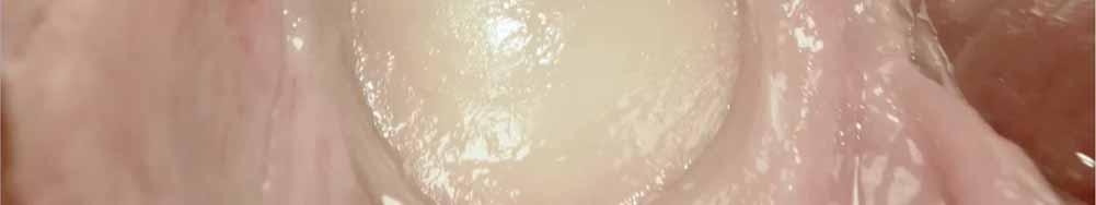
11 ARTHROSCOPY AXILLARY NERVE arm in adduction arm in abduction Jae Chul Yoo, Arthroscopy, 23: 1271, 2007
12 SUPRASCAPULAR NERVE - upper trunk of brachial plexus C5/(C6) - under transverse scapular lig. - around spinoglenoid notch - Supraspinatus - Infraspinatus

")
13 Nerve injury: SUPRASCAPULAR NERVE - Ossified transverse scapular ligament - Hypertrophied spinoglenoid ligament - SLAP lesion - Ganglion cyst - Traction injury ISP (+/- SSP) Atrophy

14 ROTATOR CUFF
 sup. subscapular nerve inf.")
15 SUBSCAPULARIS MUSCLE - origin: subscapularis fossa - insertion: lesser tuberosity - innervation: - 2 branches (C5,C6) sup. subscapular nerve inf. subscapular nerve Frick, Thieme, 1980 Bouchet, Simep, 1990

,")
16 SUBSCAPULARIS MUSCLE function: internal rotation (lift-off test) Gerber, JBJS (Br), 73(3):389, 1991

 - function: abductor")
17 SUPRASPINATUS MUSCLE - origin: supraspinatus fossa - insertion: greater tuberosity - suprascapular nerve (C5,C6) - function: abductor (Jobe test)
 - axillary nerve (C5, C6) - function: external rotation INFRASPINATUS AND TERES MINOR")
18 - origin: infraspinatus fossa laterale margin - insertion: greater tuberosity - suprascapular nerve (C5,C6) - axillary nerve (C5, C6) - function: external rotation INFRASPINATUS AND TERES MINOR MUSCLE


19 external rotation lag in adduction INFRASPINATUS AND TERES MINOR MUSCLE external rotation lag in abduction (Horn blower sign) Arthuis M., Rev Chir Orthop Reparatrice Appar, 58:124, 1972

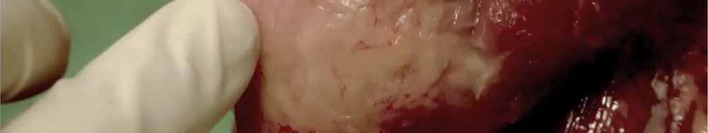
20 POSTEROSUPERIOR ROTATOR CUFF courtesy of Georg Lajtai


21 ROTATOR CUFF courtesy of Georg Lajtai
22 GLENOHUMERAL LIGAMENTS - GH joint capsule superior, anterior, inferior reinforced with glenohumeral ligaments: - superior (SGHL) - middle (MGHL) - inferior (IGHL) - posterior: thin capsule, no ligaments

23 SGHL ~ 1/3-2/3 of thickness of long biceps - in 42 % only thin / rudimentary - origin: labrum / supraglenoidal tubercle - insertion: above lesser tuberosity Warner JP, Am J Sports Med, 1992 Jost B, JSES, 2000


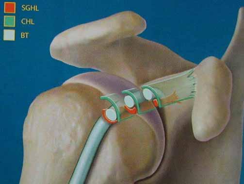
24 ROTATOR INTERVAL ANTERIOR PULLEY
 - stabilizes: - against ER / ant.")
25 - very variable - in 55 % thin Warner JP, Am J Sports Med, in % absent DePalma, AAOS, 1949 O'Brien, Saunders, 1990 MGHL - origins: - glenoid rim adjacent below SGHL - insertion: - medially of lesser tuberosity - subscapularis tendon (fused) - stabilizes: - against ER / ant. translation in neutral rotation - against inferior translation in ER


26 MGHL (ANTERIOR LABRUM) ANATOMIC VARIATIONS 12% 1.5% Williams M, Snyder S, and Buford D. Arthroscopy, 1994

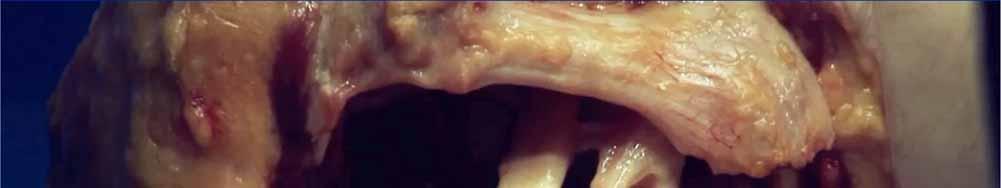
27 - always present O'Brien, Saunders, consists of: - anterior band - axillary recess - posterior band IGHL - stabilizes: - against a.p and inf. GH translation in Abd

28 GLENOHUMERAL LIGAMENTS courtesy of Georg Lajtai


29 Thank you!!! courtesy of Georg Lajtai
MUSCLES OF SHOULDER REGION
 Dr Jamila EL Medany OBJECTIVES At the end of the lecture, students should: List the name of muscles of the shoulder region. Describe the anatomy of muscles of shoulder region regarding: attachments of
THE SHOULDER JOINT T H E G L E N O H U M E R A L ( G H ) J O I N T
 J O I N T") THE SHOULDER JOINT T H E G L E N O H U M E R A L ( G H ) J O I N T CLARIFICATION OF TERMS Shoulder girdle = scapula and clavicle Shoulder joint (glenohumeral joint) = scapula and humerus Lippert, p115
THE SHOULDER JOINT T H E G L E N O H U M E R A L ( G H ) J O I N T CLARIFICATION OF TERMS Shoulder girdle = scapula and clavicle Shoulder joint (glenohumeral joint) = scapula and humerus Lippert, p115
Region of upper limb attachment to the trunk Proximal segment of limb overlaps parts of the trunk (thorax and back) and lower lateral neck.
 and lower lateral neck.") Region of upper limb attachment to the trunk Proximal segment of limb overlaps parts of the trunk (thorax and back) and lower lateral neck. includes Pectoral Scapular Deltoid regions of the upper limb
Region of upper limb attachment to the trunk Proximal segment of limb overlaps parts of the trunk (thorax and back) and lower lateral neck. includes Pectoral Scapular Deltoid regions of the upper limb
Scapular and Deltoid Regions
 M1 Gross and Developmental Anatomy Scapular and Deltoid Regions Dr. Peters 1 Outline I. Skeleton of the Shoulder and Attachment of the Upper Extremity to Trunk II. Positions and Movements of the Scapula
7/31/2012 THE SHOULDER JOINT CLARIFICATION OF TERMS OSTEOLOGY OF THE GH JOINT(BONES)
") THE SHOULDER JOINT T H E G L E N O H U M E R AL ( G H ) J O I N T CLARIFICATION OF TERMS Shoulder girdle = scapula and clavicle Shoulder joint (glenohumerual joint) = scapula and Lippert, p115 OSTEOLOGY
THE SHOULDER JOINT T H E G L E N O H U M E R AL ( G H ) J O I N T CLARIFICATION OF TERMS Shoulder girdle = scapula and clavicle Shoulder joint (glenohumerual joint) = scapula and Lippert, p115 OSTEOLOGY
Anatomy of the Shoulder Girdle. Prof Oluwadiya Kehinde FMCS (Orthop)
") Anatomy of the Shoulder Girdle Prof Oluwadiya Kehinde FMCS (Orthop) www.oluwadiya.com Bony Anatomy Shoulder Complex: Sternum(manubrium) Clavicle Scapula Proximal humerus Manubrium Sterni Upper part of
Anatomy of the Shoulder Girdle Prof Oluwadiya Kehinde FMCS (Orthop) www.oluwadiya.com Bony Anatomy Shoulder Complex: Sternum(manubrium) Clavicle Scapula Proximal humerus Manubrium Sterni Upper part of
3 Mohammad Al-Mohtasib Areej Mosleh
 3 Mohammad Al-Mohtasib Areej Mosleh ***Muscles Connecting the Upper Limb to the Vertebral Column 1.Trapezius Muscle ***The first muscle on the back is trapezius muscle, it s called so according
The shoulder girdle consists of the glenohumeral, acromioclavicular, sternoclavicular and scapulothoracic joints
 Anatomy of Shoulder Girdle The shoulder girdle consists of the glenohumeral, acromioclavicular, sternoclavicular and scapulothoracic joints Glenohumeral Joint A ball and socket synoval joint with a large
FUNCTIONAL ANATOMY OF SHOULDER JOINT
 FUNCTIONAL ANATOMY OF SHOULDER JOINT ARTICULATION Articulation is between: The rounded head of the Glenoid cavity humerus and The shallow, pear-shaped glenoid cavity of the scapula. 2 The articular surfaces
Pectoral region. Lecture 2
 Pectoral region Lecture 2 Muscle Action Each muscle has: Origin Beginning. Insertion End. Body (belly). Law: When a muscle performs its action, its insertion, moves towards its origin. Spring 2016 Dr.
Muscle Action Origin Insertion Nerve Innervation Chapter Page. Deltoid. Trapezius. Latissimus Dorsi
 Muscle Action Origin Insertion Nerve Innervation Chapter Page All Fibers Abduct the shoulder (glenohumeral joint) Deltoid Anterior Fibers Flex the shoulder (G/H joint) Horizontally adduct the shoulder
Pectoral region. Lecture 2
 Pectoral region Lecture 2 Muscle Action Each muscle has: Origin Beginning. Insertion End. Body (belly). Law: When a muscle performs its action, its insertion, moves towards its origin. Spring 2016 Dr.
I (and/or my co-authors) have something to disclose.
 have something to disclose.") Shoulder Anatomy And Biomechanics Nikhil N Verma, MD Director of Sports Medicine Professor, Department of Orthopedics Rush University Team Physician, Chicago White Sox and Bulls I (and/or my co-authors)
Shoulder Anatomy And Biomechanics Nikhil N Verma, MD Director of Sports Medicine Professor, Department of Orthopedics Rush University Team Physician, Chicago White Sox and Bulls I (and/or my co-authors)
This figure (of humerus) is from Dr. Maher's newest slides. -Its added here just for consideration-
 is from Dr. Maher's newest slides. -Its added here just for consideration-") This figure (of humerus) is from Dr. Maher's newest slides. -Its added here just for consideration- Slides of Anatomy Please note : These slides are Dr. Maher Hadidi s slides of spring 2016 and were edited
This figure (of humerus) is from Dr. Maher's newest slides. -Its added here just for consideration- Slides of Anatomy Please note : These slides are Dr. Maher Hadidi s slides of spring 2016 and were edited
SHOULDER JOINT ANATOMY AND KINESIOLOGY
 SHOULDER JOINT ANATOMY AND KINESIOLOGY SHOULDER JOINT ANATOMY AND KINESIOLOGY The shoulder joint, also called the glenohumeral joint, consists of the scapula and humerus. The motions of the shoulder joint
MUSCLES. Anconeus Muscle
 LAB 7 UPPER LIMBS MUSCLES Anconeus Muscle anconeus origin: distal end of dorsal surface of humerus insertion: lateral surface of ulna from distal margin of the semilunar notch to proximal end of the olecranon
Joint G*H. Joint S*C. Joint A*C. Labrum. Humerus. Sternum. Scapula. Clavicle. Thorax. Articulation. Scapulo- Thoracic
 A*C Joint Scapulo- Thoracic Articulation Thorax Sternum Clavicle Scapula Humerus S*C Joint G*H Joint Labrum AC Ligaments SC Ligaments SC JOINT AC Coracoacromial GH GH Ligament Complex Coracoclavicular
Upper Limb Muscles Muscles of Axilla & Arm
 Done By : Saleh Salahat Upper Limb Muscles Muscles of Axilla & Arm 1) Muscles around the axilla A- Muscles connecting the upper to thoracic wall (4) 1- pectoralis major Origin:- from the medial half of
Upper limb Pectoral region & Axilla
 Upper limb Pectoral region & Axilla 黃敏銓 mchuang@ntu.edu.tw 1 Pectoral region Intercostal nerve Anterior branch of lateral cutaneous branch Lateral cutaneous branch Anterior cutaneous branch Anterior cutaneous
Upper limb Arm & Cubital region 黃敏銓
 Upper limb Arm & Cubital region 黃敏銓 1 Arm Lateral intermuscular septum Anterior (flexor) compartment: stronger Medial intermuscular septum Posterior (extensor) compartment 2 Coracobrachialis Origin: coracoid
Lawrence Gulotta Gillian Lieberman, MD October Gillian Lieberman, MD. Shoulder Imaging. Lawrence V. Gulotta, HMS IV 10/16/02
 October 2002 Shoulder Imaging Lawrence V. Gulotta, HMS IV 10/16/02 Goals Review Anatomy of the Shoulder -Dynamic Stabilizers -> Rotator Cuff -Static Stabilizers -> Labrum and Capsule Systematic Approach
Axilla and Brachial Region
 L 4 A B O R A T O R Y Axilla and Brachial Region BRACHIAL PLEXUS 5 Roots/Rami (ventral rami C5 T1) 3 Trunks Superior (C5, C6) Middle (C7) Inferior (C8, T1) 3 Cords Lateral Cord (Anterior Superior and Anterior
Shoulder Joint Examination. Shoulder Joint Examination. Inspection. Inspection Palpation Movement. Look Feel Move
 Shoulder Joint Examination History Cuff Examination Instability Examination AC Joint Examination Biceps Tendon Examination Superior Labrum Examination Shoulder Joint Examination Inspection Palpation Movement
SHOULDER PAIN. A Real Pain in the Neck. Michael Wolk, MD Northeastern Rehabilitation Associates October 31, 2017
 SHOULDER PAIN A Real Pain in the Neck Michael Wolk, MD Northeastern Rehabilitation Associates October 31, 2017 THE SHOULDER JOINT (S) 1. glenohumeral 2. suprahumeral 3. acromioclavicular 4. scapulocostal
R. Frank Henn III, MD. Associate Professor Chief of Sports Medicine Residency Program Director
 R. Frank Henn III, MD Associate Professor Chief of Sports Medicine Residency Program Director Disclosures No financial relationships to disclose 1. Labral anatomy 2. Adaptations of the throwing shoulder
Glenohumeral Joint. Glenohumeral Joint. Glenohumeral Joint. Glenohumeral Joint. Glenohumeral Joint. Glenohumeral Joint
 The Shoulder Joint Chapter 5 The Shoulder Joint Manual of Structural Kinesiology R.T. Floyd, EdD, ATC, CSCS McGraw-Hill Higher Education. All rights reserved. 5-1 Shoulder joint is attached to axial skeleton
Anatomy and Physiology II. Review Shoulder Girdle New Material Upper Extremities - Bones
 Anatomy and Physiology II Review Shoulder Girdle New Material Upper Extremities - Bones Anatomy and Physiology II Shoulder Girdle Review Questions From Last Lecture Can you identify the following muscles?
The Shoulder. Anatomy and Injuries PSK 4U Unit 3, Day 4
 The Shoulder Anatomy and Injuries PSK 4U Unit 3, Day 4 Shoulder Girdle Shoulder Complex is the most mobile joint in the body. Scapula Clavicle Sternum Humerus Rib cage/thorax Shoulder Girdle It also includes
Patient ID. Case Conference. Physical Examination. Image examination. Treatment 2011/6/16
 Patient ID Case Conference R3 高逢駿 VS 徐郭堯 55 y/o female C.C.: recurrent right shoulder dislocation noted since falling down injury 2 years ago Came to ER because of dislocation for many times due to minor
G24: Shoulder and Axilla
 G24: Shoulder and Axilla Syllabus - Pg. 2 ANAT 6010- Medical Gross Anatomy David A. Morton, Ph.D. Objectives Upper limb Systemically: Bones (joints) Muscles Nerves Vessels (arteries/veins) Fascial compartments
26/9/2016. Anatomy. 1 Nour Erekat Wejdan Amer
 26/9/2016 Anatomy st 1 Nour Erekat Wejdan Amer Notes before we start the lecture. Bring any colored Atlas with you to the lab. The main reference is clinical anatomy by regions by Richard snell the 9 th
Shoulder: Clinical Anatomy, Kinematics & Biomechanics
 Shoulder: Clinical Anatomy, Kinematics & Biomechanics Dr. Alex K C Poon Department of Orthopaedics & Traumatology Pamela Youde Nethersole Eastern Hospital Clinical Anatomy the application of anatomy to
The Biomechanics of the Human Upper Extremity. Dr Ayesha Basharat BSPT, PP.DPT. M.PHIL
 The Biomechanics of the Human Upper Extremity Dr Ayesha Basharat BSPT, PP.DPT. M.PHIL Sternoclavicular Joint Provides major axis of rotation for movement of clavicle and scapula Freely permitted frontal
Page 1. Shoulder Injuries in Sports.
 www.schulterteam.ch Shoulder Injuries in Sports Matthias A Zumstein Shoulder, Elbow and Orthopaedic Sports Medicine Department of Orthopedic Surgery and Traumatology University of Berne, Switzerland matthias.zumstein@insel.ch
Anatomy Workshop Upper Extremity David Ebaugh, PT, PhD Workshop Leader. Lab Leaders: STATION I BRACHIAL PLEXUS
 Anatomy Workshop Upper Extremity David Ebaugh, PT, PhD Workshop Leader Lab Leaders: STATION I BRACHIAL PLEXUS A. Posterior cervical triangle and axilla B. Formation of plexus 1. Ventral rami C5-T1 2. Trunks
Musculoskeletal Ultrasound. Technical Guidelines SHOULDER
 Musculoskeletal Ultrasound Technical Guidelines SHOULDER 1 Although patient s positioning for shoulder US varies widely across different Countries and Institutions reflecting multifaceted opinions and
The Shoulder. Systematically scanning the shoulder provides extremely useful diagnostic information. The Shoulder
 1 ! The most ACCESSIBLE to sonographic exam! The most MOBILE and VULNERABLE extremity AND Systematically scanning the shoulder provides extremely useful diagnostic information! The Goal for this section
The Arm and Cubital Fossa
 The Arm and Cubital Fossa Dr. Andrew Gallagher School of Anatomical Sciences University of the Witwatersrand Introduction The ARM (BRACHIUM) is the most proximal segment of the upper limb musculoskeletal
Glenohumeral. Laura Leonetti Genna Moak Taylor Hansen
 Glenohumeral Laura Leonetti Genna Moak Taylor Hansen Surface anatomy Genna Anterior axillary fold Clavicle Clavicular head of Pectoralis major Clavipectoral triangle Acromial part of Deltoid Manubrium
SHOULDER INSTABILITY
 SHOULDER INSTABILITY Dr.KN Subramanian M.Ch Orth., FRCS (Tr & Orth), CCT Orth(UK) Consultant Orthopaedic Surgeon, Special interest: Orthopaedic Sports Injury, Shoulder and Knee Surgery, SPARSH Hospital
Shoulder Injury Evaluation.
 Shoulder Injury Evaluation www.fisiokinesiterapia.biz Basic Anatomy & Kinesiology 3 Bone Structures Clavicle Scapula Humerus Evaluation Principles Always follow a standard progression Determine the target
Sports Medicine Part II : ANATOMY OF THE SPINE, ABDOMEN AND SHOULDER COMPLEX
 Sports Medicine 25 1.1 Part II : ANATOMY OF THE SPINE, ABDOMEN AND SHOULDER COMPLEX c.w.p. Wagner High School, Sports Medicine, A. Morgan, T. Morgan & A. Eastlake, 2008 Muscles of the Upper Limbs In this
Connects arm to thorax 3 joints. Glenohumeral joint Acromioclavicular joint Sternoclavicular joint
 Connects arm to thorax 3 joints Glenohumeral joint Acromioclavicular joint Sternoclavicular joint Scapula Elevation Depression Protraction (abduction) Retraction (adduction) Downward Rotation Upward Rotation
Practical 2 Worksheet
 Practical 2 Worksheet Upper Extremity BONES 1. Which end of the clavicle is on the lateral side (acromial or sternal)? 2. Describe the difference in the appearance of the acromial and sternal ends of the
Returning the Shoulder Back to Optimal Function. Scapula. Clavicle. Humerus. Bones of the Shoulder (Osteology) Joints of the Shoulder (Arthrology)
 Joints of the Shoulder (Arthrology)") Returning the Shoulder Back to Optimal Function Sternum Clavicle Ribs Scapula Humerus Bones of the Shoulder (Osteology) By Rick Kaselj Clavicle Scapula Medial Left Anterior Clavicle Inferior View 20 degree
Returning the Shoulder Back to Optimal Function Sternum Clavicle Ribs Scapula Humerus Bones of the Shoulder (Osteology) By Rick Kaselj Clavicle Scapula Medial Left Anterior Clavicle Inferior View 20 degree
Muscles of the Upper Limb
 Muscles of the Upper Limb anterior surface of ribs 3 5 coracoid process Pectoralis minor pectoral nerves protracts / depresses scapula Serratus anterior Subclavius ribs 1-8 long thoracic nerve rib 1 ----------------
US finding of the shoulder (with live demonstration) 인제의대상계백병원 안재기
 인제의대상계백병원 안재기") US finding of the shoulder (with live demonstration) 인제의대상계백병원 안재기 Shoulder US Biceps tendon & Rotator Cuff Long Head of Biceps Tendon Subscapularis tendon Supraspinatus tendon Infraspinatus tendon Teres
US finding of the shoulder (with live demonstration) 인제의대상계백병원 안재기 Shoulder US Biceps tendon & Rotator Cuff Long Head of Biceps Tendon Subscapularis tendon Supraspinatus tendon Infraspinatus tendon Teres
WEEKEND 2 Shoulder. Shoulder Active Range of Motion Assessment
 Virginia Orthopedic Manual Physical Therapy Institute - 2016 Technique Manual WEEKEND 2 Shoulder Shoulder Active Range of Motion Assessment - Patient Positioning: Standing, appropriately undressed so that
Mastering the Musculoskeletal Exam UCSF Essentials of Women s Health July 7, 2016 Carlin Senter, M.D. Henry Crevensten, M.D.
 Mastering the Musculoskeletal Exam UCSF Essentials of Women s Health July 7, 2016 Carlin Senter, M.D. Henry Crevensten, M.D. I have nothing to disclose Outline Knee exam Shoulder exam Knee Anatomy The
The Upper Limb II. Anatomy RHS 241 Lecture 11 Dr. Einas Al-Eisa
 The Upper Limb II Anatomy RHS 241 Lecture 11 Dr. Einas Al-Eisa Sternoclavicular joint Double joint.? Each side separated by intercalating articular disc Grasp the mid-portion of your clavicle on one side
MR Arthrography of the Shoulder - A Beginner's Guide
 MR Arthrography of the Shoulder - A Beginner's Guide Poster No.: C-1034 Congress: ECR 2011 Type: Educational Exhibit Authors: A. Jain, S. Connolly; Prescot/UK Keywords: Pathology, Arthrography, MR, Musculoskeletal
Suprascapular Nerve: How to identify when it is a problem and what to do? Speaker Disclosure
 Suprascapular Nerve: How to identify when it is a problem and what to do? Eric C. McCarty, MD Associate Professor Chief of Sports Medicine and Shoulder Surgery University of Colorado School of Medicine
Netter's Anatomy Flash Cards Section 6 List 4 th Edition
 Netter's Anatomy Flash Cards Section 6 List 4 th Edition https://www.memrise.com/course/1577581/ Section 6 Upper Limb (66 cards) Plate 6-1 Humerus and Scapula: Anterior View 1.1 Acromion 1.2 Greater tubercle
Human Anatomy Biology 351
 1 Human Anatomy Biology 351 Upper Limb Exam Please place your name on the back of the last page of this exam. You must answer all questions on this exam. Because statistics demonstrate that, on average,
region of the upper limb between the shoulder and the elbow Superiorly communicates with the axilla.
 1 region of the upper limb between the shoulder and the elbow Superiorly communicates with the axilla. Inferiorly, a number of important structures pass between arm & forearm through cubital fossa. 2 medial
ARTHROSCOPIC SUPRASCAPULAR NERVE RELEASE
 ARTHROSCOPIC SUPRASCAPULAR NERVE RELEASE Laurent Lafosse & Robert Fullick Alps Surgery Institute Annecy France DISCLOSURE Royalties Depuy Consultant Depuy Stock options ITS Stock options Orthospace 1 Background
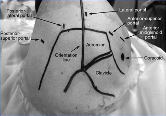
Shoulder Arthroscopy Portals
 Shoulder Arthroscopy Portals Alper Deveci and Metin Dogan 7 7.1 Bony Landmarks Before starting shoulder arthroscopy, the patient must be positioned and draping applied. Then the bony landmarks are identified
Introduction to anatomy Lecture # 2
 Introduction to anatomy Dr. Maher Hadidi Tareq AlTal 2 6/2/2013 Quick revision: The body is built on the skeleton The skeleton is divided into two parts >>>> axial skeleton (80 bones) >>>> appendicular
The suction cup mechanism is enhanced by the slightly negative intra articular pressure within the joint.
 SHOULDER INSTABILITY Stability A. The stability of the shoulder is improved by depth of the glenoid. This is determined by: 1. Osseous glenoid, 2. Articular cartilage of the glenoid, which is thicker at
Shoulder Arthroscopy Lab Manual
 Shoulder Arthroscopy Lab Manual Dalhousie University Orthopaedic Program May 5, 2017 Skills Centre OBJECTIVES 1. Demonstrate a competent understanding of the arthroscopic anatomy and biomechanics of the
Shoulder Arthroscopy Curriculum
 ARTHRO Mentor 1 Description All those with an interest in the shoulder should develop a basic level of proficiency and should be able to perform a thorough diagnostic exam, looking from both the anterior
Shoulder arthroscopy. Mohammad nasir Naderi, MD Fellowship in shoulder and arthroscopic surgery
 Shoulder arthroscopy Mohammad nasir Naderi, MD Fellowship in shoulder and arthroscopic surgery Shoulder arthroscopy Evolve understanding of anatomy and pathophysiology of shoulder This technology, allow
Shoulder examination. P Sripathi Rao Arthroscopy & Sports Injuries Unit Dean, Kasturba Medical College
 Shoulder examination P Sripathi Rao Arthroscopy & Sports Injuries Unit Dean, Kasturba Medical College Manipal University, Manipal Common symptoms Tingling Numbness Pain Loss of movements Weakness Approach
LIST OF STRUCTURES TO BE IDENTIFIED IN LAB: UPPER EXTREMITY REVIEW 2016
 LIST OF STRUCTURES TO BE IDENTIFIED IN LAB: UPPER EXTREMITY REVIEW 2016 BONES Ribs, sternum, clavicle Humerus: Head, greater tubercle, lesser tubercle, intertubercular sulcus, surgical neck, anatomical
Physical Examination of the Shoulder
 General setup Patient will be examined in both the seated and supine position so exam table needed 360 degree access to patient Expose neck and both shoulders (for comparison); female in gown or sports
SHOULDER PROBLEMS & ARTHROSCOPIC MANAGEMENT
 SHOULDER PROBLEMS & ARTHROSCOPIC MANAGEMENT DR.SHEKHAR SRIVASTAV Sr. Consultant-KNEE & SHOULDER Arthroscopy Sant Parmanand Hospital,Delhi Peculiarities of Shoulder Elegant piece of machinery It has the
WEEKEND THREE HOMEWORK
 WEEKEND THREE HOMEWORK READING ASSIGNMENTS Salvo Massage Therapy Principles and Practice 4 th Edition Muscolino The Muscular System Manual Muscolino The Muscle and Bone Palpation Manual Ch. 19 Skeletal
MRI SHOULDER WHAT TO SEE
 MRI SHOULDER WHAT TO SEE DR SHEKHAR SRIVASTAV Sr. Consultant- Knee & Shoulder Arthroscopy Sant Parmanand Hospital Normal Anatomy Normal Shoulder MRI Coronal Oblique Sagital Oblique Axial Cuts Normal Coronal
Functional Anatomy. CHAPTER 5 Functional Anatomy of the Upper Extremity. CHAPTER 6 Functional Anatomy of the Lower Extremity
 Hamill_ch05_137-186.qxd 11/2/07 3:55 PM Page 137 S E C T I O N II Functional Anatomy CHAPTER 5 Functional Anatomy of the Upper Extremity CHAPTER 6 Functional Anatomy of the Lower Extremity CHAPTER 7 Functional
The arm: *For images refer back to the slides
 The arm: *For images refer back to the slides Muscles of the arm: deltoid, triceps (which is located at the back of the arm), biceps and brachialis (it lies under the biceps), brachioradialis (it lies
Personal use only. MR Imaging Assessment of Rotator Cuff Muscle Quality
 MR Imaging Assessment of Rotator Cuff Muscle Quality Christian W. A. Pfirrmann Orthopedic University Hospital Balgrist University of Zurich Switzerland Atrophy of Infraspinatus Infraspinatus Atrophy: Diagnosis?
Shoulder Injuries. Glenoid labrum injuries. SLAP Lesions
 Shoulder Injuries functional anatomy clinical perspective impingement rotator cuff injuries glenoid labrum injuries dislocation Glenoid labrum injuries SLAP lesions stable or unstable traction/compression
SYMPOSIUM: TRIBUTE TO DR. ANTHONY F. DEPALMA, FIRST EDITOR-IN-CHIEF
 Clin Orthop Relat Res (2008) 466:543 551 DOI 10.1007/s11999-007-0103-5 SYMPOSIUM: TRIBUTE TO DR. ANTHONY F. DEPALMA, FIRST EDITOR-IN-CHIEF OF CLINICAL ORTHOPAEDICS AND RELATED RESEARCH The Classic Surgical
SHOULDER MRI OSSEOUS OUTLET
 SHOULDER MRI OSSEOUS OUTLET -Acromion: Type I flat, II curved, III hooked (spur), IV convex (@sagittal); Lateral downsloping or low lying acromion w/ respect to clavicle (@coronal) -AC (degen arthrosis,
Pectoral girdle, SUPERIEUR ARM AND HAND. Danil Hammoudi.MD
 Pectoral girdle, SUPERIEUR ARM AND HAND Danil Hammoudi.MD The pectoral girdle is the set of bones which connect the upper limb to the axial skeleton on each side. It consists of the clavicle scapula in
STRUCTURAL BASIS OF MEDICAL PRACTICE EXAMINATION 5 October 6, 2006
 STRUCTURAL BASIS OF MEDICAL PRACTICE EXAMINATION 5 October 6, 2006 PART l. Answer in the space provided. (8 pts) 1. Identify the structures. (2 pts) B C A. _pisiform B. _ulnar artery A C. _flexor carpi
The pectoral region. University of Babylon College of Medicine Dr.HaythemAli Alsayigh M.B.CH.B.-F.I.M.B.S. Surgical Clinical Anatomy
 The pectoral region University of Babylon College of Medicine Dr.HaythemAli Alsayigh M.B.CH.B.-F.I.M.B.S. Surgical Clinical Anatomy Objective Study the Bones and Joints A. Clavicle (collarbone) B. Scapula
Anatomical Considerations/ Pathophysiology The shoulder is the most mobile joint in the body. : Three bones:
 Introduction Musculoskeletal training is generally underrepresented in medical training and residency curriculums. There is a general deficit in musculoskeletal knowledge amongst current medical students,
Gross Anatomy Questions That Should be Answerable After October 27, 2017
 Gross Anatomy Questions That Should be Answerable After October 27, 2017 1. The inferior angle of the scapula of a woman who was recently in an automobile accident seems to protrude making a ridge beneath
Chapter 1 Anatomy of the Shoulder and Elbow
 Page 1 of 35 Chapter 1 Anatomy of the Shoulder and Elbow Joseph A. Abboud Matthew L. Ramsey Gerald R. Williams OVERVIEW OF SHOULDER AND ELBOW SURGERY In order to diagnose and treat problems of the shoulder
Welcome to. Not to be copied without the express permission of EDUCATA. Copyright 2014 EDUCATA. All rights reserved. 1. How to Navigate EDUCATA
 Welcome to Copyright 2014 EDUCATA. This presentation is not to be copied in whole or in part without the express permission of EDUCATA. How to Navigate EDUCATA The Shoulder Complex Mechanics, Muscle Function
*the Arm* -the arm extends from the shoulder joint (proximal), to the elbow joint (distal) - it has one bone ; the humerus which is a long bone
, to the elbow joint (distal) - it has one bone ; the humerus which is a long bone") *the Arm* -the arm extends from the shoulder joint (proximal), to the elbow joint (distal) - it has one bone ; the humerus which is a long bone - muscles in the arm : *brachialis muscle *Biceps brachii
*the Arm* -the arm extends from the shoulder joint (proximal), to the elbow joint (distal) - it has one bone ; the humerus which is a long bone - muscles in the arm : *brachialis muscle *Biceps brachii
Sports Medicine: Shoulder Arthrography. Christine B. Chung, M.D. Professor of Radiology Musculoskeletal Division UCSD and VA Healthcare System
 Sports Medicine: Shoulder Arthrography Christine B. Chung, M.D. Professor of Radiology Musculoskeletal Division UCSD and VA Healthcare System Disclosure Off-label use for gadolinium Pediatric Sports Injuries
Acknowledgement. Here are some flash cards all set up in a "pdf" format for you! Thanks to Laura H. (spring 08)
") Acknowledgement Here are some flash cards all set up in a "pdf" format for you! Thanks to Laura H. (spring 08) for her donation to all my anatomy students! t Here is her suggestion for making flashcards
Acknowledgement Here are some flash cards all set up in a "pdf" format for you! Thanks to Laura H. (spring 08) for her donation to all my anatomy students! t Here is her suggestion for making flashcards
Muscles in the Shoulder, Chest, Arm, Stomach, and Back
 Muscles in the Shoulder, Chest, Arm, Stomach, and Back Shoulder Muscles Deltoid Supraspinatus Infraspinatus Teres Major Teres Minor Subscapularis Deltoid (Delts) Function: Raises the upper arm Origin:
Shoulder Biomechanics
 Shoulder Biomechanics Lecture originally developed by Bryan Morrison, Ph.D. candidate Arizona State University Fall 2000 1 Outline Anatomy Biomechanics Problems 2 Shoulder Complex Greatest Greatest Predisposition
CLINICAL SUMMARY AND RECOMMENDATIONS 378
 Shoulder 9 CLINICAL SUMMARY AND RECOMMENDATIONS 378 Anatomy 379 Osteology 379 Arthrology 380 Scapulohumeral Rhythm 381 Ligaments 382 Muscles 384 Nerves 387 Patient History 389 Initial Hypotheses Based
SUPERIEUR ARM AND HAND
 Pectoral girdle, SUPERIEUR ARM AND HAND Danil Hammoudi.MD The pectoral girdle is the set of bones which connect the upper limb to the axial skeleton on each side. It consists of the clavicle scapula in
Continuing Education: Shoulder Stability
 Continuing Education: Shoulder Stability Anatomy & Kinesiology: The GHJ consists of the articulation of three bones: the scapula, clavicle and humerus. The scapula has three protrusions: the coracoid,
Introduction to anatomy
 Introduction to anatomy Dr. Maher Hadidi Fareed Halteh 3 7/2/2013 Subscapularis: It is located on the anterior side of the scapula. It has a triangular shape. It is like Pectoralis major and Teres major
The Clavicle Right clavicle Deltoid tubercle: Conoid tubercle, conoid ligamen Impression for the
 The Clavicle Muscle Attachment Sites in the Upper Limb Pectoralis major Right clavicle Smooth superior surface of the shaft, under the platysma muscle tubercle: attachment of the deltoid Acromial facet
Ultrasound-Guided Shoulder Injections 인제대학교일산백병원 재활의학과 임길병
 Ultrasound-Guided Shoulder Injections 인제대학교일산백병원 재활의학과 임길병 How to improve needle visibility Advantages of Ultrasound in Procedures Real-time imaging Avoids radiation exposure But, interventions without
213: HUMAN FUNCTIONAL ANATOMY: PRACTICAL CLASS 1: Proximal bones, plexuses and patterns
 213: HUMAN FUNCTIONAL ANATOMY: PRACTICAL CLASS 1: Proximal bones, plexuses and patterns CLAVICLE Examine an isolated clavicle and compare it with a clavicle on an articulated skeleton. Viewed from above,
MRI of the Shoulder What to look for and how to find it? Dr. Eric Handley Musculoskeletal Radiologist Cherry Creek Imaging
 MRI of the Shoulder What to look for and how to find it? Dr. Eric Handley Musculoskeletal Radiologist Cherry Creek Imaging MRI of the Shoulder Benefits of Ultrasound: * Dynamic * Interactive real time
Abduction of arm until your hand rich your head. Flexion of forearm at elbow joint. Extension of arm at elbow joint. Flexion of fingers 10.
 Num. answer 1. Medialy With the manubrium ( sternum ), and laterally with the acromion of the scapula 2. 1. Trapezius 2. Levator scapulae 3. Rhomboids 3. 1. Pectoralis major 2. Pectoralis minor 3. Latissiumus
Al-Balqa Applied University
 Al-Balqa Applied University Faculty Of Medicine *You can use this checklist as a guide to you for the lab. the items on this checklist represent the main features of the models that you have to know for
BRACHIAL PLEXUS. DORSAL SCAPULAR NERVE (C5) supraclavicular branch innervates rhomboids (major and minor) and levator scapulae
 supraclavicular branch innervates rhomboids (major and minor) and levator scapulae") THE BRACHIAL PLEXUS DORSAL SCAPULAR NERVE (C5) supraclavicular branch innervates rhomboids (major and minor) and levator scapulae SCHEMA OF THE BRACHIAL PLEXUS THE BRACHIAL PLEXUS PHRENIC NERVE supraclavicular
THE BRACHIAL PLEXUS DORSAL SCAPULAR NERVE (C5) supraclavicular branch innervates rhomboids (major and minor) and levator scapulae SCHEMA OF THE BRACHIAL PLEXUS THE BRACHIAL PLEXUS PHRENIC NERVE supraclavicular
MUSCLES OF THE ELBOW REGION
 MUSCLES OF THE ELBOW REGION Dr Bronwen Ackermann COMMONWEALTH OF AUSTRALIA Copyright Regulation WARNING This material has been reproduced and communicated to you by or on behalf of the University of Sydney
Rotator Cuff Conditioning Exercises with th i R ck Kaselj, MS ck K Rick Kaselj Exercises
 Rotator Cuff Conditioning Exercises with Rick Kaselj, MS Before I Start Recording - Webinar will be recorded - I will send you details on how to access the video tomorrow - Tour of Things / Questions: