Ultrasound-Guided Shoulder Injections 인제대학교일산백병원 재활의학과 임길병
|
|
|
- Alisha Ray
- 5 years ago
- Views:
Transcription
1 Ultrasound-Guided Shoulder Injections 인제대학교일산백병원 재활의학과 임길병
2 How to improve needle visibility
3 Advantages of Ultrasound in Procedures Real-time imaging Avoids radiation exposure But, interventions without adequate needle tip visualization unintentional vascular, neural, vi sceral injury
4 Procedural Needle-Related Visibility Factors 1. Basic Sonography & Needle Image Interpretati on ultrasound beam : penetrate through tissue to varying degrees depending upon tissue composition(muscle, t endon, other tissue) cannot pass dense tissue such as bone
5 Procedural Needle-Related Visibility Factors 2. Acoustic Impedance as the Basis for Procedure Needle Visualization another essential aspect of needle visualization : acous tic impedance=> dependent on density of tissue and s peed which ultrasound beam travel marked differences in acoustic impedance between tw o tissues=> the brighter or more hyperechoic the son ographic signal of the needle
6 Procedural Needle-Related Visibility Factors 3. Size of the needle and echogenicity larger caliber needle : more easily visualized than a small er size diameter needle greater surface area: more si gnificant change in acoustic impedance, intercept the ultr asound beam, reflected back to the transducer
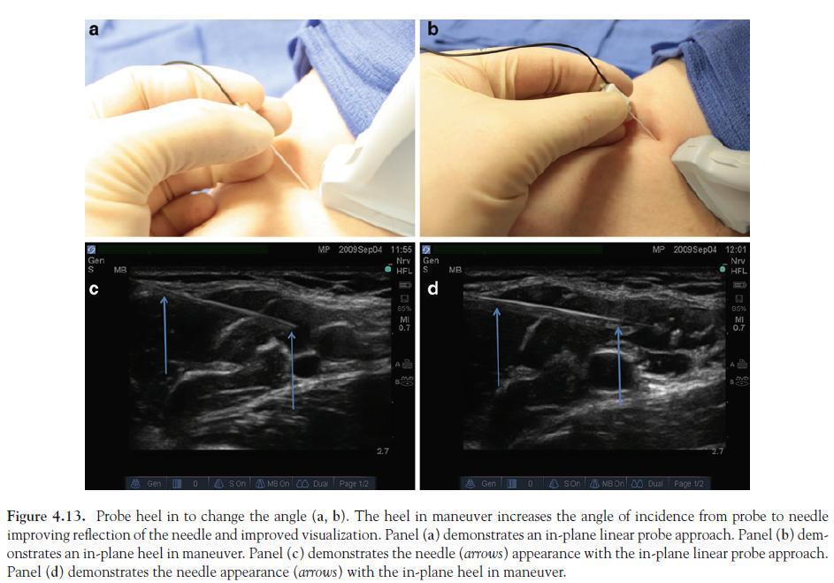
7 Procedural Needle-Related Visibility Factors The Skin Insertion Site Selected and Angle of Procedure Needle Passage : critical role in optimizing needle visualiza tion the angle is too steep=> shorter portion of the us beam will be reflected back perpendicular angle(90 ) of insertion us beam direction =>the transducer maximizes reception of reflected beam heel in maneuver : to press or tilt the opposite end of the ultrasound transducer-> close to 90 most optimal angle for a procedure needle to the skin s urface interface: 30 and 45
8 The Skin Insertion Site Selected & Angle of Procedure Needle Passage
9
10 Procedural Needle-Related Visibility Factors Echogenic Procedure Needles Small angled indentations or notches : needle shaft res ulting in an irregular surface -> increase scatter of ultr asound polymer encased procedure needle is another technol ogical advancement-> improve needle echogenicity an d ultrasound image quality
11 Procedural Needle-Related Visibility Factors Procedure Needle Tip procedure needle tip bevel : usually scatter the ultraso und beam d/t irregularity of the needle tip surface, less steep ang le of the needle tip procedure needle bevel up position : improved needle tip visualization
12 Procedure Needle Tip
13

14 Mechanical and Optical Procedure Needle Guides

15 Enhancement and Techniques to Improve Procedure Needle Localization Basic Sonographic Effect of Enhancement enhancement : improve the visualization of needle within a vascular structure or certain tissues(e.g., fat) that have lower acoustic impedance compared to needle

16 Enhancement and Techniques to Improve Procedure Needle Localization Enhancement with Priming, Insertion of Stylet or Guide Wire, & Vibration
17 Enhancement & Techniques to Improve Procedure Needle Localization Hydrolocalization of the Procedure Needle
18 Enhancement and Techniques to Improve Procedure Needle Localization Needle Visibility by Agitated Solutions or with US Contrast Agents
19 Ultrasound-Guided Shoulder Injection
20 Ultrasound (US) superficial tendons : long-head biceps, SSP, ISP-> excellent echogenicity and structural resolution visualization of soft tissue adjacent to orthopedic hardware dynamic assessment of the joint little information: interosseous structures & those shielded by bone suspicion of intra-articular pathology or osseous pathology -> plain film imaging is essential
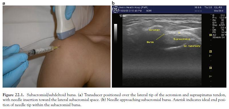
21 Subacromial/Subdeltoid Bursa subacromial bursa : m/c injected structure Ix: rotator cuff pathology, impingement syndrome, & subacromial bursitis.
22 Subacromial/Subdeltoid Bursa Anatomy subacromial & subdeltoid bursa typically communicate location: on upper surface of supraspinatus muscle, under deep surface of deltoid functions : protect the supraspinatus
23 Subacromial/Subdeltoid Bursa Clinical Presentation Shoulder abduction, IR can potentially impinge : humeral head (greater tubercle) & arch of the acromio n & coracoacromial ligament Limitations of the Blind Approach : accuracy of blind injections : as low as 29%
24 Subacromial/Subdeltoid Bursa US-Guided Technique Transducer oriented in scapular plane, positioned just over tip of acromion SSP tendon should be visualized Bursa is seen as a thin anechoic fluid layer above the tendon Seated position with the arm hanging at their sid e Elbow flexed 90, arm supinated & palm over the ipsilateral hip Triamcinolone and local anesthetics
25
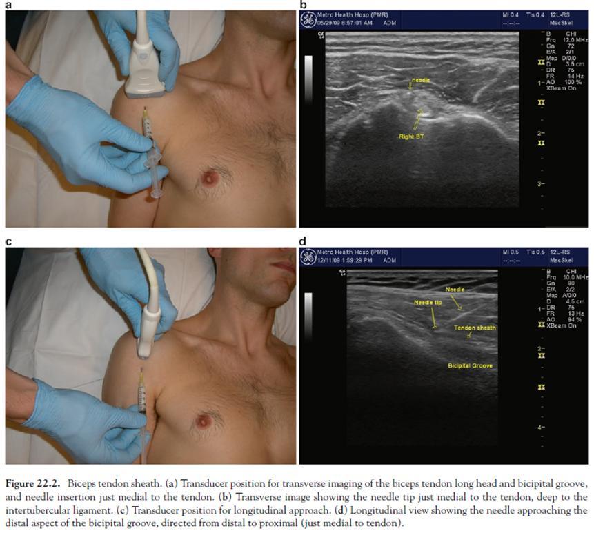
26 Biceps Tendon Sheath Anatomy long head : originates at the supraglenoid tubercle of the glenoid labrum intertubercular groove houses the bicipital tendon tendon sheath communicates proximally with the glenohumeral joint short head : originates on the coracoid process
27 Biceps Tendon Sheath Clinical Presentation long head : m/c injured portion of the muscle usually occur at the proximal end Bicipital tendinopathy: Speed test
28 Biceps Tendon Sheath US-Guided Technique short-axis approach is more common, technically easier not allow for visualization of the entire length of needle target is the small space btn the tendon & the lesser tuberosity of the humerus, just medial to the tendon inj of steroid directly into the tendon may lead to rupture longitudinal approach :more appropriate for aspiration of the fluid in the sheath triamcinolone & local anesthetic
29
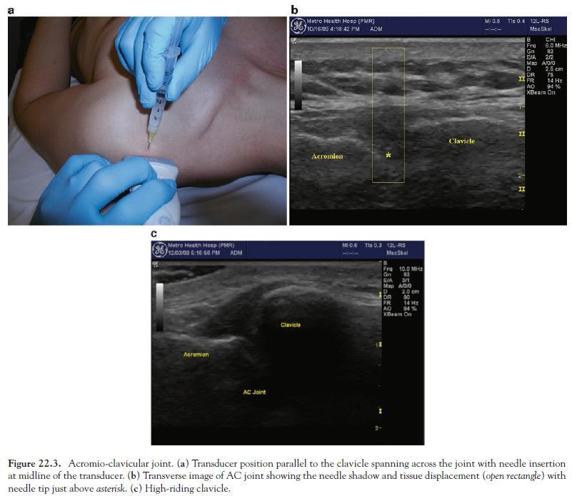
30 Acromioclavicular Joint Anatomy AC joint : formed by articulation of distal end of clavicle & acromion process easy to localize because of its superficial position, often narrowed by osteophytes => US guidance is very helpful subacromial bursa & supraspinatus tendon lie directly beneath the joint
31 Clinical Presentation Acromioclavicular Joint Pain is reproduced with active elevation of the arm or scarf test(positioned in crossed-arm adduction) AC joint : appearance is typically a V shape covered by a thin capsule (acromioclavicular ligament) distended if effusion is present, no significant vascular or neural structures
32 Acromioclavicular Joint US-Guided Technique Technique : in the seated position with the arm hanging at their side V of the joint should be positioned precisely in the middle of the image needle is inserted in short-axis orientation tip of the needle is visualized as a bright dot 0.25 ml triamcinolone (40 mg/ml) & 0.75 ml of l ocal anesthetic
33
34 Anatomy Glenohumeral Joint Glenohumeral joint true articulation surface : small and shallow joi nt surface area is greatly increased by cartilaginous glenoid labrum surrounded by a thin fibrous articular capsule strengthened by three glenohumeral ligaments joint synovium extends down bicipital sheath into intertubercular groove
35 Glenohumeral Joint Clinical Presentation Glenohumeral pathology : painful and restricted range of motion of joint (reduced external rotation) mimic cervical radiculopathy(referred pain, paresthesias down entire upper limb)
36 Glenohumeral Joint Limitations of the Blind Approach 26.8% accuracy using an anterior approach 94% using us to guide glenohumeral joint injections
37 US-Guided Technique Glenohumeral Joint glenohumeral joint is visualized by posterior view with transducer just caudal & parallel to the spine of the scapula circular humeral head, abutting glenoid fossa with less-echogenic triangular-shaped labrum between them Inj: posterior approach with the humerus adducted across the thorax. long-axis approach, 2 cm lateral to the lateral heel of the transducer depending upon shoulder size, a 3 or 4 inch needle 1 ml of triamcinolone(40 mg/ml) & 2 ml of local anesthetic
38 Glenohumeral Joint The Rotator Interval Approach Anterior visualization : difficult d/t depth & overlying dense structures rotator interval : a triangular space(corocoid process, the anterior-most portion of the ssp & the superior border of the subscapularis tendon) contain : biceps tendon, glenohumeral capsule, coracohumeral ligame nt, & glenohumeral ligament Inj : arm resting at patient's side, shoulder placed in slight external rotation transducer is positioned in the transverse plane on the sup/ant shoulder Resistance to inj-> needle tip has entered tendon or ligament
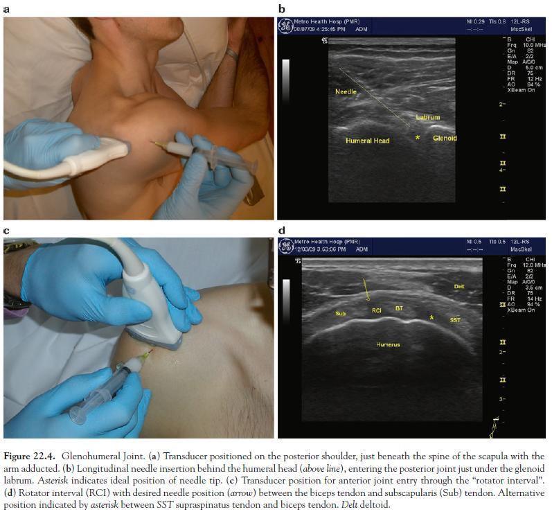
39
40
41
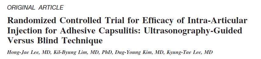
42 Subscapularis Tendon/Subscapularis Bursa Anatomy subscapularis m : originates from subscapular fossa of the scapula inserts on the lesser tuberosity of humerus only rotator cuff muscle that acts to internally rotate subscapularis bursa lies deep to the tendon against th e neck of the scapula communicates with the shoulder joint
43 Subscapularis Tendon/Subscapularis Bursa Clinical Presentation Subscapularis tendinopathy : pain in the anterior shoulder provoked with active internal rotation or passive ER diffuse shoulder pain & impingement signs Strength of the subscapularis is assessed with th e Lift off test. -> subscapularis weakness, tendo n rupture, or inadequate ROM
44 Subscapularis Tendon/Subscapularis Bursa linear transducer is held in transverse position relative to humerus & bicipital groove subscapularis : its deep, medially located muscle belly to attach to the lesser tuberosity bursa is reached by advancing the needle through the tendon, at which time a subtle pop or give-way is detected
45
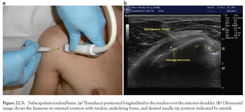
46 Sternoclavicular Joint Anatomy SC joint : formed by articulation of prox. end of clavicle with clavicular fossa in superior lateral aspect of the ster num scapular retraction -> end of the clavicle becomes more prominent great vessels of the chest & the pleura lie deep to the jo int, so care is needed
47 Sternoclavicular Joint Clinical Presentation SC joint pain, chest wall pain, swelling, tenderness dire ctly over the joint
48 Sternoclavicular Joint Tech. : placing a linear transducer in line with the clavicle small notch with clavicle projecting superficially compared to sternum covered by very thin capsule & may be distended if effusion is present needle tip is visualized as a bright dot as it enters the field of view 0.25 ml triamcinolone (40 mg/ml) & 0.75 ml of local anesthetic.
49

50 Thank you for attention!
The Shoulder. Anatomy and Injuries PSK 4U Unit 3, Day 4
 The Shoulder Anatomy and Injuries PSK 4U Unit 3, Day 4 Shoulder Girdle Shoulder Complex is the most mobile joint in the body. Scapula Clavicle Sternum Humerus Rib cage/thorax Shoulder Girdle It also includes
FUNCTIONAL ANATOMY OF SHOULDER JOINT
 FUNCTIONAL ANATOMY OF SHOULDER JOINT ARTICULATION Articulation is between: The rounded head of the Glenoid cavity humerus and The shallow, pear-shaped glenoid cavity of the scapula. 2 The articular surfaces
Musculoskeletal Ultrasound. Technical Guidelines SHOULDER
 Musculoskeletal Ultrasound Technical Guidelines SHOULDER 1 Although patient s positioning for shoulder US varies widely across different Countries and Institutions reflecting multifaceted opinions and
Returning the Shoulder Back to Optimal Function. Scapula. Clavicle. Humerus. Bones of the Shoulder (Osteology) Joints of the Shoulder (Arthrology)
 Joints of the Shoulder (Arthrology)") Returning the Shoulder Back to Optimal Function Sternum Clavicle Ribs Scapula Humerus Bones of the Shoulder (Osteology) By Rick Kaselj Clavicle Scapula Medial Left Anterior Clavicle Inferior View 20 degree
Returning the Shoulder Back to Optimal Function Sternum Clavicle Ribs Scapula Humerus Bones of the Shoulder (Osteology) By Rick Kaselj Clavicle Scapula Medial Left Anterior Clavicle Inferior View 20 degree
The Upper Limb II. Anatomy RHS 241 Lecture 11 Dr. Einas Al-Eisa
 The Upper Limb II Anatomy RHS 241 Lecture 11 Dr. Einas Al-Eisa Sternoclavicular joint Double joint.? Each side separated by intercalating articular disc Grasp the mid-portion of your clavicle on one side
Connects arm to thorax 3 joints. Glenohumeral joint Acromioclavicular joint Sternoclavicular joint
 Connects arm to thorax 3 joints Glenohumeral joint Acromioclavicular joint Sternoclavicular joint Scapula Elevation Depression Protraction (abduction) Retraction (adduction) Downward Rotation Upward Rotation
Anatomy of the Shoulder Girdle. Prof Oluwadiya Kehinde FMCS (Orthop)
") Anatomy of the Shoulder Girdle Prof Oluwadiya Kehinde FMCS (Orthop) www.oluwadiya.com Bony Anatomy Shoulder Complex: Sternum(manubrium) Clavicle Scapula Proximal humerus Manubrium Sterni Upper part of
Anatomy of the Shoulder Girdle Prof Oluwadiya Kehinde FMCS (Orthop) www.oluwadiya.com Bony Anatomy Shoulder Complex: Sternum(manubrium) Clavicle Scapula Proximal humerus Manubrium Sterni Upper part of
Joint G*H. Joint S*C. Joint A*C. Labrum. Humerus. Sternum. Scapula. Clavicle. Thorax. Articulation. Scapulo- Thoracic
 A*C Joint Scapulo- Thoracic Articulation Thorax Sternum Clavicle Scapula Humerus S*C Joint G*H Joint Labrum AC Ligaments SC Ligaments SC JOINT AC Coracoacromial GH GH Ligament Complex Coracoclavicular
Scapular and Deltoid Regions
 M1 Gross and Developmental Anatomy Scapular and Deltoid Regions Dr. Peters 1 Outline I. Skeleton of the Shoulder and Attachment of the Upper Extremity to Trunk II. Positions and Movements of the Scapula
THE SHOULDER JOINT T H E G L E N O H U M E R A L ( G H ) J O I N T
 J O I N T") THE SHOULDER JOINT T H E G L E N O H U M E R A L ( G H ) J O I N T CLARIFICATION OF TERMS Shoulder girdle = scapula and clavicle Shoulder joint (glenohumeral joint) = scapula and humerus Lippert, p115
THE SHOULDER JOINT T H E G L E N O H U M E R A L ( G H ) J O I N T CLARIFICATION OF TERMS Shoulder girdle = scapula and clavicle Shoulder joint (glenohumeral joint) = scapula and humerus Lippert, p115
7/31/2012 THE SHOULDER JOINT CLARIFICATION OF TERMS OSTEOLOGY OF THE GH JOINT(BONES)
") THE SHOULDER JOINT T H E G L E N O H U M E R AL ( G H ) J O I N T CLARIFICATION OF TERMS Shoulder girdle = scapula and clavicle Shoulder joint (glenohumerual joint) = scapula and Lippert, p115 OSTEOLOGY
THE SHOULDER JOINT T H E G L E N O H U M E R AL ( G H ) J O I N T CLARIFICATION OF TERMS Shoulder girdle = scapula and clavicle Shoulder joint (glenohumerual joint) = scapula and Lippert, p115 OSTEOLOGY
The Shoulder. Systematically scanning the shoulder provides extremely useful diagnostic information. The Shoulder
 1 ! The most ACCESSIBLE to sonographic exam! The most MOBILE and VULNERABLE extremity AND Systematically scanning the shoulder provides extremely useful diagnostic information! The Goal for this section
SHOULDER JOINT ANATOMY AND KINESIOLOGY
 SHOULDER JOINT ANATOMY AND KINESIOLOGY SHOULDER JOINT ANATOMY AND KINESIOLOGY The shoulder joint, also called the glenohumeral joint, consists of the scapula and humerus. The motions of the shoulder joint
US finding of the shoulder (with live demonstration) 인제의대상계백병원 안재기
 인제의대상계백병원 안재기") US finding of the shoulder (with live demonstration) 인제의대상계백병원 안재기 Shoulder US Biceps tendon & Rotator Cuff Long Head of Biceps Tendon Subscapularis tendon Supraspinatus tendon Infraspinatus tendon Teres
US finding of the shoulder (with live demonstration) 인제의대상계백병원 안재기 Shoulder US Biceps tendon & Rotator Cuff Long Head of Biceps Tendon Subscapularis tendon Supraspinatus tendon Infraspinatus tendon Teres
MUSCLES OF SHOULDER REGION
 Dr Jamila EL Medany OBJECTIVES At the end of the lecture, students should: List the name of muscles of the shoulder region. Describe the anatomy of muscles of shoulder region regarding: attachments of
Region of upper limb attachment to the trunk Proximal segment of limb overlaps parts of the trunk (thorax and back) and lower lateral neck.
 and lower lateral neck.") Region of upper limb attachment to the trunk Proximal segment of limb overlaps parts of the trunk (thorax and back) and lower lateral neck. includes Pectoral Scapular Deltoid regions of the upper limb
Region of upper limb attachment to the trunk Proximal segment of limb overlaps parts of the trunk (thorax and back) and lower lateral neck. includes Pectoral Scapular Deltoid regions of the upper limb
Shoulder joint Assessment and General View
 Shoulder joint Assessment and General View Done by; Mshari S. Alghadier BSc Physical Therapy RHPT 366 m.alghadier@sau.edu.sa http://faculty.sau.edu.sa/m.alghadier/ Functional anatomy The shoulder contains
Shoulder Joint Examination. Shoulder Joint Examination. Inspection. Inspection Palpation Movement. Look Feel Move
 Shoulder Joint Examination History Cuff Examination Instability Examination AC Joint Examination Biceps Tendon Examination Superior Labrum Examination Shoulder Joint Examination Inspection Palpation Movement
Ultrasound of Shoulder Pathology and Intervention 서울대학교병원재활의학과 김기원
 Ultrasound of Shoulder Pathology and Intervention 서울대학교병원재활의학과 김기원 Ultrasound for Shoulder Disorder Advantage Dynamic evaluation Immediate clinical correlation + Intervention Weakness Diagnostic accuracy?
Evidence Based Approach to Shoulder Injections
 Evidence Based Approach to Shoulder Injections Bradley Sandella, DO Christiana Care Sports Medicine Joseph Straight, MD First State Orthopaedics Objectives Relevant Anatomy Indications for injections Injection
Ultrasound Guided Injections
 Ultrasound Guided Injection Technique More accurate injections Better Results! 1 Benefits: Increased Level of Certainty ie : really know how accurate PRP/Prolotherapy Avoid damage to articular cartilage
Anatomical Considerations/ Pathophysiology The shoulder is the most mobile joint in the body. : Three bones:
 Introduction Musculoskeletal training is generally underrepresented in medical training and residency curriculums. There is a general deficit in musculoskeletal knowledge amongst current medical students,
Physical Examination of the Shoulder
 General setup Patient will be examined in both the seated and supine position so exam table needed 360 degree access to patient Expose neck and both shoulders (for comparison); female in gown or sports
Introduction. Rarely does a single muscle act in isolation at the shoulder complex.
 Shoulder complex 1 Introduction Our study of the upper limb begins with the shoulder complex, a set of four articulations involving the sternum, clavicle, ribs, scapula, and humerus. Rarely does a single
Chapter 8. The Pectoral Girdle & Upper Limb
 Chapter 8 The Pectoral Girdle & Upper Limb Pectoral Girdle pectoral girdle (shoulder girdle) supports the arm consists of two on each side of the body // clavicle (collarbone) and scapula (shoulder blade)
Structure and Function of the Bones and Joints of the Shoulder Girdle
 Structure and Function of the Bones and Joints of the Shoulder Girdle LEARNING OBJECTIVES: At the end of this laboratory exercise the student will be able to: Palpate the important skeletal landmarks of
The Shoulder Complex. Anatomy. Articulations 12/11/2017. Oak Ridge High School Conroe, Texas. Clavicle Collar Bone Scapula Shoulder Blade Humerus
 The Shoulder Complex Oak Ridge High School Conroe, Texas Anatomy Clavicle Collar Bone Scapula Shoulder Blade Humerus Articulations Sternoclavicular SC joint. Sternum and Clavicle. Acromioclavicular AC
Ultrasound Guided Therapeutic Injections in the Treatment of Shoulder Pain: A Multimedia Review
 Ultrasound Guided Therapeutic Injections in the Treatment of Shoulder Pain: A Multimedia Review Poster No.: P-0127 Congress: ESSR 2015 Type: Educational Poster Authors: A. Karsandas, J. Tuckett, R. Sinha,
26/9/2016. Anatomy. 1 Nour Erekat Wejdan Amer
 26/9/2016 Anatomy st 1 Nour Erekat Wejdan Amer Notes before we start the lecture. Bring any colored Atlas with you to the lab. The main reference is clinical anatomy by regions by Richard snell the 9 th
Muscle Action Origin Insertion Nerve Innervation Chapter Page. Deltoid. Trapezius. Latissimus Dorsi
 Muscle Action Origin Insertion Nerve Innervation Chapter Page All Fibers Abduct the shoulder (glenohumeral joint) Deltoid Anterior Fibers Flex the shoulder (G/H joint) Horizontally adduct the shoulder
Review shoulder anatomy Review the physical exam of the shoulder Discuss some common causes of acute shoulder pain Discuss some common causes of
 Review shoulder anatomy Review the physical exam of the shoulder Discuss some common causes of acute shoulder pain Discuss some common causes of chronic shoulder pain Review with some case questions Bones:
Upper Limb Muscles Muscles of Axilla & Arm
 Done By : Saleh Salahat Upper Limb Muscles Muscles of Axilla & Arm 1) Muscles around the axilla A- Muscles connecting the upper to thoracic wall (4) 1- pectoralis major Origin:- from the medial half of
P V S MEMORIAL HOSPITAL LTD.
 SHOULDER XRAYS Instability Series o True AP (Grashey s) o Axillary o Stryker Notch view o True AP in Internal rotation o Scapular Y view o West Point view for Bony Bankart ( looks like modif axillary view)
The Shoulder. By Patrick Ryan, Bobby Law, Jack Beaty, Alex Newhouse and Chuck Nelson
 The Shoulder By Patrick Ryan, Bobby Law, Jack Beaty, Alex Newhouse and Chuck Nelson Learning Objectives/Agenda Review the anatomy of the shoulder Describe the main diseases of the shoulder Describe the
Case study # 6 Sharon P
 Patient is a morbidly obese 70 year old female presenting with left shoulder pain after a severe fall. Patient is in moderate to severe pain with extremely limited range of motion due to extensive shoulder
Ultrasound of the Shoulder
 Ultrasound of the Shoulder Patrick Battaglia, DC, DACBR Logan University, Department of Radiology Outline Review ultrasound appearance of NMSK tissues Present indications for ultrasound of the shoulder.
Gross Anatomy Questions That Should be Answerable After October 27, 2017
 Gross Anatomy Questions That Should be Answerable After October 27, 2017 1. The inferior angle of the scapula of a woman who was recently in an automobile accident seems to protrude making a ridge beneath
Common Applications for Sonography and Guided Intervention: Shoulder
 Common Applications for Sonography and Guided Intervention: Shoulder Jon A. Jacobson, M.D. Professor of Radiology Director, Division of Musculoskeletal Radiology University of Michigan Disclosures: Consultant:
Pectoral region. Lecture 2
 Pectoral region Lecture 2 Muscle Action Each muscle has: Origin Beginning. Insertion End. Body (belly). Law: When a muscle performs its action, its insertion, moves towards its origin. Spring 2016 Dr.
MUSCLES. Anconeus Muscle
 LAB 7 UPPER LIMBS MUSCLES Anconeus Muscle anconeus origin: distal end of dorsal surface of humerus insertion: lateral surface of ulna from distal margin of the semilunar notch to proximal end of the olecranon
Shoulder: Clinical Anatomy, Kinematics & Biomechanics
 Shoulder: Clinical Anatomy, Kinematics & Biomechanics Dr. Alex K C Poon Department of Orthopaedics & Traumatology Pamela Youde Nethersole Eastern Hospital Clinical Anatomy the application of anatomy to
THE SKELETAL SYSTEM. Focus on the Pectoral Girdle
 THE SKELETAL SYSTEM Focus on the Pectoral Girdle Appendicular Skeleton 126 bones Includes bones of the limbs (arms and legs) Pectoral girdle (shoulder) Pelvic girdle (hip) Pectoral Girdle (the shoulder)
SHOULDER PAIN. A Real Pain in the Neck. Michael Wolk, MD Northeastern Rehabilitation Associates October 31, 2017
 SHOULDER PAIN A Real Pain in the Neck Michael Wolk, MD Northeastern Rehabilitation Associates October 31, 2017 THE SHOULDER JOINT (S) 1. glenohumeral 2. suprahumeral 3. acromioclavicular 4. scapulocostal
Mastering the Musculoskeletal Exam UCSF Essentials of Women s Health July 7, 2016 Carlin Senter, M.D. Henry Crevensten, M.D.
 Mastering the Musculoskeletal Exam UCSF Essentials of Women s Health July 7, 2016 Carlin Senter, M.D. Henry Crevensten, M.D. I have nothing to disclose Outline Knee exam Shoulder exam Knee Anatomy The
Continuing Education: Shoulder Stability
 Continuing Education: Shoulder Stability Anatomy & Kinesiology: The GHJ consists of the articulation of three bones: the scapula, clavicle and humerus. The scapula has three protrusions: the coracoid,
UPPER EXTREMITY INJURIES. Recognizing common injuries to the upper extremity
 UPPER EXTREMITY INJURIES Recognizing common injuries to the upper extremity ANATOMY BONES Clavicle Scapula Spine of the scapula Acromion process Glenoid fossa/cavity Humerus Epicondyles ANATOMY BONES Ulna
Stefan C Muzin, MD PM&R Attending Physician, Beth Israel Deaconess Medical Center, Harvard Medical School Onsite Physiatrist, GE Aviation, Lynn, MA
 Stefan C Muzin, MD PM&R Attending Physician, Beth Israel Deaconess Medical Center, Harvard Medical School Onsite Physiatrist, GE Aviation, Lynn, MA Consultant, OEHN (Occupational and Environmental Network)
Upper limb Arm & Cubital region 黃敏銓
 Upper limb Arm & Cubital region 黃敏銓 1 Arm Lateral intermuscular septum Anterior (flexor) compartment: stronger Medial intermuscular septum Posterior (extensor) compartment 2 Coracobrachialis Origin: coracoid
Figure 1: Bones of the upper limb
 BONES OF THE APPENDICULAR SKELETON The appendicular skeleton is composed of the 126 bones of the appendages and the pectoral and pelvic girdles, which attach the limbs to the axial skeleton. Although the
WEEKEND THREE HOMEWORK
 WEEKEND THREE HOMEWORK READING ASSIGNMENTS Salvo Massage Therapy Principles and Practice 4 th Edition Muscolino The Muscular System Manual Muscolino The Muscle and Bone Palpation Manual Ch. 19 Skeletal
Shoulder Biomechanics
 Shoulder Biomechanics Lecture originally developed by Bryan Morrison, Ph.D. candidate Arizona State University Fall 2000 1 Outline Anatomy Biomechanics Problems 2 Shoulder Complex Greatest Greatest Predisposition
Kinesiology of the Upper Extremity
 P A R T II Kinesiology of the Upper Extremity Pectoralis Latissimus dorsi UNIT 1: SHOULDER UNIT: THE SHOULDER COMPLEX Chapter 8: Structure and Function of the Bones and Joints of the Shoulder Girdle Chapter
A&P 1 Skeletal Lab Guide Week 2 - Appendicular Skeleton and Joints Lab Exercises: Pectoral Girdle
 A&P 1 Skeletal Lab Guide Week 2 - Appendicular Skeleton and Joints Lab Exercises: Pectoral Girdle PLEASE NOTE: Your group will need an articulated skeleton, a disarticulated skeleton, and the joint models
This figure (of humerus) is from Dr. Maher's newest slides. -Its added here just for consideration-
 is from Dr. Maher's newest slides. -Its added here just for consideration-") This figure (of humerus) is from Dr. Maher's newest slides. -Its added here just for consideration- Slides of Anatomy Please note : These slides are Dr. Maher Hadidi s slides of spring 2016 and were edited
This figure (of humerus) is from Dr. Maher's newest slides. -Its added here just for consideration- Slides of Anatomy Please note : These slides are Dr. Maher Hadidi s slides of spring 2016 and were edited
10/12/2010. Upper Extremity. Pectoral (Shoulder) Girdle. Clavicle (collarbone) Skeletal System: Appendicular Skeleton
 Girdle. Clavicle (collarbone) Skeletal System: Appendicular Skeleton") Skeletal System: Appendicular Skeleton Pectoral girdle Pelvic girdle Upper limbs Lower limbs 8-1 Pectoral (Shoulder) Girdle Consists of scapula and clavicle Clavicle articulates with sternum (Sternoclavicular
Skeletal System: Appendicular Skeleton Pectoral girdle Pelvic girdle Upper limbs Lower limbs 8-1 Pectoral (Shoulder) Girdle Consists of scapula and clavicle Clavicle articulates with sternum (Sternoclavicular
Benefits of Aspiration and Injection JOINT INJECTIONS. Injection Indications. Mechanism of Action 1/11/2016
 Benefits of Aspiration and Injection JOINT INJECTIONS Mark Niedfeldt, M.D. Medical College of Wisconsin Decrease or resolution of pain Decrease or resolution of inflammation Decrease or resolution of effusion
The Arm and Cubital Fossa
 The Arm and Cubital Fossa Dr. Andrew Gallagher School of Anatomical Sciences University of the Witwatersrand Introduction The ARM (BRACHIUM) is the most proximal segment of the upper limb musculoskeletal
Pectoral region. Lecture 2
 Pectoral region Lecture 2 Muscle Action Each muscle has: Origin Beginning. Insertion End. Body (belly). Law: When a muscle performs its action, its insertion, moves towards its origin. Spring 2016 Dr.
RN(EC) ENC(C) GNC(C) MN ACNP *** MECHANISM OF INJURY.. MOST IMPORTANT ***
 ENC(C) GNC(C) MN ACNP *** MECHANISM OF INJURY.. MOST IMPORTANT ***") HISTORY *** MECHANISM OF INJURY.. MOST IMPORTANT *** Age - Certain conditions are more prevalent in particular age groups (i.e. Full rotator cuff tears are more common over the age of 45, traumatic injuries
HISTORY *** MECHANISM OF INJURY.. MOST IMPORTANT *** Age - Certain conditions are more prevalent in particular age groups (i.e. Full rotator cuff tears are more common over the age of 45, traumatic injuries
Anatomy and Physiology II. Review Shoulder Girdle New Material Upper Extremities - Bones
 Anatomy and Physiology II Review Shoulder Girdle New Material Upper Extremities - Bones Anatomy and Physiology II Shoulder Girdle Review Questions From Last Lecture Can you identify the following muscles?
Netter's Anatomy Flash Cards Section 6 List 4 th Edition
 Netter's Anatomy Flash Cards Section 6 List 4 th Edition https://www.memrise.com/course/1577581/ Section 6 Upper Limb (66 cards) Plate 6-1 Humerus and Scapula: Anterior View 1.1 Acromion 1.2 Greater tubercle
Vol 3, 2008 CEC ARTICLE: Special Medical Conditions Part 2: Shoulder Maintenance and Rehab C. Eggers
 Vol 3, 2008 CEC ARTICLE: Special Medical Conditions Part 2: Shoulder Maintenance and Rehab C. Eggers SHOULDER GIRDLE STABILIZATION Knowledge of the anatomy and biomechanics of the shoulder girdle is essential
region of the upper limb between the shoulder and the elbow Superiorly communicates with the axilla.
 1 region of the upper limb between the shoulder and the elbow Superiorly communicates with the axilla. Inferiorly, a number of important structures pass between arm & forearm through cubital fossa. 2 medial
3 Mohammad Al-Mohtasib Areej Mosleh
 3 Mohammad Al-Mohtasib Areej Mosleh ***Muscles Connecting the Upper Limb to the Vertebral Column 1.Trapezius Muscle ***The first muscle on the back is trapezius muscle, it s called so according
WEEKEND 2 Shoulder. Shoulder Active Range of Motion Assessment
 Virginia Orthopedic Manual Physical Therapy Institute - 2016 Technique Manual WEEKEND 2 Shoulder Shoulder Active Range of Motion Assessment - Patient Positioning: Standing, appropriately undressed so that
Tendinosis & Subacromial Impingement Syndrome. Gene Desepoli, LMT, D.C.
 Tendinosis & Subacromial Impingement Syndrome Gene Desepoli, LMT, D.C. What is the shoulder joint? Shoulder joint or shoulder region? There is an interrelatedness of all moving parts of the shoulder and
SHOULDER ANATOMY Karl Wieser, MD Department of Orthopedics, University of Zurich, Balgrist, Switzerland
 20th Course in Shoulder Surgery Balgrist SHOULDER ANATOMY Karl Wieser, MD Department of Orthopedics, University of Zurich, Balgrist, Switzerland www.balgrist.ch ANATOMY OVERVIEW courtesy of Georg Lajtai
Sports Medicine Part II : ANATOMY OF THE SPINE, ABDOMEN AND SHOULDER COMPLEX
 Sports Medicine 25 1.1 Part II : ANATOMY OF THE SPINE, ABDOMEN AND SHOULDER COMPLEX c.w.p. Wagner High School, Sports Medicine, A. Morgan, T. Morgan & A. Eastlake, 2008 Muscles of the Upper Limbs In this
Practical 2 Worksheet
 Practical 2 Worksheet Upper Extremity BONES 1. Which end of the clavicle is on the lateral side (acromial or sternal)? 2. Describe the difference in the appearance of the acromial and sternal ends of the
Ultrasound assessment of most frequent shoulder disorders
 Ultrasound assessment of most frequent shoulder disorders Poster No.: C-2026 Congress: ECR 2014 Type: Educational Exhibit Authors: S. P. Ivanoski; Ohrid/MK Keywords: Trauma, Athletic injuries, Arthritides,
The examination of the painful knee. Maja K Artandi, MD, FACP Clinical Associate Professor of Medicine Stanford University
 The examination of the painful knee Maja K Artandi, MD, FACP Clinical Associate Professor of Medicine Stanford University Objectives of the talk By the end of this talk you will know The important anatomy
Bony Thorax. Anatomy and Procedures of the Bony Thorax Edited by M. Rhodes
 Bony Thorax Anatomy and Procedures of the Bony Thorax 10-526-191 Edited by M. Rhodes Anatomy Review Bony Thorax Formed by Sternum 12 pairs of ribs 12 thoracic vertebrae Conical in shape Narrow at top Posterior
Anatomy Workshop Upper Extremity David Ebaugh, PT, PhD Workshop Leader. Lab Leaders: STATION I BRACHIAL PLEXUS
 Anatomy Workshop Upper Extremity David Ebaugh, PT, PhD Workshop Leader Lab Leaders: STATION I BRACHIAL PLEXUS A. Posterior cervical triangle and axilla B. Formation of plexus 1. Ventral rami C5-T1 2. Trunks
Shoulder and Elbow ORTHOPAEDIC SYPMPOSIUM APRIL 8, 2017 DANIEL DOTY MD
 Shoulder and Elbow ORTHOPAEDIC SYPMPOSIUM APRIL 8, 2017 DANIEL DOTY MD Shoulder Articulations Glenohumeral Joint 2/3 total arc of motion Shallow Ball and Socket Joint Allows for excellent ROM Requires
Diagnostic and Management Approach to the Painful Shoulder
 Diagnostic and Management Approach to the Painful Shoulder Introduction What conditions causing shoulder pain commonly present in General Practice? Subacromial impingement Rotator cuff tears AC joint pathology
The shoulder girdle consists of the glenohumeral, acromioclavicular, sternoclavicular and scapulothoracic joints
 Anatomy of Shoulder Girdle The shoulder girdle consists of the glenohumeral, acromioclavicular, sternoclavicular and scapulothoracic joints Glenohumeral Joint A ball and socket synoval joint with a large
1. The coordinated action of a scapular upward rotation and humeral abduction is known as the:
 1 1. The coordinated action of a scapular upward rotation and humeral abduction is known as the: a. Carrying angle of the arm b. Scapulohumeral rhythm c. Glenohumeral capsular pattern d. Abduction resistance
The skeleton consists of: Bones: special connective tissue, hard. Cartilage: special connective tissue, less hard than bones. Joints: joint is the
 The skeleton consists of: Bones: special connective tissue, hard. Cartilage: special connective tissue, less hard than bones. Joints: joint is the location at witch two bones make contact, whereas ligaments
Lab Activity 11: Group II
 Lab Activity 11: Group II Muscles Martini Chapter 11 Portland Community College BI 231 Origin and Insertion Origin: The place where the fixed end attaches to a bone, cartilage, or connective tissue. Insertion:
Orthopedics - Dr. Ahmad - Lecture 2 - Injuries of the Upper Limb
 The shoulder and the upper arm Fractures of the clavicle 1. Fall on the shoulder. 2. Fall on outstretched hand. In mid shaft fractures, the outer fragment is pulled down by the weight of the arm and the
Shoulder Injury Evaluation.
 Shoulder Injury Evaluation www.fisiokinesiterapia.biz Basic Anatomy & Kinesiology 3 Bone Structures Clavicle Scapula Humerus Evaluation Principles Always follow a standard progression Determine the target
1. Occupation; Right or left handed, Age
 SHOULDER HISTORY 1. Occupation; Right or left handed, Age 2. Pain: Site. Any referred pain to the deltoid insertion Any localizing pain at Acromio-clavicular joint How long? Continuous or not Night pain
Glenohumeral Joint. Glenohumeral Joint. Glenohumeral Joint. Glenohumeral Joint. Glenohumeral Joint. Glenohumeral Joint
 The Shoulder Joint Chapter 5 The Shoulder Joint Manual of Structural Kinesiology R.T. Floyd, EdD, ATC, CSCS McGraw-Hill Higher Education. All rights reserved. 5-1 Shoulder joint is attached to axial skeleton
THE SHORT DESCRIPTION OF THE JOINTS 1. THE UPPER LIMB (Dr. Dóra Reglődi*, version )
") THE SHORT DESCRIPTION OF THE JOINTS 1. THE UPPER LIMB (Dr. Dóra Reglődi*, version 02-2007) Shoulder girdle The shoulder girdle consists of the clavicle and scapula on both sides. The two sides are connected
THE SHORT DESCRIPTION OF THE JOINTS 1. THE UPPER LIMB (Dr. Dóra Reglődi*, version 02-2007) Shoulder girdle The shoulder girdle consists of the clavicle and scapula on both sides. The two sides are connected
I (and/or my co-authors) have something to disclose.
 have something to disclose.") Shoulder Anatomy And Biomechanics Nikhil N Verma, MD Director of Sports Medicine Professor, Department of Orthopedics Rush University Team Physician, Chicago White Sox and Bulls I (and/or my co-authors)
Shoulder Anatomy And Biomechanics Nikhil N Verma, MD Director of Sports Medicine Professor, Department of Orthopedics Rush University Team Physician, Chicago White Sox and Bulls I (and/or my co-authors)
Joints. Judi Laprade. Illustrations from: Essential Clinical Anatomy 3 rd ed. (ECA3) Moore, K. and Agur, A. Lippincott Williams and Wilkins, 2007
 Moore, K. and Agur, A. Lippincott Williams and Wilkins, 2007") Slide 1 Joints Judi Laprade Illustrations from: Essential Clinical Anatomy 3 rd ed. (ECA3) Moore, K. and Agur, A. Lippincott Williams and Wilkins, 2007 Grant s Atlas of Anatomy 12 th ed. (GA12) Agur, A.
Slide 1 Joints Judi Laprade Illustrations from: Essential Clinical Anatomy 3 rd ed. (ECA3) Moore, K. and Agur, A. Lippincott Williams and Wilkins, 2007 Grant s Atlas of Anatomy 12 th ed. (GA12) Agur, A.
Calcific Tendinitis of the Long Head of the Biceps Brachii Distal to the Glenohumeral Joint: Plain Film
 1011 Calcific Tendinitis of the Long Head of the Biceps Brachii Distal to the Glenohumeral Joint: Plain Film Radiographic Findings Amy Beth Goldman1 Calcific tendinitis is a painful condition related to
Osteology of the Elbow and Forearm Complex. The ability to perform many activities of daily living (ADL) depends upon the elbow.
 depends upon the elbow.") Osteology of the Elbow and Forearm Complex The ability to perform many activities of daily living (ADL) depends upon the elbow. Activities of Daily Living (ADL) Can you think of anything that you do to
Osteology of the Elbow and Forearm Complex The ability to perform many activities of daily living (ADL) depends upon the elbow. Activities of Daily Living (ADL) Can you think of anything that you do to
A Patient s Guide to Shoulder Anatomy
 A Patient s Guide to Shoulder Anatomy Glendale Adventist Medical Center 1509 Wilson Terrace Glendale, CA 91206 Phone: (818) 409-8000 DISCLAIMER: The information in this booklet is compiled from a variety
An Introduction to the Appendicular Skeleton
 An Introduction to the Appendicular Skeleton The Appendicular Skeleton is composed of the 126 bones of the appendages (limbs) and the pectoral and pelvic girdles, which attach to the axial skeleton. Each
Biomechanics of the Upper Extremity Shoulder and Hip
 Biomechanics of the Upper Extremity Shoulder and Hip www.fisiokinesiterapia.biz The Shoulder Common Injuries Shoulder Joint - Bones Anatomical Structures Bursa - Fibrous, fluid-filled sac that reduces
REFERENCE DIAGRAMS OF UPPER LIMB MUSCLES: NAMES, LOCATIONS, ATTACHMENTS, FUNCTIONS MUSCLES CONNECTING THE UPPER LIMB TO THE AXIAL SKELETON
 REFERENCE DIAGRAMS OF UPPER LIMB MUSCLES: NAMES, LOCATIONS, ATTACHMENTS, FUNCTIONS MUSCLES CONNECTING THE UPPER LIMB TO THE AXIAL SKELETON A25LAB EXERCISES: UPPER LIMB MUSCLES Page 1 MUSCLES CONNECTING
Musculoskeletal Examination Benchmarks
 Musculoskeletal Examination Benchmarks _ The approach to examining the musculoskeletal system is the same no matter what joint or limb is being examined. The affected and contralateral region should both
Introduction to anatomy Lecture # 2
 Introduction to anatomy Dr. Maher Hadidi Tareq AlTal 2 6/2/2013 Quick revision: The body is built on the skeleton The skeleton is divided into two parts >>>> axial skeleton (80 bones) >>>> appendicular
Dr. Stefan C. Muzin, MD PM&R Beth Israel Deaconess Medical Center Harvard Medical School Consultant, GE Aviation, OEHN.
 Dr. Stefan C. Muzin, MD PM&R Beth Israel Deaconess Medical Center Harvard Medical School Consultant, GE Aviation, OEHN Work Related Workshop WorkInjuries Related Injuries Workshop Think of the Big Picture
Imaging of the shoulder
 ISBN: PPI201402DC4571 WWW.BOTICA.COM.VE ISSN: 2443-4388 Imaging of the shoulder ABSTRACT It is difficult to precisely define which imaging method is the gold standard in the evolution of numerous problems
The Elbow and the cubital fossa. Prof Oluwadiya Kehinde
 The Elbow and the cubital fossa Prof Oluwadiya Kehinde www.oluwadiya.com Elbow and Forearm Anatomy The elbow joint is formed by the humerus, radius, and the ulna Bony anatomy of the elbow Distal Humerus
Learning Objectives. 07 Aug 12. Article E-1. At the end of this section the learner will be able to:
 Module 1: Comparative Functional Anatomy and Biomechanics Article E-1 Learning Objectives At the end of this section the learner will be able to: Describe the bones of the equine thoracic Describe the