Introduction. Rarely does a single muscle act in isolation at the shoulder complex.
|
|
|
- Briana Floyd
- 5 years ago
- Views:
Transcription
1 Shoulder complex 1
2 Introduction Our study of the upper limb begins with the shoulder complex, a set of four articulations involving the sternum, clavicle, ribs, scapula, and humerus. Rarely does a single muscle act in isolation at the shoulder complex. Muscles work in teams to produce a highly coordinated action that is expressed over multiple joints. The very cooperative nature of shoulder muscles increases the versatility, control, and range of active movements.

3 Four joints within the shoulder complex Sternoclavicular joint Acromioclavicular joint Scapulothoracic joint Glenohumeral joint

4 Sternum The sternum consists of the manubrium, body, and xiphoid process.

5 Clavicle When looking from above, the shaft of the clavicle is curved with its anterior surface being generally convex medially and concave laterally.

6 Superior view of both shoulders in the anatomic position A. The orientation of the clavicle deviated about 20 posterior to the frontal plane. B. The orientation of the scapula deviated about 35 anterior to the frontal plane. C. Retroversion of the humeral head about 30 posterior to the mediallateral axis at the elbow.

7 Scapula The triangular-shaped scapula has three angles: inferior, superior, and lateral.

8 Scapula Anterior view of the right scapula showing an approximate 5 upward tilt of the glenoid fossa relative to the medial border of the scapula.

The retroversion of the humeral head")
9 (A)The right hummerus showing a 135 angle of inclination between the shaft and head of the humerus in the frontal plane. (B)The retroversion of the humeral head relative to the distal humerus.
10 Arthrology The most proximal articulation within the shoulder complex is the sternoclavicular joint. The clavicle, through its attachment to the sternum, functions as a mechanical strut, or prop, holding the scapula at a relatively constant distance from the trunk. Located at the lateral end of the clavicle is the acromioclavicular joint. This joint, and associated ligaments, firmly attaches the scapula to the clavicle. Motions of the right scapula against the posterior-lateral surface of the thorax, forming the scapulothoracic joint.
11 Arthrology Movements at the scapulothoracic joint are mechanically linked to the movements at both the sternoclavicular and acromioclavicular joints. The position of the scapula on the thorax provides a base of operation for the glenohumeral joint, the most distal and mobile link of the complex. The term shoulder movement describes the combined motions at both the glenohumeral and the scapulothoracic joints. 11
12 Primary movements at the scapulothoracic joint Elevation The scapula slides superiorly on the thorax, such as in the shrugging of the shoulders. Depression From an elevated position, the scapula slides inferiorly on the thorax. 12
13 Primary movements at the scapulothoracic joint Protraction The medial border of the scapula slides anterior-laterally on the thorax away from the midline. Retraction The medial border of the scapula slides posterior-medially on the thorax toward the midline, such as occurs during the pinching of the shoulder blades together. 13
14 Primary movements at the scapulothoracic joint Upward Rotation The inferior angle of the scapula rotates in a superior-lateral direction such that the glenoid fossa faces upward. This rotation occurs as a natural component of the arm reaching upward. Downward Rotation The inferior angle of the scapula rotates in an inferior-medial direction such that the glenoid fossa faces downward. This motion occurs as a natural component of the lowering of the arm to the side from the elevated position. 14
15 Sternoclavicular joint The sternoclavicular(sc) joint is a complex articulation, involving the medial end of the clavicle, the clavicular facet on the sternum, and the superior border of the cartilage of the first rib. The SC joint functions as the basiar joint of the entire upper extremity, linking the appendicular skeleton with the axial skeleton. The joint therefore must be firmly attached while simultaneously allowing considerable range of movement.

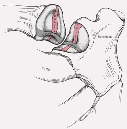
16 Sternoclavicular joint Complex saddle shaped articular surface
17 Periarticular connective tissue The SC joint is enclosed by a capsule reinforced by anterior and posterior sternoclavicular ligaments. The interclavicular ligament spans the jugular notch, connecting the medial end of the right and left clavicles. 17
18 Periarticular connective tissue The costoclavicular ligament is a strong structure extending from the cartilage of the first rib to the costal tuberosity on the inferior surface of the clavicle. The articular disc at the SC joint separates the joint into distinct medial and lateral joint cavities. The disc not only strengthens the articulation but functions as a shock absorber by increasing the surface area of joint contact. 18
19 kinematics The osteokinematics of the clavicle involve a rotation in all 3 of freedom. The primary purpose of these movements is to place the scapula in an optimal position to accept the head of the humerus.
20 kinematics Elevation and depression of the clavicle occur approximmately parallel to the frontal plane, around a near anterior-posterior axis of rotation. Maximums of approximately 45 degrees of elevation and 10 degrees of depression.

21 Arthrokinematics Elavation and depression 21
22 kinematics Protraction and retraction of the clavicle occur nearly parallel to the horizontal plane around a vertical axis of rotation.

23 Arthrokinematics Protraction and retraction 23
24 Axial(longitudinal)rotation of the clavicle The 3 rd degree of freedom at the SC joint is a rotation of the clavicle around the bone s longitudinal axis. During shoulder abduction or flexion, a point on the superior aspect of the clavicle rotates posteriorly 20 to 35 degrees. The arthrokinematics of clavicular rotation involve a spin of the head of the clavicle about the lateral surface of the articular disc.

25 Acromioclavicular Joint The acromioclavicular(ac) joint is the articulation between the lateral end of the clavicle and the acromion of the scapula. A. an anterior view showing the sloping nature. B. a posterior view of the joint opened up from behind, showing the clavicular facet on the acromion and the disc.

26 Periarticular connective tissue The AC joint is surrounded by a capsule that is direectly reinforced by superior and inferior ligaments. The coracoclavicular ligament provides an important extrinsic source of stability to the AC joint. The articular surfaces at the AC joint are lined with a layer of fibrocartilage and often separated by a complete or incomplete articular disc. 26

27 kinematics Upward rotation of the scapula at the AC joint occurs as the scapula swing upwardly and outwardly in relation to the lateral edge of the clavicle. Downward rotation at the AC joint returns the scapula back to its anatomic position, a motion mechanically associated with shoulder adduction or extension.

28 Acromioclavicular joint dislocation The resulting medially directed ground force may displace the acromion medially and under the sloped articular facet of the wellstabilized clavicle.
29 Scapulothoracic joint In the anatomic position, the scapula is typically positioned between the second and the seventh rib, with the medial border located about 6 cm lateral to the spine. Although highly variable, the average resting posture of the scapula is about 10 degrees of anterior tilt, 5 to 10 degrees of upward rotation, and about 35 degrees of internal rotation-a position consistent with the previously described plane of the scapula.

30 kinematics Scapular elevation at the scapulothoracic joint occurs as a composite of SC and AC joint rotations. 30

31 kinematics Protraction of the scapula occurs through a summation of horizontal plane rotations at both the SC and AC joints.
32 kinematics Upward rotation of the scapulothoracic joint is an integral part of raising the arm over the head. Downward rotation of the scapula occurs as the arm is returned to the side from a raised position
The Shoulder. Anatomy and Injuries PSK 4U Unit 3, Day 4
 The Shoulder Anatomy and Injuries PSK 4U Unit 3, Day 4 Shoulder Girdle Shoulder Complex is the most mobile joint in the body. Scapula Clavicle Sternum Humerus Rib cage/thorax Shoulder Girdle It also includes
Returning the Shoulder Back to Optimal Function. Scapula. Clavicle. Humerus. Bones of the Shoulder (Osteology) Joints of the Shoulder (Arthrology)
 Joints of the Shoulder (Arthrology)") Returning the Shoulder Back to Optimal Function Sternum Clavicle Ribs Scapula Humerus Bones of the Shoulder (Osteology) By Rick Kaselj Clavicle Scapula Medial Left Anterior Clavicle Inferior View 20 degree
Returning the Shoulder Back to Optimal Function Sternum Clavicle Ribs Scapula Humerus Bones of the Shoulder (Osteology) By Rick Kaselj Clavicle Scapula Medial Left Anterior Clavicle Inferior View 20 degree
Main Menu. Shoulder Girdle click here. The Power is in Your Hands. 1:07:11 PM]
![Main Menu. Shoulder Girdle click here. The Power is in Your Hands. 1:07:11 PM]](/thumbs/86/94418253.jpg "Main Menu. Shoulder Girdle click here. The Power is in Your Hands. 1:07:11 PM]") 1 The Shoulder Girdle click here Main Menu K.2 http://www.handsonlineeducation.com/classes//k2entry.htm[3/23/18, 1:07:11 PM] Bones Scapula and Clavicle Move as a unit Clavicle s articulation with sternum
1 The Shoulder Girdle click here Main Menu K.2 http://www.handsonlineeducation.com/classes//k2entry.htm[3/23/18, 1:07:11 PM] Bones Scapula and Clavicle Move as a unit Clavicle s articulation with sternum
Shoulder: Clinical Anatomy, Kinematics & Biomechanics
 Shoulder: Clinical Anatomy, Kinematics & Biomechanics Dr. Alex K C Poon Department of Orthopaedics & Traumatology Pamela Youde Nethersole Eastern Hospital Clinical Anatomy the application of anatomy to
Connects arm to thorax 3 joints. Glenohumeral joint Acromioclavicular joint Sternoclavicular joint
 Connects arm to thorax 3 joints Glenohumeral joint Acromioclavicular joint Sternoclavicular joint Scapula Elevation Depression Protraction (abduction) Retraction (adduction) Downward Rotation Upward Rotation
Kinesiology of the Upper Extremity
 P A R T II Kinesiology of the Upper Extremity Pectoralis Latissimus dorsi UNIT 1: SHOULDER UNIT: THE SHOULDER COMPLEX Chapter 8: Structure and Function of the Bones and Joints of the Shoulder Girdle Chapter
Anatomy of the Shoulder Girdle. Prof Oluwadiya Kehinde FMCS (Orthop)
") Anatomy of the Shoulder Girdle Prof Oluwadiya Kehinde FMCS (Orthop) www.oluwadiya.com Bony Anatomy Shoulder Complex: Sternum(manubrium) Clavicle Scapula Proximal humerus Manubrium Sterni Upper part of
Anatomy of the Shoulder Girdle Prof Oluwadiya Kehinde FMCS (Orthop) www.oluwadiya.com Bony Anatomy Shoulder Complex: Sternum(manubrium) Clavicle Scapula Proximal humerus Manubrium Sterni Upper part of
Structure and Function of the Bones and Joints of the Shoulder Girdle
 Structure and Function of the Bones and Joints of the Shoulder Girdle LEARNING OBJECTIVES: At the end of this laboratory exercise the student will be able to: Palpate the important skeletal landmarks of
Copyright 2010 Pearson Education, Inc.
 E. VERTEBRAL COLUMN 1. The vertebral column extends from the skull to the pelvis and forms the vertical axis of the skeleton. 2. The vertebral column is composed of vertebrae that are separated by intervertebral
The Thoracic Cage ANATOMY 2: THORACIC CAGE AND VERTEBRAL COLUMN
 ANATOMY 2: THORACIC CAGE AND VERTEBRAL COLUMN PSK 4U Mr. S. Kelly North Grenville DHS The Thoracic Cage 7 true ribs 3 false ribs 2 floating ribs Clavicle = collarbone Manubrium Sternum Xiphoid Process
SHOULDER JOINT ANATOMY AND KINESIOLOGY
 SHOULDER JOINT ANATOMY AND KINESIOLOGY SHOULDER JOINT ANATOMY AND KINESIOLOGY The shoulder joint, also called the glenohumeral joint, consists of the scapula and humerus. The motions of the shoulder joint
The Thoracic Cage. Role of the Thoracic Cage 2/13/2019. Anatomy 2: Thoracic Cage and Vertebral Column
 PSK 4U Mr. S. Kelly North Grenville DHS Anatomy 2: Thoracic Cage and Column The Thoracic Cage 7 true ribs 3 false ribs 2 floating ribs Clavicle = collarbone Manubrium Sternum Xiphoid Process 12 thoracic
6.4 The Ankle. Body Divided into Planes. Health Services: Unit 6 Arms and Legs. Body Movement Vocabulary
 6.4 The Ankle Body Movement Vocabulary When fitness professionals refer to movement of the body, the pattern of movement is described from the anatomical position This position can best be described as
Region of upper limb attachment to the trunk Proximal segment of limb overlaps parts of the trunk (thorax and back) and lower lateral neck.
 and lower lateral neck.") Region of upper limb attachment to the trunk Proximal segment of limb overlaps parts of the trunk (thorax and back) and lower lateral neck. includes Pectoral Scapular Deltoid regions of the upper limb
Region of upper limb attachment to the trunk Proximal segment of limb overlaps parts of the trunk (thorax and back) and lower lateral neck. includes Pectoral Scapular Deltoid regions of the upper limb
Chapter 8. The Pectoral Girdle & Upper Limb
 Chapter 8 The Pectoral Girdle & Upper Limb Pectoral Girdle pectoral girdle (shoulder girdle) supports the arm consists of two on each side of the body // clavicle (collarbone) and scapula (shoulder blade)
Shoulder Biomechanics
 Shoulder Biomechanics Lecture originally developed by Bryan Morrison, Ph.D. candidate Arizona State University Fall 2000 1 Outline Anatomy Biomechanics Problems 2 Shoulder Complex Greatest Greatest Predisposition
The online version of this article, along with updated information and services, can
 Functional Anatomy of the Shoulder Complex Malcolm Peat PHYS THER. 1986; 66:1855-1865. The online version of this article, along with updated information and services, can be found online at: http://ptjournal.apta.org/content/66/12/1855
Continuing Education: Shoulder Stability
 Continuing Education: Shoulder Stability Anatomy & Kinesiology: The GHJ consists of the articulation of three bones: the scapula, clavicle and humerus. The scapula has three protrusions: the coracoid,
The Upper Limb II. Anatomy RHS 241 Lecture 11 Dr. Einas Al-Eisa
 The Upper Limb II Anatomy RHS 241 Lecture 11 Dr. Einas Al-Eisa Sternoclavicular joint Double joint.? Each side separated by intercalating articular disc Grasp the mid-portion of your clavicle on one side
Treatment of the Shoulder Girdle for Functional Outcomes. Postural Alignment and it s Effect on the Shoulder Girdle. Left Anterior Rotation of Pelvis
 Treatment of the Shoulder Girdle for Functional Outcomes Gail Ritchie, OTR/L Postural Alignment and it s Effect on the Shoulder Girdle Floating system Relies on the alignment of the axial skeleton Left
Motion of Left Upper Extremity During A Right- Handed Golf Swing
 Motion of Left Upper Extremity During A Right- Handed Golf Swing Description of Movement While the movement required for a golf swing requires many muscles, joints, & ligaments throughout the body, the
THE SHOULDER JOINT T H E G L E N O H U M E R A L ( G H ) J O I N T
 J O I N T") THE SHOULDER JOINT T H E G L E N O H U M E R A L ( G H ) J O I N T CLARIFICATION OF TERMS Shoulder girdle = scapula and clavicle Shoulder joint (glenohumeral joint) = scapula and humerus Lippert, p115
THE SHOULDER JOINT T H E G L E N O H U M E R A L ( G H ) J O I N T CLARIFICATION OF TERMS Shoulder girdle = scapula and clavicle Shoulder joint (glenohumeral joint) = scapula and humerus Lippert, p115
Joints. Judi Laprade. Illustrations from: Essential Clinical Anatomy 3 rd ed. (ECA3) Moore, K. and Agur, A. Lippincott Williams and Wilkins, 2007
 Moore, K. and Agur, A. Lippincott Williams and Wilkins, 2007") Slide 1 Joints Judi Laprade Illustrations from: Essential Clinical Anatomy 3 rd ed. (ECA3) Moore, K. and Agur, A. Lippincott Williams and Wilkins, 2007 Grant s Atlas of Anatomy 12 th ed. (GA12) Agur, A.
Slide 1 Joints Judi Laprade Illustrations from: Essential Clinical Anatomy 3 rd ed. (ECA3) Moore, K. and Agur, A. Lippincott Williams and Wilkins, 2007 Grant s Atlas of Anatomy 12 th ed. (GA12) Agur, A.
JOINT MOBILITY Joint Mobility of Upper Extremity
 Kinesiology 2017#5: JOINT MOBILITY Joint Mobility of Upper Extremity Huei-Ming Chai, Ph.D., PT School of Physical Therapy National Taiwan University Functions of Synovial Joints Joint Mobility Osteokinematic
7/31/2012 THE SHOULDER JOINT CLARIFICATION OF TERMS OSTEOLOGY OF THE GH JOINT(BONES)
") THE SHOULDER JOINT T H E G L E N O H U M E R AL ( G H ) J O I N T CLARIFICATION OF TERMS Shoulder girdle = scapula and clavicle Shoulder joint (glenohumerual joint) = scapula and Lippert, p115 OSTEOLOGY
THE SHOULDER JOINT T H E G L E N O H U M E R AL ( G H ) J O I N T CLARIFICATION OF TERMS Shoulder girdle = scapula and clavicle Shoulder joint (glenohumerual joint) = scapula and Lippert, p115 OSTEOLOGY
Scapular and Deltoid Regions
 M1 Gross and Developmental Anatomy Scapular and Deltoid Regions Dr. Peters 1 Outline I. Skeleton of the Shoulder and Attachment of the Upper Extremity to Trunk II. Positions and Movements of the Scapula
Copyright 2010 Pearson Education, Inc. Copyright 2010 Pearson Education, Inc. Figure Sectioned spinous process. Interspinous.
 PowerPoint Lecture Slides prepared by Janice Meeking, Mount Royal College C H A P T E R 7 The Skeleton: Part B Vertebral Column Transmits weight of trunk to lower limbs Surrounds and protects spinal cord
Pectoral girdle, SUPERIEUR ARM AND HAND. Danil Hammoudi.MD
 Pectoral girdle, SUPERIEUR ARM AND HAND Danil Hammoudi.MD The pectoral girdle is the set of bones which connect the upper limb to the axial skeleton on each side. It consists of the clavicle scapula in
Sports Medicine Part I : ANATOMY OF THE SPINE, ABDOMEN AND SHOULDER COMPLEX
 Sports Medicine 25 1.1 Part I : ANATOMY OF THE SPINE, ABDOMEN AND SHOULDER COMPLEX c.w.p. Wagner High School, Sports Medicine, A. Morgan, T. Morgan 2008 Anatomy of the Upper Body In this section of the
Figure 1: Bones of the upper limb
 BONES OF THE APPENDICULAR SKELETON The appendicular skeleton is composed of the 126 bones of the appendages and the pectoral and pelvic girdles, which attach the limbs to the axial skeleton. Although the
SUPERIEUR ARM AND HAND
 Pectoral girdle, SUPERIEUR ARM AND HAND Danil Hammoudi.MD The pectoral girdle is the set of bones which connect the upper limb to the axial skeleton on each side. It consists of the clavicle scapula in
I (and/or my co-authors) have something to disclose.
 have something to disclose.") Shoulder Anatomy And Biomechanics Nikhil N Verma, MD Director of Sports Medicine Professor, Department of Orthopedics Rush University Team Physician, Chicago White Sox and Bulls I (and/or my co-authors)
Shoulder Anatomy And Biomechanics Nikhil N Verma, MD Director of Sports Medicine Professor, Department of Orthopedics Rush University Team Physician, Chicago White Sox and Bulls I (and/or my co-authors)
The Skeletal System. Dr. Naim Kittana. Faculty of Medicine & Health Sciences An-Najah National University
 The Skeletal System Dr. Naim Kittana Faculty of Medicine & Health Sciences An-Najah National University 1 Declaration The content and the figures of this seminar were directly adopted from the text book
Types of Body Movements
 Types of Body Movements Bởi: OpenStaxCollege Synovial joints allow the body a tremendous range of movements. Each movement at a synovial joint results from the contraction or relaxation of the muscles
Muscle Tissue. Isometric Contraction. Isotonic Contractions 11/22/2016. Muscles. Anatomy Two Joints And Movements
 Muscles Anatomy Two Joints And Movements Structure of a Muscle Organ Copyright 2008 by Saunders Muscle Tissue Highly elastic and vascularized, produces movement through elongation and contraction Types
Assignment 2: Human Anatomy
 Assignment 2: Human Anatomy Chapter 2 Quiz: How Much Do You Know About Anatomy? 1. Which of the following is not a feature of the anatomical position: A) The body stands erect. B) The body is facing forward.
Axial Skeleton: Vertebrae and Thorax
 Axial Skeleton: Vertebrae and Thorax Function of the vertebral column (spine or backbone): 1) 2) 3) Composition of Vertebral column The vertebral column is formed by 33 individual vertebrae (some of which
9/26/2012. Osteokinematics (how the bones move) & Arthrokinematics (how the joints move) Planes & Axes. Planes & Axes continued
 & Arthrokinematics (how the joints move) Planes & Axes. Planes & Axes continued") Osteokinematics (how the bones move) & (how the joints move) Planes & Axes Planes of Action = Three fixed lines of reference along which the body is divided. Each plane is at right angles (or perpendicular)
Osteokinematics (how the bones move) & (how the joints move) Planes & Axes Planes of Action = Three fixed lines of reference along which the body is divided. Each plane is at right angles (or perpendicular)
The Skeletal System. Dr. Naim Kittana Dr. Suhaib Hattab. Faculty of Medicine & Health Sciences An-Najah National University
 The Skeletal System Dr. Naim Kittana Dr. Suhaib Hattab Faculty of Medicine & Health Sciences An-Najah National University 1 Declaration The content and the figures of this seminar were directly adopted
UNIT 2 - CHAPTER 8: JOINTS OF THE SKELETAL SYSTEM LEARNING OUTCOMES:
 LEARNING OUTCOMES: 8.1 Introduction 1. List the functions of joints. 2. Explain how joints can be classified according to the type of tissue that binds the bones together and the degree of movement possible
Anatomy. Anatomy deals with the structure of the human body, and includes a precise language on body positions and relationships between body parts.
 Anatomy deals with the structure of the human body, and includes a precise language on body positions and relationships between body parts. Proper instruction on safe and efficient exercise technique requires
Human Anatomy - Problem Drill 06: The Skeletal System Axial Skeleton & Articualtions
 Human Anatomy - Problem Drill 06: The Skeletal System Axial Skeleton & Articualtions Question No. 1 of 10 Instructions: (1) Read the problem and answer choices carefully, (2) Work the problems on paper
UNIT 2 - CHAPTER 8: JOINTS OF THE SKELETAL SYSTEM LEARNING OUTCOMES:
 LEARNING OUTCOMES: 8.1 Types of Joints 1. Explain how joints can be classified according to the type of tissue that binds the bones together and the degree of movement possible at the joint. (p. 268) 2.
Mercer County Community College Donna Doulong March 8, 2013
 Mercer County Community College Donna Doulong March 8, 2013 Why strengthen the scapular muscles? The scapular thoracic articulation is the true core of the upper extremity. (Shankman & Manske, 2011, p.
REMINDER. an exercise program. Senior Fitness Obtain medical clearance and physician s release prior to beginning
 Functional Forever: Exercise for Independent Living REMINDER Obtain medical clearance and physician s release prior to beginning an exercise program for clients with medical or orthopedic concerns. What
THE SKELETAL SYSTEM. Focus on the Pectoral Girdle
 THE SKELETAL SYSTEM Focus on the Pectoral Girdle Appendicular Skeleton 126 bones Includes bones of the limbs (arms and legs) Pectoral girdle (shoulder) Pelvic girdle (hip) Pectoral Girdle (the shoulder)
Functional Anatomy. CHAPTER 5 Functional Anatomy of the Upper Extremity. CHAPTER 6 Functional Anatomy of the Lower Extremity
 Hamill_ch05_137-186.qxd 11/2/07 3:55 PM Page 137 S E C T I O N II Functional Anatomy CHAPTER 5 Functional Anatomy of the Upper Extremity CHAPTER 6 Functional Anatomy of the Lower Extremity CHAPTER 7 Functional
Glenohumeral Joint. Glenohumeral Joint. Glenohumeral Joint. Glenohumeral Joint. Glenohumeral Joint. Glenohumeral Joint
 The Shoulder Joint Chapter 5 The Shoulder Joint Manual of Structural Kinesiology R.T. Floyd, EdD, ATC, CSCS McGraw-Hill Higher Education. All rights reserved. 5-1 Shoulder joint is attached to axial skeleton
The study of the internal workings of the human body and how it moves. A user s guide
 DEFINITION The study of the internal workings of the human body and how it moves. A user s guide OUR FOCUS Bones: structure, protection, levers Joints: allow for movement Muscles: cause movement Anatomical
Upper Limb Muscles Muscles of Axilla & Arm
 Done By : Saleh Salahat Upper Limb Muscles Muscles of Axilla & Arm 1) Muscles around the axilla A- Muscles connecting the upper to thoracic wall (4) 1- pectoralis major Origin:- from the medial half of
Joint G*H. Joint S*C. Joint A*C. Labrum. Humerus. Sternum. Scapula. Clavicle. Thorax. Articulation. Scapulo- Thoracic
 A*C Joint Scapulo- Thoracic Articulation Thorax Sternum Clavicle Scapula Humerus S*C Joint G*H Joint Labrum AC Ligaments SC Ligaments SC JOINT AC Coracoacromial GH GH Ligament Complex Coracoclavicular
Muscle Action Origin Insertion Nerve Innervation Chapter Page. Deltoid. Trapezius. Latissimus Dorsi
 Muscle Action Origin Insertion Nerve Innervation Chapter Page All Fibers Abduct the shoulder (glenohumeral joint) Deltoid Anterior Fibers Flex the shoulder (G/H joint) Horizontally adduct the shoulder
The skeleton consists of: Bones: special connective tissue, hard. Cartilage: special connective tissue, less hard than bones. Joints: joint is the
 The skeleton consists of: Bones: special connective tissue, hard. Cartilage: special connective tissue, less hard than bones. Joints: joint is the location at witch two bones make contact, whereas ligaments
An Introduction to the Appendicular Skeleton
 An Introduction to the Appendicular Skeleton The Appendicular Skeleton is composed of the 126 bones of the appendages (limbs) and the pectoral and pelvic girdles, which attach to the axial skeleton. Each
Biomechanics of the Upper Extremity Shoulder and Hip
 Biomechanics of the Upper Extremity Shoulder and Hip www.fisiokinesiterapia.biz The Shoulder Common Injuries Shoulder Joint - Bones Anatomical Structures Bursa - Fibrous, fluid-filled sac that reduces
Anatomy Lecture #19 AN INTRODUCTION TO THE THORAX April 3, 2012
 Page 1 بسم الله الرحمن الرحيم The Thoracic Wall Firstly, when we talk about thorax, we should begin with the thorax wall which means not only bones that construct the thorax but also the muscles which
FUNCTIONAL ANATOMY OF SHOULDER JOINT
 FUNCTIONAL ANATOMY OF SHOULDER JOINT ARTICULATION Articulation is between: The rounded head of the Glenoid cavity humerus and The shallow, pear-shaped glenoid cavity of the scapula. 2 The articular surfaces
What is Kinesiology? Basic Biomechanics. Mechanics
 What is Kinesiology? The study of movement, but this definition is too broad Brings together anatomy, physiology, physics, geometry and relates them to human movement Lippert pg 3 Basic Biomechanics the
Rehabilitation after Shoulder Arthroscopy
 Rehabilitation after Shoulder Arthroscopy Pavlos G. Matsangos Bachelor Thesis Faculty of Physical Education and Sport Charles University of Prague Academic year: 2011 Supervisor: Miroslava Jalovcova Mgr.
The pectoral region. University of Babylon College of Medicine Dr.HaythemAli Alsayigh M.B.CH.B.-F.I.M.B.S. Surgical Clinical Anatomy
 The pectoral region University of Babylon College of Medicine Dr.HaythemAli Alsayigh M.B.CH.B.-F.I.M.B.S. Surgical Clinical Anatomy Objective Study the Bones and Joints A. Clavicle (collarbone) B. Scapula
P V S MEMORIAL HOSPITAL LTD.
 SHOULDER XRAYS Instability Series o True AP (Grashey s) o Axillary o Stryker Notch view o True AP in Internal rotation o Scapular Y view o West Point view for Bony Bankart ( looks like modif axillary view)
Relationships Between Humeral Retroversion, Anterior Glenohumeral Laxity, and Forward Scapular Posture in Collegiate Baseball Players
 Illinois State University ISU ReD: Research and edata Theses and Dissertations 3-13-2014 Relationships Between Humeral Retroversion, Anterior Glenohumeral Laxity, and Forward Scapular Posture in Collegiate
By Dr.Sanaa Alshaarawy
 By Dr.Sanaa Alshaarawy OBJECTIVES By the end of the lecture, students should be able to: Define the term Joint. Describe the classification of the 3 types of joints & give an example of each. Describe
Chapter 9 Articulations Articulations joints where two bones interconnect. Two classification methods are used to categorize joints:
 Chapter 9 Articulations Articulations joints where two bones interconnect Two classification methods are used to categorize joints: Functional classification Structural classification Functional classification
Yoga Straps for Scapular Strength
 ACE Pro Source Yoga Straps for Scapular Strength By Elizabeth R. Kovar M.A. In a well-rounded yoga practice, it is important to demonstrate scapular or shoulder blade strength. For some people, however,
Bony Thorax. Anatomy and Procedures of the Bony Thorax Edited by M. Rhodes
 Bony Thorax Anatomy and Procedures of the Bony Thorax 10-526-191 Edited by M. Rhodes Anatomy Review Bony Thorax Formed by Sternum 12 pairs of ribs 12 thoracic vertebrae Conical in shape Narrow at top Posterior
PowerPoint Lecture Slides prepared by Janice Meeking, Mount Royal College C H A P T E R. Joints: Part A. Copyright 2010 Pearson Education, Inc.
 PowerPoint Lecture Slides prepared by Janice Meeking, Mount Royal College C H A P T E R 8 Joints: Part A Warm Up 11/28/16 Happy Thanksgiving welcome back! J (be ready to share something fun you did over
The Biomechanics of the Human Upper Extremity. Dr Ayesha Basharat BSPT, PP.DPT. M.PHIL
 The Biomechanics of the Human Upper Extremity Dr Ayesha Basharat BSPT, PP.DPT. M.PHIL Sternoclavicular Joint Provides major axis of rotation for movement of clavicle and scapula Freely permitted frontal
The Shoulder Complex. Anatomy. Articulations 12/11/2017. Oak Ridge High School Conroe, Texas. Clavicle Collar Bone Scapula Shoulder Blade Humerus
 The Shoulder Complex Oak Ridge High School Conroe, Texas Anatomy Clavicle Collar Bone Scapula Shoulder Blade Humerus Articulations Sternoclavicular SC joint. Sternum and Clavicle. Acromioclavicular AC
Overview of the Skeleton: Bone Markings
 Name Overview of the Skeleton: Bone Markings Match the terms in column B with the appropriate description in column A. Column A 1. sharp, slender process* 2. small rounded projection* 3. narrow ridge of
Body Organizations Flashcards
 1. What are the two main regions of the body? 2. What three structures are in the Axial Region? 1. Axial Region (Goes down midline of the body) 2. Appendicular Region (limbs) 3. Axial Region (Goes down
Student Objectives. When you have completed the exercises in this chapter, you will have accomplished the following objectives:
 Student Objectives When you have completed the exercises in this chapter, you will have accomplished the following objectives: Classification of Joints 1. Define joint or articulation. 2. Classify joints
26/9/2016. Anatomy. 1 Nour Erekat Wejdan Amer
 26/9/2016 Anatomy st 1 Nour Erekat Wejdan Amer Notes before we start the lecture. Bring any colored Atlas with you to the lab. The main reference is clinical anatomy by regions by Richard snell the 9 th
THE THORACIC WALL. Boundaries Posteriorly by the thoracic part of the vertebral column. Anteriorly by the sternum and costal cartilages
 THE THORACIC WALL Boundaries Posteriorly by the thoracic part of the vertebral column Anteriorly by the sternum and costal cartilages Laterally by the ribs and intercostal spaces Superiorly by the suprapleural
Anatomy and Physiology II. Review Shoulder Girdle New Material Upper Extremities - Bones
 Anatomy and Physiology II Review Shoulder Girdle New Material Upper Extremities - Bones Anatomy and Physiology II Shoulder Girdle Review Questions From Last Lecture Can you identify the following muscles?
Dr.Israa H. Mohsen. Lecture 5. The vertebral column
 Anatomy Lecture 5 Dr.Israa H. Mohsen The vertebral column The vertebral column a flexible structure consisting of 33 vertebrae holds the head and torso upright, serves as an attachment point for the legs,
To classify the joints relative to structure & shape
 To classify the joints relative to structure & shape To describe the anatomy of the hip joint To describe the ankle joint To memorize their blood & nerve supply JOINTS: Joints are sites where skeletal
Matt Woronczak Advanced Practice Musculoskeletal Physiotherapist Emergency Department, Dandenong Hospital
 Matt Woronczak Advanced Practice Musculoskeletal Physiotherapist Emergency Department, Dandenong Hospital Only joint holding the upper limb to the rest of the skeleton Classified as a diarthroidal saddle
Arthrology the study of joint structure, function and dysfunction. Sentenced to Life in the Joint
 Arthrology Arthrology the study of joint structure, function and dysfunction Sentenced to Life in the Joint Kinesiology study of musculo-skeletal movement Articulations any point where two bones meet (joint)
Sports Medicine Part II : ANATOMY OF THE SPINE, ABDOMEN AND SHOULDER COMPLEX
 Sports Medicine 25 1.1 Part II : ANATOMY OF THE SPINE, ABDOMEN AND SHOULDER COMPLEX c.w.p. Wagner High School, Sports Medicine, A. Morgan, T. Morgan & A. Eastlake, 2008 Muscles of the Upper Limbs In this
Understand the skeletal system:
 Understand the skeletal system: Including axial and appendicular skeleton All joints in the body All major bones Development of bones & bone growth Training effects on the skeletal system All movements
10/12/2010. Upper Extremity. Pectoral (Shoulder) Girdle. Clavicle (collarbone) Skeletal System: Appendicular Skeleton
 Girdle. Clavicle (collarbone) Skeletal System: Appendicular Skeleton") Skeletal System: Appendicular Skeleton Pectoral girdle Pelvic girdle Upper limbs Lower limbs 8-1 Pectoral (Shoulder) Girdle Consists of scapula and clavicle Clavicle articulates with sternum (Sternoclavicular
Skeletal System: Appendicular Skeleton Pectoral girdle Pelvic girdle Upper limbs Lower limbs 8-1 Pectoral (Shoulder) Girdle Consists of scapula and clavicle Clavicle articulates with sternum (Sternoclavicular
The Shoulder Girdle: Kinesiology Review Pamela K Levangie, PT, DSC, and Ellen Cook Humphrey, PT, MAPT, OCS, ATC
 The Shoulder Girdle: Kinesiology Review Pamela K Levangie, PT, DSC, and Ellen Cook Humphrey, PT, MAPT, OCS, ATC Objectives After reading this continuing education (CE) article and reviewing shoulder girdle
Anatomical Considerations/ Pathophysiology The shoulder is the most mobile joint in the body. : Three bones:
 Introduction Musculoskeletal training is generally underrepresented in medical training and residency curriculums. There is a general deficit in musculoskeletal knowledge amongst current medical students,
2. The vertebral arch is composed of pedicles (projecting from the body) and laminae (uniting arch posteriorly).
 and laminae (uniting arch posteriorly).") VERTEBRAL COLUMN 2018zillmusom I. VERTEBRAL COLUMN - functions to support weight of body and protect spinal cord while permitting movements of trunk and providing for muscle attachments. A. Typical vertebra
VERTEBRAL COLUMN 2018zillmusom I. VERTEBRAL COLUMN - functions to support weight of body and protect spinal cord while permitting movements of trunk and providing for muscle attachments. A. Typical vertebra
Describe methods to evaluate for scapular. Perform a scapular dyskinesis examination. With humeral elevation, the scapula:
 Describe methods to evaluate for scapular dyskinesis Perform a scapular dyskinesis examination Lori Michener, PhD, PT, ATC Virginia Commonwealth University Richmond, VA Ant / Post Tilting Internal / External
CHAPTER 3 What Is Anatomy?
 CHAPTER 3 What Is Anatomy? Kinesiology Books Publisher 1 TABLE OF CONTENTS The Language of Anatomy Anatomical Position Directional Terms Body Planes Movements Musculoskeletal System Human Skeleton Types
Introduction. The primary function of the ankle and foot is to absorb shock and impart thrust to the body during walking.
 The ankle 1 Introduction The primary function of the ankle and foot is to absorb shock and impart thrust to the body during walking. OSTEOLOGRY The term ankle refers primarily to the talocrural joint,
2 skull, vertebral column, thoracic cage
 CHAPTER 7-SKELTON FILL-IN NOTES 2 skull, vertebral column, thoracic cage 3 Fig. 7.1 pg. 199 4 I. Skull: A. : Encloses and the brain - 8 bones B. : 14 bones Cranium A. Forehead (brain) Anterior part of
Terms of Movements by Prof. Dr. Muhammad Imran Qureshi
 Terms of Movements by Prof. Dr. Muhammad Imran Qureshi Three systems of the body work in coordination to perform various movements of the body. These are: A System of Bones (Osteology), A System of Muscles
Clinical Assessment of Scapular Motion
 Clinical Assessment of Scapular Motion BRADY L. TRIPP, MEd, ATC, and TIM L. UHL, PhD, ATC, PT University of Kentucky Key Points Many athletic therapists appreciate the role that scapular function plays
CHAPTER 2: MUSCULOSKELETAL SYSTEM: FRAMEWORK AND MOVEMENTS
 CHAPTER 2: MUSCULOSKELETAL SYSTEM: FRAMEWORK AND MOVEMENTS KINESIOLOGY Scientific Basis of Human Motion, 12 th edition Hamilton, Weimar & Luttgens Presentation Created by TK Koesterer, Ph.D., ATC Humboldt
Osteology of the Thorax. Prof Oluwadiya K S
 Osteology of the Thorax Prof Oluwadiya K S www.oluwadiya.com The thoracic skeleton consists of the following: 12 pairs of ribs and associated costal cartilages 12 thoracic vertebrae and their intervertebral
Joints Dr. Ali Ebneshahidi
 Joints Dr. Ali Ebneshahidi Function of Joints 1. Serve as functional junctions between bones. 2. Bind bones, strokes, and other related tissues together. 3. Allow bone growth to occur. 4. Permit certain
Copyright 2003 Pearson Education, Inc. publishing as Benjamin Cummings. Dr. Nabil khouri
 Dr. Nabil khouri Appendicular Skeleton The appendicular skeleton is made up of the bones of the upper and lower limbs and their girdles Two girdles: Pectoral girdles attach the upper limbs to the body
MUSCLES OF SHOULDER REGION
 Dr Jamila EL Medany OBJECTIVES At the end of the lecture, students should: List the name of muscles of the shoulder region. Describe the anatomy of muscles of shoulder region regarding: attachments of
Vol 3, 2008 CEC ARTICLE: Special Medical Conditions Part 2: Shoulder Maintenance and Rehab C. Eggers
 Vol 3, 2008 CEC ARTICLE: Special Medical Conditions Part 2: Shoulder Maintenance and Rehab C. Eggers SHOULDER GIRDLE STABILIZATION Knowledge of the anatomy and biomechanics of the shoulder girdle is essential
The shoulder girdle consists of the glenohumeral, acromioclavicular, sternoclavicular and scapulothoracic joints
 Anatomy of Shoulder Girdle The shoulder girdle consists of the glenohumeral, acromioclavicular, sternoclavicular and scapulothoracic joints Glenohumeral Joint A ball and socket synoval joint with a large
Introduction. Physiology. Classification of Bones. Anatomy of a Long Bone. Anatomy of a Long Bone. Skeletal System and Joint Movements.
 Chapter 13 Skeletal System and Joint Movements Susan G. Salvo Introduction Skeletal system is composed of bones, cartilage, ligaments, and joints 206 bones in the body Bone is living tissue Skeletal system