Pelvis and hip joints: from anatomy to sports trauma
|
|
|
- Blake Miller
- 5 years ago
- Views:
Transcription
1 Pelvis and hip joints: from anatomy to sports trauma Sports Medicine Program, Sackler Faculty of Medicine, Tel-Aviv University, Israel Iftach Hetsroni, MD Sports Medicine Injuries Service Meir General Hospital, Kfar Saba, Israel Sackler Faculty of Medicine, Tel Aviv University, Tel-Aviv, Israel
2 We will discuss 1. Uniqueness and source of complexity in diagnosing and managing pain syndromes at the pelvis and hip joints. 2. Musculo-skeletal anatomy of the pelvis and hip joints. 3. Sports-related injuries at the pelvis and hip joints: - Femoro-acetabular impingement (FAI) - Snapping hip disorders - Tendon/apophyseal avulsions (ASIS, AIIS, Ischium) - Chronic tendinopathies (adductors, abductors, hamstrings) - Stress fractures - Posterior inguinal wall insufficiency (sportsman hernia)
3 Pelvis and hip joint pain syndromes: Uniqueness and diagnostic complexity
4 Background - The musculoskeletal portion of the pelvis and hip joints is considered one of the most challenging fields in sports trauma in terms of: 1. Establishment of accurate diagnosis. 2. Providing optimal management (either operative or non-operative). 3. Understanding phases of rehabilitation. 4. Deciding if and when return to sports activities is advisable. - This area is considered the fastest growing field in sports trauma in terms of number of studies published in recent years, surgical techniques developed, and outcome studies, compared to other joints such as the knee, ankle, shoulder.
5 Why is diagnosis and management of pain at the pelvis and hip so complex? 1. Multiple source for pain: - Multiple joints: sacro-iliac, symphysis pubis, lumbo-sacral, hip joint; - Multiple layers of muscles (some of which cross two joints): Rectus femoris, Iliopsoas, Sartorius, hip abductors, hip adductors, TFL/ITB, Hamstrings; - Lower abdominal structures (muscles and others): Rectus abdominis, oblique muscles, inguinal canal, genitalia; - Nerves : lumbo-sacral plexus, and specifically the Sciatic and Femoral nerves and their branches.
: - Knee: direct force on the meniscus, patellofemoral joint, retropatellar fat pad, etc.")
6 Why is diagnosis and management of pain at the pelvis and hip so complex? 2. The hip joint is a deep-seated joint (very different from the knee, shoulder, ankle, elbow, etc): - Knee: direct force on the meniscus, patellofemoral joint, retropatellar fat pad, etc. - Shoulder: direct force on the GT, ACJ, LBT, etc. - Hip: Since the joint is covered by multiple layers of muscles, as well a very thick joint capsule, it is impossible to apply direct force on intraarticular sources for pain, such as: articular cartilage, labrum, Ligamentum Teres. Solution : more frequent use of diagnostic injections along with advanced imaging and high index of suspicion.


7 Why is diagnosis and management of pain at the pelvis and hip so complex? 3. Intra-osseous pathologies in the hip area are not uncommon in young athletic population and should be diagnosed ASAP: - Femoral neck stress fractures; - AVN of the femoral head; - Transient osteoporosis of the hip; - Never forget tumors and infections in the D.D.!.


8 Why is diagnosis and management of pain at the pelvis and hip so complex? 4. In terms of arthroscopic surgical solutions, it is considered a much youngest field compared to other joints. Long-term outcome studies of surgery, as well as phases of rehab and optimal timing for return to competitive activity are still needed, and this is still being learned and investigated.
9 Pelvis and hip joint: Basic musculoskeletal anatomy
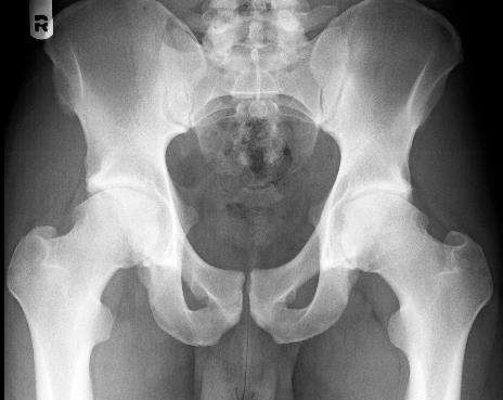
10 Basic musculoskeletal anatomy of the pelvis and hip joint - Bony structures: ASIS Ilium AIIS L5 Sacrum GT LT


 2.")
11 Basic musculoskeletal anatomy of the pelvis and hip joint - Intra-articular hip joint anatomy: 1. Hyaline cartilage surfaces (femoral head & acetabulum) 2. Acetabular labrum 3. Ligamentum Teres and cotyloid fossa


 2.")
12 Basic musculoskeletal anatomy of the pelvis and hip joint - Hip joint capsule: Has 2 roles: 1. Providing joint stability. 2. Providing vascular supply for the femoral head and acetabulum. Has 3 parts: 1. Iliofemoral ligament (anterior+lateral) 2. Pubofemoral ligament (medial) 3. Ischiofemoral ligament (posterior)

13 Basic musculoskeletal anatomy of the pelvis and hip joint - Non-synovial joints at the pelvis: LUMBO-SACRAL SYMPHYSIS PUBIS
14 Basic musculoskeletal anatomy of the pelvis and hip joint - Apophyseal prominences of the pelvis: ASIS (Insertion of Sartorius) AIIS (Insertion of direct head of Rectus Femoris) Ischium (Insertion of Hamstrings) Iliac crest (Insertion of lower lumbar spine muscles)

.")
15 Basic musculoskeletal anatomy of the pelvis and hip joint - Musculo-tendinous anatomy of anterior aspect of the hip: Deep anterior, overlying capsule: Iliopsoas, Iliocapsularis, Glut. minimus Mid-anterior: Rectus femoris (reflected and direct heads). Superficial anterior: Sartorius

16 Basic musculoskeletal anatomy of the pelvis and hip joint - Musculo-tendinous anatomy of medial aspect of the hip: Medial: Pectineus, Adductors.


17 Basic musculoskeletal anatomy of the pelvis and hip joint - Musculo-tendinous anatomy of lateral and posterolateral aspects of the hip: Lateral and posterolateral: Gluteus medius, TFL/ITB, Gluteus maximus.

18 Basic musculoskeletal anatomy of the pelvis and hip joint - Musculo-tendinous anatomy of posterior aspect of the hip: Deep posterior: Piriformis and short external rotators. Superficial posterior: Hamstrings, Gluteus maximus.
19 Basic musculoskeletal anatomy of the pelvis and hip joint - Musculo-tendinous anatomy of lower abdominal wall: Rectus abdominis Oblique muscles Apponeurosis overlying symphysis pubis


20 Pelvis and hip joint: Sports-related injuries
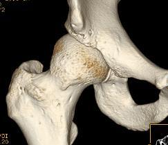
21 Femoroacetabular impingement (FAI) - Definition: Abnormal contacts between the femoral head-neck and the acetabular rim/labrum when the hip is in motion Lead to activity-related hip and groin pain and can result in progressive joint cartilage wear. Considered a potential cause for the development of hip arthrosis. Two morphological variants which are believed to result in such abnormal intra-articular mechanics have been described: 1. Aspheric femoral head ( Cam-type impingement). 2. Acetabular rim over-coverage ( Rim-type or Pincer-type impingement). Commonly, both types are believed to co-exist to a certain extent.
 - Abnormal contact")
22 Femoroacetabular impingement (FAI) - Abnormal contact mechanics:

 maneuver.")
23 Femoroacetabular impingement (FAI) - Physical examination: Hallmark: Hip impingement sign = Anterior hip and groin pain during passive FADIR (Flexion-Adduction-Internal rotation) maneuver. Often C-sign, indicating pain distribution.
.")
24 Femoroacetabular impingement (FAI) - Important diagnostic tool: Intra-articular injection of a local-anesthetic, followed by repeating the physical exam provocative test (FADIR). Assist in confirming intra-articular source for the pain.
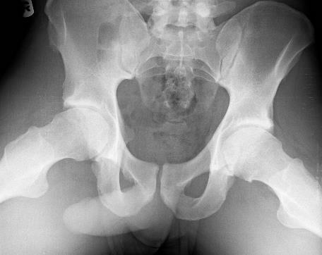
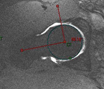
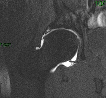
25 Femoroacetabular impingement (FAI) - Imaging:


26 Femoroacetabular impingement (FAI) - Treatment options: Activity modification Physical therapy Analgesics Surgery, aimed for: 1. Labral repair (to preserve negative pressure seal mechanism). 2. Femoral head-neck osteochondroplasty (to recreate head sphericity and eliminate impingement). 3. Focal rim trimming (to decrease impingement resulting from over-coverage).
: 1. Labral repair. 2.")
27 Case example: Bilateral FAI in a young soccer player Bilateral arthroscopic hip surgery (separate procedures): 1. Labral repair. 2. Femoral head-neck osteochondroplasty (to recreate head sphericity).


28 Femoroacetabular impingement (FAI) Surgery, aimed for: Improving hip kinematics decreasing further impingement decreasing risk for ongoing chondrolabral damage. Bedi A, Dolan M, Hetsroni I, Magennis E, Lipman J, Buly R, Kelly BT. Surgical treatment of femoroacetabular impingement improves hip kinematics: a computer-assisted model. Am J Sports Med, 2011
29 Snapping hip disorders (internal vs. external) - Internal snapping hip (coxa saltans interna): A condition where pain in the groin is elicited dynamically in conjunction with iliopsoas tendon snapping when the hip extends and internally rotates from a flexed, abducted, and externally rotated position (= circumduction maneuver). The tendon snapping can be over the iliopectineal eminence, the femoral head, loose body, or a para-labral cyst. - External snapping hip (coxa saltans externa): A condition where the posterior band of the ITB or the anterior band of the Gluteus maximus facia is snapping over the posterior edge of the greater trochanter when the hip moves from flexion to extension with or without rotation. - An unusual cause for external snap : Ischiofemoral impingement. Impingement of the Quadratus femoris between the lesser trochanter and Ischium during extension and external rotation (during walking).
. This anteversion can also be a source for overload on the antero-superior labrum.")
30 Internal snapping hip (iliopsoas snapping) - The snap is clearly heard. - More common in females (and in dancers). - Source for pain: inflamed iliopsoas tendon, or a chondro-labral injury. - Possibly related to hip anteversion (mild dysplasia). This anteversion can also be a source for overload on the antero-superior labrum. - Physical examination: The snap can be reproduced with hip circumduction.


31 Internal snapping hip (iliopsoas snapping) - Treatment options: 1. Snapping without pain should be treated with stretching only and no surgery!!! 2. Activity modification, stretching and core strengthening, injections.


32 Internal snapping hip (iliopsoas snapping) - Treatment options: 3. Iliopsoas surgical release at the lesser trochanter or at the joint level. - Downsides of surgical release: 1. Flexors weakness. 2. The tight iliopsoas is a potential secondary stabilizer of a mildly anteverted dysplastic hip. Therefore, release can increase instability (Fabricant PD, et al. Clinical outcomes after arthroscopic psoas lengthening: the effect of femoral version. Arthroscopy 28; , 2012 July).
33 External snapping hip (ITB/ Gluteus maximus) - The snap can be clearly seen (sometimes misinterpreted as joint subluxation). - Source for snap: either the posterior aspect of the ITB or the anterior aspect of the Gluteus maximus facia. The snap is over the greater trochanter. - The snapping occurs when the hip moves from flexion to extension with or without rotation. - Physical examination: The snap can be reproduced by moving the hip from flexion to extension when the patient is in the lateral decubitus position.


34 External snapping hip (ITB/ Gluteus maximus) - Treatment options: 1. Activity modification, stretching, and core stability. 2. Surgery: release of the posterior ITB/ anterior Gluteus maximus facia. - Downsides of surgery: possible deformity and abductors overload in the absence of part of the ITB. Therefore, some investigators described releasing the Gluteus maximus insertion at the linea aspera on the femur, instead of removing part of the ITB.

. 2.")
. 3.")
35 Tendon-apophyseal avulsions Occurs when muscle is stronger then physis, usually during eccentric overload. Three common tendon apophyseal avulsions in the pelvis: 1. ASIS avulsion of the Sartorius. Related to kicking (soccer). 2. AIIS avulsion of the direct head of the Rectus femoris. Related to kicking (soccer). 3. Ischium tuberosity avulsion of the Hamstrings. Common in water-skiing.
36 Tendon-apophyseal avulsions Treatment: 1. Nonoperative treatment results in good recovery and return to all activities within a few weeks in the majority of cases. 2. Rare indications for surgery: - Hamstrings avulsion gap of more than 3-4cm. - Schiatic nerve irritation by a scar tissue after hamstrings avulsion. - Prominent bone formation at the buttock after hamstrings avulsion. - AIIS avulsion leading to spur deformity and hip impingement in flexion.
 in patients with hip")
37 The anterior inferior iliac spine (AIIS) in patients with hip impingement: *


38 Hetsroni I, Poultsides L, Bedi A, Larson CM, Kelly BT. Anterior inferior iliac spine morphology correlates with hip range of motion: a classification system and dynamic model. CORR 2013:471(8);


39 Hetsroni I, Larson CM, Dela Torre K, Zbeda R, Magennis E, Kelly BT. Anterior inferior iliac spine deformity as an extra-articular source for hip impingement: a series of 10 patients treated with arthroscopic decompression. Arthroscopy 2012:28; Demonstrated the reproducibility of arthroscopic AIIS decompression in a series of 10 young adult males. - Function significantly improved after surgery.
. Pain: located at the Groin (insertion of the adductor longus to the pubic bone). 2.")
40 Chronic tendinopathies at the pelvis area Definition: Degenerative process within the tendon as a result of aging and prolonged overuse, leading to micro-tears, pain, and chronic inflammation. Examples: 1. CHAT = chronic hip adductor tendinopathy. (Common in soccer players). Pain: located at the Groin (insertion of the adductor longus to the pubic bone). 2. Hamstrings tendinopathy at the Ischium insertion. (Common in runners). Pain: located at the upper posterior thigh (insertion of hamstrings to Ischium). 3. GTPS (Greater Trochanteric Pain Syndrome) related to Gluteus medius tendinopathy. (Common in middle-age women). 4. Others? Iliopsoas


.")
41 Chronic tendinopathies at the pelvis area Diagnosis: Pain on direct palpation. Pain when applying force against resistance. Imaging: X-rays, US, MRI. Treatment: Activity modification, stretching, core stability. Injections (ultrasound-guided, if needed). Rarely surgery, for either release (CHAT) or repair (Hamstrings, Gluteus med.).


42 Stress fractures at the pelvis Femoral neck stress fracture: 1. Symptoms: Groin pain. Suspect when history reveals a non-gradual increase in running distance, or in female runners with nutrition imbalance. 2. Imaging: X-rays, MRI, Bone scan. 3. Tension vs. compression type. 4. Intra-articular. Therefore, high risk of nonunion and severe disability if not diagnosed ASAP and treated as needed.

43 Stress fractures and stress reactions at the pelvis Femoral neck stress fracture: Treatment: 1. Stop running immediately!!! Consider PWB with crutches. 2. Aqua-running possible. 3. Low-intensity ultra-sound bone stimulator possible. 4. Hyper-baric oxygenation therapy possible. 5. In tension type : surgery is commonly recommended (internal fixation with screws) to avoid displacement of the fragments and subsequent non-union or AVN of the femoral head. 6. Repeat MRI after 3 months before resuming running.

44 Stress fractures at the pelvis Sacral stress fracture: 1. Symptoms: Low-back pain. Suspect when history reveals a non-gradual increase in running distance, or in female runners with nutrition imbalance. 2. Imaging: X-rays, MRI, Bone scan. 3. Should not lead to displacement or significant long-term disability. 4. Treatment: Activity modification (6-12 weeks), consider low-intensity ultra-sound. 5. Surgery is not indicated.

45 Stress fractures at the pelvis Pubic bone stress fracture: 1. Symptoms: Groin pain or near the symphysis pubis. Suspect when history reveals a nongradual increase in running distance, or in female runners with nutrition imbalance. 2. Imaging: X-rays, MRI, Bone scan. 3. Should not lead to displacement or significant long-term disability. 4. Treatment: Activity modification (6-12 weeks), consider low-intensity ultra-sound. 5. Surgery is not indicated.

46 Stress fractures at the pelvis As a general rule for stress fractures work-up - consider: 1. CBC 2. TSH 3. Calcium, Phosphate, Alkaline phosphatase 4. PTH 5. Vitamin D

47 Aponeurosis syndrome Also termed: 1. Sportsman s hernia. 2. Gilmor s hernia. 3. PIWI = posterior inguinal wall insufficiency - May be diagnosed with dynamic ultra-sound. - May require surgical repair. - May co-exist with FAI, and therefore examine thoroughly and use diagnostic tests (intra-articular injection) and imaging as needed.
48 Summary and take-home message 1. The pelvis is a complex structure and has multiple potential sources for pain in athletes. 2. Accurate diagnosis frequently requires multiple diagnostic tests and advanced imaging. 3. Multiple sources for pain may co-exist (adductor tendinopathy and FAI, etc.). 4. Remember: Low lumbar spine may cause radicular symptoms to the hip. 5. Remember: Hip joint pathology can present as knee pain due to similar nerve innervations. 4. Never overlook femoral neck stress fracture. 5. Always remember that athletes may present with a non-sports related source for their symptoms, such as tumors, infections, metabolic diseases, and others.
49 Thank you
The Hip (Iliofemoral) Joint. Presented by: Rob, Rachel, Alina and Lisa
 Joint. Presented by: Rob, Rachel, Alina and Lisa") The Hip (Iliofemoral) Joint Presented by: Rob, Rachel, Alina and Lisa Surface Anatomy: Posterior Surface Anatomy: Anterior Bones: Os Coxae Consists of 3 Portions: Ilium Ischium Pubis Bones: Pubis Portion
The Hip (Iliofemoral) Joint Presented by: Rob, Rachel, Alina and Lisa Surface Anatomy: Posterior Surface Anatomy: Anterior Bones: Os Coxae Consists of 3 Portions: Ilium Ischium Pubis Bones: Pubis Portion
THE HIP. Cooler than cool, the pinnacle of what is "it". Beyond all trends and conventional coolness.
 THE HIP Cooler than cool, the pinnacle of what is "it". Beyond all trends and conventional coolness. Objectives Hip anatomy Causes of hip pain Hip exam Anatomy Bones Ilium Anterior Superior Iliac Spine
Hip Injuries & Arthroscopy in Athletes
 Hip Injuries & Arthroscopy in Athletes John P Salvo, MD Sports Medicine Rothman Institute Philadelphia, PA EATA Annual Meeting January, 2011 Hip Injuries & Arthroscopy in Anatomy History Physical Exam
Hip Pain in the Athlete: A Diagnostic Challenge
 : A Diagnostic Challenge Matthew Gimre MD Sports Medicine 11 th Annual Sports Medicine Conference Presented June 17, 2017 on: Month day, Year Presented to: Insert relevant presenter information Calibri
Lesson 24. A & P Hip
 Lesson 24 A & P Hip 1 Aims of the Session This session will allow candidates to have an understanding of the bony prominences and soft tissues of the hip 2 Learning Outcomes By the end of the lesson the
FUNCTIONAL ANATOMY AND EXAM OF THE HIP, GROIN AND THIGH
 FUNCTIONAL ANATOMY AND EXAM OF THE HIP, GROIN AND THIGH Peter G Gerbino, MD, FACSM Orthopedic Surgeon Monterey Joint Replacement and Sports Medicine Monterey, CA TPC, San Diego, 2017 The lecturer has no
Hip & Groin pain. M Hassabi (MD) Assistant professor Department of Sports & Exercise Medicine Shahid Beheshti University of Medical Sciences
 Assistant professor Department of Sports & Exercise Medicine Shahid Beheshti University of Medical Sciences") Hip & Groin pain M Hassabi (MD) Assistant professor Department of Sports & Exercise Medicine Shahid Beheshti University of Medical Sciences EPIDEMIOLOGY Groin pain and injury is common with sports that
Hip & Groin pain M Hassabi (MD) Assistant professor Department of Sports & Exercise Medicine Shahid Beheshti University of Medical Sciences EPIDEMIOLOGY Groin pain and injury is common with sports that
Overview. Overview. Introduction. Introduction Anatomy History Examination Common Disorders. Introduction Anatomy History Examination Common Disorders
 Common Hip Disorders in Figure Skaters 14 th Annual Meeting of Sports Medicine and Science in Figure Skating January 25, 2009 8:15-8:45am Robert J. Dimeff, MD Medical Director of Sports Medicine Overview
MR Imaging in Athlete s Hip/Pelvis
 MR Imaging in Athlete s Hip/Pelvis Tara Lawrimore, MD FRCPC Department of Radiology Musculoskeletal Division Massachusetts General Hospital Harvard Medical School No disclosures MR and Hip Pain in the
What s Hip: Common Hip Problems and Kids and Adults
 What s Hip: Common Hip Problems and Kids and Adults Alan Zhang MD Assistant Professor Sports Medicine and Hip Arthroscopy UCSF Department of Orthopaedic Surgery I have no relevant disclosures. 2 1 Most
The Hip Joint. Shenequia Howard David Rivera
 The Hip Joint Shenequia Howard David Rivera Topics Of Discussion Movement Bony Anatomy Ligamentous Anatomy Muscular Anatomy Origin/Insertion/Action/Innervation Common Injuries MOVEMENT Flexion Extension
Figure 1 - Hip and Pelvis
 Hip Figure 1 - Hip and Pelvis The terms hip and pelvis are frequently used interchangeably, but strictly speaking, the pelvis is a girdle of bones and the hip is a joint. The pelvis consists of The sacrum
CLINICS IN SPORTS MEDICINE
 Clin Sports Med 25 (2006) 365 369 CLINICS IN SPORTS MEDICINE A Acetabular labrum, tears of, hip arthroscopy in, 264 Acetabular rim, trimming of, and labral repair, new method for, 293 297 Acetabulum, femoral
Main Menu. Joint and Pelvic Girdle click here. The Power is in Your Hands
 1 Hip Joint and Pelvic Girdle click here Main Menu K.6 http://www.handsonlineeducation.com/classes//k6entry.htm[3/23/18, 2:01:12 PM] Hip Joint (acetabular femoral) Relatively stable due to : Bony architecture
Mr Simon Jennings BSc, MB BS, FRCS, Dip Sports Med FRCS (Trauma & Orthopaedics)
") Mr Simon Jennings BSc, MB BS, FRCS, Dip Sports Med FRCS (Trauma & Orthopaedics) Consultant Orthopaedic Surgeon Northwick Park Hospital 107 Harley Street RSM 16 th September 2010 Orthopaedic Surgeon Knee
Mr Simon Jennings BSc, MB BS, FRCS, Dip Sports Med FRCS (Trauma & Orthopaedics) Consultant Orthopaedic Surgeon Northwick Park Hospital 107 Harley Street RSM 16 th September 2010 Orthopaedic Surgeon Knee
Sports Medicine and Radiology
 Sports Medicine and Radiology The judicious utilization of a thorough history and physical examination and appropriately applied imaging studies will allow for accurate diagnosis and treatment of athletic
Lectures of Human Anatomy
 Lectures of Human Anatomy Lower Limb Gluteal Region and Hip Joint By DR. ABDEL-MONEM AWAD HEGAZY M.B. with honor 1983, Dipl."Gynecology and Obstetrics "1989, Master "Anatomy and Embryology" 1994, M.D.
Non-arthritic anterior hip pain in the younger patient: examination and intervention strategies
 Non-arthritic anterior hip pain in the younger patient: examination and intervention strategies Melodie Kondratek, PT, DScPT, OMPT Bryan Kuhlman, PT, DPT, OMPT Oakland University Orthopedic Spine and Sports
Hip Anatomy. Bony. The Athletic Hip: Anatomy and Common Injuries. Kyle Wilkens MSPAS, PA-C, ATC/L October 8, 2013
 The Athletic Hip: Anatomy and Common Injuries Kyle Wilkens MSPAS, PA-C, ATC/L October 8, 2013 Hip Anatomy Bony Femur Ischium Ilium Pubis Sacrum Coccyx Soft Tissue Joints Muscles -Pubic Symphisis Labrum
Young Adult Hip problems. Aresh Hashemi-Nejad FRCS(Orth)
") Young Adult Hip problems Aresh Hashemi-Nejad FRCS(Orth) RNOH founded 1837 by William Little 14 year old presenting with limp Knee pain on and off 4 months Limps Aresh Hashemi-Nejad FRCS(Orth) The Royal
Young Adult Hip problems Aresh Hashemi-Nejad FRCS(Orth) RNOH founded 1837 by William Little 14 year old presenting with limp Knee pain on and off 4 months Limps Aresh Hashemi-Nejad FRCS(Orth) The Royal
Evaluation of the Hip
 Evaluation of the Hip Adam Lewno, DO PCSM Fellow, University of Michigan Primary Care Sports Update 2017 Disclosures Financial: None Images: I would like to acknowledge the work of the original owners
Stephanie W. Mayer, MD. Director of Child and Young Adult Hip Preservation Sports Medicine Center Children s Hospital Colorado
 Stephanie W. Mayer, MD Director of Child and Young Adult Hip Preservation Sports Medicine Center Children s Hospital Colorado University of Colorado Sports Medicine Assistant Team Physician, Colorado Avalanche
Joints of the lower limb
 Joints of the lower limb 1-Type: Hip joint Synovial ball-and-socket joint 2-Articular surfaces: a- head of femur b- lunate surface of acetabulum Which is deepened by the fibrocartilaginous labrum acetabulare
The Lower Limb. Anatomy RHS 241 Lecture 2 Dr. Einas Al-Eisa
 The Lower Limb Anatomy RHS 241 Lecture 2 Dr. Einas Al-Eisa The bony pelvis Protective osseofibrous ring for the pelvic viscera Transfer of forces to: acetabulum & head of femur (when standing) ischial
Snapping Hip and Impingement
 Snapping Hip and Impingement Jon A. Jacobson, M.D. Professor of Radiology Director, Division of Musculoskeletal Radiology University of Michigan Disclosures: Consultant: Bioclinica Advisory Board: GE,
The Evaluation of Hip pain in the Athlete
 The Evaluation of Hip pain in the Athlete DREW ROGERS,MD The Evaluation of Hip pain in the Athlete Andrew Rogers, MD (Drew) Orthopedic Care Physician Network Chief of Orthopedics Morton Hospital Team Physician
The Young Adult Hip: FAI. Jason Snibbe, M.D. Snibbe Orthopedics Team Physician, University of Southern California
 The Young Adult Hip: FAI Jason Snibbe, M.D. Snibbe Orthopedics Team Physician, University of Southern California Introduction Femoroacetabular Impingment(FAI) Presentation and Exam Imaging Surgical Management
STAIRS. What s Hip: Top 5 Hip Problems in Primary Care. I have no relevant disclosures. Top 5 (or 6) Pathologies. Big 3- Questions to Ask
 Pathologies. Big 3- Questions to Ask") I have no relevant disclosures. What s Hip: Top 5 Hip Problems in Primary Care Alan Zhang MD Assistant Professor Sports Medicine and Hip Arthroscopy UCSF Department of Orthopaedic Surgery December, 2015
I have no relevant disclosures. What s Hip: Top 5 Hip Problems in Primary Care Alan Zhang MD Assistant Professor Sports Medicine and Hip Arthroscopy UCSF Department of Orthopaedic Surgery December, 2015
Bony Anatomy. Femur. Femoral Head Femoral Neck Greater Trochanter Lesser Trochanter Intertrochanteric Crest Intertrochanteric Line Gluteal Tuberosity
 Hip Anatomy Bony Anatomy Femur Femoral Head Femoral Neck Greater Trochanter Lesser Trochanter Intertrochanteric Crest Intertrochanteric Line Gluteal Tuberosity Bony Anatomy Pelvic Girdle Acetabulum 3 bones
GET HIP! CAPA 2015 Annual Conference WHAT IS HIP? HIP JOINT. Bradford H. Stiles, M.D., FAAFP
 GET HIP! Bradford H. Stiles, M.D., FAAFP WHAT IS HIP? HIP JOINT Synovial ball-and-socket joint Articulation between femoral head and acetabulum Acetabulum formed by the confluence of pelvis bones (ilium,
A Guide for Patients with Hip and Groin Pain. By - Rob Lawton & Ajay Malviya. Overview
 A Guide for Patients with Hip and Groin Pain By - Rob Lawton & Ajay Malviya Overview - Introduction - Hip Anatomy - Is the pain coming from the hip joint? - Intra-articular causes of hip pain o Impingement
The hip: Built for endurance and mobility
 The hip: Built for endurance and mobility The hip joint Some anatomical landmarks Innominate Ilium, pubis, ischium Sacrum Iliac crests Asis Psis Pubic tubercle Acetabulum Femur Head of femur Neck of femur
Labral Tears/FAI. Andrew Parker, MD
 Labral Tears/FAI Andrew Parker, MD Athletic Hip Injuries Incidence of hip injuries has increased dramatically over the last decade In part due to better recognition with improved imaging and arthroscopy,
The psoas minor is medial to the psoas major. The iliacus is a fan-shaped muscle that when contracted helps bring the swinging leg forward in walking
 1 p.177 2 3 The psoas minor is medial to the psoas major. The iliacus is a fan-shaped muscle that when contracted helps bring the swinging leg forward in walking and running. The iliopsoas and adductor
Imaging in Groin Pain What the Team Physician Needs to Know
 Imaging in Groin Pain What the Team Physician Needs to Know Üstün Aydıngöz, MD Professor of Radiology Hacettepe University School of Medicine Ankara, Turkey ustunaydingoz@yahoo.com No conflicts of interest
CONSERVATIVE MANAGEMENT OF FEMOROACETABULAR IMPINGEMENT
 SPORTS REHABILITATION CONSERVATIVE MANAGEMENT OF FEMOROACETABULAR IMPINGEMENT A case study and rationale for treatment Written by Joanne Kemp and Kay Crossley, Australia BACKGROUND The hip joint and FAI
3/18/18. Adolescent Hip Injuries. Adolescents with Hip Injuries DISCLOSURES
 Adolescent Hip Injuries Henry Bone Ellis, Jr., MD DFW Sports Medicine Symposium March 24, 2018 DISCLOSURES Royalties and stock options Consulting income Smith and Nephew Other support Research on Osteochondritis
Labral Tears / Femoro- Acetabular Impingement / Hip Arthroscopy/THA. Dr Allen Turnbull Hip and Knee Surgery
 Labral Tears / Femoro- Acetabular Impingement / Hip Arthroscopy/THA Hip Anatomy Labrum Fovea Femoral Head Articular Cartilage Ligamentum Teres Labral Tears Function of Labrum Deepens acetabulum by 20%
Hip joint and pelvic girdle. Lower Extremity. Pelvic Girdle 6/5/2017
 Hip joint and pelvic girdle Lower Extremity The relationship between the pelvic girdle and hip is similar to that between the shoulder girdle and shoulder joint. The lower limbs are attached to the axial
rotation of the hip Flexion of the knee Iliac fossa of iliac Lesser trochanter Femoral nerve Flexion of the thigh at the hip shaft of tibia
 Anatomy of the lower limb Anterior & medial compartments of the thigh Dr. Hayder The fascia lata encloses the entire thigh like a sleeve/stocking. Three intramuscular fascial septa (lateral, medial, and
Apophysis. Apophyseal Avulsion. Apophyseal avulsion injuries 3/2/2017
 Apophysis 0 Differentiate from Epiphysis: The end of long bones which undergo endochondral ossification to produce longitudinal growth of the bones. i.e. growth plates 0 Apophysis refers to any eminence,
Applied anatomy of the hip and buttock
 CHAPTER CONTENTS The hip joint e9 Capsule and ligaments e9 s e0 Flexor muscles................... e0 Extensor muscles.................. e Abductor muscles.................. e Adductor muscles..................
APPLICATION OF THE MOVEMENT SYSTEMS MODEL TO THE MANAGEMENT COMMON HIP PATHOLOGIES
 APPLICATION OF THE MOVEMENT SYSTEMS MODEL TO THE MANAGEMENT COMMON HIP PATHOLOGIES Tracy Porter, PT, DPT Des Moines University Department of Physical Therapy Objectives Review current literature related
SURGICAL AND APPLIED ANATOMY
 Página 1 de 6 Copyright 2001 Lippincott Williams & Wilkins Bucholz, Robert W., Heckman, James D. Rockwood & Green's Fractures in Adults, 5th Edition SURGICAL AND APPLIED ANATOMY Part of "37 - HIP DISLOCATIONS
Gluteal region DR. GITANJALI KHORWAL
 Gluteal region DR. GITANJALI KHORWAL Gluteal region The transitional area between the trunk and the lower extremity. The gluteal region includes the rounded, posterior buttocks and the laterally placed
The University Of Jordan Faculty Of Medicine THE LOWER LIMB. Dr.Ahmed Salman Assistant Prof. of Anatomy. The University Of Jordan
 The University Of Jordan Faculty Of Medicine THE LOWER LIMB Dr.Ahmed Salman Assistant Prof. of Anatomy. The University Of Jordan Gluteal Region Cutaneous nerve supply of (Gluteal region) 1. Lateral cutaneous
Hip Injuries in the Workers Compensation Arena: Diagnosis and Treatment. Joshua S Hornstein, MD TOG Institute
 Hip Injuries in the Workers Compensation Arena: Diagnosis and Treatment Joshua S Hornstein, MD TOG Orthopaedics@Rothman Institute Disclosures No Relevant Disclosures Objectives Basic Anatomy Pathology
Doc, I've done my groin. Groin Pain. Peter Brukner. Doc, I've done my groin 1. acute chronic
 Doc, I've done my groin Peter Brukner Associate Professor in Sports Medicine Centre for Sports Medicine Research and Education School of Physiotherapy 9/22/2006 The University of Melbourne Groin Pain acute
APPROACH TO THE DIAGNOSIS OF GROIN PAIN. Alexandra Myers, D.O., M.S.H.S. February 22, 2018 OPSC Annual Convention
 APPROACH TO THE DIAGNOSIS OF GROIN PAIN Alexandra Myers, D.O., M.S.H.S. February 22, 2018 OPSC Annual Convention OVERVIEW Review the entities that may contribute to groin pain Discuss the approach to making
Diagnostic Approach to Hip Pain. Zoë J. Foster, MD October 3, 2018
 Diagnostic Approach to Hip Pain Zoë J. Foster, MD October 3, 2018 Disclosures I have nothing to disclose. Objectives Review examination of the hip, including special tests Discuss differential diagnosis
lesser trochanter of femur lesser trochanter of femur iliotibial tract (connective tissue) medial surface of proximal tibia
 medial surface of proximal tibia") LOWER LIMB MUSCLES OF THE APPENDICULAR SKELETON The muscles that act on the lower limb fall into three groups: those that move the thigh, those that move the lower leg, and those that move the ankle, foot,
LOWER LIMB MUSCLES OF THE APPENDICULAR SKELETON The muscles that act on the lower limb fall into three groups: those that move the thigh, those that move the lower leg, and those that move the ankle, foot,
Anatomy & Examination of the Athlete s Hip
 Anatomy & Examination of the Athlete s Hip Shane J. Nho, MD, MS Hip Preservation Center, Division of Sports Medicine, Department of Orthopedic Surgery, Rush University Medical Center Layered Anatomical
The thigh. Prof. Oluwadiya KS
 The thigh Prof. Oluwadiya KS www.oluwadiya.com The Thigh: Boundaries The thigh is the region of the lower limb that is approximately between the hip and knee joints Anteriorly, it is separated from the
RN(EC) ENC(C) GNC(C) MN ACNP *** MECHANISM OF INJURY.. MOST IMPORTANT ***
 ENC(C) GNC(C) MN ACNP *** MECHANISM OF INJURY.. MOST IMPORTANT ***") HISTORY *** MECHANISM OF INJURY.. MOST IMPORTANT *** Age of patient - Certain conditions are more prevalent in particular age groups (Hip pain in children may refer to the knee from Legg-Calve-Perthes
HISTORY *** MECHANISM OF INJURY.. MOST IMPORTANT *** Age of patient - Certain conditions are more prevalent in particular age groups (Hip pain in children may refer to the knee from Legg-Calve-Perthes
Baraa Ayed حسام أبو عوض. Ahmad Salman. 1 P a g e
 4 Baraa Ayed حسام أبو عوض Ahmad Salman 1 P a g e Today we are going to cover these concepts: Iliotibial tract Anterior compartment of the thigh and the hip Medial compartment of the thigh Femoral triangle
hip pathology w mccormick 2017 mccormickortho.com
 hip pathology w mccormick 2017 mccormickortho.com overview classification common hip pathologies FAI GT pain snapping workup treatments sample cases rehabilitation outcomes/complications hip pathology
FAI syndrome with or without labral tear.
 Case This 16-year-old female, soccer athlete was treated for pain in the right groin previously. Now has acute onset of pain in the left hip. The pain was in the groin that was worse with activities. Diagnosis
ANATYOMY OF The thigh
 ANATYOMY OF The thigh 1- Lateral cutaneous nerve of the thigh Ι) Skin of the thigh Anterior view 2- Femoral branch of the genitofemoral nerve 5- Intermediate cutaneous nerve of the thigh 1, 2 and 3 are
Acland's DVD Atlas of Human Anatomy. Transcript for Volume Robert D Acland
 Acland's DVD Atlas of Human Anatomy Transcript for Volume 2 2007 Robert D Acland This free downloadable pdf file is to be used for individual study only. It is not to be reproduced in any form without
Review causes of hip and groin pain in athlete Discuss indications for hip arthroscopy Review, if any, history & physical findings of a patient who
 Review causes of hip and groin pain in athlete Discuss indications for hip arthroscopy Review, if any, history & physical findings of a patient who may benefit from hip arthroscopy Review portal placement
Muscles of the lower extremities. Dr. Nabil khouri MD, MSc, Ph.D
 Muscles of the lower extremities Dr. Nabil khouri MD, MSc, Ph.D Posterior leg Popliteal fossa Boundaries Biceps femoris (superior-lateral) Semitendinosis and semimembranosis (superior-medial) Gastrocnemius
Identify the muscles associated with the medial compartment of the thigh. Identify the attachment points of the medial thigh muscles.
 L 8 A B O R A T O R Y Thigh MEDIAL THIGH Identify the muscles associated with the medial compartment of the thigh. Identify the attachment points of the medial thigh muscles. Identify the actions of these
Hip Arthroscopy Indications and Latest Techniques
 Transcript Details This is a transcript of an educational program accessible on the ReachMD network. Details about the program and additional media formats for the program are accessible by visiting: https://reachmd.com/programs/clinicians-roundtable/hip-arthroscopy-indications-and-latesttechniques/3801/
Human Anatomy Biology 351
 Human Anatomy Biology 351 Lower Limb Please place your name on the back of the last page of this exam. You must answer all questions on this exam. Because statistics demonstrate that, on average, between
MSK Radiology Interesting Case Presentation
 MSK Radiology Interesting Case Presentation Sitt Ching Man Jacqueline 27 th Feb 2015 Patient 1: YPY F/27 Professional badminton player C/o sudden onset of right shoulder pain after a very vigorous match
Human Anatomy Biology 351
 Human Anatomy Biology 351 Lower Limb Please place your name on the back of the last page of this exam. You must answer all questions on this exam. Because statistics demonstrate that, on average, between
Hip Arthroscopy. Christopher J. Utz, MD. Assistant Professor of Orthopaedic Surgery University of Cincinnati
 Hip Arthroscopy Christopher J. Utz, MD Assistant Professor of Orthopaedic Surgery University of Cincinnati Disclosures I have no disclosures relevant to this topic. Outline 1. Brief History 2. Review of
Thigh, Hip, & Low Back Evaluation.
 Thigh, Hip, & Low Back Evaluation www.fisiokinesiterapia.biz Thigh Injuries Quad contusions - Myositis Ossificans Trochanteric bursitis - snapping hip Ischial bursitis - bench-warmer s bursitis Strains
A Patient s Guide to Femoroacetabular Impingement (FAI) of the Hip
 of the Hip") A Patient s Guide to Femoroacetabular Impingement (FAI) of the Hip 651 Old Country Road Plainview, NY 11803 Phone: 5166818822 Fax: 5166813332 p.lettieri@aol.com DISCLAIMER: The information in this booklet
A Patient s Guide to Femoroacetabular Impingement (FAI) of the Hip 651 Old Country Road Plainview, NY 11803 Phone: 5166818822 Fax: 5166813332 p.lettieri@aol.com DISCLAIMER: The information in this booklet
First practical session. Bones of the gluteal region
 First practical session 2017 Bones of the gluteal region The Hip bone The hip bone is made of: 1 The ilium: superior in position 2 The ischium:postero-inferior in position 3 The pubis: antero-inferior
Female Athlete Hip Injuries: Exploring the CORE of Patterns and Prevention
 Female Athlete Hip Injuries: Exploring the CORE of Patterns and Prevention Kelly C. McInnis DO Irene Davis, PhD, PT, FAPTA, FACSM, FASB David Nolan, PT, DPT, MS, OCS, SCS, CSCS Your name and credentials
Hip Impingement and Arthritis: Preservation vs. Total Hip Arthroplasty. Faculty Disclosures. Objectives 11/17/2017
 Hip Impingement and Arthritis: Preservation vs. Total Hip Arthroplasty Jonathan R. Schiller, MD Assistant Professor of Orthopedics Warren Alpert Medical School of Brown University Director, Adolescent
Lower limb summary. Anterior compartment of the thigh. Done By: Laith Qashou. Doctor_2016
 Lower limb summary Done By: Laith Qashou Doctor_2016 Anterior compartment of the thigh Sartorius Anterior superior iliac spine Upper medial surface of shaft of tibia 1. Flexes, abducts, laterally rotates
ANATYOMY OF The thigh
 ANATYOMY OF The thigh 1- Lateral cutaneous nerve of the thigh Ι) Skin of the thigh Anterior view 2- Femoral branch of the genitofemoral nerve 5- Intermediate cutaneous nerve of the thigh 1, 2 and 3 are
ANATYOMY OF The thigh
 ANATYOMY OF The thigh 1- Lateral cutaneous nerve of the thigh Ι) Skin of the thigh Anterior view 2- Femoral branch of the genitofemoral nerve 1, 2 and 3 are From the lumber plexus 5- Intermediate cutaneous
11/11/2016. Hip FAI & Core Muscle Deficiency: Diagnosis and Treatment. Disclosures. Differential Diagnosis. Consultant, Smith and Nephew
 Hip FAI & Core Muscle Deficiency: Diagnosis and Treatment FORE Baseball Sports Medicine Game-Changing Concepts November 4, 2016 T. Sean Lynch, MD Assistant Professor New York-Presbyterian/ Columbia University
emoryhealthcare.org/ortho
 COMMON SOCCER INJURIES Oluseun A. Olufade, MD Assistant Professor, Department of Orthopedics and PM&R 1/7/18 GOALS Discuss top soccer injuries and treatment strategies Simplify hip and groin injuries in
To classify the joints relative to structure & shape
 To classify the joints relative to structure & shape To describe the anatomy of the hip joint To describe the ankle joint To memorize their blood & nerve supply JOINTS: Joints are sites where skeletal
The Muscular System. Chapter 10 Part D. PowerPoint Lecture Slides prepared by Karen Dunbar Kareiva Ivy Tech Community College
 Chapter 10 Part D The Muscular System Annie Leibovitz/Contact Press Images PowerPoint Lecture Slides prepared by Karen Dunbar Kareiva Ivy Tech Community College Table 10.14: Muscles Crossing the Hip and
MRI of the Hips and Pelvis
 MRI of the Hips and Pelvis Hips and Pelvis Protocols Vascular abnormalities Fractures Soft tissues Labrum and FAI Hips and Pelvis Protocols Vascular abnormalities Fractures Soft tissues Labrum and FAI
Muscles of Gluteal Region
 1 The Gluteal Region In the gluteal region the skin is tough with many layers underneath. Directly under it is the superficial fascia followed by the deep fascia then the muscles and the bones of the thigh.
Dynamic Snapping of the Hip: Sonographic Evaluation. Adam M. Pourcho DO Swedish Sports Medicine
 Dynamic Snapping of the Hip: Sonographic Evaluation Adam M. Pourcho DO Swedish Sports Medicine Special Thanks Jay Smith, MD Disclosures Objectives Brief review of anatomy of anterior, lateral and posterior
Muscles of the Thigh. 6.1 Identify, describe the attachments of and deduce the actions of the muscles of the thigh: Anterior group
 Muscles of the Thigh 6.1 Identify, describe the attachments of and deduce the actions of the muscles of the thigh: Anterior group Sartorius: This is a long strap like muscle with flattened tendons at each
Anterior and Medial compartments of the thigh. Dr. Heba Kalbouneh Associate Professor of Anatomy and Histology
 Anterior and Medial compartments of the thigh Dr. Heba Kalbouneh Associate Professor of Anatomy and Histology Terms Related to Movements Movement Flexion Extension Abduction Adduction Medial (internal)
Sports Hernias. Matthew Gimre, MD ATC Conference, June 20, 2015
 Sports Hernias Matthew Gimre, MD ATC Conference, June 20, 2015 Sports hernia: So what is it? An injury to the rectus abdominis-common adductor aponeurosis, at the anterior/inferior aspect of the pubic
Femoroacetabular Impingement in the Throwing Athlete. Michael Banffy, MD Sports Medicine, Hip Preservation Kerlan Jobe Institute
 Femoroacetabular Impingement in the Throwing Athlete Michael Banffy, MD Sports Medicine, Hip Preservation Kerlan Jobe Institute Disclosures None Baseball Hip Injuries - Background Abdominal/groin injuries
Surgical Anatomy of the Hip. Joseph H. Dimon
 Surgical Anatomy of the Hip Joseph H. Dimon The hip joint is a deep joint surrounded by large and powerful muscles necessary for its proper function. Essential neurovascular structures lie in front and
CHAPTER 8: THE BIOMECHANICS OF THE HUMAN LOWER EXTREMITY
 CHAPTER 8: THE BIOMECHANICS OF THE HUMAN LOWER EXTREMITY _ 1. The hip joint is the articulation between the and the. A. femur, acetabulum B. femur, spine C. femur, tibia _ 2. Which of the following is
When the patient presents at the GP surgery with Hip pain. Investigations, Input & Referral
 When the patient presents at the GP surgery with Hip pain Investigations, Input & Referral GP with an interest in Pain Management How common is hip pain? Around 450 patients per 100,000 population will
A Patient s Guide to Labral Tears of the Hip
 A Patient s Guide to Labral Tears of the Hip Sports-related injuries require specialized care to promote optimum healing. Whether you are a weekend jogger or tennis player, a professional soccer player
Evaluation of Posterior Hip Pain
 Evaluation of Posterior Hip Pain Anthony J. Ferretti, D.O., MHSA Hip Pain in the Adult Various etiologies: Traumatic Infectious Neurovascular Degenerative Congenital Pathologic 1 Hip Pain Complex interaction
Lecture 08 THIGH MUSCLES ANTERIOR COMPARTMENT. Dr Farooq Khan Aurakzai. Dated:
 Lecture 08 THIGH MUSCLES ANTERIOR COMPARTMENT BY Dr Farooq Khan Aurakzai Dated: 11.02.2017 INTRODUCTION to the thigh Muscles. The musculature of the thigh can be split into three sections by intermuscular
LAB Notes#1. Ahmad Ar'ar. Eslam
 LAB Notes#1 Ahmad Ar'ar Eslam 1 P a g e Anatomy lab Notes Lower limb bones :- Pelvic girdle: It's the connection between the axial skeleton and the lower limb; it's made up of one bone called the HIP BONE
lower limb Anterior Compartment: lecture 3 The deep fascia ( fascia lata) divides the thigh into 3 compartments:
 divides the thigh into 3 compartments:") lower limb lecture 3 The deep fascia ( fascia lata) divides the thigh into 3 compartments: 1. Anterior Extensor compartment 2. Medial Adductor compartment 3. Posterior Flexor compartment Anterior Compartment:
lower limb lecture 3 The deep fascia ( fascia lata) divides the thigh into 3 compartments: 1. Anterior Extensor compartment 2. Medial Adductor compartment 3. Posterior Flexor compartment Anterior Compartment:
Anatomage Table Instructors Guide- Lower Limb
 The Lower Limb Anatomage Table Instructors Guide- Lower Limb Table of Contents Lower Limb 1- The Skeletal System...3 1: Hip Bone...3 2: Hip Joint and Femur...4 3: Patella and Knee Joint...7 4: Tibia, Fibula,
F.A.I. indications. E. Sabetta. Arcispedale S. Maria Nuova Struttura Complessa Ortopedia e Traumatologia Direttore: Ettore Sabetta
 F.A.I. indications E. Sabetta Arcispedale S. Maria Nuova Struttura Complessa Ortopedia e Traumatologia Direttore: Ettore Sabetta !.FAI treatment might become a mainstay in joint-preserving treatment of
Specialists in Joint Replacement, Spinal Surgery, Orthopaedics and Sport Injuries. The Hip.
 Specialists in Joint Replacement, Spinal Surgery, Orthopaedics and Sport Injuries The Hip INTRODUCTION THE HIP The hip is a ball-and-socket joint. The socket is formed by the acetabulum, which is part
Hip Cases from Clinic: Refining your history and physical
 Hip Cases from Clinic: Refining your history and physical Alan Zhang MD Assistant Professor Sports Medicine and Hip Arthroscopy UCSF Department of Orthopaedic Surgery 11/20/2017 Case #1 Healthy 21 M College
Hip Region. PHTY2020: Lecture
 Region PHTY2020: Lecture 2.1 29.02.16 Functional Overview Transfer body weight form trunk to legs Allows leg to adopt numerous positions needed for standing, walking running, stairs, sitting and other
What is FAI? And Why are we getting Hip Problems?
 What is FAI? And Why are we getting Hip Problems? Why is Hip pain so common NOW? Greater awareness More accurate imaging MRI Fashionable?? Geography Arthroscopy used to be OP now FAI! Why is Hip pain so