Angle Stable Distal Radial Plate System WINSTA-R
|
|
|
- Imogen Campbell
- 5 years ago
- Views:
Transcription

1 Angle Stable Distal Radial Plate System WINSTA-R Priv.-Doz.Dr.med. Martin Walz Dr. med. Felix Menzinger Prof.Dr.med. Jürgen Rudigier


2 General The problem posed by metaphyseal and intra-articular fractures lies in the fact that even though implants can be well-anchored in the distal section of the shaft, fixation stability is however limited in the spongiose bones of the fragment close to the joint. Multifragmentary, intra-articular fractures as well as senile osteoporosis exacerbate this problem. In relation to the long proximal shaft section, the lever arm of the short distal joint fragment mechanically unfavourable during functional treatment places high demands on the stability of the implant itself as well as on its fixation in the distal fragment. In contrast to conventional implants, angle stable ones enable early functional treatment (especially desirable in the case of fractures close to joints), which is of great importance for the rapid restoration of a joint's functional integrity. Adaptation of the plate form and of the direction of the distal angle stable plate screws or pins to the physiological articular surface axes and curvatures facilitates correct reposition as well as the ideal repositioning of the implant components, thus increasing postoperative fracture stability. Product Characteristics High stability in the case of small implant dimensions. Anatomically correct form with volar bending to reconstruct the "palmar tilt", and secure support of the volar contour of the distal radius. The four distal plate holes are adjusted to the radioulnar curvature of the articular surface, and enable secure fixation of the distal radius even in the case of intra-articular fractures via subchondral positioning of the stabilization screws and pins. The given direction of the four angle stable screws or pins at the distal plate end corresponds to the physiological dorsovolar inclination of the articular surface, and enables the screws or pins to be placed in the ulnar fragment as well as in the styloid process of the radius. For fixation in the distal fragment, either self-cutting angle stable Ø 3.0 mm stabilization screws or angle stable Ø 2.0 mm stabilization pins are available. For fixation of the plate, self-cutting Ø 2.7mm corticalis screws are available. In the case of osteoporotic bones, angle stable Ø 3.0 stabilization screws can be used as an alternative. Precision drilling and length measurement for angle stable screws/pins via screw-in drill sleeve. For all screws and pins, only a Ø 2.0 mm drill is required. A cross-oval plate-hole enables reposition of intra-articular fragments as well as possible defect filling with spongiosa or bone substitute after plate fixation. The clearly laid-out instrumentarium facilitates handling. Various different plate lengths also suit the requirements of multiple metaphyseal fractures. Special surface treatment of plates and angle stable screws and pins byanodisation typeii(dotize ) Properties of processed products: - reduced tendency of cold welding of Titanium implants by screw in the angle stable pins and screws. - hardened Titanium surface. - increased fatigue resistance of implants. - significant reduction of AL and V release Reconstruction of the palmar tilt Anatomical radioulnar curvature Page


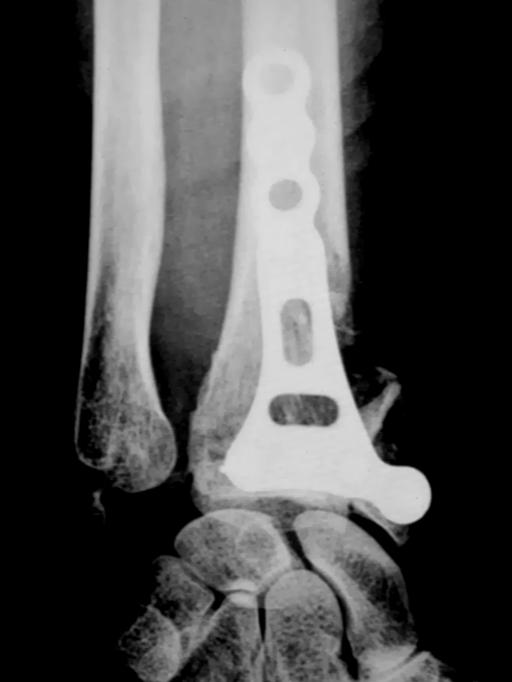
3 Indications Extra-articular fractures of AO-Type 23-A2 and A3 Partial intra-articular fractures of AO-Type 23-B and B3 Total intra-articular fractures of AO-Type 23-C to C3 03 Radiological Procedure, Case Study 0 AO-TYP 23-A3 Radiological Procedure, Case Study 2 05 AO-TYP 23-C Radiological Procedure, Case Study 3 AO-TYP 23-C3 Page 2


4 Operative Technique Two different techniques of volar bone-plate osteosynthesis can be applied: Possibility I: Primary fixation of the plate on the radius shaft. Reposition of the joint fragment on the distal plate section. 06 Possibility II: Primary fixation of the plate on the joint fragment. Reposition via fixation of plate onto radius shaft. 07 Possibility I Possibility II Access / Reposition of Fracture After partial deprivation of blood supply to the upper arm, a straight, volar skin incision of 5 to 0 cm is made parallel to the flexor carpi radialis as far as the antebrachial fascia. The ensuing procedure can either be radial, along the flexor carpi radialis, or medial. Radially, the A. radialis must be protected, and medially the N. medianus. Detach the M. pronator quadratus on the radial side from the radius shaft. Note that the distal fragment should only be displayed in so far as it is necessary for attaching the plate. If the joint fragment is multifragmentary, the fragments should be left together to facilitate ligamentotaxis. Then, reposition of the fracture, whereby the accident mechanism has to be repeated one more time. In the case of an extension fracture this results in extension at first, then in traction with transition to flexion; in the case of a flexion fracture, initial flexion is followed by traction with transition to extension. An image intensifier check of the reposition then results in two planes. Positioning and Fixation of the Plate on the Radius Shaft 08 After allocation of the correct plate length and the respective plate side, the plate is placed centrally onto the radius shaft, with image intensifier check. Make sure here that the longitudinal axis of the radius shaft and the plate match each other. Now position the plate, with image intensifier check, in the proximal-distal direction under preliminary fracture reposition via extension and volar flexion of the wrist, in order to estimate the original length of the radius. Here, the distal plate-end should be placed only a few millimetres proximally to the volar radial labrum, in order to achieve optimal support of the articular surfaces via subchondral screw positioning. Page 3
 in the oblong hole of the plate shaft. Using the Ø 2.0 mm twist drill (REF: 0.2000.020), drill the core hole through the double drill bush (REF: 02.20060.")
 Screw in the Ø 2.7 mm corticalis screw with the 2.5 mm hexagonal screwdriver. (REF: 03.2000.")
5 A Ø 2.7 mm corticalis screw is now placed into the oblong hole of the plate shaft. This results in the following procedure: Central placement of the double drill bush (REF: ) in the oblong hole of the plate shaft. Using the Ø 2.0 mm twist drill (REF: ), drill the core hole through the double drill bush (REF: ) Determine screw length by means of depth gauge ( ) Screw in the Ø 2.7 mm corticalis screw with the 2.5 mm hexagonal screwdriver. (REF: ) 09 Note here that the screw must not yet be tightened completely, so that the plate position can still be adjusted distally and proximally. Check again, and correct the plate position if necessary, with image intensifier check. Once the plate is correctly positioned, tighten the screw, thus fixing the plate to the shaft of the radius. If definitive fixation of the plate to the radius shaft is now required, a screw can also be applied through the other screw-holes in the shaft area. Use of the Distal Plate-Holes By means of pulling at the fingers and volar flexion of the wrist, the fracture is reduced until the joint fragment lies on the distal plate-end. In the case of a dorsal fracture zone it can be helpful to dorsally exert pressure with a finger on this zone while using the distal plate-holes, in order to secure the retention of these fragments in anatomical position by means of the applied stabilization screws or pins. It is recommended to begin by using the marginal ulnar platehole. This fixes the ulnar bone fragment and guarantees the congruence of the distal radioulnar joint. This results in the following procedure: Screw the drill sleeve (REF: ) into the marginal ulnar plate-hole. Using a Ø 2.0 mm twist drill (REF: ), drill the core hole through the drill sleeve (REF: ), during optimal reposition. Determine screw length by means of depth gauge (REF: ) through the screwed-on drill sleeve (REF: ). Screw in the angle stable stabilization screw or pin with the 2.5 mm hexagonal screwdriver (REF: ) during optimal reposition. 0 With an image intensifier check, check the implant location in two planes as well as the reposition result. If necessary, and if no definite fixation of the plate on the radius shaft has been undertaken, correction of plate location and reposition can be carried out by loosening the screw in the oblong plate-hole. Page


6 The recommended next step is to make use of the terminal radial plate-hole. This generally means that the distal joint fragment is adequately fixed, so that the fingers no longer need to be pulled. One now has the choice of using either stabilization screws or pins in the two central plate-holes. As a result of this depending on the type of fracture a further stabilization of intermediate fragments can be achieved, if necessary by repositioning them separately, via the crossoval plate-hole. Defect filling can also be carried out through this if necessary. Follow with an image intensifier check in two planes. The remaining holes in the shaft region now have to be filled if this has not occurred already with Ø 2.7 mm corticalis screws. However there is also a possibility, especially in the case of bad bones, to use angle stable corticalis screws in the remaining holes. This is left to the surgeon to decide, on the basis of the respective bone quality. 2 Follow with an image intensifier check in two planes, plus a function check with regard to free mesial rotation of the forearm, upward turning of the palm, extension and flexion. Now bathe the operating site thoroughly, if necessary adding wound drainage proximally to the skin incision, and follow with subcutaneous and cutaneous sutures. Any required immobilization in plaster needs to be decided in individual cases. As a rule this is unnecessary, both because of the degree of stability achieved and because of any intended postoperative functional therapy. During the first few days, an elastic pressure bandage above cotton-wool padding is frequently all the protection that is required. 3 Additional Information In the case of a very broad distal radius, use the form of the intra-articular fracture components to ascertain whether the plate position should be more radial or more ulnar, in order to guarantee retention either of an ulnar bone fragment or a fractured styloid process of the radius. Here the correspondence between the longitudinal axes of the radius shaft and the plate shaft should be retained, in order not to change the screw direction adapted to the anatomical form especially where the marginal radial and ulnar plate-holes are involved. In the case of a very narrow distal radius, by screwing in each drill sleeve onto the terminal radial and ulnar plate-hole and using image intensification with orthograde focus, check to see whether the relevant screw or stabilization pin is lying in a secure intraossal position. Should this not be the case, one of the marginal plate-holes can remain unused. Page 5

 No.")

7 Angle stable supporting pins Ø 2.0 mm Screw length 6 mm 8 mm 20 mm 22 mm 2 mm 26 mm 28 mm 30 mm Corticalis screws Ø 2.7 mm, self-cutting Screw length 0 mm 2 mm mm 6 mm 8 mm 20 mm 22 mm 2 mm Angle stable cortical screws Ø 3.0mm, self-cutting Screw length 2 mm mm 6 mm 8 mm 20 mm 22 mm 2 mm 26 mm mm mm Angle stable radial plates right / left No. of holes in head (without oblong hole) No. of holes in shaft (without oblong hole) Right Left Right Left Right Left Right Left Right Left Left Right Left Right Page 6
8 Angle Stable Distal Radial Plate System WINSTA-R GRAPHICS CASSETTE Item description Graphics cassette + implant rack empty Graphics cassette + implant rack with contents Implant rack empty Container, only, empty Lid for container INSTRUMENTS IN SET Quantity Item description Twist drill Ø 2.0 mm, 2 mm Screwdriver 2.5 mm hexagonal Screw holding sleeve Double drill sleeve.7 mm / 2.0 mm Drill sleeve Depth gauge 50 mm Depth gauge 60 mm Screw-retaining forceps Dieter Marquardt Medizintechnik GmbH Robert-Bosch-Str Spaichingen Telephone: +9 (0) 72 / 9580 Telefax: +9 (0) 72 / 50 info@marquardt-medizintechnik.de Date: Rev. C
Dieter Marquardt Medizintechnik GmbH 10 Hand System
 TWIN TWIN Screw Ø 3.0 / 3.9mm Thread Diameter: 3.00mm Core Diameter: 2.05mm Head diameter: 3.90mm Thread pitch: 1.25mm Cannulation: 1.15mm 8 30mm Hexagon socket: 2.4 mm I (gold) General description: Reconstruction
WINSTA-R. Distal Radius System
 Distal Radius System Table of Contents Introduction WINSTA-R System 2 Indication 2 Surgical Technique Palmar Access for Radius Plate 3 Dorsal Access for Radius Plate 3 Positioning of the Radius Plate
WINSTA-C. Clavicle Plating System
 Clavicle Plating System Clinical Advisor Michael Kurer FRCS FRCS (Orth) Consultant Orthopaedic and Shoulder Surgeon North Middlesex University Hospital NHS Trust Table of Contents Introduction Indication
Angle-stable Foot plate system Pedus-O and Pedus-U
 www.marquardt-medizintechnik.de Angle-stable Foot plate system Pedus-O and Pedus-U Angle-stable Pedus-O and Pedus-U foot plate system > Angle-stable Pedus-O foot plate system 1. Product characteristics
2.4 mm Variable Angle LCP Volar Extra-Articular Distal Radius System. For fragment-specific fracture fixation with variable angle locking technology.
 Technique Guide 2.4 mm Variable Angle LCP Volar Extra-Articular Distal Radius System. For fragment-specific fracture fixation with variable angle locking technology. Table of Contents Introduction 2.4
PipTree. Arthrodesis of the PIP joint
 Arthrodesis of the PIP joint Table of Contents Introduction PipTree 2 System Characteristics 2 Indication 2 Surgical Technique Access 3 Osteotomy 3 Preparation of the proximal phalanx 3 Preparation of
Technique Guide. 2.4 mm Variable Angle LCP Distal Radius System. For fragment-specific fracture fixation with variable angle locking technology.
 Technique Guide 2.4 mm Variable Angle LCP Distal Radius System. For fragment-specific fracture fixation with variable angle locking technology. Table of Contents Introduction 2.4 mm Variable Angle LCP
Distal Ulnar Locking Plate
 INDEX Indications Patient Position Surgical Technique - Step 1 Approach - Step 2 Plate Contouring - Step 3 Fracture Reduction - Step 4 Distal Plate Fixation - Step 5 Confirm Proper Reconstruction - Step
Aesculap Orthopaedics Targon DR
 Aesculap Orthopaedics Targon DR Intramedullary nail for the distal radius Stability from within Targon DR stability from within PD Dr. med. Georg Gradl, Dept. for Accident and Reconstructive Surgery at
Instrument and Implant for wrist fracture
 Instrument and Implant for wrist fracture Jansri Janpanya Product specialist The Bangkok Unitrade Co,.ltd. Objectives Type of LCP for distal radius Fx. The new LCP design for distal radius Fx. Have knowledge
Distal Radius Plate Instrument and Implant Set. Discontinued December 2017 DSUS/TRM/0916/1063(1)
") Distal Radius Plate Instrument and Implant Set Surgical Technique Discontinued December 2017 DSUS/TRM/0916/1063(1) The Distal Radius Plates Indications For fixation of fractures and osteotomies, including
Distal Radius Plate Instrument and Implant Set Surgical Technique Discontinued December 2017 DSUS/TRM/0916/1063(1) The Distal Radius Plates Indications For fixation of fractures and osteotomies, including
Distal Radius Plate 2.4/2.7 dorsal and volar
 Distal Radius Plate 2.4/2.7 dorsal and volar Surgical Technique This publication is not intended for distribution in the USA. Instruments and implants approved by the AO Foundation. Distal Radius Plate
PEDUS-L. Locking Plantar Lapidus Plate
 PEDUS-L Locking Plantar Lapidus Plate Page 1 PEDUS-L - Locking Plantar Lapidus Plate Table of Contents Implants 3 System 4 Operation manual 5 Approach 5 Identification of the TMT 1 joint with a cannula
Long Volar Plates for Diaphyseal-Metaphyseal Radius Fractures LCP. Dia-Meta Volar Distal Radius Plates. Surgical Technique
 Long Volar Plates for Diaphyseal-Metaphyseal Radius Fractures LCP Dia-Meta Volar Distal Radius Plates Surgical Technique Table of Contents Introduction LCP Dia-Meta Volar Distal Radius Plates 2 AO Principles
LCP Proximal Radius Plates 2.4. Plates for radial head rim and for radial head neck address individual fracture patterns of the proximal radius.
 Technique Guide LCP Proximal Radius Plates 2.4. Plates for radial head rim and for radial head neck address individual fracture patterns of the proximal radius. Table of Contents Introduction LCP Proximal
The NBX Non-Bridging External Fixator A Non-Bridging External Fixator/Locking Plate capturing a series of.062mm K-wires and 3mm half-pins that are
 The NBX Non-Bridging External Fixator A Non-Bridging External Fixator/Locking Plate capturing a series of.062mm K-wires and 3mm half-pins that are inserted in a multiplanar and multi-directional fashion
Complex process High costs
 With a non sterile standard kit Calling on medical staff Constraints Complex traceability Contracted out sterilization Suppliers deadline Complex process High costs 1 Delivery 2 Storage 3 Unpacking 4 Control
Variable Angle LCP Volar Rim Distal Radius Plate 2.4. For fragment-specific fracture fixation with variable angle locking technology.
 Technique Guide Variable Angle LCP Volar Rim Distal Radius Plate 2.4. For fragment-specific fracture fixation with variable angle locking technology. Image intensifier control Warning This description
2.4 mm Variable Angle LCP Volar Extra-Articular Distal Radius System. For fragment-specific fracture fixation with variable angle locking technology.
 2.4 mm Variable Angle LCP Volar Extra-Articular Distal Radius System. For fragment-specific fracture fixation with variable angle locking technology. Surgical Technique This publication is not intended
LCP Medial Distal Tibia Plate, without Tab. The Low Profile Anatomic Fixation System with Angular Stability and Optimal Screw Orientation.
 LCP Medial Distal Tibia Plate, without Tab. The Low Profile Anatomic Fixation System with Angular Stability and Optimal Screw Orientation. Technique Guide LCP Small Fragment System Table of Contents Introduction
NCB Distal Femur System. Surgical Technique
 NCB Distal Femur System Surgical Technique NCB Distal Femur System Surgical Technique 3 Surgical Technique NCB Distal Femur System Table of Contents Introduction 4 Indications 8 Preoperative Planning
Distal Radius and Distal Ulna Plates System Self-Tapping Spherical Locking Screw Self-Tapping Conical Locking Screw Cortex Screw
 DISTAL RADIUS AND ULNA LOCKING PLATE SYSTEM Surgical Technique Distal Radius and Distal Ulna Plates System Self-Tapping Spherical Locking Screw Self-Tapping Conical Locking Screw Cortex Screw Approved
External Distal Radius Fixator. Supplement to the 8 mm rod fixator system
 External Distal Radius Fixator. Supplement to the 8 mm rod fixator system Surgical technique This publication is not intended for distribution in the USA. Instruments and implants approved by the AO Foundation
Humerus Block. Discontinued December 2016 DSEM/TRM/0115/0296(1) Surgical Technique. This publication is not intended for distribution in the USA.
 Surgical Technique. This publication is not intended for distribution in the USA.") Humerus Block Surgical Technique Discontinued December 2016 DSEM/TRM/0115/0296(1) This publication is not intended for distribution in the USA. Instruments and implants approved by the AO Foundation. Contents
Humerus Block Surgical Technique Discontinued December 2016 DSEM/TRM/0115/0296(1) This publication is not intended for distribution in the USA. Instruments and implants approved by the AO Foundation. Contents
Wrist Fixation System
 Wrist Fixation System Anatomy / Fracture Implant EXTRA & SIMPLE ARTICULAR Volar Radius Volar Fixed Angle Plate Volar Bearing Plate Radial Peg Plate Volar Hook Plate Volar Buttress Pin Volar Shear Plate
2.4 mm LCP Radial Head Plates. Part of the Synthes LCP Distal Radius Plate System.
 2.4 mm LCP Radial Head Plates. Part of the Synthes LCP Distal Radius Plate System. Technique Guide Instruments and Implants approved by the AO Foundation Table of Contents Introduction 2.4 mm LCP Radial
LCP Distal Humerus Plates
 The anatomic fixation system for the distal humerus with angular stability Surgical technique LCP Locking Compression Plate Contents Indications and contraindications 2 Implants 3 Instruments 5 Preparation
Technique Guide. Adjustable Distal Radius Fixator. Part of the Synthes External Fixation Systems.
 Technique Guide Adjustable Distal Radius Fixator. Part of the Synthes External Fixation Systems. Table of Contents Introduction Adjustable Distal Radius Fixator 2 Indications 3 Surgical Technique Configure
LCP Distal Tibia Plate
 Surgical Technique LCP Locking Compression Plate Original Instruments and Implants of the Association for the Study of Internal Fixation AO/ASIF Table of contents Indications 3 Implants/Instruments 5 Surgical
Introduction Basics MIS Screw 2 System Characteristics 2 Indication 2
 Clinical Advisor M. Walther. M.D., Ph.D. Professor of Orthopedic Surgery Head of Department Centre for Foot and Ankle Surgery Schön Klinik München Harlaching FIFA Medical Centre Table of Contents Introduction
Technique Guide. Locking Attachment Plate. For treatment of periprosthetic fractures.
 Technique Guide Locking Attachment Plate. For treatment of periprosthetic fractures. Table of Contents Introduction Locking Attachment Plate 2 Indications 4 Surgical Technique Patient Positioning 5 Preparation
NCB Proximal Humerus Plating System
 NCB Proximal Humerus Plating System Surgical Technique The right locking option for tough fractures Disclaimer This document is intended exclusively for experts in the field, i.e. physicians in particular,
2.4 mm LCP Volar Column Distal Radius Plates. Part of the 2.4 mm LCP Distal Radius System.
 2.4 mm LCP Volar Column Distal Radius Plates. Part of the 2.4 mm LCP Distal Radius System. 12 anatomically shaped volar plates Multiple screw options for fixedangle support to articular surface Combi holes
PROXIMAL TIBIAL PLATE
 SURGICAL NÁSTROJE TECHNIQUE PRO ARTROSKOPII PROXIMAL INSTRUMENTS TIBIAL FOR PLATE ARTHROSCOPY Proximal Tibial Plate Description of medical device The Proximal Tibial Plate is used in epyphyseal and metaphyseal
Technique Guide. 3.5 mm LCP Olecranon Plates. Part of the Synthes locking compression plate (LCP) system.
 system.") Technique Guide 3.5 mm LCP Olecranon Plates. Part of the Synthes locking compression plate (LCP) system. Table of Contents Introduction 3.5 mm LCP Olecranon Plates 2 AO Principles 3 Indications 3 Clinical
Technique Guide 3.5 mm LCP Olecranon Plates. Part of the Synthes locking compression plate (LCP) system. Table of Contents Introduction 3.5 mm LCP Olecranon Plates 2 AO Principles 3 Indications 3 Clinical
VariAx TM Distal Radius Locking Plate System
 Osteosynthesis VariAx TM Distal Radius Locking Plate System Operative Technique Anatomical & Universal Volar Plates Dorsal Plates Fragment Specific Plates Introduction -15 +15 The NEW VariAx Distal Radius
VariAx Distal Radius Locking Plate System
 Osteosynthesis VariAx Distal Radius Locking Plate System Operative Technique Anatomical & Universal Volar Plates Dorsal Plates Fragment Specific Plates Introduction The VariAx Distal Radius Plating System
Open reduction; plate fixation 1 Principles
 Executive Editor: Peter Trafton Authors: Martin Jaeger, Frankie Leung, Wilson Li Proximal humerus 11-A2 Open reduction, plate fixation Search search... Shortcuts All Preparations All Approaches All Reductions
A locking plate system that expands a surgeon s options in trauma surgery. Zimmer NCB Plating System
 A locking plate system that expands a surgeon s options in trauma surgery Zimmer NCB Plating System The Power of Choice The power of having true intraoperative options is at your fingertips. Using standard
QUICK REFERENCE GUIDE. The Pennig Dynamic Wrist Fixator. Part A: Trans-articular application
 10 The Pennig Dynamic Wrist Fixator Part A: Trans-articular application B1 B2 B3 III IV TRANS-ARTICULAR APPLICATION The fractures that can be treated with this technique include AO type B and C fractures,
Conventus CAGE PH Surgical Techniques
 Conventus CAGE PH Surgical Techniques Conventus Orthopaedics The Conventus CAGE PH (PH Cage) is a permanent implant comprised of an expandable scaffold, made from nitinol and titanium, which is deployed
LCP Proximal Radius Plates 2.4. Plates for radial head rim and for radial head neck address individual fracture patterns of the proximal radius.
 LCP Proximal Radius Plates 2.4. Plates for radial head rim and for radial head neck address individual fracture patterns of the proximal radius. Surgical Technique This publication is not intended for
3.5 mm LCP Distal Humerus Plates
 Part of the DePuy Synthes Locking Compression Plate (LCP ) System 3.5 mm LCP Distal Humerus Plates Surgical Technique Table of Contents Introduction 3.5 mm LCP Distal Humerus Plates 2 AO Principles 4 Indications
Acu-Loc Wrist Plating System. Surgical Technique
 Acu-Loc Wrist Plating System Surgical Technique Acumed is a global leader of innovative orthopaedic and medical solutions. We are dedicated to developing products, service methods, and approaches that
OBSOLETED. LCP Medial Distal Tibia Plate, without Tab. The Low Profile Anatomic Fixation System with Angular Stability and Optimal Screw Orientation.
 LCP Medial Distal Tibia Plate, without Tab. The Low Profile Anatomic Fixation System with Angular Stability and Optimal Screw Orientation. Surgical Technique LCP Small Fragment System This publication
LCP Anterolateral Distal Tibia Plate 3.5. The low profile anatomic fixation system with optimal plate placement and angular stability.
 LCP Anterolateral Distal Tibia Plate 3.5. The low profile anatomic fixation system with optimal plate placement and angular stability. Technique Guide LCP Small Fragment System Table of Contents Introduction
2.4 mm Variable Angle LCP Dorsal Distal Radius Plate
 For Fragment-Specific Fracture Fixation With Variable Angle (VA) Locking Technology 2.4 mm Variable Angle LCP Dorsal Distal Radius Plate Surgical Technique Table of Contents Introduction 2.4 mm VA LCP
MINIMALLY INVASIVE PLATE OSTEOSYNTHESIS FOR DISTAL RADIUS FRACTURES: SURGICAL TECHNIQUE M. TOBE 1, K. MIZUTANI 1, Y. TSUBUKU 1, Y.
 Riv Chir Mano - Vol. 43 (3) 2006 MINIMALLY INVASIVE PLATE OSTEOSYNTHESIS FOR DISTAL RADIUS FRACTURES: SURGICAL TECHNIQUE M. TOBE 1, K. MIZUTANI 1, Y. TSUBUKU 1, Y. YANAGIHARA 2 1 Department of 2nd Orthopaedic
MICRONAIL. Intramedullary Distal Radius System SURGICAL TECHNIQUE
 MICRONAIL II Intramedullary Distal Radius System SURGICAL TECHNIQUE Contents Introduction 3 4 Chapter 1 5 Chapter 2 6 Appendix A 18 Appendix B 19 Surgeon Design Team Introduction Product Information Surgical
LCP Medial Proximal Tibial Plate 4.5/5.0. Part of the Synthes LCP periarticular plating system.
 LCP Medial Proximal Tibial Plate 4.5/5.0. Part of the Synthes LCP periarticular plating system. Technique Guide This publication is not intended for distribution in the USA. Instruments and implants approved
Technique Guide. 3.5 mm LCP Low Bend Medial Distal Tibia Plate Aiming Instruments. Part of the 3.5 mm LCP Percutaneous Instrument System.
 Technique Guide 3.5 mm LCP Low Bend Medial Distal Tibia Plate Aiming Instruments. Part of the 3.5 mm LCP Percutaneous Instrument System. Table of Contents Introduction 3.5 mm LCP Low Bend Medial Distal
Aesculap Targon FN. Head Preserving Solution for Medial Femoral Neck Fractures. Aesculap Orthopaedics
 Aesculap Targon FN Head Preserving Solution for Medial Femoral Neck Fractures Aesculap Orthopaedics Targon FN Operating Technique Indications for Targon FN AO 3 B. AO 3 B.2 AO 3 B.3 Undisplaced intracapsular
A locking plate system that expands a surgeon s options in trauma surgery. Zimmer NCB Plating System
 A locking plate system that expands a surgeon s options in trauma surgery Zimmer NCB Plating System The Power of Choice The power of having true intraoperative options is at your fingertips. Using standard
Technique Guide. 3.5 mm LCP Proximal Tibia Plate. Part of the Synthes Small Fragment LCP System.
 Technique Guide 3.5 mm LCP Proximal Tibia Plate. Part of the Synthes Small Fragment LCP System. Table of Contents AO ASIF Principles of Internal Fixation 4 Indications/Contraindications 5 Surgical Technique
Olecranon Osteotomy Nail. For simple fractures and osteotomies of the olecranon.
 Olecranon Osteotomy Nail. For simple fractures and osteotomies of the olecranon. Technique Guide Discontinued June 2016; AVAILABLE FOR IMPLANT REMOVAL PURPOSES ONLY DSEM/TRM/0517/0843 Table of Contents
Technique Guide. Compact 2.0 LOCK Mandible. The locking system for the mandible.
 Technique Guide Compact 2.0 LOCK Mandible. The locking system for the mandible. Table of Contents Introduction Compact 2.0 LOCK Mandible 2 AO Principles 4 Indications and Contraindications 5 Surgical
Technique Guide. SureLock Distal Targeting Device. C-arm guided targeting for trochanteric fixation nail.
 Technique Guide SureLock Distal Targeting Device. C-arm guided targeting for trochanteric fixation nail. Table of Contents Introduction SureLock Distal Targeting Device 2 Surgical Technique Preoperative
Technique Guide. 3.5 mm LCP Low Bend Medial Distal Tibia Plates. Part of the Synthes locking compression plate (LCP) system.
 system.") Technique Guide 3.5 mm LCP Low Bend Medial Distal Tibia Plates. Part of the Synthes locking compression plate (LCP) system. Table of Contents Introduction 3.5 mm LCP Low Bend Medial Distal Tibia Plates
Technique Guide 3.5 mm LCP Low Bend Medial Distal Tibia Plates. Part of the Synthes locking compression plate (LCP) system. Table of Contents Introduction 3.5 mm LCP Low Bend Medial Distal Tibia Plates
2.4 mm LCP Distal Radius System
 A Comprehensive Plating System to Address a Variety of Fracture Patterns 2.4 mm LCP Distal Radius System Surgical Technique Table of Contents Introduction 2.4 mm LCP Distal Radius System 2 AO Principles
The Wrist Fusion Set. Stainless Steel and Titanium TECHNIQUE GUIDE. Instruments and implants approved by the AO Foundation
 The Wrist Fusion Set Stainless Steel and Titanium TECHNIQUE GUIDE Instruments and implants approved by the AO Foundation Three Plate Options Stainless Steel or Titanium* Standard Bend Stainless Steel [242.510]
NCB Proximal Humerus System. Surgical Technique
 NCB Proximal Humerus System Surgical Technique NCB Proximal Humerus System Surgical Technique 3 Surgical Technique NCB Proximal Humerus System Table of Contents Introduction 4 Cable Fixation Options 5
LCP Low Bend Medial Distal Tibia Plates 3.5 mm. Anatomic plates with low profile head for intra- and extraarticular fractures.
 LCP Low Bend Medial Distal Tibia Plates 3.5 mm. Anatomic plates with low profile head for intra- and extraarticular fractures. Surgical Technique This publication is not intended for distribution in the
2.4 mm Variable Angle LCP Volar Rim Distal Radius Plates
 For Fragment-Specific Fracture Fixation With Variable Angle Locking Technology 2.4 mm Variable Angle LCP Volar Rim Distal Radius Plates Surgical Technique Table of Contents Introduction 2.4 mm Variable
3.5 mm LCP Olecranon Plates
 Part of the DePuy Synthes Locking Compression Plate (LCP ) System 3.5 mm LCP Olecranon Plates Surgical Technique Table of Contents Introduction 3.5 mm LCP Olecranon Plates 2 AO Principles 3 Indications
Pre-Operative Planning. Positioning of the Patient
 Surgical Technique Pre-Operative Planning Decide upon the size and angle of the barrel plate to be used from measuring the x-rays. To maximise the sliding action when using shorter lag screws, the Short
Surgical Technique. Elbow Plating System
 Surgical Technique Elbow Plating System Acumed is a global leader of innovative orthopaedic and medical solutions. We are dedicated to developing products, service methods, and approaches that improve
Surgical Technique. CONQUEST FN Femoral Neck Fracture System
 Surgical Technique CONQUEST FN Femoral Neck Fracture System Table of Contents Introduction... 3 Indications... 3 Product Overview... 4 Surgical Technique... 5 Patient Positioning... 5 Reduce the Fracture...
LCP Medial Proximal Tibial Plate 3.5. Part of the Synthes small fragment Locking Compression Plate (LCP) system.
 system.") LCP Medial Proximal Tibial Plate 3.5. Part of the Synthes small fragment Locking Compression Plate (LCP) system. Technique Guide This publication is not intended for distribution in the USA. Instruments
LCP Medial Proximal Tibial Plate 3.5. Part of the Synthes small fragment Locking Compression Plate (LCP) system. Technique Guide This publication is not intended for distribution in the USA. Instruments
VariAx. Hand & Wrist. Distal Radius Locking Plate System
 Hand & Wrist VariAx Distal Radius Locking Plate System Hand & Wrist Operative Technique Anatomical & Universal Volar Plates Dorsal Plates Fragment Specific Plates XXL Anatomical Volar Plates VariAx Distal
Elbow Plating System Surgical Technique
 Locking Compression Technology by aap 1 Disclaimer This surgical technique is exclusively intended for medical professionals, especially physicians, and therefore may not be regarded as a source of information
3.5 mm LCP Extra-articular Distal Humerus Plate
 Part of the DePuy Synthes Locking Compression Plate (LCP ) System 3.5 mm LCP Extra-articular Distal Humerus Plate Surgical Technique Table of Contents Introduction 3.5 mm LCP Extra-articular Distal Humerus
Small External Fixator Nonspanning Wrist Frame. For the treatment of wrist fractures.
 Small External Fixator Nonspanning Wrist Frame. For the treatment of wrist fractures. Technique Guide Part of the Small External Fixation System Small External Fixator Nonspanning Wrist Frame When to use
Extron External Fixator
 Operative Technique Extron External Fixator The disposable set for a Distal Radius Fracture The EXTRON-External Fixator made by tantum provides you with a new generation of supply engineering for distal
Surgical Technique. Olecranon Locking Plate
 Surgical Technique Olecranon Locking Plate PERI-LOC Locked Plating System Olecranon Locking Plate Surgical Techniquealog Infor Table of Contents Introduction...2 Indications...3 Plate Features...3 Patient
Small External Fixator Wrist Spanning Frame. For the treatment of wrist fractures.
 Small External Fixator Wrist Spanning Frame. For the treatment of wrist fractures. Technique Guide Part of the Small External Fixation System Small External Fixator Wrist Spanning Frame When to use The
Zimmer MIS Periarticular 3.5mm Proximal Tibial Locking Plate
 Zimmer MIS Periarticular 3.5mm Proximal Tibial Locking Plate Surgical Technique The Science of the Landscape Zimmer MIS Periarticular 3.5mm Proximal Tibial Locking Plate Surgical Technique 1 Zimmer MIS
3.5 mm LCP Hook Plate
 Part of the DePuy Synthes Locking Compression Plate (LCP ) System 3.5 mm LCP Hook Plate Surgical Technique Table of Contents Introduction 3.5 mm LCP Hook Plate 2 AO Principles 4 Indications 5 Clinical
Clavicle Hook Locking Plate
 990210003 Clavicle Hook Locking Plate Clavicle Hook Locking Plate Clavicle Hook Locking Plate INDEX Indications Patient Position Surgical Technique Step 1 Approach Step 2 Reduction Step 3 Temporary Fixation
Ixos. Radius Plating System Simply clever!
 Ixos Radius Plating System Simply clever! www.klsmartin.com In the field of hand surgery we not only offer you solutions for standard restorations, but also products for unusual and difficult situations.
3. Insert Tocar Sleeves Insert the NCB tissue protection sleeve assembly 1.6 to 10mm through a skin incision (Fig. 38).
.") NCB Proximal Humerus Plating System Surgical Technique 19 2. Temporary Plate Fixation The plate can be temporary fixed to the bone with 1.6mm K-wire through the proximal cannulated fixation screw of the
NCB Proximal Humerus Plating System Surgical Technique 19 2. Temporary Plate Fixation The plate can be temporary fixed to the bone with 1.6mm K-wire through the proximal cannulated fixation screw of the
Olecranon Locking Plate II
 INDEX Indications Patient Position Fracture Reduction and Fixation Surgical Technique Step 1 Surgical Approach Step 2 Implantation Step 3 Proximal Locking Screw Insertion Step 4 Distal Screw Insertion
Surgical Technique. Proximal Humerus Locking Plate
 Surgical Technique Proximal Humerus Locking Plate PERI-LOC Upper Extremity Locked Plating System 3.5mm & 4.5mm Proximal Humerus Locking PlatesCatalog Infor Table of Contents Introduction.........................................................2
3.5 MM VA-LCP PROXIMAL TIBIA PLATE SYSTEM
 3.5 MM VA-LCP PROXIMAL TIBIA PLATE SYSTEM Part of the DePuy Synthes Variable Angle Periarticular Plating System SURGICAL TECHNIQUE TABLE OF CONTENTS INTRODUCTION 3.5 mm VA-LCP Proximal Tibial Plate 2 AO
AcUMEDr. FoREARM ROD SYSTEM
 AcUMEDr FoREARM ROD SYSTEM FoREARM ROD SYSTEM Since 1988 Acumed has been designing solutions to the demanding situations facing orthopedic surgeons, hospitals and their patients. Our strategy has been
Femur Condylar Plate System Procedural Steps.
 Femur Condylar Plate System Procedural Steps www.carbo-fix.com 1 Table of Contents Introduction..3 Instrumentation Set... 8 Procedural Steps:...... 12 Ordering Information 19 2 Introduction The CarboFix
Technique Guide. Small Fragment Locking Compression Plate (LCP) System. Stainless steel and titanium.
 System. Stainless steel and titanium.") Technique Guide Small Fragment Locking Compression Plate (LCP) System. Stainless steel and titanium. Table of Contents Introduction Small Fragment Locking Compression Plate (LCP) System 2 AO Principles
Technique Guide Small Fragment Locking Compression Plate (LCP) System. Stainless steel and titanium. Table of Contents Introduction Small Fragment Locking Compression Plate (LCP) System 2 AO Principles
LCP Wrist Fusion Set. Anatomic plates for total wrist fusion.
 LCP Wrist Fusion Set. Anatomic plates for total wrist fusion. Technique Guide This publication is not intended for distribution in the USA. Instruments and implants approved by the AO Foundation. Table
Surgical Technique. Targeter Systems Overview
 Surgical Technique Targeter Systems Overview PERI-LOC Locked Plating System Targeter Systems Overview Table of contents Product overview... 2 Introduction... 2 Indications... 2 Design features and benefits...
Surgical Technique. Distal Humerus Locking Plate
 Surgical Technique Distal Humerus Locking Plate PERI-LOC Locked Plating System Distal Humerus Locking Plate Surgical Technique Table of Contents Introduction...2 Indications...3 Plate Features...3 Patient
MetaCun DuoMetaCun. Midfoot Fusion Locking Plate System. Surgical Technique and Ordering Information
 MetaCun DuoMetaCun Midfoot Fusion Locking Plate System Surgical Technique and Ordering Information 2 Table of contents Table of contents Description... 4 Indications for use...5 Contraindications...5 Surgical
3.5 mm LCP Clavicle Hook Plates
 Part of the Synthes Locking Compression Plate (LCP ) System 3.5 mm LCP Clavicle Hook Plates Surgical Technique Table of Contents Introduction 3.5 mm LCP Clavicle Hook Plates 2 AO Principles 4 Indications
Biomet Large Cannulated Screw System
 Biomet Large Cannulated Screw System s u r g i c a l t e c h n i q u e A Complete System for Simplified Fracture Fixation 6.5mm & 7.3mm The Titanium, Self-drilling, Self-tapping Large Cannulated Screw
Elbow Plating System
 Elbow Plating System Elbow Plating System Acumed is a global leader of innovative orthopaedic and medical solutions. We are dedicated to developing products, service methods and approaches that improve
Technique Guide. 3.5 mm LCP Periarticular Proximal Humerus Plate. Part of the Synthes locking compression plate (LCP) system.
 system.") Technique Guide 3.5 mm LCP Periarticular Proximal Humerus Plate. Part of the Synthes locking compression plate (LCP) system. Table of Contents Introduction 3.5 mm LCP Proximal Humerus Plate 2 AO Principles
Technique Guide 3.5 mm LCP Periarticular Proximal Humerus Plate. Part of the Synthes locking compression plate (LCP) system. Table of Contents Introduction 3.5 mm LCP Proximal Humerus Plate 2 AO Principles
Orthopedic Bone Nail System - Distal Femoral Nail Surgical Technique Manual
 Orthopedic Bone Nail System - Distal Femoral Nail Surgical Technique Manual Note: The surgical procedures should be performed under the guidance of qualified skilled orthopedic surgeons, and this surgical
The Locking Calcaneal Plate Instrument and Implant Sets
 Part of the DePuy Synthes Locking Compression Plate (LCP ) System The Locking Calcaneal Plate Instrument and Implant Sets Surgical Technique Table of Contents Introduction Locking Calcaneal Plate 2 AO
SWEMAC CHS. Compression Hip Screw System
 SWEMAC CHS Compression Hip Screw System Swemac CHS Compression Hip Screw System This system provides a simple and easy-to-use solution for all surgeons facing hip fractures. Offering a wide choice of hip