_CH01redo.qxd 9/24/07 3:07 PM Page 1. [Half-Title to come]
|
|
|
- Rosalind Boyd
- 5 years ago
- Views:
Transcription
1 _CH01redo.qxd 9/24/07 3:07 PM Page 1 [Half-Title to come]

2 _CH01redo.qxd 9/24/07 3:07 PM Page 2 THE BACK Lippincott Williams & Wilkins atlas of ANATOMY CHAPTER 1 Plate 1-01 Palpable Structures of the Back Plate 1-02 Vertebral Column, Lateral View Plate 1-03 Cervical Vertebrae Plate 1-04 Articulated Cervical Vertebrae Plate 1-05 Thoracic and Lumbar Vertebrae Plate 1-06 Articulated Thoracic Vertebrae Plate 1-07 Articulated Lumbar Vertebrae Plate 1-08 and Coccyx Plate 1-09 Ligaments of the Cervical Vertebrae Plate 1-10 Ligaments of the Thoracic Vertebrae Plate 1-11 Ligaments of the Lumbar Vertebrae and Plate 1-12 Cutaneous Innervation of the Back Plate 1-13 Superficial Muscles of the Back Plate 1-14 Deep Back Muscles, Superficial Dissection Plate 1-15 Deep Back Muscles, Deeper Dissection Plate 1-16 Suboccipital Region Plate 1-17 Pattern of a Typical Spinal Nerve Plate 1-18 Spinal Cord, Posterior View Plate 1-19 Portion of the Spinal Cord Plate 1-20 Inferior Portion of the Spinal Cord Plate 1-21 Blood Supply of the Spinal Cord, Anterior View Plate 1-22 Venous Drainage of the Vertebral Column and Spinal Cord Plate 1-23 Dermatomes
3 _CH01redo.qxd 9/24/07 3:07 PM Page 4 Palpable Structures of the Back PLATE 1-01 Palpable bony structures nuchal line External occipital protuberance border of trapezius muscle Vertebra prominens () Clavicle Acromioclavicular joint Greater tubercle of humerus Acromion of scapula Spine of scapula Posterior axillary fold es of vertebrae in vertebral furrow Inferior angle of scapula Rib Iliac crest Bulge of erector spinae muscles Posterior superior iliac spine Hip bone Greater trochanter of femur Coccyx Ischial tuberosity PAGE 5

, inferior view Anterior arch Anterior tubercle Articular facet for dens")
 Vertebral articular facet for occipital condyle Groove for vertebral artery and")


, lateral view Sacral curvature : Anterior tubercle Posterior")
4 _CH01redo.qxd 9/24/07 3:07 PM Page 6 PLATE 1-02 Vertebral Column, Lateral View Cervical Vertebrae PLATE 1-03 Vertebral Column, Lateral View A. Atlas (C1), superior view B. Atlas (C1), inferior view Anterior arch Anterior tubercle Articular facet for dens Posterior arch Anterior a rch Posterior tubercle Vertebral Atlas (C1) Cervical curvature Axis (C2) Vertebral articular facet for occipital condyle Groove for vertebral artery and C1 ventral ramus Posterior tubercle Posterior arch Inferior articular facet for axis Anterior arch Anterior tubercle Articular facet for dens C. Axis (C2), superior view Anterior arch Dens Anterior articular facet for arch of atlas Thoracic curvature articular facet Inferior articular 2 Posterior articular facet for transverse of atlas Lumbar curvature D. Cervical vertebra (C4), superior view Body E. Cervical vertebra (C4), lateral view Sacral curvature : Anterior tubercle Posterior tubercle Vertebral Uncinate articular facet articular facet and Uncinate Inferior articular Body : Anterior tubercle Posterior tubercle Coccyx PAGE 6 PAGE 7

, superior view Body B.")
 Posterior arch of atlas (C1) Intervertebral disc Zygapophyseal joint")


5 _CH01redo.qxd 9/24/07 3:07 PM Page 8 PLATE 1-04 Articulated Cervical Vertebrae Thoracic and Lumbar Vertebrae PLATE 1-05 A. Lateral view B. Radiograph of cervical vertebrae, lateral view A. Thoracic vertebra (T6), superior view Body B. Thoracic vertebra (T6), lateral view C1 C2 C3 C4 C5 Occipital bone Anterior arch of atlas (C1) Posterior arch of atlas (C1) Intervertebral disc Zygapophyseal joint Intervertebral C1 C2 C3 C4 C5 Vertebral vertebral notch articular facet costal facet costal facet costal facet Inferior articular articular facet and vertebral notch costal facet Inferior vertebral notch Inferior costal facet Body of C. Intervertebral disc, superior view Anulus fibrosus Nucleus pulposus C. Posterior view D. Radiograph of cervical vertebrae, posterior view C1 C2 C3 C4 C5 Mandible of C4 es of of C3 C4 C5 D. Lumbar vertebra (L3), superior view articular and facet Mammillary Body Vertebral E. Lumbar vertebra (L3), lateral view Inferior articular articular Inferior articular facet vertebral notch Inferior vertebral notch Body PAGE 8 PAGE 9


6 _CH01redo.qxd 9/24/07 3:07 PM Page 10 PLATE 1-06 Articulated Thoracic Vertebrae Articulated Lumbar Vertebrae PLATE 1-07 A. Lateral view B. Posterior view A. Lateral view B. Radiograph of lumbar vertebrae, lateral view 2 Inferior articular 2 articular Intervertebral disc es L2 L2 costal facets Intervertebral foramina L3 Inferior vertebral notch Intervertebral L3 vertebral notch Inferior costal facets of e Body costal facets es C. Posterior view D. Radiograph of lumbar vertebrae, posterior view 2 2 s Intervertebral discs L2 Zygapophyseal joint Inferior articular articular L2 L3 L3 2 PAGE 10 PAGE 11

 Lumbosacral articular surface Promontory Body of axis (C2) Vertebral artery ridges")
 Intervertebral discs (C4 C5 and ) B. Posterior view B.")
 Atlas (C1) Axis (C2) Median sacral crest Lateral sacral crest Posterior sacral")
7 _CH01redo.qxd 9/24/07 3:07 PM Page 12 PLATE 1-08 and Coccyx Ligaments of the Cervical Vertebrae PLATE 1-09 A. Anterior view Base of sacrum A. Lateral view Anterior atlanto-occipital membrane Ala (lateral part) articular Posterior atlanto-occipital membrane Capsule of atlanto-occipital joint Anterior arch of atlas (C1) Lumbosacral articular surface Promontory Body of axis (C2) Vertebral artery ridges Anterior (pelvic) sacral foramina Ligamenta flava Ligamentum nuchae Capsules of zygapophyseal joints (C3 C4 and C4 C5) Anterior longitudinal Coccyx Apex of sacrum Interspinous of vertebra (vertebra prominens) Intervertebral discs (C4 C5 and ) B. Posterior view B. Posterior view Occipital bone articular facet Auricular surface Capsule of atlanto-occipital joint Capsule of lateral atlantoaxial joint Posterior atlanto-occipital membrane of atlas (C1) Atlas (C1) Axis (C2) Median sacral crest Lateral sacral crest Posterior sacral foramina Ligamenta flava Ligamentum nuchae Capsules of zygapophyseal joints Vertebral artery Sacral hiatus Coccyx Supraspinous PAGE 12 PAGE 13


8 _CH01redo.qxd 9/24/07 3:07 PM Page 14 PLATE 1-10 Ligaments of the Thoracic Vertebrae Ligaments of the Lumbar Vertebrae and PLATE 1-11 A. Lateral view Body of 2 Supraspinous A. Lateral view Ligamenta flava Intervertebral Body of Posterior longitudinal Anulus fibrosus Nucleus pulposus Tip of conus medullaris (cut) surrounded by nerve roots Interspinous Intervertebral Intertransverse s costotransverse s Posterior longitudinal Anterior longitudinal B. Posterior view Zygapophyseal joint capsules Supraspinous Interspinous L2 spinal nerve Anterior longitudinal Intervertebral disc Radiate s Ribs costotransverse Supraspinous Intervertebral discs Ribs Lateral costotransverse Dorsal rami: Ventral rami: B. Posterior view Intertransverse Posterior longitudinal (cut) 2 Anterior longitudinal Intertransverse Ligamenta flava Zygopophyseal joint capsule Iliolumbar Supraspinous 2 Posterior sacroiliac PAGE 14 PAGE 15
 Third occipital nerve")
 Deltoid muscle Trapezius muscle Deltoid muscle Infraspinatus fascia Teres major")
9 _CH01redo.qxd 9/24/07 3:07 PM Page 16 PLATE 1-12 Cutaneous Innervation of the Back Superficial Muscles of the Back PLATE 1-13 nuchal line Greater occipital nerve (dorsal ramus of C2) Third occipital nerve (dorsal ramus of C3) Sternocleidomastoid muscle of Splenius capitis muscle Accessory nerve (CN XI) Splenius cervicis muscle cervical artery Trapezius muscle Levator scapulae muscle Skin Trapezius muscle Rhomboid minor muscle Spine of scapula Rhomboid major muscle Posterior cutaneous branches (dorsal rami of C4 T6) Deltoid muscle Trapezius muscle Deltoid muscle Infraspinatus fascia Teres major muscle Infraspinatus fascia Teres major muscle Thoracodorsal artery and nerve Latissimus dorsi muscle Posterior cutaneous branches (dorsal rami of T7 2) Triangle of auscultation Superficial fascia External abdominal oblique muscle Erector spinae muscles (deep to thoracolumbar fascia) Latissimus dorsi muscle Latissimus dorsi muscle (cut) Lateral cutaneous branches of ventral rami (intercostal nerves) External abdominal oblique muscle of 2 Lumbar triangle Serratus anterior muscle Serratus posterior inferior muscle cluneal nerves (dorsal rami of L3) Iliac crest 12th rib Thoracolumbar fascia Iliac crest Middle cluneal nerves (dorsal rami of S1 S3) PAGE 16 PAGE 17
10 _CH01redo.qxd 9/24/07 3:07 PM Page 18 PLATE 1-14 Deep Back Muscles, Superficial Dissection Deep Back Muscles, Deeper Dissection PLATE 1-15 External occipital protuberance nuchal line External occipital protuberance Rectus capitis posterior muscles: Minor Major of nuchal line Posterior tubercle of atlas Semispinalis capitis muscle Splenius capitis and cervicis muscles Longissimus cervicis muscle Iliocostalis cervicis muscle Posterior tubercle of atlas (C1) of axis (C2) Semispinalis muscles: Capitis Thoracis Obliquus capitis superior muscle of altas (C1) Obliquus capitis inferior muscle Interspinalis cervicis muscle Rotatores cervicis muscles: Longus Brevis Iliocostalis thoracis muscle (retracted) of Serratus posterior superior muscle Rotatores thoracis muscles: Longus Brevis External intercostal muscles Erector spinae muscle: Iliocostalis muscle Longissimus muscle Spinalis muscle Spinalis thoracis muscle Longissimus thoracis muscle Multifidus muscles Interspinalis thoracis muscle Levatores costarum muscles: Longus Brevis Iliocostalis lumborum muscle Serratus posterior inferior muscle Interspinalis lumborum muscle External abdominal oblique muscle Internal abdominal oblique muscle Lateral intertransversarius muscle Iliac crest Iliac crest PAGE 18 PAGE 19


 Suboccipital nerve (dorsal ramus")

 Sympathetic")
 White and")
 Spinal cord")
 Dorsal")
11 _CH01redo.qxd 9/24/07 3:07 PM Page 20 PLATE 1-16 Suboccipital Region Pattern of a Typical Spinal Nerve PLATE 1-17 A. Orientation B. Oblique section Anterior cutaneous branch of 6th intercostal nerve: Medial branch Lateral branch Trapezius muscles (cut) Occipital artery Greater occipital nerve Parietal pleura Superficial fascia Rectus capitis posterior minor muscle Skin Rectus capitis posterior major muscle Sternum Vertebral artery Obliquus capitis superior muscle External intercostal muscle Internal intercostal muscle External intercostal membrane Transversus thoracis muscle Sternocleidomastoid muscle (cut) Innermost intercostal muscle Sternocleidomastoid muscle (cut) Suboccipital nerve (dorsal ramus of C1 spinal nerve) Obliquus capitis inferior muscle Lateral cutaneous branch of 6th intercostal nerve: Anterior branch Posterior branch Body of 6th thoracic vertebra Lesser occipital nerve Splenius capitis muscle (cut and reflected) Sympathetic ganglion Semispinalis capitis muscle Splenius capitis muscle Greater occipital nerve (dorsal ramus of C2 spinal nerve) White and gray rami communicantes Ventral root Trapezius muscle (cut) 3rd occipital nerve (dorsal ramus of C3 spinal nerve) Spinal cord Dorsal root Dorsal root ganglion Longissimus capitis muscle Scapula 6th thoracic spinal nerve Ventral ramus (intercostal nerve) Dorsal ramus Posterior cutaneous branch of dorsal ramus of 6th thoracic spinal nerve: Lateral branch Medial branch Deep back muscles PAGE 20 PAGE 21
 C2 vertebra (axis, cut) Denticulate C1 spinal nerve Splenius capitis and splenius cervicis muscles (cut and reflected) B.")
 Arachnoid mater Pia mater on surface of spinal cord Erector spinae muscle: Iliocostalis muscle Longissimus muscle (spinalis muscle removed) Roots of spinal nerves T2 1 Ventral ramus Dorsal")
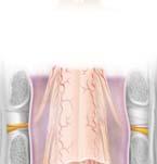
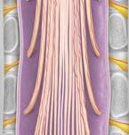
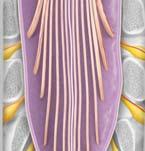
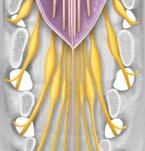
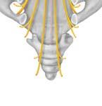
12 _CH01redo.qxd 9/24/07 3:07 PM Page 22 PLATE 1-18 Spinal Cord, Posterior View Portion of the Spinal Cord PLATE 1-19 Occipital bone A. Orientation C1 vertebra (atlas, cut) C2 vertebra (axis, cut) Denticulate C1 spinal nerve Splenius capitis and splenius cervicis muscles (cut and reflected) B. Dissection Serratus posterior superior muscle (cut and reflected) vertebra (cut) vertebra (cut) Dorsal root ganglion spinal nerve C5 pedicle (cut) Dura mater Dura and arachnoid mater (cut and reflected) Arachnoid mater Pia mater on surface of spinal cord Erector spinae muscle: Iliocostalis muscle Longissimus muscle (spinalis muscle removed) Roots of spinal nerves T2 1 Ventral ramus Dorsal median sulcus Serratus posterior inferior muscle (cut and reflected) Conus medullaris Dorsal ramus Dorsal rootlets 2 vertebra (cut) vertebra (cut) spinal nerve Termination of spinal cord at intervertebral disc /L2 Cauda equina Dorsal root of spinal nerve Denticulate Posterior radicular artery vertebra (cut) Filum terminale internum spinal nerve Posterior spinal arteries Ventral root of spinal nerve (cut) Hip bone S1 spinal nerve Termination of dural sac at S2 vertebral level Spinal branches of posterior intercostal artery Dorsal root ganglion S5 spinal nerve T3 pedicle (cut) Filum terminale externum Coccygeal nerve Coccyx PAGE 22 PAGE 23


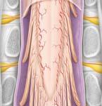


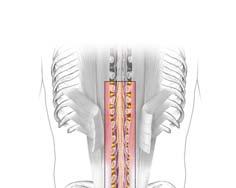

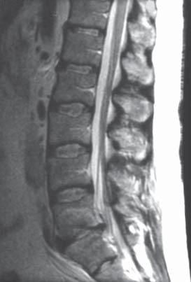
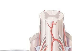

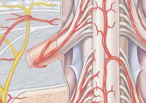
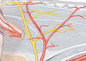
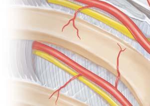
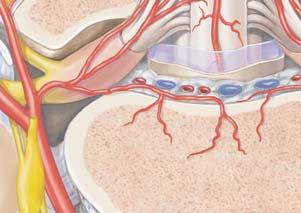

13 _CH01redo.qxd 9/24/07 3:08 PM Page 24 PLATE 1-20 Inferior Portion of the Spinal Cord Blood Supply of the Spinal Cord, Anterior View PLATE 1-21 A. Orientation B. Dissection Thoracic vertebrae Anterior spinal artery Posterior spinal arteries Spinal cord Arachnoid mater Dura mater Dura and arachnoid mater (cut and reflected) Conus medullaris Radicular arteries Lumbar vertebrae Spinal branch Denticulate (cut) Branch to vertebral body and dura mater Arachnoid mater Posterior radicular artery Anterior radicular artery Posterior intercostal artery Cauda equina s (cut) Filum terminale internum Dorsal root ganglia Filum terminale externum C. MRI (T2 weighted), sagittal view Basivertebral veins Conus medullaris Dorsal branch of posterior intercostal artery Intercostal nerves Accessory hemiazygos vein Vertebral bodies (L2 and L3) Cerebrospinal fluid in lumbar cistern Sympathetic trunk and ganglion Posterior intercostal arteries Descending thoracic aorta Thoracic duct Intervertebral disc Cauda equina Accessory hemiazygos vein Azygos vein PAGE 24 PAGE 25


14 _CH01redo.qxd 9/24/07 3:08 PM Page 26 PLATE 1-22 Venous Drainage of the Vertebral Column and Spinal Cord Dermatomes PLATE 1-23 A. Sagittal view A. Anterior view B. Posterior view CN V Internal vertebral venous plexus Cutaway sections of: Epidural fat Dura mater Arachnoid mater Spinal cord Posterior external vertebral venous plexus Anterior external vertebral venous plexus Basivertebral vein vertebral body C8 C5 S2 S3 C2 L2 C3 C4 T2 T3 T4 T5 T6 T7 T8 T C2 C3 C4 C5 C8 T2 T3 T4 T5 T6 T7 T8 T L2 L3 S1 S2 S3 S4 S5 C8 L3 B. view External vertebral venous plexus S1 Basivertebral vein Internal vertebral venous plexus Intervertebral vein Anterior median spinal vein Anterior lateral spinal vein Anterior radicular vein Posterior radicular vein S2 Epidural fat External vertebral venous plexus Posterior lateral spinal vein Posterior median spinal vein S1 PAGE 26 PAGE 27
2 Back and Spinal Cord
 2 Back and Spinal Cord Cards 2-1 to 2-24 Bones and Joints 2-1 Vertebral Column 2-2 Cervical Vertebrae 2-3 Thoracic Vertebrae 2-4 Lumbar Vertebra 2-5 Lumbar Vertebrae 2-6 Vertebral Ligaments: Lumbar Region
1TRUNK: BODY WALL AND SPINE
 TRUNK: BODY WALL AND SPINE SURFACE ANATOMY SKELETON JOINTS & LIGAMENTS MUSCLES VASCULATURE NERVES SPINAL CORD & VERTEBRAL CANAL ANTERIOR BODY WALL & MAMMARY GLAND LATERAL BODY WALL INGUINAL REGION SUPERFICIAL
Muscles of the Upper Limb that are dissected in the Back Region Muscle Origin Insertion Action Innervation Artery Notes
 Muscles of Upper Limb that are dissected in Back Region Muscle Origin Insertion Action Innervation Artery Notes floor of thoraco thoraco inserting spines from intertubercular arm nerve (C7,8) a. tendon
Welcome to the Structure & Development Dissector. Section I
 Welcome to the Structure & Development Dissector The vast majority of questions will be drawn from structures present in the checklist; however, we reserve the right to use a structure or two that is not
The Back. Anatomy RHS 241 Lecture 9 Dr. Einas Al-Eisa
 The Back Anatomy RHS 241 Lecture 9 Dr. Einas Al-Eisa The spine has to meet 2 functions Strength Mobility Stability of the vertebral column is provided by: Deep intrinsic muscles of the back Ligaments
The Back OUTLINE. Vertebral Column (review) Craniovertebral Joints Dorsal Scapular Region(review) Muscles of the Back Suboccipital Region
 Craniovertebral Joints Dorsal Scapular Region(review) Muscles of the Back Suboccipital Region") The Back OUTLINE Vertebral Column (review) Craniovertebral Joints Dorsal Scapular Region(review) Muscles of the Back Suboccipital Region Dept. of Human Anatomy, Si Chuan University Zhou hongying eaglezhyxzy@163.com
The Back OUTLINE Vertebral Column (review) Craniovertebral Joints Dorsal Scapular Region(review) Muscles of the Back Suboccipital Region Dept. of Human Anatomy, Si Chuan University Zhou hongying eaglezhyxzy@163.com
Clarification of Terms
 Clarification of Terms The Spine, Spinal Column, and Vertebral Column are synonymous terms referring to the bony components housing the spinal cord Spinal Cord = made of nervous tissue Facet = a small,
Clarification of Terms
 Clarification of Terms The Spine, Spinal Column, and Vertebral Column are synonymous terms referring to the bony components housing the spinal cord Spinal Cord = made of nervous tissue Facet = a small,
Thoracic and Lumbar Spine Anatomy.
 Thoracic and Lumbar Spine Anatomy www.fisiokinesiterapia.biz Thoracic Vertebrae Bodies Pedicles Laminae Spinous Processes Transverse Processes Inferior & Superior Facets Distinguishing Feature Costal Fovea
Clarification of Terms
 Clarification of Terms The Spine, Spinal Column, and Vertebral Column are synonymous terms referring to the bony components housing the spinal cord Spinal Cord = made of nervous tissue Facet = a small,
Chest cavity, vertebral column and back muscles. Respiratory muscles. Sándor Katz M.D., Ph.D.
 Chest cavity, vertebral column and back muscles. Respiratory muscles. Sándor Katz M.D., Ph.D. Chest cavity - bony structures Chest cavity- bony structures Sternum Ribs True ribs: first seven pairs connect
Thoracolumbar Anatomy Eric Shamus Catherine Patla Objectives
 1 2 Thoracolumbar Anatomy Eric Shamus Catherine Patla Objectives List the muscular and ligamentous attachments of the thoracic and lumbar spine Describe how the muscles affect the spine and upper extremity
Anatomy and Physiology II. Spine
 Anatomy and Physiology II Spine Bones and Other Structures Vertibrae Contains Cervical, Thoracic, Lumbar, Sacral and Coccygeal regions We use Capital letters to refer to these (C, T, L, S, and Co) and
Structure and Function of the Vertebral Column
 Structure and Function of the Vertebral Column Posture Vertebral Alignment Does it really matter? Yes it does! Postural Curves The vertebral column has a series of counterbalancing curves posterior anterior
Copyright 2010 Pearson Education, Inc.
 E. VERTEBRAL COLUMN 1. The vertebral column extends from the skull to the pelvis and forms the vertical axis of the skeleton. 2. The vertebral column is composed of vertebrae that are separated by intervertebral
Main Menu. Trunk and Spinal Column click here. The Power is in Your Hands
 1 The Trunk and Spinal Column click here Main Menu K.9 http://www.handsonlineeducation.com/classes/k9/k9entry.htm[3/27/18, 2:00:55 PM] The Trunk and Spinal Column Vertebral column complex 24 intricate
Copyright 2010 Pearson Education, Inc. Copyright 2010 Pearson Education, Inc. Figure Sectioned spinous process. Interspinous.
 PowerPoint Lecture Slides prepared by Janice Meeking, Mount Royal College C H A P T E R 7 The Skeleton: Part B Vertebral Column Transmits weight of trunk to lower limbs Surrounds and protects spinal cord
Muscle Action Origin Insertion Nerve Innervation Chapter Page. Deltoid. Trapezius. Latissimus Dorsi
 Muscle Action Origin Insertion Nerve Innervation Chapter Page All Fibers Abduct the shoulder (glenohumeral joint) Deltoid Anterior Fibers Flex the shoulder (G/H joint) Horizontally adduct the shoulder
Transitioning to the Suboccipital Triangle. Suboccipital Triangle
 Transitioning to the Suboccipital Triangle Syllabus p. 14-15 Suboccipital Triangle Borders -Rectus capitis posterior major -Obliquus capitis superior -Obliquus capitis inferior Contents -Vertebral artery
The vault bones Frontal Parietals Occiput Temporals Sphenoid Ethmoid
 The Vertebral Column Head, Neck and Spine Bones of the head Some consider the bones of the head in terms of the vault bones and the facial bones hanging off the front of them The vault bones Frontal Parietals
Bony framework of the vertebral column Structure of the vertebral column
 5.1: Vertebral column & back. Overview. Bones o vertebral column. o typical vertebra. o vertebral canal. o spinal nerves. Joints o Intervertebral disc. o Zygapophyseal (facet) joint. Muscles o 2 compartments:
2. The vertebral arch is composed of pedicles (projecting from the body) and laminae (uniting arch posteriorly).
 and laminae (uniting arch posteriorly).") VERTEBRAL COLUMN 2018zillmusom I. VERTEBRAL COLUMN - functions to support weight of body and protect spinal cord while permitting movements of trunk and providing for muscle attachments. A. Typical vertebra
VERTEBRAL COLUMN 2018zillmusom I. VERTEBRAL COLUMN - functions to support weight of body and protect spinal cord while permitting movements of trunk and providing for muscle attachments. A. Typical vertebra
The Biomechanics of the Human Spine. Basic Biomechanics, 6 th edition By Susan J. Hall, Ph.D.
 Chapter 9 The Biomechanics of the Human Spine Structure of the Spine The spine is a curved stack of 33 vertebrae structurally divided into five regions: cervical region - 7 vertebrae thoracic region -
Upper Limb Muscles Muscles of Axilla & Arm
 Done By : Saleh Salahat Upper Limb Muscles Muscles of Axilla & Arm 1) Muscles around the axilla A- Muscles connecting the upper to thoracic wall (4) 1- pectoralis major Origin:- from the medial half of
VERTEBRAL COLUMN VERTEBRAL COLUMN
 VERTEBRAL COLUMN FUNCTIONS: 1) Support weight - transmits weight to pelvis and lower limbs 2) Houses and protects spinal cord - spinal nerves leave cord between vertebrae 3) Permits movements - *clinical
The posterior abdominal wall. Prof. Oluwadiya KS
 The posterior abdominal wall Prof. Oluwadiya KS www.oluwadiya.sitesled.com Posterior Abdominal Wall Lumbar vertebrae and discs. Muscles opsoas, quadratus lumborum, iliacus, transverse, abdominal wall
Overview of the Skeleton: Bone Markings
 Name Overview of the Skeleton: Bone Markings Match the terms in column B with the appropriate description in column A. Column A 1. sharp, slender process* 2. small rounded projection* 3. narrow ridge of
MD Bones & Joints of the Back. A/Prof Chris Briggs Department of Anatomy & Neuroscience
 MD 2017 Bones & Joints of the Back A/Prof Chris Briggs Department of Anatomy & Neuroscience WARNING This material has been provided to you pursuant to section 49 of the Copyright Act 1968 (the Act) for
3 Mohammad Al-Mohtasib Areej Mosleh
 3 Mohammad Al-Mohtasib Areej Mosleh ***Muscles Connecting the Upper Limb to the Vertebral Column 1.Trapezius Muscle ***The first muscle on the back is trapezius muscle, it s called so according
THE THORACIC WALL. Boundaries Posteriorly by the thoracic part of the vertebral column. Anteriorly by the sternum and costal cartilages
 THE THORACIC WALL Boundaries Posteriorly by the thoracic part of the vertebral column Anteriorly by the sternum and costal cartilages Laterally by the ribs and intercostal spaces Superiorly by the suprapleural
THE VERTEBRAL COLUMN. Average adult length: In male: about 70 cms. In female: about 65 cms.
 THE VERTEBRAL COLUMN Average adult length: In male: about 70 cms. In female: about 65 cms. 1 Vertebral Column (Regions and Curvatures) Curvatures of the vertebral column: A. Primary curvature: C-shaped;
Anatomy and Physiology II. Review Spine and Neck
 Anatomy and Physiology II Review Spine and Neck Spine regions How many cervical vertibrae are there? 7 The curvature is the cervical region posterior? Concave posterior How many thoracic? And curvature?
Vertebral Column. Backbone consists of 26 vertebrae. Five vertebral regions. Cervical
 Vertebral Column Backbone consists of 26 vertebrae. Five vertebral regions Cervical vertebrae (7) in the neck. Thoracic vertebrae (12) in the thorax. Lumbar vertebrae (5) in the lower back. Sacrum (5,
THEME 2. VERTEBRAE (GENERAL DATA). CERVICAL, THORACIC AND LUMBAR VERTEBRAE. SACRUM. COCCYX. THE VERTEBRAL COLUMN AS A WHOLE
. CERVICAL, THORACIC AND LUMBAR VERTEBRAE. SACRUM. COCCYX. THE VERTEBRAL COLUMN AS A WHOLE") THEME 2. VERTEBRAE (GENERAL DATA). CERVICAL, THORACIC AND LUMBAR VERTEBRAE. SACRUM. COCCYX. THE VERTEBRAL COLUMN AS A WHOLE Osteology of the Vertebral Column Bone Description vertebra Notes a vertebra
THEME 2. VERTEBRAE (GENERAL DATA). CERVICAL, THORACIC AND LUMBAR VERTEBRAE. SACRUM. COCCYX. THE VERTEBRAL COLUMN AS A WHOLE Osteology of the Vertebral Column Bone Description vertebra Notes a vertebra
Anatomy of the Shoulder Girdle. Prof Oluwadiya Kehinde FMCS (Orthop)
") Anatomy of the Shoulder Girdle Prof Oluwadiya Kehinde FMCS (Orthop) www.oluwadiya.com Bony Anatomy Shoulder Complex: Sternum(manubrium) Clavicle Scapula Proximal humerus Manubrium Sterni Upper part of
Anatomy of the Shoulder Girdle Prof Oluwadiya Kehinde FMCS (Orthop) www.oluwadiya.com Bony Anatomy Shoulder Complex: Sternum(manubrium) Clavicle Scapula Proximal humerus Manubrium Sterni Upper part of
Axial Skeleton: Vertebrae and Thorax
 Axial Skeleton: Vertebrae and Thorax Function of the vertebral column (spine or backbone): 1) 2) 3) Composition of Vertebral column The vertebral column is formed by 33 individual vertebrae (some of which
Lab Activity 11: Group I
 Lab Activity 11: Group I Muscles Martini Chapter 11 Portland Community College BI 231 Origin and Insertion Origin: The place where the fixed end attaches to a bone, cartilage, or connective tissue. Insertion:
Spine & Upper Extremity. Chapter 2: Spine & Upper Extremity
 Chapter 2: Spine & Upper Extremity Spine I. Osteology A. Overview Consists of stacks of vertebrae forming the vertebral column Five sections: Cervical: 7 vertebrae Thoracic: 12 vertebrae Lumbar: 5 vertebrae
Lamina: meet the pedicles Superior & inferior articular processes: former above pedicle, latter extends from spinous process.
 Spinal cord (anatomy and internal composition), back, vertebrae Vertebral Column (Moore pp 432, Netter Plate 142) The vertebral column can be divided into individual parts namely: cervical (7), thoracic
VERTEBRAL COLUMN ANATOMY IN CNS COURSE
 VERTEBRAL COLUMN ANATOMY IN CNS COURSE Vertebral body Sections of the spine Atlas (C1) Axis (C2) What type of joint is formed between atlas and axis? Pivot joint What name is given to a fracture of both
thoracic cage inlet and outlet landmarks of the anterior chest wall muscles of the thoracic wall sternum joints ribs intercostal spaces diaphragm
 Thoracic Wall Lecture Objectives Describe the shape and outline of the thoracic cage including inlet and outlet. Describe the anatomical landmarks of the anterior chest wall. List various structures making
Biology 218 Human Anatomy. Adapted from Martini Human Anatomy 7th ed. Chapter 12 Surface Anatomy and Cross-Sectional Anatomy
 Adapted from Martini Human Anatomy 7th ed. Chapter 12 Surface Anatomy and Introduction Surface anatomy is the study of anatomical landmarks on the exterior of the human body Knowledge of surface anatomy
The Thoracic wall including the diaphragm. Prof Oluwadiya KS
 The Thoracic wall including the diaphragm Prof Oluwadiya KS www.oluwadiya.com Components of the thoracic wall Skin Superficial fascia Chest wall muscles (see upper limb slides) Skeletal framework Intercostal
Chapter 14. The Nervous System. The Spinal Cord and Spinal Nerves. Lecture Presentation by Steven Bassett Southeast Community College
 Chapter 14 The Nervous System The Spinal Cord and Spinal Nerves Lecture Presentation by Steven Bassett Southeast Community College Introduction The Central Nervous System (CNS) consists of: The spinal
Upper limb Arm & Cubital region 黃敏銓
 Upper limb Arm & Cubital region 黃敏銓 1 Arm Lateral intermuscular septum Anterior (flexor) compartment: stronger Medial intermuscular septum Posterior (extensor) compartment 2 Coracobrachialis Origin: coracoid
STRUCTURAL BASIS OF MEDICAL PRACTICE EXAMINATION 5. September 30, 2011
 STRUCTURAL BASIS OF MEDICAL PRACTICE EXAMINATION 5 September 30, 2011 PART l. Answer in the space provided. (12 pts) 1. Identify the structures. (2 pts) EXAM NUMBER A. Suprascapular nerve B. Axillary nerve
Anatomy - Reconnect with your Spine Muscles by NFPT Idea World 2016 : Session 449 Friday July 15th 9:40-11:30am Beverly Hosford, MA
 Anatomy - Reconnect with your Spine Muscles by NFPT Idea World 2016 : Session 449 Friday July 15th 9:40-11:30am Beverly Hosford, MA Posture Core Anatomy Awareness Action 1. Anatomy *Know the muscle attachments.
Ligaments of the vertebral column:
 In the last lecture we started talking about the joints in the vertebral column, and we said that there are two types of joints between adjacent vertebrae: 1. Between the bodies of the vertebrae; which
Human Anatomy Biology 351
 nnnnn 1 Human Anatomy Biology 351 Exam #2 Please place your name on the back of the last page of this exam. You must answer all questions on this exam. Because statistics demonstrate that, on average,
THE BACK THE SPINAL CORD
 THE BACK THE SPINAL CORD The structures in the vertebral canal: the spinal cord spinal nerve roots spinal meninges the neurovascular structures THE SPINAL CORD The spinal cord occupies the superior 2/3
ANATOMY OF SPINAL CORD. Khaleel Alyahya, PhD, MEd King Saud University School of
 ANATOMY OF SPINAL CORD Khaleel Alyahya, PhD, MEd King Saud University School of Medicine @khaleelya OBJECTIVES At the end of the lecture, students should be able to: Describe the external anatomy of the
Gross Anatomy Faculty: Gross Anatomy Faculty: Gross Anatomy Faculty: Dr. Melissa McGinn. Welcome to Gross and Developmental Anatomy
 Welcome to Gross and Developmental Anatomy M1 Anatomy Gross Anatomy Faculty: Dr. Richard Krieg Dr. Milton Sholley Dr. David Simpson 1 2 Gross Anatomy Faculty: Gross Anatomy Faculty: Dr. Steve Gudas Dr.
Posterior Triangle of the Neck By Prof. Dr. Muhammad Imran Qureshi
 Posterior Triangle of the Neck By Prof. Dr. Muhammad Imran Qureshi For the purpose of anatomical description the neck is sub divided into two major triangles, the Anterior and the Posterior by muscle bellies
Sports Medicine Part II : ANATOMY OF THE SPINE, ABDOMEN AND SHOULDER COMPLEX
 Sports Medicine 25 1.1 Part II : ANATOMY OF THE SPINE, ABDOMEN AND SHOULDER COMPLEX c.w.p. Wagner High School, Sports Medicine, A. Morgan, T. Morgan & A. Eastlake, 2008 Muscles of the Upper Limbs In this
STERNUM. Lies in the midline of the anterior chest wall It is a flat bone Divides into three parts:
 STERNUM Lies in the midline of the anterior chest wall It is a flat bone Divides into three parts: 1-Manubrium sterni 2-Body of the sternum 3- Xiphoid process The body of the sternum articulates above
Scapula Spine Lateral edge of clavicle. Medial border Scapula. Medial border of Scapula, between superior angle and root of spine. Scapula.
 Muscle attachments and actions answer sheet Muscle Origins insertions Movements Joints crossed Trapezius Base of skull Spinous process of C7 Thoracic Spine Lateral edge of clavicle Elevation Retraction
The Trunk and Spinal Column Kinesiology Cuneyt Mirzanli Istanbul Gelisim University
 The Trunk and Spinal Column Kinesiology Cuneyt Mirzanli Istanbul Gelisim University The Trunk and Spinal Column Vertebral column 24 articulating vertebrae 31 pairs of spinal nerves Abdominal muscles some
Cranium Facial bones. Sternum Rib
 Figure 7.1 The human skeleton. Skull Thoracic cage (ribs and sternum) Cranium Facial bones Sternum Rib Bones of pectoral girdle Vertebral column Sacrum Vertebra Bones of pelvic girdle (a) Anterior view
Chapter 7: Skeletal System: Gross Anatomy
 Chapter 7: Skeletal System: Gross Anatomy I. General Considerations A. How many bones in an average adult skeleton? B. Anatomic features of bones are based on II. Axial Skeleton A. Skull 1. Functionally
Anatomy of the Nervous System. Brain Components
 Anatomy of the Nervous System Brain Components NERVOUS SYSTEM INTRODUCTION Is the master system of human body, controlling the functions of rest of the body systems Nervous System CLASSIFICATION A. Anatomical
Spinal nerves and cervical plexus Prof. Abdulameer Al Nuaimi. E mail: a.al E. mail:
 Spinal nerves and cervical plexus Prof. Abdulameer Al Nuaimi E mail: a.al nuaimi@sheffield.ac.uk E. mail: abdulameerh@yahoo.com Branches of ophthalmic artery Muscles of face A spinal nerve Spinal
Human Anatomy. Spinal Cord and Spinal Nerves
 Human Anatomy Spinal Cord and Spinal Nerves 1 The Spinal Cord Link between the brain and the body. Exhibits some functional independence from the brain. The spinal cord and spinal nerves serve two functions:
The trunk and spinal column. Functions of Spine. Bones 6/5/2017. Chapter 10. Consider the complexity of functions. 33 bones of the spine
 The trunk and spinal column Chapter 10 Functions of Spine Consider the complexity of functions provides stability to a cylinder permits movement in all directions supports structures of considerable weight
Anatomy & Physiology II. Trunk
 Anatomy & Physiology II Trunk Bones and Landmarks of the Vertebral column 24 vertebrae Sacrum - consists of 5 vertebrae that fuse into one bone Median sacral crest Sacral hiatus 4 sacral foramina Coccyx
Prime movers provide the major force for producing a specific movement Antagonists oppose or reverse a particular movement Synergists
 Dr. Gary Mumaugh Prime movers provide the major force for producing a specific movement Antagonists oppose or reverse a particular movement Synergists Add force to a movement Reduce undesirable or unnecessary
bio4165 lab quiz 1 Posterior View Anterior View Lateral View Anterior View bio fall.quarter lab.quiz.1...page.1 of 6
 B A Posterior View D C E Lateral View bio.4165...fall.quarter.2005...lab.quiz.1...page.1 of 6 F I G 35 Posterior View H bio.4165...fall.quarter.2005...lab.quiz.1...page.2 of 6 J Posterior View L K Inferior
3 Movements of the Trunk. Flexion Rotation Extension
 3 Movements of the Trunk Flexion Rotation Extension 1 TRUNK FLEXION 2 TRUNK FLEXION: Rectus Abdominalis O: Crest of Pubis & ligaments covering front of symphysis pubis. I: By «3 portions into cartilages
CHAPTER 9: THE SPINAL COLUMN AND THORAX KINESIOLOGY Scientific Basis of Human Motion, 12 th edition Hamilton, Weimar & Luttgens
 CHAPTER 9: THE SPINAL COLUMN AND THORAX KINESIOLOGY Scientific Basis of Human Motion, 12 th edition Hamilton, Weimar & Luttgens Presentation Created by TK Koesterer, Ph.D., ATC Humboldt State University
STRUCTURAL BASIS OF MEDICAL PRACTICE EXAMINATION 5 October 6, 2006
 STRUCTURAL BASIS OF MEDICAL PRACTICE EXAMINATION 5 October 6, 2006 PART l. Answer in the space provided. (8 pts) 1. Identify the structures. (2 pts) B C A. _pisiform B. _ulnar artery A C. _flexor carpi
213: HUMAN FUNCTIONAL ANATOMY: PRACTICAL CLASS 1: Proximal bones, plexuses and patterns
 213: HUMAN FUNCTIONAL ANATOMY: PRACTICAL CLASS 1: Proximal bones, plexuses and patterns CLAVICLE Examine an isolated clavicle and compare it with a clavicle on an articulated skeleton. Viewed from above,
The Thoracic Cage ANATOMY 2: THORACIC CAGE AND VERTEBRAL COLUMN
 ANATOMY 2: THORACIC CAGE AND VERTEBRAL COLUMN PSK 4U Mr. S. Kelly North Grenville DHS The Thoracic Cage 7 true ribs 3 false ribs 2 floating ribs Clavicle = collarbone Manubrium Sternum Xiphoid Process
THE DEVELOPMENT OF THE VERTEBRA AND THE INTERVERTEBRAL DISC
 THE DEVELOPMENT OF THE VERTEBRA AND THE INTERVERTEBRAL DISC Precartilage Stage (mesenchimal stage) The vertebral column develops from the mesenchimal cells that accumulated around the notochord during
Muscles in the Shoulder, Chest, Arm, Stomach, and Back
 Muscles in the Shoulder, Chest, Arm, Stomach, and Back Shoulder Muscles Deltoid Supraspinatus Infraspinatus Teres Major Teres Minor Subscapularis Deltoid (Delts) Function: Raises the upper arm Origin:
locomotice system Plastinated specimensⅠ: Silicone specimens Regional specimens and organs
 locomotice system Plastinated specimensⅠ: Silicone specimens Regional specimens and organs Art-No. Name Description The locomotor system SL001 Two hundred pieces of plastinated bones (without six The bones
Note : I put the sheet's info within the slides to easily understand this lecture Done by : Zaid Al-Ghnaneem
 Note : I put the sheet's info within the slides to easily understand this lecture Done by : Zaid Al-Ghnaneem Thoracic Wall Lecture Objectives Describe the shape and outline of the thoracic cage including
Gross Morphology of Spinal Cord
 Gross Morphology of Spinal Cord Done By : Rahmeh Alsukkar ** I did my best and sorry for any mistake ** the sheet does not contain pictures, tables and some slides so please be careful and go back to slides
Upper limb Pectoral region & Axilla
 Upper limb Pectoral region & Axilla 黃敏銓 mchuang@ntu.edu.tw 1 Pectoral region Intercostal nerve Anterior branch of lateral cutaneous branch Lateral cutaneous branch Anterior cutaneous branch Anterior cutaneous
Note: Please refer to handout Spinal Plexuses and Representative Spinal Nerves for
 Chapter 13 Outline Note: Please refer to handout Spinal Plexuses and Representative Spinal Nerves for what you need to know from Exhibits 13.1 13.4 I. INTRODUCTION A. The spinal cord and spinal nerves
Workshop on thoracic diagnostic procedures
 Workshop on thoracic diagnostic procedures SAMM / U. Böhni Kyphosis is in correlation with lordosis of lumbar and cervicle spine 20-40 (Hefti et al. 1997) Extrinsic dorsal muscles superficial layer a.
Gross Morphology of Spinal Cord
 Gross Morphology of Spinal Cord Lecture Objectives Describe the gross anatomical features of the spinal cord. Describe the level of the different spinal segments compared to the level of their respective
Anatomy of the Thorax
 Anatomy of the Thorax A) THE THORACIC WALL Boundaries Posteriorly by the thoracic part of the vertebral column Anteriorly by the sternum and costal cartilages Laterally by the ribs and intercostal spaces
Morphological Classification of the Muscular Tubercles of the Vertebrae
 Okajimas Folia Anat. Jpn., 58(4-6) : 1167-1186, March 1982 Morphological Classification of the Muscular Tubercles of the Vertebrae By TATSUO SATO and SUSUMU NAKAZAWA Department of Anatomy, School of Medicine,
Fig Cervical spinal nerves. Cervical enlargement C7. Dural sheath. Subarachnoid space. Thoracic. Spinal cord Vertebra (cut) spinal nerves
 spinal nerves") Fig. 13.1 C1 Cervical enlargement C7 Cervical spinal nerves Dural sheath Subarachnoid space Thoracic spinal nerves Spinal cord Vertebra (cut) Lumbar enlargement Medullary cone T12 Spinal nerve Spinal nerve
Fig. 13.1 C1 Cervical enlargement C7 Cervical spinal nerves Dural sheath Subarachnoid space Thoracic spinal nerves Spinal cord Vertebra (cut) Lumbar enlargement Medullary cone T12 Spinal nerve Spinal nerve
The Thoracic Cage. Role of the Thoracic Cage 2/13/2019. Anatomy 2: Thoracic Cage and Vertebral Column
 PSK 4U Mr. S. Kelly North Grenville DHS Anatomy 2: Thoracic Cage and Column The Thoracic Cage 7 true ribs 3 false ribs 2 floating ribs Clavicle = collarbone Manubrium Sternum Xiphoid Process 12 thoracic
Region of upper limb attachment to the trunk Proximal segment of limb overlaps parts of the trunk (thorax and back) and lower lateral neck.
 and lower lateral neck.") Region of upper limb attachment to the trunk Proximal segment of limb overlaps parts of the trunk (thorax and back) and lower lateral neck. includes Pectoral Scapular Deltoid regions of the upper limb
Region of upper limb attachment to the trunk Proximal segment of limb overlaps parts of the trunk (thorax and back) and lower lateral neck. includes Pectoral Scapular Deltoid regions of the upper limb
The Anatomy Coloring Book Wynn Kapit Lawrence M. Elson Fourth Edition
 The Anatomy Coloring Book Wynn Kapit Lawrence M. Elson Fourth Edition Pearson Education Limited Edinburgh Gate Harlow Essex CM20 2JE England and Associated Companies throughout the world Visit us on the
Chapter 28: The neck. Fascia of the neck
 Chapter 28: The neck Fascia of the neck The superficial fascia is a fatty areolar layer between the skin and the more obvious deep fascia. It contains the platysma muscles and the external jugular veins
Thoracic Cooled-RF Training Presentation
 Thoracic Cooled-RF Training Presentation Patient Selection Anatomy Overview Neuroanatomy Lesion targets Technique Diagnostic Block Cooled-RF Precautions Summary Appendix AGENDA Patient Selection Thoracic
Biology 2401 The Skeletal System
 Biology 2401 The Skeletal System Purpose: The lab will describe the microscopic and gross anatomy of bone, identify bones of the body, and identify important bone markings. I. Overview of the Skeleton
Classification of the nervous system. Prof. Dr. Nikolai Lazarov 2
 1 1. Formation and general organization 2. Spinal ganglia 3. Zonal and segmental innervation 4. Dorsal rami of the spinal nerves 5. Ventral rami of the spinal nerves 6. Cervical plexus Classification of
LEVEL II MUSCLE CHART NB: Needle length varies with tissue depth, this chart acts as a guide only. Side lye or prone.25 x 30-50mm Inferior to ilium
 LUMBAR SPINE LEVEL II MUSCLE CHART NB: Needle length varies with tissue depth, this chart acts as a guide only Muscle/ Innervation Comments Position Quadratus Lumborum T12-L3/4 segmentally PSIS Comments.
Chapter 7 Part B The Skeleton
 Chapter 7 Part B The Skeleton 7.2 The Vertebral Column General Characteristics Extends from skull to pelvis Also called spine or spinal column Functions to transmit weight of trunk to lower limbs, surround
TEST YOURSELF- Chapter 7
 TEST YOURSELF- Chapter 7 Cranial Bones 1. Give the name of the bone for each of the following markings. Some of the markings are found on more than one bone. List all that apply. Cranium a. Frontal squama:
MEDICAL IMAGING OF THE VERTEBRAE
 MEDICAL IMAGING OF THE VERTEBRAE Vertebrae are your friends Matthew Harper MS-IV LECTURE OBJECTIVES INTRODUCE THE MOST COMMON MODALITIES OF MEDICAL IMAGING AND BASIC TECHNIQUES FOR READING THESE IMAGES
SKELETAL MUSCLES. Objectives
 Objectives Body Wall Muscles Remember the pattern Hypaxial Muscle Overview Cervical body wall muscles Hypaxial Muscle Overview Thoracic body wall muscles Hypaxial Muscle Overview Abdominal body wall muscles
Dr. Weyrich G07: Superior and Posterior Mediastina. Reading: 1. Gray s Anatomy for Students, chapter 3
 Dr. Weyrich G07: Superior and Posterior Mediastina Reading: 1. Gray s Anatomy for Students, chapter 3 Objectives: 1. Subdivisions of mediastinum 2. Structures in Superior mediastinum 3. Structures in Posterior
Synapse Homework. Back page last question not counted. 4 pts total, each question worth 0.18pts. 26/34 students answered correctly!
 Synapse Homework Back page last question not counted 26/34 students answered correctly! 4 pts total, each question worth 0.18pts Business TASS hours extended! MWF 1-2pm, Willamette 204 T and Th 9:30-10:30am,
Axial skeleton bones and markings
 Axial skeleton bones and markings Skull Cranial bones Frontal x 1 Supraorbital foramen Occipital x 1 Foramen magnum Occipital condyles Superior nuchal line Inferior nuchal line Anterior cranial fossa External
Biology 218 Human Anatomy. Adapted from Martini Human Anatomy 7th ed. Chapter 10 The Muscular System Axial Musculature
 Adapted from Martini Human Anatomy 7th ed. Chapter 10 The Muscular System Axial Musculature Introduction The skeletal muscle of the body can be subdivided into: Axial musculature Muscles that position
human anatomy 2015 lecture four Dr meethak ali ahmed neurosurgeon
 The Vertebral Column the vertebral columnis central pillar of the body.it serve to protect the spinal cord and support the weight of the head trunk, which it transmits to the hip bones & the lower limbs.