ELBOW MRI BASICS BONES/CARTILAGE
|
|
|
- Laurel Hart
- 6 years ago
- Views:
Transcription
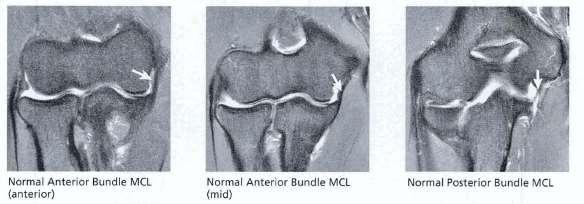
1 ELBOW MRI BASICS supine vs prone (superman) imaging Coronal for collateral lig and bones Sagittal for biceps/triceps tendons and cartilage Axial for muscles and nerves FABS (flexed elbow, abducted shoulder, and supinated forearm) axial/sag PD for biceps Arthrogram (do for OCD, loose bodies, undersurface tears of radial/ulnar collateral lig) BONES/CARTILAGE -Capitellar pseudodefect (posterior non-articular capitellum has abrupt notch-like slope on coronal image) -Bare area trochlear cortical notch or groove (devoid of cartilage) and transverse trochlear ridge of olecranon seen on sagittal -Radiocapitellar joint -Ulnohumeral joint (ulnar coronoidtrochlea; olecranon process) -Prox RUJ (radial head rotates w/in sigmoid notch of ulna for pronation/supination) Panner s (5-12yo; fragmented/mottled capitellar epiphysis w/ patchy low signal T1 w/ abnl contours & no loose body; resolve upon F/U w/ no residual deformity) OCL (12-16yo; anterior convex aspect of capitellum in dominant hand of thrower due to chronic microtrauma assess for stability of fragment, loose body, and overlying hyaline cartilage integrity; F/U shows residual deformity) unstable if surrounding T2 signal/cyst deep to fragment/t2 edema in fragment (do arthrogram) -MIMIC=pseudodefect posterior non-articular capitellum has abrupt notch-like slope on coronal image (but no edema) Epicondylitis (adults; medial>lateral; implies partial tear/tendinopathy); Apophysitis (kids; widening of physis on plain film c/w SH-I fx; Little league elbow) Stress fx (middle third of olecranon) Posterior dislocation (assoc w/ tear of radial collateral lig and common extensor tendon) LIGAMENTS (coronal) -Radial (lateral) collateral ligaments ( tennis elbow ; generally not torn unless posterior dislocation vs after radial head resection vs after common extensor tendon or tennis elbow release) RESIST VARUS STRESS RCL (radial collateral lig; coronal; triangular; arises from anterior aspect of epicondyle and inserts onto annular lig & fascia of supinator muscle; may be hard to distinguish from overlying common extensor tendon unless joint effusion or arthrogram) LUCL (lateral ulnar collateral lig; coronal; most important posterolateral stabilizer; located posterior and superficial; oriented obliquely; inserts onto crista supinatoris of ulna; absent in 10%; tear leads to posterolateral rotatory insufficiencytransient rotatory sublux ulnohumeral jt and secondary sublux/disloc of radiohumeral jt) Annular ligament (axial; arises and inserts on sigmoid notch of ulna; primary stabilizer of prox RUJ) Evaluate integrity of common extensor tendon (superficial to radial collateral lig) Lateral synovial fringe (radiocapitellar meniscus; seen on coronal) Tennis elbow (radial collateral lig injury + common extensor tendon injury esp partial avulsion of extensor carpi radialis brevis + lat epicondylitis) MIMIC=radial nerve entrapment -Ulnar (medial) collateral ligaments more commonly injured (even though lateral epicondylitis more common) RESIST VALGUS STRESS Anterior bundle UCL (thicker and most imp; coronal; prox flared and distal tapered; from epicondyle to sublime tubercle of coronoid process which can avulse off; should be taut; normal to have sl signal w/in prox flared portion; midsubstance tear> distal>proximal; chronic degen or remodelling if thickened +/- dystrophic calc; partial-thickness deep undersurface tears very difficult w/o gadsubtle fluid beneath distal extent of bundle separating lig from bone T-sign on coronal c/w partial tear or stripping) tears treated surgically only in elite athletes aka Tommy John s surgery (docking technique w/ autograft) Other 2 bundles not well seen (together form floor of cubital tunnel) -Posterior bundle (fan-shaped; from epicondyle to olecranon; best seen when elbow flexed at 90deg) -Transverse bundle (least imp; horizontal from olecranon to coranoid; join inf aspects of anterior and posterior bundles) Evaluate integrity of common flexor tendon Golfer s elbow (ulnar collateral lig injury + common flexor tendon injury + medial epicondylitis; Little league elbow =avulsion of medial epicondylar apophysis) Valgus instability (etiologies include: medial epicoylopathy; ulnar neuropathy; posteromedial olecranon impingement +/- loose bodies; radiocapitellar overload w/ OCD) MUSCLES (axial and sagittal)
2 Anterior compartment -Biceps brachii tendon (short head inserts distally on radial tuberosity with larger footprint while long head inserts slightly proximally with smaller footprint; no synovial lining; bicipital aponeurosis aka lacertus fibrosus keeps it in place at antecubital fossa; distal tendon covered by extrasynovial partenon; radiobicipital bursa underneath it at radial tuberosity normally not visualized; partial tear less common than complete tear; injury occurs with abrupt overloading of muscle at midlfexion; complete tear at radial tuberositylook for popeye prox retraction and integrity of aponeurosis; may not retract unless aponeurosis is torn) mimickers of biceps tear include biceps tendonitis vs radiobicipital bursitis (usually seen with partial tear) vs lateral antebrachial cutaneous nerve entrapment -Brachialis (deep to biceps; inserts on ulnar tuberosity; tendon surrounded by muscle so not commonly injured; tendonitis/sprain aka climber s elbow ) Posterior compartment -Triceps brachii (broad insertion on prox olecranon; ok to have high T1/T2 signal striated signal at insertion; tendon rupture is least common; may have degeneration if associated olecranon bursitis; dialysis/lupus/hpt predispose to rupture; laxity of tendon ok if elbow fully extended) -Anconeus (triangular/curvilinear; arises from posterior aspect of lateral epicondyle and inserts laterally on olecranon; helps identify lateral from medial elbow on axial) Medial compartment (more commonly injured than lateral compartment even though lateral epicondylitis more common) -Common flexor tendon (conjoined tendon insert on medial epicondyle; disruption more common than common extensor tendon; golfers or pitchers elbow) -Flexors of wrist/hand (deepest; flexor carpi radialis, flexor carpi ulnaris, flexor digitorum superficialis) -Palmaris longus -Pronator teres (most superficial) Lateral compartment -Common extensor tendon (conjoined tendon inserts on lateral epicondyle) -Extensors of wrist/hand (extensor carpi radialis brevis=most common culprit in lateral epicondylopathy, extensor digitorum, extensor digiti minimi, and extensor carpi ulnaris originate for lateral epicondyle however, extensor carpi radialis longus originates from lower supracondylar ridge of humerus ) -Supinator (wraps around radial neck) -Brachioradialis (big muscle anterolateral; arises from upper supracondylar ridge of humerus) NERVES (axial T1 and T2) -Neuropathy (nerve=increased T2 signal, thickened nerve, indistinct fascicles, fluid around nerve; muscle=acute neurogenic edema, late fatty infiltration and muscle atrophy) Ulnar nerve (injury more common than other nerves; located posterior to medial epicondyle) -Cubital tunnel (floor=post/transverse bundles of ulnar collateral lig; roof= proximally by CT retinaculum aka osborne lig extending from olecranon to medial epicondyle but it may be absent/incomplete and distally by flexor carpi ulnaris aponeurosis aka arcuate lig) -Cubital tunnel syndrome=paraesthesia ring and little finger (thickened CT retinaculum; thickened ulnar collateral lig; bone spur at medial epicondyle; anomalous anconeus epitrochlearis muscle replacing CT retinaculum; anomalous lig of spinner; absent CT retinaculum resulting in subluxation/friction; thickened CT retinaculum; pressure from wheelchair or OR table compression; masses)travels thru guyon s canal in wrist -Look for surrounding increased T2 signal with enlarged or flattened or irregular nerve (may have magic angle on PD imaging) Median nerve (not important) -Best seen on prone imaging; located over brachialis muscle and deep to bicipital aponeurosis of biceps tendonthen passes btwn ulnar and humeral heads of pronator teresthen gives rise to anterior interosseous nerve branch (motor only) at inferior margin of pronator teres -Pronator syndrome (dynamic compression btwn two heads of pronator teres muscle) -Median neuropathy (pronator syndrome most common; other etiology includes thickened bicipital aponeurosis, biceps tears, radiobicipital bursitis, supracondylar process of humerus) -Anterior interosseous syndrome (aka Kiloh-Nevin syndrome; rare; anterior interosseous nerve is motor branch of median nerve; cant flex distal joint of thumb/index, look for classic muscle edema of flexor pollicis longus, pronator quadratus, and a part of flexor digitorum profundus) Radial nerve -Located btwn brachialis and brachioradialis muscle (anterior to lateral epicondyle); divides at elbow joint near capitellum into superficial radial nerve (sensory; superficial to supinator muscle; follows course of radial artery) and posterior interosseous nerve (motor; penetrates btwn deep and superficial fibers of supinator muscle to course along interosseous membrane); arcade of Frohse is fibrous arch btwn brachialis and brachioradialis seen in 35-50% -Radial neuropathy (proximal to elbow joint; secondary to trauma, crutches, tourniquet etc; increased signal) -Radial tunnel syndrome or Posterior interosseous syndrome (aka supinator syndrome or deep radial nerve syndrome; at elbow joint; motor only; secondary to thickened arcade of Frohse, fx/disloc of prox radius, abnormal recurrent vessel along interosseous membrane at elbow joint; increased signal seen w/in posterior compartment muscles w/ edema/fatty atrophy of deep fibers of supinator muscle) MISCELLANEOUS -Effusion -Loose bodies (axial and sagittal views esp GRE; look for donor site and synovitis; may be secondary to OCD vs acute trauma vs posteromedial olecranon impingement in throwing athletes seen within post olecranon fossa) -Bursitis -bicipitoradial bursa (BRB)=deep to biceps tendon (r/o radial artery aneurysm) AND interosseous bursa (IOB)=superficial to biceps tendon (both located in AC region on elbow; both communicate w/ each other and can affect median nerve or posterior interosseous branch of radial nerve) -olecranon bursa (may be seen in Gout) -Epitrochlear adenopathy (cat-scratch disease; Bartonella henselae) -Steroid injection for epicondylitis (peritendinous approach); PRP injection (intratendinous approach)


3


4


5


6

7

8

9
The Elbow and the cubital fossa. Prof Oluwadiya Kehinde
 The Elbow and the cubital fossa Prof Oluwadiya Kehinde www.oluwadiya.com Elbow and Forearm Anatomy The elbow joint is formed by the humerus, radius, and the ulna Bony anatomy of the elbow Distal Humerus
Functional Anatomy of the Elbow
 Functional Anatomy of the Elbow Orthopedic Institute Daryl C. Osbahr, M.D. Chief of Sports Medicine, Orlando Health Chief Medical Officer, Orlando City Soccer Club Orthopedic Consultant, Washington Nationals
Elbow Elbow Anatomy. Flexion extension. Pronation Supination. Anatomy. Anatomy. Romina Astifidis, MS., PT., CHT
 Elbow Elbow Anatomy Romina Astifidis, MS., PT., CHT Curtis National Hand Center Baltimore, MD October 6-8, 2017 Link between the arm and forearm to position the hand in space Not just a hinge Elbow = 70%
MUSCLES OF THE ELBOW REGION
 MUSCLES OF THE ELBOW REGION Dr Bronwen Ackermann COMMONWEALTH OF AUSTRALIA Copyright Regulation WARNING This material has been reproduced and communicated to you by or on behalf of the University of Sydney
ARM Brachium Musculature
 ARM Brachium Musculature Coracobrachialis coracoid process of the scapula medial shaft of the humerus at about its middle 1. flexes the humerus 2. assists to adduct the humerus Blood: muscular branches
Lecture 9: Forearm bones and muscles
 Lecture 9: Forearm bones and muscles Remember, the region between the shoulder and the elbow = brachium/arm, between elbow and wrist = antebrachium/forearm. Forearm bones : Humerus (distal ends) Radius
Lab Activity 11: Group II
 Lab Activity 11: Group II Muscles Martini Chapter 11 Portland Community College BI 231 Origin and Insertion Origin: The place where the fixed end attaches to a bone, cartilage, or connective tissue. Insertion:
Levels of the anatomical cuts of the upper extremity RADIUS AND ULNA right
 11 CHAPTER 2 Levels of the anatomical cuts of the upper extremity AND right CUT 1 CUT 4 1 2 3 4 5 6 Isolated fixation of the radius is difficult at this level because of the anterolateral vessels and the
MCQWeek2. All arise from the common flexor origin. The posterior aspect of the medial epicondyle is the common flexor origin.
 MCQWeek2. 1. Regarding superficial muscles of anterior compartment of the forearm: All arise from the common flexor origin. The posterior aspect of the medial epicondyle is the common flexor origin. Flexor
I (and/or my co-authors) have something to disclose.
 have something to disclose.") Elbow Anatomy And Biomechanics Nikhil N Verma, MD Director, Division of Sports Medicine Professor, Department of Orthopedics Rush University Medical Center Team Physician, Chicago White Sox and Bulls I
Elbow Anatomy And Biomechanics Nikhil N Verma, MD Director, Division of Sports Medicine Professor, Department of Orthopedics Rush University Medical Center Team Physician, Chicago White Sox and Bulls I
region of the upper limb between the shoulder and the elbow Superiorly communicates with the axilla.
 1 region of the upper limb between the shoulder and the elbow Superiorly communicates with the axilla. Inferiorly, a number of important structures pass between arm & forearm through cubital fossa. 2 medial
Elbow. Chapter 2 LISTEN. Mechanism of Injury (If Applicable) Pain
 Pain") Chapter 2 Elbow LISTEN Mechanism of Injury (If Applicable) Patient usually remembers their position at the time of injury Certain mechanisms of injury result in characteristic patterns Fall on outstretched
Chapter 2 Elbow LISTEN Mechanism of Injury (If Applicable) Patient usually remembers their position at the time of injury Certain mechanisms of injury result in characteristic patterns Fall on outstretched
Fascial Compartments of the Upper Arm
 Fascial Compartments of the Upper Arm The upper arm is enclosed in a sheath of deep fascia and has two fascial septa: 1- Medial fascial septum (medial intermuscular septum): attached to the medial supracondylar
The Elbow and Radioulnar Joints Kinesiology. Dr Cüneyt Mirzanli Istanbul Gelisim University
 The Elbow and Radioulnar Joints Kinesiology Dr Cüneyt Mirzanli Istanbul Gelisim University 1 The Elbow & Radioulnar Joints Most upper extremity movements involve the elbow & radioulnar joints. Usually
divided by the bones ( redius and ulna ) and interosseous membrane into :
 and interosseous membrane into :") fossa Cubital Has: * floor. * roof : - Skin - superficial fasica - deep fascia ( include bicipital aponeurosis ) Structures within the roof : -cephalic and basilic veins -and between them median cubital
fossa Cubital Has: * floor. * roof : - Skin - superficial fasica - deep fascia ( include bicipital aponeurosis ) Structures within the roof : -cephalic and basilic veins -and between them median cubital
Key Relationships in the Upper Limb
 Key Relationships in the Upper Limb This list contains some of the key relationships that will help you identify structures in the lab. They are organized by dissection assignment as defined in the syllabus.
Ligaments of Elbow hinge: sagittal plane so need lateral and medial ligaments
 Ligaments of Elbow hinge: sagittal plane so need lateral and medial ligaments Ulnar Collateral ligament on medial side; arising from medial epicondyle and stops excess valgus movement (lateral movement)
Joints of the upper limb II
 Joints of the upper limb II Prof. Abdulameer Al-Nuaimi E-mail: a.al-nuaimi@sheffield.ac.uk E. mail: abdulameerh@yahoo.com Elbow joint The elbow joint is connecting the upper arm to the forearm. It is classed
The Elbow 3/5/2015. The Elbow Scanning Sequence. * Anterior Joint (The anterior Pyramid ) * Lateral Epicondyle * Medial Epicondyle * Posterior Joint
 * Lateral Epicondyle * Medial Epicondyle * Posterior Joint") Scanning Sequence * Anterior Joint (The anterior Pyramid ) * Lateral Epicondyle * Medial Epicondyle * Posterior Joint Anterior Elbow Pyramid Courtesy of Jay Smith, MD. Vice chair PMR Mayo Clinic Rochester,
Scanning Sequence * Anterior Joint (The anterior Pyramid ) * Lateral Epicondyle * Medial Epicondyle * Posterior Joint Anterior Elbow Pyramid Courtesy of Jay Smith, MD. Vice chair PMR Mayo Clinic Rochester,
The Muscular System. Chapter 10 Part C. PowerPoint Lecture Slides prepared by Karen Dunbar Kareiva Ivy Tech Community College
 Chapter 10 Part C The Muscular System Annie Leibovitz/Contact Press Images PowerPoint Lecture Slides prepared by Karen Dunbar Kareiva Ivy Tech Community College Table 10.9: Muscles Crossing the Shoulder
Sports Medicine Unit 16 Elbow
 Sports Medicine Unit 16 Elbow I. Bones a. b. c. II. What movements does the elbow perform? a. Flexion b. c. Pronation d. III. Muscles in motion a. FLEXION (supinated) i Brachialis (pronated) ii (neutral)
Muscular Nomenclature and Kinesiology - One
 Chapter 16 Muscular Nomenclature and Kinesiology - One Lessons 1-3 (with lesson 4) 1 Introduction 122 major muscles covered in this chapter Chapter divided into nine lessons Kinesiology study of human
Connects arm to thorax 3 joints. Glenohumeral joint Acromioclavicular joint Sternoclavicular joint
 Connects arm to thorax 3 joints Glenohumeral joint Acromioclavicular joint Sternoclavicular joint Scapula Elevation Depression Protraction (abduction) Retraction (adduction) Downward Rotation Upward Rotation
Forearm and Wrist Regions Neumann Chapter 7
 Forearm and Wrist Regions Neumann Chapter 7 REVIEW AND HIGHLIGHTS OF OSTEOLOGY & ARTHROLOGY Radius dorsal radial tubercle radial styloid process Ulna ulnar styloid process ulnar head Carpals Proximal Row
Netter's Anatomy Flash Cards Section 6 List 4 th Edition
 Netter's Anatomy Flash Cards Section 6 List 4 th Edition https://www.memrise.com/course/1577581/ Section 6 Upper Limb (66 cards) Plate 6-1 Humerus and Scapula: Anterior View 1.1 Acromion 1.2 Greater tubercle
The Forearm 2. Extensor & lateral Compartments of the Forearm
 The Forearm 2 Extensor & lateral Compartments of the Forearm 1-Lateral Fascial Compartment (at the lateral side of the forearm ) *Some books mention the lateral compartment contain just the Brachioradialis
Osteology of the Elbow and Forearm Complex. The ability to perform many activities of daily living (ADL) depends upon the elbow.
 depends upon the elbow.") Osteology of the Elbow and Forearm Complex The ability to perform many activities of daily living (ADL) depends upon the elbow. Activities of Daily Living (ADL) Can you think of anything that you do to
Osteology of the Elbow and Forearm Complex The ability to perform many activities of daily living (ADL) depends upon the elbow. Activities of Daily Living (ADL) Can you think of anything that you do to
Biceps Brachii. Muscles of the Arm and Hand 4/4/2017 MR. S. KELLY
 Muscles of the Arm and Hand PSK 4U MR. S. KELLY NORTH GRENVILLE DHS Biceps Brachii Origin: scapula Insertion: radius, fascia of forearm (bicipital aponeurosis) Action: supination and elbow flexion Innervation:
MR of the adult elbow: what a resident needs to know
 MR of the adult elbow: what a resident needs to know Poster No.: C-0423 Congress: ECR 2013 Type: Educational Exhibit Authors: R. Cano Alonso, L. Herraiz Hidalgo, J. Carrascoso Arranz, E. 1 1 2 3 1 1 Alvarez
Nerves of the upper limb Prof. Abdulameer Al-Nuaimi. E. mail:
 Nerves of the upper limb Prof. Abdulameer Al-Nuaimi E-mail: a.al-nuaimi@sheffield.ac.uk E. mail: abdulameerh@yahoo.com Brachial plexus Median nerve After originating from the brachial plexus in the axilla,
Nerves of Upper limb. Dr. Brijendra Singh Professor & Head Department of Anatomy AIIMS Rishikesh
 Nerves of Upper limb Dr. Brijendra Singh Professor & Head Department of Anatomy AIIMS Rishikesh 1 Objectives Origin, course & relation of median & ulnar nerves. Motor & sensory distribution Carpal tunnel
#12. Joint نبيل خوري
 #12 30 Anatomy Joint هيام الر جال 9/10/2015 نبيل خوري Salam Awn Some notes before starting : ** Not all slides are included, so I recommend having a look at the slides beside this sheet ** If you find
STRUCTURAL BASIS OF MEDICAL PRACTICE EXAMINATION 5 October 6, 2006
 STRUCTURAL BASIS OF MEDICAL PRACTICE EXAMINATION 5 October 6, 2006 PART l. Answer in the space provided. (8 pts) 1. Identify the structures. (2 pts) B C A. _pisiform B. _ulnar artery A C. _flexor carpi
Elbow joint ultrasonography standard procedure
 Elbow joint ultrasonography standard procedure Poster No.: C-2997 Congress: ECR 2018 Type: Educational Exhibit Authors: A. I. Aguiar, J. A. Torres de Abreu Macedo, M. Barros, P. Gomes, F. Caseiro Alves;
Elbow. Chapter 2 LISTEN. Mechanism of Injury (If Applicable) Pain
 Pain") Preface The first decade of the twenty-first century has witnessed the continuation of an explosion in our knowledge and understanding of all aspects of disease. Accompanying this has been the increasing
Preface The first decade of the twenty-first century has witnessed the continuation of an explosion in our knowledge and understanding of all aspects of disease. Accompanying this has been the increasing
MSK Imaging Conference. 07/22/2016 Eman Alqahtani, MD, MPH R3/PGY4 UCSD Radiology
 MSK Imaging Conference 07/22/2016 Eman Alqahtani, MD, MPH R3/PGY4 UCSD Radiology A 51 years old female with chronic thumb pain, and inability to actively flex the thumb interphalyngeal joint Possible trigger
The Biomechanics of the Human Upper Extremity-The Elbow Joint C. Mirzanli Istanbul Gelisim University
 The Biomechanics of the Human Upper Extremity-The Elbow Joint C. Mirzanli Istanbul Gelisim University Structure of The Elbow Joint A simple hinge joint, actually categorized as a trochoginglymus joint
REFERENCE DIAGRAMS OF UPPER LIMB MUSCLES: NAMES, LOCATIONS, ATTACHMENTS, FUNCTIONS MUSCLES CONNECTING THE UPPER LIMB TO THE AXIAL SKELETON
 REFERENCE DIAGRAMS OF UPPER LIMB MUSCLES: NAMES, LOCATIONS, ATTACHMENTS, FUNCTIONS MUSCLES CONNECTING THE UPPER LIMB TO THE AXIAL SKELETON A25LAB EXERCISES: UPPER LIMB MUSCLES Page 1 MUSCLES CONNECTING
The Elbow Scanning Protocol
 The Elbow Scanning Protocol Diagnostic Imaging of the Elbow: Introduction The elbow maybe considered as consisting of four quadrants, anterior, medial, lateral and posterior. Ultrasound would normally
compartments of the forearm
 " forearm posterior compartment " compartments of the forearm Posterior Fascial compartment Muscles: ** The superficial group 1. Extensor carpi radialis brevis 2. Ex. digitorum 3. Ex. digiti minimi 4.
Elbow injuries in athletes
 Elbow injuries in athletes Babette Pluim IOC Advanced Team Physician s Course, Oslo Case # 1 13 yr old junior elite tennis player Medial and lateral elbow pain 24-month history with episodes of elbow pain,
Ultrasound of the elbow joint - anatomical review of normal structures
 Ultrasound of the elbow joint - anatomical review of normal structures Poster No.: C-2089 Congress: ECR 2015 Type: Educational Exhibit Authors: D. Castelo, E. Matos, F. C. Pires ; Vila Nova de Gaia/PT,
Elbow & Forearm H O W V I T A L I S T H E E L B O W T O O U R D A I L Y L I V E S?
 Elbow & Forearm H O W V I T A L I S T H E E L B O W T O O U R D A I L Y L I V E S? Clarification of Terms The elbow includes: 3 bones (humerus, radius, and ulna) 2 joints (humeroulnar and humeroradial)
Practical 2 Worksheet
 Practical 2 Worksheet Upper Extremity BONES 1. Which end of the clavicle is on the lateral side (acromial or sternal)? 2. Describe the difference in the appearance of the acromial and sternal ends of the
Human Anatomy Biology 351
 1 Human Anatomy Biology 351 Upper Limb Exam Please place your name on the back of the last page of this exam. You must answer all questions on this exam. Because statistics demonstrate that, on average,
David G. Simpson, Ph.D.
 David G. Simpson, Ph.D. ARM & CUBITAL FOSSA Revised 7/08 Text References Moores 3 rd ed., p402 408, 436 439, 439 443, 478, 481 LEARNING OBJECTIVES: 1. Describe the humerus, indicating the sites of muscle
Main Menu. Elbow and Radioulnar Joints click here. The Power is in Your Hands
 1 The Elbow and Radioulnar Joints click here Main Menu K.4 http://www.handsonlineeducation.com/classes//k4entry.htm[3/23/18, 1:29:53 PM] Bones Ulna is much larger proximally than radius Radius is much
Ultrasonography of Peripheral Nerve -upper extremity
 Ultrasonography of Peripheral Nerve -upper extremity Department of Physical Medicine and Rehabilitation Korea University Guro Hospital Korea University College of Medicine Yoon Joon Shik Normal median
The Clavicle Right clavicle Deltoid tubercle: Conoid tubercle, conoid ligamen Impression for the
 The Clavicle Muscle Attachment Sites in the Upper Limb Pectoralis major Right clavicle Smooth superior surface of the shaft, under the platysma muscle tubercle: attachment of the deltoid Acromial facet
Elbow Pain. Lateral Elbow Pain. Lateral Elbow Pain. tennis elbow lateral epicondylitis extensor tendinopathy
 Elbow Pain Peter Brukner OAM, FACSP Associate Professor in Sports Medicine Centre for Health, Exercise and Sports Medicine University of Melbourne Lateral Elbow Pain tennis elbow lateral epicondylitis
forearm posterior compartment
 Quick revision: The anterior compartment of the forearm contains of 8 muscles... -4 superficial -1 intermediate -3 deep *All supplied by median nerve except 1 and 1/2 muscle (by ulnar N.) forearm posterior
The arm: *For images refer back to the slides
 The arm: *For images refer back to the slides Muscles of the arm: deltoid, triceps (which is located at the back of the arm), biceps and brachialis (it lies under the biceps), brachioradialis (it lies
Elbow Anatomy, Growth and Physical Exam. Donna M. Pacicca, MD Section of Sports Medicine Division of Orthopaedic Surgery Children s Mercy Hospital
 Elbow Anatomy, Growth and Physical Exam Donna M. Pacicca, MD Section of Sports Medicine Division of Orthopaedic Surgery Children s Mercy Hospital Contributing Factors to Elbow Injury The elbow is affected
Lateral Elbow Pathology
 Lateral Elbow Pathology Jon A. Jacobson, M.D. Professor of adiology Director, Division of Musculoskeletal adiology University of Michigan Disclosures: Consultant: Bioclinica Advisory Board: GE, Philips
Elbow Joint Anatomy ELBOW ANATOMY, BIOMECHANICS. Bone Anatomy. Bone Anatomy. Property of VOMPTI, LLC
 ELBOW ANATOMY, BIOMECHANICS AND PATHOLOGY Kristin Kelley, DPT, OCS, FAAOMPT Elbow Joint Anatomy Joint articulations Humeroulnar Radiohumeral Radioulnar (proximal and distal) Orthopaedic Manual Physical
Slide 1. Slide 2. Slide 3. The Thrower s Elbow: When to Operate. Medial Elbow Pain in the Athlete. Goal of This Talk
 Slide 1 The Thrower s Elbow: When to Operate Luke S. Oh, MD Massachusetts General Hospital Team Physician, Boston Red Sox Team Physician, New England Revolution Consultant, Harvard University Athletics
Figure 27: The synovial membrane of the shoulder joint (anterior view)
") The coracoacromial ligament; is an accessory ligament that protects the superior aspect of the joint extending from the coracoid process to the acromion over the tendon of supraspinatus. The synovial membrane
The coracoacromial ligament; is an accessory ligament that protects the superior aspect of the joint extending from the coracoid process to the acromion over the tendon of supraspinatus. The synovial membrane
Cubital fossa and forearm
 Cubital fossa and forearm Cubital fossa is the triangular space in front of elbow joint. - The Cubital fossa has boundaries: apex, base, roof and floor and it has contents. The base: an imaginary horizontal
Grundkurs SGSM-SSMS Sion Sports Elbow. Dr Stéphane Kämpfen
 Grundkurs SGSM-SSMS Sion 2015 Sports Elbow Dr Stéphane Kämpfen Type of pathologies! Acute:! Fractures - dislocation! Sprain! Chronic:! Overload syndrome Fractures - Radial head: Mason classification Mason
medial half of clavicle; Sternum; upper six costal cartilages External surfaces of ribs 3-5
 MUSCLE ORIGIN INSERTION ACTION NERVE Pectoralis Major medial half of clavicle; Sternum; upper six costal cartilages Lateral lip of intertubercular groove of horizontal adduction Medial and lateral pectoral
Practical Reporting of Musculoskeletal Imaging Studies: MRI Elbow
 Practical Reporting of Musculoskeletal Imaging Studies: MRI Elbow James F Griffith History Where is pain located? For how long? Trauma if so, what and when Radiographers can get this info Grade. Don t
Muscles of the Upper Limb
 Muscles of the Upper Limb anterior surface of ribs 3 5 coracoid process Pectoralis minor pectoral nerves protracts / depresses scapula Serratus anterior Subclavius ribs 1-8 long thoracic nerve rib 1 ----------------
Sports related injuries of the elbow. Dr. B. The, MD, PhD Upper Limb Unit Amphia Hospital Breda
 Sports related injuries of the elbow Dr. B. The, MD, PhD Upper Limb Unit Amphia Hospital Breda bthe@amphia.nl A short intro Work at hand Thrower s elbow First report 1941 (Bennet, JAMA) a possible complication
Elbow, Wrist & Hand Evaluation.
 Elbow, Wrist & Hand Evaluation www.fisiokinesiterapia.biz Common Injuries to the Elbow, Wrist, Hand & Fingers Lateral epicondylitis tennis elbow Medial epicondylitis golfer s s elbow, little league elbow
Dr. Mahir Alhadidi Anatomy Lecture #9 Feb,28 th 2012
 Quick Revision: Upper arm is divided into two compartments: 1. Anterior Compartment: Contains three muscles (Biceps brachii, Coracobrachialis, Brachialis). Innervated by Musculocutaneous nerve. 2. Posterior
Module 7 - The Muscular System Muscles of the Arm and Trunk
 Module 7 - The Muscular System Muscles of the Arm and Trunk This Module will cover the muscle anatomy of the arms and trunk. We have already seen the muscles that move the humerus, so this module will
LIST OF STRUCTURES TO BE IDENTIFIED IN LAB: UPPER EXTREMITY REVIEW 2016
 LIST OF STRUCTURES TO BE IDENTIFIED IN LAB: UPPER EXTREMITY REVIEW 2016 BONES Ribs, sternum, clavicle Humerus: Head, greater tubercle, lesser tubercle, intertubercular sulcus, surgical neck, anatomical
Magnetic Resonance Imaging of the Elbow
 JOURNAL OF MAGNETIC RESONANCE IMAGING 31:1036 1053 (2010) Review Magnetic Resonance Imaging of the Elbow Kathryn J. Stevens, MB, BS* Elbow pain is frequently encountered in clinical practice and can result
Osteology of the Elbow and Forearm Complex
 Osteology of the Elbow and Forearm Complex The ability to perform m any activities of daily living (ADL) d epends upon the elbow. Activities of Daily Living (ADL) Can you think of anything that you do
MUSCLES. Anconeus Muscle
 LAB 7 UPPER LIMBS MUSCLES Anconeus Muscle anconeus origin: distal end of dorsal surface of humerus insertion: lateral surface of ulna from distal margin of the semilunar notch to proximal end of the olecranon
Anatomy Workshop Upper Extremity David Ebaugh, PT, PhD Workshop Leader. Lab Leaders: STATION I BRACHIAL PLEXUS
 Anatomy Workshop Upper Extremity David Ebaugh, PT, PhD Workshop Leader Lab Leaders: STATION I BRACHIAL PLEXUS A. Posterior cervical triangle and axilla B. Formation of plexus 1. Ventral rami C5-T1 2. Trunks
Copy Right- Hongqi ZHANG-Department of Anatomy-Fudan University. Systematic Anatomy. Locomotor system - Part 6
 Systematic Anatomy Locomotor system - Part 6 Muscles of abdomen Muscles of the upper limb Dr.Hongqi Zhang ( 张红旗 ) Email: zhanghq58@126.com 1 Muscles of abdomen Muscles of the upper limb Muscles of abdomen
Supplied in part by the musculocutaneous nerve. Forms the axis of rotation in movements of pronation and supination
 Anatomy: Upper limb (15 questions) 1. Latissimus Dorsi: Is innervated by the dorsal scapular nerve Lies above feres major muscle Medially rotates the humerus All of the above 2. Supinator muscle is: Deep
Peripheral Nerve Ultrasound
 Peripheral Nerve Ultrasound Jon A. Jacobson, M.D. Professor of Radiology Director, Division of Musculoskeletal Radiology University of Michigan Normal Peripheral Nerve Ultrasound appearance: Hypoechoic
Al-Balqa Applied University
 Al-Balqa Applied University Faculty Of Medicine *You can use this checklist as a guide to you for the lab. the items on this checklist represent the main features of the models that you have to know for
Disclaimer. Evaluation & Treatment of Shoulder and Elbow Pain in the Adult Patient. Objectives. Anatomy
 Evaluation & Treatment of Shoulder and Elbow Pain in the Adult Patient William T. Crowe, RN-C, FNP, MSN, MBA Disclaimer! I, William T Crowe, have relevant financial relationships to be discussed, directly
Interesting Case Series. Posterior Interosseous Nerve Compression
 Interesting Case Series Posterior Interosseous Nerve Compression Jeon Cha, BMedSci, MBBS, Blair York, MBChB, and John Tawfik, MBBS, BPharm, FRACS The Sydney Hospital Hand Unit, Sydney Hospital and Sydney
The Elbow: Diagnosis and Treatment of Common Injuries
 Prim Care Clin Office Pract 32 (2005) 1 16 The Elbow: Diagnosis and of Common Injuries Robb Sellards, MD a, Chris Kuebrich, MD b a Department of Orthopaedic Surgery, Section of Sports Medicine, Louisiana
Other Elbow Concerns in Overhead Athletes
 Other Elbow Concerns in Overhead Athletes John A. Steubs, M.D. Team Physician, Minnesota Twins TRIA Orthopaedic Center Disclosures None relevant to this presentation. Other Elbow Problems Valgus extension
Index. radiologic.theclinics.com. Note: Page numbers of article titles are in boldface type.
 Index Note: Page numbers of article titles are in boldface type. A Acromioclavicular joint injuries in football players, 318, 319 ALPSA. See Anterior labroligamentous periosteal sleeve avulsion. Anterior
Systematic Anatomy (For international students)
") Systematic Anatomy (For international students) Department of Anatomy,Fudan University Teaching contents Muscles of abdomen & upper limbs Dr.Hongqi Zhang ( 张红旗 ) Email: zhanghq58@126.com 1 Muscles of abdomen
Systematic Anatomy (For international students) Department of Anatomy,Fudan University Teaching contents Muscles of abdomen & upper limbs Dr.Hongqi Zhang ( 张红旗 ) Email: zhanghq58@126.com 1 Muscles of abdomen
Chapter 6 The Elbow and Radioulnar Joints
 The Elbow & Radioulnar Chapter 6 The Elbow and Radioulnar Manual of Structural Kinesiology R.T. Floyd, EdD, ATC, CSCS Most upper extremity movements involve the elbow & radioulnar joints Usually grouped
Upper limb Arm & Cubital region 黃敏銓
 Upper limb Arm & Cubital region 黃敏銓 1 Arm Lateral intermuscular septum Anterior (flexor) compartment: stronger Medial intermuscular septum Posterior (extensor) compartment 2 Coracobrachialis Origin: coracoid
# Anatomy. Upper Extremities Muscles and anatomy of axilla. Tiba Al-Ani 9/10/2015 Nabil. Page 0 of 16
 #10 25 Anatomy Upper Extremities Muscles and anatomy of axilla Tiba Al-Ani 9/10/2015 Nabil Page 0 of 16 Salam AWN Today s lecture is divided into two parts, the first part is the continuation of the upper
[[Sally Leaning Towards Peter To Take Cold Hand]]
![[[Sally Leaning Towards Peter To Take Cold Hand]]](/thumbs/84/91174469.jpg "[[Sally Leaning Towards Peter To Take Cold Hand]]") In this lecture we will talk about the bones of the hand, and the muscles and contents of the forearm. *The hand bones are: - Carpal bones. -Metacarpals. -Phalanges. *The carpal bones (wrist bones): They
In this lecture we will talk about the bones of the hand, and the muscles and contents of the forearm. *The hand bones are: - Carpal bones. -Metacarpals. -Phalanges. *The carpal bones (wrist bones): They
Nerve Injury. 1) Upper Lesions of the Brachial Plexus called Erb- Duchene Palsy or syndrome.
 Upper Lesions of the Brachial Plexus called Erb- Duchene Palsy or syndrome.") Nerve Injury - Every nerve goes to muscle or skin so if the nerve is injured this will cause paralysis in the muscle supplied from that nerve (paralysis means loss of function) then other muscles and other
Nerve Injury - Every nerve goes to muscle or skin so if the nerve is injured this will cause paralysis in the muscle supplied from that nerve (paralysis means loss of function) then other muscles and other
Axilla and Brachial Region
 L 4 A B O R A T O R Y Axilla and Brachial Region BRACHIAL PLEXUS 5 Roots/Rami (ventral rami C5 T1) 3 Trunks Superior (C5, C6) Middle (C7) Inferior (C8, T1) 3 Cords Lateral Cord (Anterior Superior and Anterior
Main Menu. Wrist and Hand Joints click here. The Power is in Your Hands
 1 The Wrist and Hand Joints click here Main Menu K.5 http://www.handsonlineeducation.com/classes/k5/k5entry.htm[3/23/18, 1:40:40 PM] Bones 29 bones, including radius and ulna 8 carpal bones in 2 rows of
BLUE SKY SCHOOL OF PROFESSIONAL MASSAGE AND THERAPEUTIC BODYWORK. Musculoskeletal Anatomy & Kinesiology II REVIEW
 BLUE SKY SCHOOL OF PROFESSIONAL MASSAGE AND THERAPEUTIC BODYWORK Musculoskeletal Anatomy & Kinesiology II REVIEW MSAK101-II Session 4 LEARNING OBJECTIVES: By the end of this session, the student will be
Acknowledgement. Here are some flash cards all set up in a "pdf" format for you! Thanks to Laura H. (spring 08)
") Acknowledgement Here are some flash cards all set up in a "pdf" format for you! Thanks to Laura H. (spring 08) for her donation to all my anatomy students! t Here is her suggestion for making flashcards
Acknowledgement Here are some flash cards all set up in a "pdf" format for you! Thanks to Laura H. (spring 08) for her donation to all my anatomy students! t Here is her suggestion for making flashcards
The Arm and Cubital Fossa
 The Arm and Cubital Fossa Dr. Andrew Gallagher School of Anatomical Sciences University of the Witwatersrand Introduction The ARM (BRACHIUM) is the most proximal segment of the upper limb musculoskeletal
STRUCTURAL BASIS OF MEDICAL PRACTICE EXAMINATION 5. September 30, 2011
 STRUCTURAL BASIS OF MEDICAL PRACTICE EXAMINATION 5 September 30, 2011 PART l. Answer in the space provided. (12 pts) 1. Identify the structures. (2 pts) EXAM NUMBER A. Suprascapular nerve B. Axillary nerve
Elbow Injuries in the Adult Athlete. Tamara A. Scerpella, MD Professor, Orthopedic Surgery University of Wisconsin
 Elbow Injuries in the Adult Athlete Tamara A. Scerpella, MD Professor, Orthopedic Surgery University of Wisconsin Acute Elbow Dislocation Fracture Distal humerus Olecranon Radial head Distal Biceps Rupture
THE ANATOMY of the canine elbow has been fully
 Veterinary Surgery 38:135 143, 2009 INVITED REVIEW A Clinically Oriented Comprehensive Pictorial Review of Canine Elbow Anatomy GHEORGHE M. CONSTANTINESCU, DVM, PhD, mult Dr h c and ILEANA A. CONSTANTINESCU,
The Elbow. The Elbow. The Elbow 12/11/2017. Oak Ridge High School Conroe, Texas. Compose of three bones. Ligaments of the Elbow
 Oak Ridge High School Conroe, Texas Compose of three bones The humerus The radius The ulna Ligaments of the Elbow Ulnar collateral ligament Radial collateral ligament Annular ligament 1 The elbow is considered
The hand is full with sweat glands, activated at times of stress. In Slide #2 there was a mistake where the doctor mentioned lateral septum twice.
 We should only know: Name, action & nerve supply Layers - Skin - Superficial fascia - Deep fascia The hand is full with sweat glands, activated at times of stress. Deep fascia In Slide #2 there was a mistake
1/13/2013. Anatomy Guy Dissection Sheet Extensor Forearm and Hand. Eastern Virginia Medical School
 Dr. Craig Goodmurphy Anatomy Guy Superficial Extensor Muscles Complete skin removal if necessary then remove the antebrachial fascia starting at the extensor retinaculum and working proximally. Define
Anatomy and Physiology II. Review Shoulder Girdle New Material Upper Extremities - Bones
 Anatomy and Physiology II Review Shoulder Girdle New Material Upper Extremities - Bones Anatomy and Physiology II Shoulder Girdle Review Questions From Last Lecture Can you identify the following muscles?
Medial Collateral Instability of the Elbow. CSES Residents Course Calgary AB February 1-3, 2017 WD Regan MD
 Medial Collateral Instability of the Elbow CSES Residents Course Calgary AB February 1-3, 2017 WD Regan MD Disclosures I have no disclosures to report Anatomy Medial Collateral Ligament Anterior Oblique