Gross Morphology of Spinal Cord
|
|
|
- Ashley Chandler
- 5 years ago
- Views:
Transcription
1 Gross Morphology of Spinal Cord
2 Lecture Objectives Describe the gross anatomical features of the spinal cord. Describe the level of the different spinal segments compared to the level of their respective vertebrae. Identify important gross features of spinal cord, nerve roots, and spinal ganglia. Describe the internal features of spinal cord (gray matter and white matter) in the different regions. Summarize the location, origin, course and termination of the important ascending and descending tracts of spinal cord.
 nerve impulses highway for upward and downward travel of sensory and motor")
3 The Spinal Cord Together with brain forms the CNS Functions spinal cord reflexes integration (summation of inhibitory and excitatory) nerve impulses highway for upward and downward travel of sensory and motor information
 upper limbs Lumbar enlargement (L2 S3) lower")
4 Spinal Cord Flattened cylinder Inches long & 3/4 inch diameter In adult ends at L2 In newborn ends at L4 Growth of cord stops at age 5 Cervical enlargement (C4 T1) upper limbs Lumbar enlargement (L2 S3) lower limbs
5 Inferior End of Spinal Cord Conus medullaris cone shaped end of spinal cord Filum terminale thread like extension of pia mater stabilizes spinal cord in canal Caudae equinae (horse s tail) dorsal & ventral roots of lowest spinal nerves Spinal segment area of cord from which each pair of spinal nerves arises
 12 pairs of")
 1 pair of coccygeal nerves Exit through the")
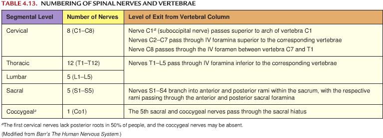
6 Spinal Nerves 31 Pairs of spinal nerves Named & numbered by the cord level of their origin 8 pairs of cervical nerves (C1 to C8) 12 pairs of thoracic nerves (T1 to T12) 5 pairs of lumbar nerves (L1 to L5) 5 pairs of sacral nerves (S1 to S5) 1 pair of coccygeal nerves Exit through the IVF


7 Spinal Cord & Spinal Nerves Spinal nerves begin as roots Dorsal or posterior root is incoming sensory fibers dorsal root ganglion (swelling) = cell bodies of sensory nerves Ventral or anterior root is outgoing motor fibers

8 Structures Covering the Spinal Cord Vertebrae Epidural space filled with fat Dura mater Dense irregular CT tube Ends at the lower border of S2 Follows the nerve roots and become continuous with epineurium Subdural space filled with interstitial fluid Arachnoid = spider web of collagen fibers Ends at the lower border of S2 Follows the nerve roots into the IVF Subarachnoid space = CSF Lumbar cistern (enlargement in subarachnoid space) L2 S2 Pia mater Thin layer covers BV Denticulate ligaments hold SC in place

9 Cervical Vertebral Canal: Content Meninges Dura matter Continuous with cranial dura matter (meningeal layer) Arachnoid matter Pia matter Lower part of medulla oblongata Cervical segments of the spinal cord Contain the upper motor neurons for the upper and lower limbs Other descending fibers to the spinal cord (e.g. reticulospianl fibers) Contain the ascending (sensory) fibers from the neck below Cervical enlargement Innervation for the upper limb Lower motor neurons Cervical spinal nerves C1 C8 C1 C7 exit above the corresponding vertebra C1 exit between the atlas and the occipital bone C8 exit between the C7 and T1 vertebrae C1 C4 form the cervical plexus C5 T1 form the brachial plexus

10 Caudal epidural anesthesia during delivery Into sacral hiatus Sacral and coccygeal cornua are important landmarks Anesthetize S2 Co1 spinal nerves Caudal Epidural Anesthesia
 L2 S2 Pia matter Filum terminale thread like extension of pia")
11 Inferior End of vertebral canal: Content Conus medullaris In adult ends at L2 In newborn ends at L4 Cauda equina (horse s tail) dorsal & ventral roots of lowest spinal nerves (L1 Co1) Spinal meninges Dura matter Ends at S2 S3 Arachnoid matter Ends at S2 S3 Subarachnoid space = CSF Lumbar cistern (enlargement in subarachnoid space) L2 S2 Pia matter Filum terminale thread like extension of pia mater stabilizes spinal cord in canal


12 Joints of Vertebral Bodies Cartilaginous joint Symphysis Vertebral bodies covered with thin plates of hyaline cartilage IVD Ligaments Anterior longitudinal ligaments Wider & stronger Attached to the vertebral bodies and the IVD Posterior longitudinal ligaments Weak and narrow Nerve supply: meningeal branches of the spinal nerves


13 Joints of Vertebral Arches Also called zygapophysial joints Plane synovial joint between the superior & inferior articular processes Articular facets Capsular ligament Ligaments Supraspinous ligament Between tips of spins Intraspinous ligament Between spines Intertransverse ligaments Between transverse processes Ligamentum flavum Between laminae Nerve supply: articular branches from posterior rami of the spinal nerves

14 Lumbar Puncture Lumbar puncture is used to withdraw CSF for diagnostic purposes LP performed from lumbar cistern to avoid the damage to the spinal cord LP approached mostly in L3 L4 or L4 L5 Epidural anesthesia Target the epidural space Same approach as LP Could be approached from the sacral hiatus


15 Spinal Nerves: Level of Exit From T1 to L5, spinal nerves exit from the IVF below their encountered vertebrae S1 S4 rami exit from their encountered sacral foramens S5 & Co1 exit from sacral hiatus
16
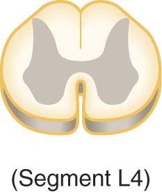
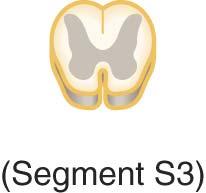
 lateral horns (motor autonomic neurons) only present in thoracic spinal cord gray commissure crosses the midline Central canal continuous with 4th ventricle of")
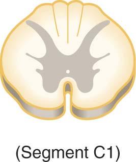
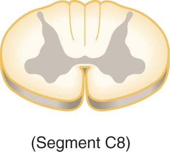
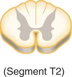
17 Gray Matter of the Spinal Cord Gray matter is shaped like the letter H or a butterfly contains neuron cell bodies, unmyelinated axons & dendrites dorsal gray horns (sensory neurons) ventral gray horns (motor somatic neurons) lateral horns (motor autonomic neurons) only present in thoracic spinal cord gray commissure crosses the midline Central canal continuous with 4th ventricle of brain

18 Nerve Cell Columns in the Gray Matter Motor Medial motor nucleus (cell column) Axial muscles Entire SC Lateral motor nucleus Limb muscles Enlargements Intermediolateral cell column Autonomic T1 L2, S2 4
, two point discrimination & vibration Nucleus dorsalis (Clarke s column) C8 L2")
19 Nerve Cell Columns in the Gray Matter Sensory Substantia gelatinosa Entire SC Pain, temperature & touch Nucleus proprius Entire SC Proprioception (sense of position & movement), two point discrimination & vibration Nucleus dorsalis (Clarke s column) C8 L2 Proprioceptive endings

20 Cell columns in the anterior gray horn of the spinal cord: somatotopic organization

21 White Matter of the Spinal Cord White matter covers gray matter Anterior median fissure deeper than Posterior median sulcus Anterior, Lateral and Posterior White Columns contain axons that form ascending & descending tracts
22
23 Tracts of the Spinal Cord Function of tracts highway for sensory & motor information sensory tracts ascend motor tracts descend Naming of tracts indicates position & direction of signal example = anterior spinothalamic tract impulses travel from spinal cord towards brain (thalamus) found in anterior part of spinal cord

24 Location of Tracts inside Cord Motor/descending tracts pyramidal tract (corticospinal) extrapyramidal tracts Sensory/ascending tracts spinothalamic tract posterior column spinocerebellar?
25 Functions of Spinal Tracts Sensory Spinothalamic tract pain, temperature, deep pressure & crude touch Posterior columns proprioception, discriminative touch, two point discrimination, pressure and vibration Motor Direct pathways (corticospinal & corticobulbar) precise, voluntary movements Indirect pathways (rubrospinal, vestibulospinal) programming automatic movements, posture & muscle tone, equilibrium & coordination of visual reflexes
 Intersegmental fibers (fasciculus")
26 White Matter of the Spinal Cord Ventral white commissure Lissaur s tract (dorsolateral fasciculus) Intersegmental fibers (fasciculus proprius)

27 Blood Supply to SC One anterior spinal a. Vertebral aa. Two posterior spinal aa. Vertebral aa. 25% PICA 75% Anterior & posterior radicular aa. Arise at every spinal level Serve their respective roots & ganglia

28 Anterior & posterior spinal medullary aa. Arise at intermittent levels Serve to augment the BS to SC Artery of Adamkierwicz Unusually large spinal medullary a. Usually on the left In low thoracic or upper lumber levels Blood Supply to SC
 Affects Corticospinal tracts Paraplegia below lesion Spinothalamic tracts Thermoanesthesia and analgesia")
29 Spinal cord Ischemia Anterior spinal a. Small & tenuous Occlusion produces bilateral damage (below lesion) Affects Corticospinal tracts Paraplegia below lesion Spinothalamic tracts Thermoanesthesia and analgesia Descending autonomic tracts Loss of bladder & bowel control Anterior gray horn Near enlargement weakness of limb muscles Blood Supply to SC
Gross Morphology of Spinal Cord
 Gross Morphology of Spinal Cord Done By : Rahmeh Alsukkar ** I did my best and sorry for any mistake ** the sheet does not contain pictures, tables and some slides so please be careful and go back to slides
Chapter 13. The Spinal Cord & Spinal Nerves. Spinal Cord. Spinal Cord Protection. Meninges. Together with brain forms the CNS Functions
 Spinal Cord Chapter 13 The Spinal Cord & Spinal Nerves Together with brain forms the CNS Functions spinal cord reflexes integration (summation of inhibitory and excitatory) nerve impulses highway for upward
Spinal Cord Protection. Chapter 13 The Spinal Cord & Spinal Nerves. External Anatomy of Spinal Cord. Structures Covering the Spinal Cord
 Spinal Cord Protection Chapter 13 The Spinal Cord & Spinal Nerves We are only going to cover Pages 420-434 and 447 Together with brain forms the CNS Functions spinal cord reflexes integration (summation
The Spinal Cord. The Nervous System. The Spinal Cord. The Spinal Cord 1/2/2016. Continuation of CNS inferior to foramen magnum.
 The Nervous System Spinal Cord Continuation of CNS inferior to foramen magnum Simpler than the brain Conducts impulses to and from brain Two way conduction pathway Reflex actions Passes through vertebral
The Spinal Cord & Spinal Nerves
 The Spinal Cord & Spinal Nerves Together with brain forms the CNS Functions spinal cord reflexes integration (summation of inhibitory and excitatory) nerve impulses highway for upward and downward travel
ANATOMY OF SPINAL CORD. Khaleel Alyahya, PhD, MEd King Saud University School of
 ANATOMY OF SPINAL CORD Khaleel Alyahya, PhD, MEd King Saud University School of Medicine @khaleelya OBJECTIVES At the end of the lecture, students should be able to: Describe the external anatomy of the
Spinal Cord H. Ruth Clemo, Ph.D.
 Spinal Cord H. Ruth Clemo, Ph.D. OBJECTIVES After studying the material of this lecture, the student should be familiar with: 1. Surface anatomy of the spinal cord. 2. Internal structure and organization
Introduction and Basic structural organization of the nervous system
 Introduction and Basic structural organization of the nervous system **the slides are in bold and the book is in red Done by : razan krishan & marah marahleh INTRODUCTION The nervous system, along with
BIOH111. o Cell Module o Tissue Module o Integumentary system o Skeletal system o Muscle system o Nervous system o Endocrine system
 BIOH111 o Cell Module o Tissue Module o Integumentary system o Skeletal system o Muscle system o Nervous system o Endocrine system Endeavour College of Natural Health endeavour.edu.au 1 Textbook and required/recommended
Note: Please refer to handout Spinal Plexuses and Representative Spinal Nerves for
 Chapter 13 Outline Note: Please refer to handout Spinal Plexuses and Representative Spinal Nerves for what you need to know from Exhibits 13.1 13.4 I. INTRODUCTION A. The spinal cord and spinal nerves
Human Anatomy. Spinal Cord and Spinal Nerves
 Human Anatomy Spinal Cord and Spinal Nerves 1 The Spinal Cord Link between the brain and the body. Exhibits some functional independence from the brain. The spinal cord and spinal nerves serve two functions:
Chapter 12b. Overview
 Chapter 12b Spinal Cord Overview Spinal cord gross anatomy Spinal meninges Sectional anatomy Sensory pathways Motor pathways Spinal cord pathologies 1 The Adult Spinal Cord About 18 inches (45 cm) long
Fig Cervical spinal nerves. Cervical enlargement C7. Dural sheath. Subarachnoid space. Thoracic. Spinal cord Vertebra (cut) spinal nerves
 spinal nerves") Fig. 13.1 C1 Cervical enlargement C7 Cervical spinal nerves Dural sheath Subarachnoid space Thoracic spinal nerves Spinal cord Vertebra (cut) Lumbar enlargement Medullary cone T12 Spinal nerve Spinal nerve
Fig. 13.1 C1 Cervical enlargement C7 Cervical spinal nerves Dural sheath Subarachnoid space Thoracic spinal nerves Spinal cord Vertebra (cut) Lumbar enlargement Medullary cone T12 Spinal nerve Spinal nerve
Anatomy of the Nervous System. Brain Components
 Anatomy of the Nervous System Brain Components NERVOUS SYSTEM INTRODUCTION Is the master system of human body, controlling the functions of rest of the body systems Nervous System CLASSIFICATION A. Anatomical
Chapter 13! Chapter 13 Spinal Cord and Spinal Nerves! The Spinal Cord and Spinal Nerves!
 Chapter 13! The Spinal Cord and Spinal Nerves! SECTION 13-1! The brain and spinal cord make up the central nervous system, and the cranial nerves and spinal nerves constitute the peripheral nervous system!
Spinal cord. We have extension of the pia mater below L1-L2 called filum terminale
 Spinal cord Part of the CNS extend from foramen magnum to the level of L1-L2 (it is shorter than the vertebral column) it is covered by spinal meninges. It is cylindrical in shape. It s lower end become
ANATOMY OF THE SPINAL CORD. Structure of the spinal cord Tracts of the spinal cord Spinal cord syndromes
 SPINAL CORD ANATOMY OF THE SPINAL CORD Structure of the spinal cord Tracts of the spinal cord Spinal cord syndromes The Nervous System Coordinates the activity of muscles, organs, senses, and actions Made
CHAPTER 13 LECTURE OUTLINE
 CHAPTER 13 LECTURE OUTLINE I. INTRODUCTION A. The spinal cord and spinal nerves mediate reactions to environmental changes. B. The spinal cord has several functions. 1. It processes reflexes. 2. It is
Chapter 13: The Spinal Cord and Spinal Nerves
 Chapter 13: The Spinal Cord and Spinal Nerves Spinal Cord Anatomy Protective structures: Vertebral column and the meninges protect the spinal cord and provide physical stability. a. Dura mater, b. Arachnoid,
Human Anatomy - Problem Drill 11: The Spinal Cord and Spinal Nerves
 Human Anatomy - Problem Drill 11: The Spinal Cord and Spinal Nerves Question No. 1 of 10 Instructions: (1) Read the problem statement and answer choices carefully, (2) Work the problems on paper as needed,
Central Nervous System: Part 2
 Central Nervous System: Part 2 1. Meninges 2. CSF 3. Spinal Cord and Spinal Nerves Explain spinal cord anatomy, including gray and white matter and meninges (give the general functions of this organ).
Spinal Cord- Medulla Spinalis. Cuneyt Mirzanli Istanbul Gelisim University
 Spinal Cord- Medulla Spinalis Cuneyt Mirzanli Istanbul Gelisim University Spinal Column Supports the skull, pectoral girdle, upper limbs and thoracic cage by way of the pelvic girdle. Transmits body weight
Overview. Spinal Anatomy Spaces & Meninges Spinal Cord. Anatomy of the dura. Anatomy of the arachnoid. Anatomy of the spinal meninges
 European Course in Neuroradiology Module 1 - Anatomy and Embryology Dubrovnik, October 2018 Spinal Anatomy Spaces & Meninges Spinal Cord Johan Van Goethem Overview spinal meninges & spaces spinal cord
The Spinal Cord and Spinal Nerves!
 Chapter 13! The Spinal Cord and Spinal Nerves! SECTION 13-1! The brain and spinal cord make up the central nervous system, and the cranial nerves and spinal nerves constitute the peripheral nervous system!
The CNS Part II pg
 The CNS Part II pg. 455-474 Protection of the Brain Objectives Describe how the meninges, cerebrospinal fluid, and the blood brain barrier protect the CNS. Explain how Cerebrospinal fluid is formed, and
THE BACK THE SPINAL CORD
 THE BACK THE SPINAL CORD The structures in the vertebral canal: the spinal cord spinal nerve roots spinal meninges the neurovascular structures THE SPINAL CORD The spinal cord occupies the superior 2/3
The Spinal Cord, Spinal Nerves, and Spinal Reflexes
 13 The Spinal Cord, Spinal Nerves, and Spinal Reflexes PowerPoint Lecture Presentations prepared by Jason LaPres Lone Star College North Harris An Introduction to the Spinal Cord, Spinal Nerves, and Spinal
Lecture 14: The Spinal Cord
 Lecture 14: The Spinal Cord M/O Chapters 16 69. Describe the relationship(s) between the following structures: root, nerve, ramus, plexus, tract, nucleus, and ganglion. 70. Trace the path of information
Chapter 14. The Nervous System. The Spinal Cord and Spinal Nerves. Lecture Presentation by Steven Bassett Southeast Community College
 Chapter 14 The Nervous System The Spinal Cord and Spinal Nerves Lecture Presentation by Steven Bassett Southeast Community College Introduction The Central Nervous System (CNS) consists of: The spinal
THE BACK. Dr. Ali Mohsin. Spinal Cord
 Spinal Cord THE BACK Dr. Ali Mohsin The spinal cord is the elongated caudal part of the CNS. It starts as the inferior continuation of the medulla oblongata at the level of foramen magnum, & ends as an
Department of Neurology/Division of Anatomical Sciences
 Spinal Cord I Lecture Outline and Objectives CNS/Head and Neck Sequence TOPIC: FACULTY: THE SPINAL CORD AND SPINAL NERVES, Part I Department of Neurology/Division of Anatomical Sciences LECTURE: Monday,
Lecturer. Prof. Dr. Ali K. Al-Shalchy MBChB/ FIBMS/ MRCS/ FRCS 2014
 Lecturer Prof. Dr. Ali K. Al-Shalchy MBChB/ FIBMS/ MRCS/ FRCS 2014 Dorsal root: The dorsal root carries both myelinated and unmyelinated afferent fibers to the spinal cord. Posterior gray column: Long
Lecture - Chapter 13: Central Nervous System
 Lecture - Chapter 13: Central Nervous System 1. Describe the following structures of the brain, what is the general function of each: a. Cerebrum b. Diencephalon c. Brain Stem d. Cerebellum 2. What structures
 Spinal Cord Tracts DESCENDING SPINAL TRACTS: Are concerned with somatic motor function, modification of ms. tone, visceral innervation, segmental reflexes. Main tracts arise form cerebral cortex and others
Organization of The Nervous System PROF. SAEED ABUEL MAKAREM
 Organization of The Nervous System PROF. SAEED ABUEL MAKAREM Objectives By the end of the lecture, you should be able to: List the parts of the nervous system. List the function of the nervous system.
Copyright McGraw-Hill Education. Permission required for reproduction or display. C1. Cervical spinal ner ves. Thor acic. T12 Spinal nerve rootlets
 Fig. 13.1 C1 Cervical enlar gem ent C7 Cervical spinal ner ves Dural sheath Subarachnoi d space Thor acic spinal ner ves Vertebra (cut) Lum bar enlar gem ent Medullar y T12 rootlets cone Posterior median
SENSORY (ASCENDING) SPINAL TRACTS
 SPINAL TRACTS") SENSORY (ASCENDING) SPINAL TRACTS Dr. Jamila El-Medany Dr. Essam Eldin Salama OBJECTIVES By the end of the lecture, the student will be able to: Define the meaning of a tract. Distinguish between the different
SENSORY (ASCENDING) SPINAL TRACTS Dr. Jamila El-Medany Dr. Essam Eldin Salama OBJECTIVES By the end of the lecture, the student will be able to: Define the meaning of a tract. Distinguish between the different
Spinal Cord Organization. January 12, 2011
 Spinal Cord Organization January 12, 2011 Spinal Cord 31 segments terminates at L1-L2 special components - conus medullaris - cauda equina no input from the face Spinal Cord, Roots & Nerves Dorsal root
Gross Anatomy of Lower Spinal Cord
 Chapter 13 Spinal Cord, Spinal Nerves and Somatic Reflexes Spinal cord Spinal nerves Somatic reflexes Gross Anatomy of Lower Spinal Cord Meninges of Vertebra & Spinal Cord Spina Bifida Congenital defect
IV. THE SPINAL CORD BLOOD SUPPLY
 IV. THE SPINAL CORD Spinal cord is covered by o Pia Mater Spinalis Film Teminale Denticulate Ligament ---------------------- Cordotomy o Arachnoid Membrane Subarachnoid Space ----------------------- Lumbar
Blood Supply of the CNS
 Blood Supply of the CNS Lecture Objectives Describe the four arteries supplying the CNS. Follow up each artery to its destination. Describe the circle of Willis and its branches. Discuss the principle
Histology of the CNS
 Histology of the CNS Lecture Objectives Describe the histology of the cerebral cortex layers. Describe the histological features of the cerebellum; layers and cells of cerebellar cortex. Describe the elements
BIOH111. o Cell Module o Tissue Module o Skeletal system o Muscle system o Nervous system o Endocrine system o Integumentary system
 BIOH111 o Cell Module o Tissue Module o Skeletal system o Muscle system o Nervous system o Endocrine system o Integumentary system Endeavour College of Natural Health endeavour.edu.au 1 Textbook and required/recommended
VERTEBRAL COLUMN ANATOMY IN CNS COURSE
 VERTEBRAL COLUMN ANATOMY IN CNS COURSE Vertebral body Sections of the spine Atlas (C1) Axis (C2) What type of joint is formed between atlas and axis? Pivot joint What name is given to a fracture of both
Arterial Blood Supply
 Arterial Blood Supply Brain is supplied by pairs of internal carotid artery and vertebral artery. The four arteries lie within the subarachnoid space Their branches anastomose on the inferior surface of
Copy Right Hongqi ZHANG Department of Anatomy Fudan University 1. Systematic Anatomy. Nervous system Spinal cord. Dr.Hongqi Zhang ( 张红旗 )
") Systematic Anatomy Nervous system Spinal cord Dr.Hongqi Zhang ( 张红旗 ) Email: zhanghq58@126.com 1 Spinal cord Central nervous system (CNS) Brain Telencephalon Diencephalon Cerebellum Brain stem 1-Midbrain
Systematic Anatomy Nervous system Spinal cord Dr.Hongqi Zhang ( 张红旗 ) Email: zhanghq58@126.com 1 Spinal cord Central nervous system (CNS) Brain Telencephalon Diencephalon Cerebellum Brain stem 1-Midbrain
With other members of your lab group, discuss the following questions: - The spinal cord connects directly to which part of the brain?
 BIOLOGY 211: HUMAN ANATOMY & PHYSIOLOGY ************************************************************************************************************************* SPINAL CORD, SPINAL NERVES, AND REFLEXES
The Nervous System: Sensory and Motor Tracts of the Spinal Cord
 15 The Nervous System: Sensory and Motor Tracts of the Spinal Cord PowerPoint Lecture Presentations prepared by Steven Bassett Southeast Community College Lincoln, Nebraska Introduction Millions of sensory
Cranial Nerves and Spinal Cord Flashcards
 1. Name the cranial nerves and their Roman numeral. 2. What is Cranial Nerve I called, and what does it 3. Scientists who are trying to find a way to make neurons divide to heal nerve injuries often study
Pain classifications slow and fast
 Pain classifications slow and fast Fast Pain Slow Pain Sharp, pricking (Aδ) fiber Short latency Well localized Short duration Dull, burning (C) fiber Slower onset Diffuse Long duration Less emotional Emotional,
Meninges. Connective tissue membranes
 Meninges Connective tissue membranes Dura mater: -outermost layer; continuous with epineuriumof the spinal nerves - dense irregular connective tissue - from the level of the foramen magnum to S2 Arachnoid
The Nervous System: The
 C h a p t e r 14 The Nervous System: The Spinal Cord and Spinal Nerves PowerPoint Lecture Slides prepared by Jason LaPres North Harris College Houston, Texas Copyright 2009 Pearson Education, Inc., publishing
Cerebral hemisphere. Parietal Frontal Occipital Temporal
 Cerebral hemisphere Sulcus / Fissure Central Precental gyrus Postcentral gyrus Lateral (cerebral) Parieto-occipital Cerebral cortex Frontal lobe Parietal lobe Temporal lobe Insula Amygdala Hippocampus
General Sensory Pathways of the Trunk and Limbs
 General Sensory Pathways of the Trunk and Limbs Lecture Objectives Describe gracile and cuneate tracts and pathways for conscious proprioception, touch, pressure and vibration from the limbs and trunk.
Spinal Cord and Spinal Nerves. Spinal Cord. Chapter 12
 Chapter 12 Spinal Cord and Spinal Nerves 1 Spinal Cord Extends from foramen magnum to second lumbar vertebra Segmented: Cervical, Thoracic, Lumbar & Sacral Gives rise to 31 pairs of spinal nerves Not uniform
Chapter 14 Lecture Outline
 Chapter 14 Lecture Outline See separate PowerPoint slides for all figures and tables preinserted into PowerPoint without notes. Copyright McGraw-Hill Education. Permission required for reproduction or
Unit Three. The brain includes: cerebrum, diencephalon, brain stem, & cerebellum. The brain lies within the cranial cavity of the skull.
 Human Anatomy & Physiology 11 Divisions of the Nervous System Karen W. Smith, Instructor Unit Three BRAIN & SPINAL CORD Refer to the following URLs. Be sure to study these along with your book. http://www.sirinet.net/~jgjohnso/nervous.html
NERVOUS SYSTEM. Academic Resource Center. Forskellen mellem oscillator og krystal
 NERVOUS SYSTEM Academic Resource Center Forskellen mellem oscillator og krystal Overview of the Nervous System Peripheral nervous system-pns cranial nerves spinal nerves ganglia peripheral nerves enteric
TEST BANK FOR FUNDAMENTALS OF ANATOMY & PHYSIOLOGY 9TH EDITION BY MARTINI
 Link download :https://testbankservice.com/download/test-bank-forfundamentals-of-anatomy-and-physiology-9th-edition-by-martini TEST BANK FOR FUNDAMENTALS OF ANATOMY & PHYSIOLOGY 9TH EDITION BY MARTINI
Anatomy Lecture Notes Chapter 13
 I. embryonic development of the CNS A. neurulation is the formation of the CNS in the embryo invagination of dorsal ectoderm (outer layer of embryo cells) this process is induced (caused) by the notochord
Organization of The Nervous System PROF. MOUSAED ALFAYEZ & DR. SANAA ALSHAARAWY
 Organization of The Nervous System PROF. MOUSAED ALFAYEZ & DR. SANAA ALSHAARAWY Objectives At the end of the lecture, the students should be able to: List the parts of the nervous system. List the function
The Nervous System PART C. PowerPoint Lecture Slide Presentation by Patty Bostwick-Taylor, Florence-Darlington Technical College
 PowerPoint Lecture Slide Presentation by Patty Bostwick-Taylor, Florence-Darlington Technical College The Nervous System 7 PART C Protection of the Central Nervous System Scalp and skin Skull and vertebral
Anatomy of the Spine. Figure 1. (left) The spine has three natural curves that form an S-shape; strong muscles keep our spine in alignment.
 The spine has three natural curves that form an S-shape; strong muscles keep our spine in alignment.") 1 2 Anatomy of the Spine Overview The spine is made of 33 individual bony vertebrae stacked one on top of the other. This spinal column provides the main support for your body, allowing you to stand upright,
1 2 Anatomy of the Spine Overview The spine is made of 33 individual bony vertebrae stacked one on top of the other. This spinal column provides the main support for your body, allowing you to stand upright,
I: To describe the pyramidal and extrapyramidal tracts. II: To discuss the functions of the descending tracts.
 Descending Tracts I: To describe the pyramidal and extrapyramidal tracts. II: To discuss the functions of the descending tracts. III: To define the upper and the lower motor neurons. 1. The corticonuclear
_CH01redo.qxd 9/24/07 3:07 PM Page 1. [Half-Title to come]
![_CH01redo.qxd 9/24/07 3:07 PM Page 1. [Half-Title to come]](/thumbs/81/84146690.jpg "_CH01redo.qxd 9/24/07 3:07 PM Page 1. [Half-Title to come]") 10752-01_CH01redo.qxd 9/24/07 3:07 PM Page 1 [Half-Title to come] 10752-01_CH01redo.qxd 9/24/07 3:07 PM Page 2 THE BACK Lippincott Williams & Wilkins atlas of ANATOMY CHAPTER 1 Plate 1-01 Palpable Structures
10752-01_CH01redo.qxd 9/24/07 3:07 PM Page 1 [Half-Title to come] 10752-01_CH01redo.qxd 9/24/07 3:07 PM Page 2 THE BACK Lippincott Williams & Wilkins atlas of ANATOMY CHAPTER 1 Plate 1-01 Palpable Structures
Spinal Cord and Properties of Cerebrospinal Fluid: Options for Drug Delivery. SMA Foundation New York
 Spinal Cord and Properties of Cerebrospinal Fluid: Options for Drug Delivery New York Why Do We Need to Know about the Spinal Cord Anatomy and Properties of Cerebrospinal Fluid? SMA therapeutics need to
Spinal Cord Workbook. Learning objec&ves
 Spinal Cord Workbook Direc&ons. Watch the following video tutorials and complete this workbook: YouTubeèTheNotedAnatomistèPlaylistsèSpinal cord and nervesèwatch videos with the names of ObjecCves A-E.
The functional Anatomy of the Nervous System. DR. OKSANA PETRICHKO Department of Human Anatomy
 The functional Anatomy of the Nervous System DR. OKSANA PETRICHKO Department of Human Anatomy Coordination and Regulation of Body Systems Nervous system. Conducts nerve impulses maintaining homeostasis
NERVOUS SYSTEM ANATOMY
 INTRODUCTION to NERVOUS SYSTEM ANATOMY M1 - Gross and Developmental Anatomy Dr. Milton M. Sholley Professor of Anatomy and Neurobiology and Dr. Michael H. Peters Professor of Chemical and Life Science
The neurvous system senses, interprets, and responds to changes in the environment. Two types of cells makes this possible:
 NERVOUS SYSTEM The neurvous system senses, interprets, and responds to changes in the environment. Two types of cells makes this possible: the neuron and the supporting cells ("glial cells"). Neuron Neurons
Biological Bases of Behavior. 3: Structure of the Nervous System
 Biological Bases of Behavior 3: Structure of the Nervous System Neuroanatomy Terms The neuraxis is an imaginary line drawn through the spinal cord up to the front of the brain Anatomical directions are
Spinal Column. Anatomy Of The Spine
 Anatomy Of The Spine The spine is a flexible column, composed of a stack of individual bones. Each bone is called a vertebra. There are seven vertebrae in the neck (cervical vertebrae) twelve in the thoracic
SPINAL CORD AND PROPERTIES OF CEREBROSPINAL FLUID: OPTIONS FOR DRUG DELIVERY
 SPINAL CORD AND PROPERTIES OF CEREBROSPINAL FLUID: OPTIONS FOR DRUG DELIVERY WHY DO WE NEED TO KNOW ABOUT THE SPINAL CORD ANATOMY AND PROPERTIES OF CEREBROSPINAL FLUID? SMA therapeutics need to reach cells
Cheat Sheet Anatomy 2
 Cheat Sheet Anatomy 2 - CNS è Brian and spinal cord - PNS è 12 Cranial/ 31 Spinal + associated ganglia - In Histological preparation è Myelin sheaths are lost leaving round empty spaces. - Gray matter
Motor tracts Both pyramidal tracts and extrapyramidal both starts from cortex: Area 4 Area 6 Area 312 Pyramidal: mainly from area 4 Extrapyramidal:
 Motor tracts Both pyramidal tracts and extrapyramidal both starts from cortex: Area 4 Area 6 Area 312 Pyramidal: mainly from area 4 Extrapyramidal: mainly from area 6 area 6 Premotorarea: uses external
Nervous System C H A P T E R 2
 Nervous System C H A P T E R 2 Input Output Neuron 3 Nerve cell Allows information to travel throughout the body to various destinations Receptive Segment Cell Body Dendrites: receive message Myelin sheath
Chapter 9. Nervous System
 Chapter 9 Nervous System Central Nervous System (CNS) vs. Peripheral Nervous System(PNS) CNS Brain Spinal cord PNS Peripheral nerves connecting CNS to the body Cranial nerves Spinal nerves Neurons transmit
Nervous System: Spinal Cord and Spinal Nerves (Chapter 13)
") Nervous System: Spinal Cord and Spinal Nerves (Chapter 13) Lecture Materials for Amy Warenda Czura, Ph.D. Suffolk County Community College Eastern Campus Primary Sources for figures and content: Marieb,
Nervous System: Spinal Cord and Spinal Nerves (Chapter 13) Lecture Materials for Amy Warenda Czura, Ph.D. Suffolk County Community College Eastern Campus Primary Sources for figures and content: Marieb,
Posterior White Column-Medial Lemniscal Pathway
 Posterior White Column-Medial Lemniscal Pathway Modality: Discriminative Touch Sensation (include Vibration) and Conscious Proprioception Receptor: Most receptors except free nerve endings Ist Neuron:
Brainstem. Steven McLoon Department of Neuroscience University of Minnesota
 Brainstem Steven McLoon Department of Neuroscience University of Minnesota 1 Course News Change in Lab Sequence Week of Oct 2 Lab 5 Week of Oct 9 Lab 4 2 Goal Today Know the regions of the brainstem. Know
THE VERTEBRAL COLUMN. Average adult length: In male: about 70 cms. In female: about 65 cms.
 THE VERTEBRAL COLUMN Average adult length: In male: about 70 cms. In female: about 65 cms. 1 Vertebral Column (Regions and Curvatures) Curvatures of the vertebral column: A. Primary curvature: C-shaped;
SHORT ANSWER. Write the word or phrase that best completes each statement or answers the question.
 Exam Name 1) A change in the conditions in the synaptic terminal can influence the soma as a result of axoplasmic transport. 2) The nervous system is composed of the brain and spinal cord. A) efferent
NERVOUS SYSTEM ANATOMY
 NTRODUCTON to NERVOUS SYSTEM ANATOMY M1 - Gross and Developmental Anatomy Dr. Milton M. Sholley Professor of Anatomy and Neurobiology and Dr. Michael H. Peters Professor of Chemical and Life Science Engineering
Biology 218 Human Anatomy
 Chapter 21 Adapted form Tortora 10 th ed. LECTURE OUTLINE A. Overview of Sensations (p. 652) 1. Sensation is the conscious or subconscious awareness of external or internal stimuli. 2. For a sensation
Lecture VIII. The Spinal Cord, Reflexes and Brain Pathways!
 Reflexes and Brain Bio 3411! Monday!! 1! Readings! NEUROSCIENCE 5 th ed: Review Chapter 1 pp. 11-21;!!Read Chapter 9 pp. 189-194, 198! THE BRAIN ATLAS 3 rd ed:! Read pp. 4-17 on class web site! Look at
Ligaments of the vertebral column:
 In the last lecture we started talking about the joints in the vertebral column, and we said that there are two types of joints between adjacent vertebrae: 1. Between the bodies of the vertebrae; which
Synapse Homework. Back page last question not counted. 4 pts total, each question worth 0.18pts. 26/34 students answered correctly!
 Synapse Homework Back page last question not counted 26/34 students answered correctly! 4 pts total, each question worth 0.18pts Business TASS hours extended! MWF 1-2pm, Willamette 204 T and Th 9:30-10:30am,
Lab Activity 13. Spinal Cord. Portland Community College BI 232
 Lab Activity 13 Spinal Cord Portland Community College BI 232 Definitions Tracts: collections of axons in CNS Nerves:collections of axons in PNS Ganglia: collections of neuron cell bodies in PNS Nucleus
Spinal nerves and cervical plexus Prof. Abdulameer Al Nuaimi. E mail: a.al E. mail:
 Spinal nerves and cervical plexus Prof. Abdulameer Al Nuaimi E mail: a.al nuaimi@sheffield.ac.uk E. mail: abdulameerh@yahoo.com Branches of ophthalmic artery Muscles of face A spinal nerve Spinal
Chapter 17 Nervous System
 Chapter 17 Nervous System 1 The Nervous System Two Anatomical Divisions Central Nervous System (CNS) Brain and Spinal Cord Peripheral Nervous System (PNS) Two Types of Cells Neurons Transmit nerve impulses
Chapter 13 Lecture Outline
 Chapter 13 Lecture Outline See separate PowerPoint slides for all figures and tables preinserted into PowerPoint without notes. Copyright McGraw-Hill Education. Permission required for reproduction or
Chapter 12 The Central Nervous System Chapter Outline
 Chapter 12 The Central Nervous System Chapter Outline Module 12.1 Overview of the Central Nervous System (Figures 12.1, 12.2, 12.3) A. The central nervous system (CNS) includes the and, and is involved
Bony framework of the vertebral column Structure of the vertebral column
 5.1: Vertebral column & back. Overview. Bones o vertebral column. o typical vertebra. o vertebral canal. o spinal nerves. Joints o Intervertebral disc. o Zygapophyseal (facet) joint. Muscles o 2 compartments:
Human Anatomy Biology 351
 nnnnn 1 Human Anatomy Biology 351 Exam #2 Please place your name on the back of the last page of this exam. You must answer all questions on this exam. Because statistics demonstrate that, on average,
Internal Organisation of the Brainstem
 Internal Organisation of the Brainstem Major tracts and nuclei of the brainstem (Notes) The brainstem is the major pathway for tracts and houses major nuclei, that contain sensory, motor and autonomics
The Spinal Cord, Spinal Nerves, and Spinal Reflexes
 12 The Spinal Cord, Spinal Nerves, and Spinal Reflexes Lecture Presentation by Lori Garrett Section 1: Functional Organization of the Spinal Cord Learning Outcomes 12.1 Describe how the spinal cord can
THEME 2. VERTEBRAE (GENERAL DATA). CERVICAL, THORACIC AND LUMBAR VERTEBRAE. SACRUM. COCCYX. THE VERTEBRAL COLUMN AS A WHOLE
. CERVICAL, THORACIC AND LUMBAR VERTEBRAE. SACRUM. COCCYX. THE VERTEBRAL COLUMN AS A WHOLE") THEME 2. VERTEBRAE (GENERAL DATA). CERVICAL, THORACIC AND LUMBAR VERTEBRAE. SACRUM. COCCYX. THE VERTEBRAL COLUMN AS A WHOLE Osteology of the Vertebral Column Bone Description vertebra Notes a vertebra
THEME 2. VERTEBRAE (GENERAL DATA). CERVICAL, THORACIC AND LUMBAR VERTEBRAE. SACRUM. COCCYX. THE VERTEBRAL COLUMN AS A WHOLE Osteology of the Vertebral Column Bone Description vertebra Notes a vertebra
Nervous Systems: Diversity & Functional Organization
 Nervous Systems: Diversity & Functional Organization Diversity of Neural Signaling The diversity of neuron structure and function allows neurons to play many roles. 3 basic function of all neurons: Receive
INDEPENDENT LEARNING: DISC HERNIATION IN THE NATIONAL FOOTBALL LEAGUE: ANATOMICAL FACTORS TO CONSIDER IN REVIEW
 INDEPENDENT LEARNING: DISC HERNIATION IN THE NATIONAL FOOTBALL LEAGUE: ANATOMICAL FACTORS TO CONSIDER IN REVIEW CDC REPORT - CAUSES OF DISABILITY, 2005 REVIEW QUESTIONS ABOUT DISC HERNIATION IN THE NATIONAL
Development of Spinal Cord & Vertebral Column. Dr. Sanaa Alshaarawi & Prof. Ahmed Fathalla
 Development of Spinal Cord & Vertebral Column Dr. Sanaa Alshaarawi & Prof. Ahmed Fathalla OBJECTIVES At the end of the lecture, students should be able to: q Describe the development of the spinal cord