USRA OF THE LOWER EXTREMITY
|
|
|
- Kerry Higgins
- 5 years ago
- Views:
Transcription
1 USRA OF THE LOWER EXTREMITY Christian R. Falyar, CRNA, DNAP Department of Nurse Anesthesia Virginia Commonwealth University Disclosure Statement of Financial Interest I, Christian Falyar, DO NOT have a financial interest/arrangement or affiliation with one or more organizations that could be perceived as a real or apparent conflict of interest in the context of the subject of this presentation. Disclosure Statement of Financial Interest I, Christian Falyar, DO NOT have a financial interest/arrangement or affiliation with one or more organizations that could be perceived as a real or apparent conflict of interest in the context of the subject of this presentation. Disclosure Statement of Unapproved/Investigative Use I, Christian Falyar, DO NOT anticipate discussing the unapproved/investigative use of a commercial product/device during this activity or presentation.
2 Objectives State the indications for each lower extremity block Describe the ultrasound landmarks for each lower extremity block Review the ultrasound-guided needle insertion plane and local anesthetic requirements for each lower extremity block Discuss the side-effects and complications related to lower extremity blocks Lower Extremity Blocks Lumbar Plexus Femoral Adductor Canal Saphenous (thigh and ankle) Sacral Plexus Sciatic (gluteal level) Sciatic (popliteal level) Ankle Blocks Tibial Peroneal (Deep and Superficial) Sural Indications Regional anesthesia has many indications, including: Primary anesthetic Pain Management History of severe PONV or risk of MH Patient is too ill for general anesthesia Surgeon preference Contraindications In certain instances regional anesthesia should not be considered. Absolute contraindications include: Patient refusal Local infection at the site of the proposed block Active bleeding an anticoagulated patient Proven allergy to a local anesthetic
3 Contraindications Most contraindications to regional anesthesia are relative. The provider must determine the risk vs. benefit. Respiratory compromise Inability to cooperate/understand procedure An anesthetized patient (adult population) Bleeding diathesis secondary to an anticoogulant or genetic defect Bloodstream infection Preexisting peripheral neuropathy Pre-Procedure Prior to beginning any procedure: Verify the correct patient Obtain informed consent Verify the correct procedure Verify the correct extremity Gather all equipment Place the patient on oxygen Obtain baseline VS and monitor during the procedure Administer proper/adequate sedation Complications Regional anesthesia can result in complications such as: Local anesthetic toxicity Intra-arterial injection Respiratory compromise Parathesias and nerve damage Risks and benefits of regional anesthesia should always be discussed beforehand Local Anesthetics How much is enough? Most references recommend mls per block Some authors have demonstrated successful, complete blocks with less volume Amount and type of local anesthetic depends: Patient factors Timing of the procedure Procedure Purpose of the block


4 Local Anesthetics Drug Max Dose (mg/kg) Max Dose with Epi (mg/kg) Lidocaine*+ Drug 4.5mg/kg Max Dose (mg/kg) 7mg/kg Max Dose with Epi (mg/kg) Lidocaine* Mepivacaine*+ 4.5mg/kg 4mg/kg 7mg/kg 7mg/kg Mepivacaine* Bupivacaine* 2.5mg/kg 4mg/kg N/A 7mg/kg Bupivacaine* 2.5mg/kg 3.2mg/kg Ropivacaine* 5mg/kg N/A Ropivacaine* 3mg/kg 3.5mg/kg Procaine+ 12mg/kg 12mg/kg N/A N/A Chloroprocaine* 11mg/kg 11mg/kg 14mg/kg 14mg/kg Prilocaine* 8mg/kg 7mg/kg 8.5mg/kg 8.5mg/kg Tetracaine+ Tetracaine+ 3mg/kg 3mg/kg N/A N/A Femoral Block The femoral block targets the major branch of the lumbar plexus Provides anesthesia to the anterior thigh, knee and a small part of the lower leg The nerve is lateral to the femoral artery and deep to the fascia lata and iliaca, and superior to the iliopsoas muscle * - Nagelhout & Plaus, 5 th ed., pg Morgan & Mikhail, 5 th ed., pg. 272 Femoral Anatomy USRA Technique Fascia lata Fascia iliaca Pt. supine with slight external rotation of femur Transducer is placed at the inguinal crease, over the femoral pulse High-frequency linear array transducer Short-axis image, needle inserted in-plane Femoral nerve is a hyperechoic circle that lies lateral to the femoral artery 5 cm B bevel needle is used 20 to 30 cc s of local anesthetic injected


5 Pre-Procedure Scan USRA Technique Needle approach is lateral to medial Nerve stimulation can be used with ultrasound; patellar snap Major benefit of real-time imaging is visualizing needle movement and local anesthetic spread FI FL IPSM FN FA USRA Technique Femoral If more than one artery is visible, scan cephalad and identify a singular femoral artery Doppler can be used to verify femoral vessels Experience suggests that if the needle and the local anesthetic are placed below the fascia iliaca and lateral the artery, successful blocks occur despite the lack of twitches


6 Femoral Femoral Too low Just right Complications such as vascular puncture and local anesthetic injection are best avoided by observing the needle tip throughout the procedure Lymph nodes in the groin appear as nerves ; scanning proximal and distal will help distinguish the lymph node as they are not continuous and can be seen only at specific locations Femoral Adductor Canal The femoral nerve block (FNB) has been the gold-standard for pain relief following TKA Although effective for pain relief, it is associated with risk of falls Adductor canal block provides sensory nerve blockade with minimal motor involvement


, owing to the large hypoechoic superficial femoral artery and vein The vastus medialis will be blocked in some patients, however")
7 Adductor Canal Anatomy USRA Technique Rectus Femoris Adductor Longus Adductor Canal Gracilis Vastus Medialis Sartorius Pt. supine, with slight external rotation of the operative leg High frequency linear array transducer placed midthigh in short axis orientation The superficial femoral artery and vein appear as hypoechoic circles bordered superiorly by the sartorius, laterally by the vastus medialis and medially by the adductor longus muscles Needle inserted using in-plane image Up to 20 ml of local anesthetic injected in the fascial plane bordering the superficial femoral artery Adductor Canal Adductor Canal Vastus Medialis Sartorius Adductor Longus Studies show consistent nerve identification and block success (100%), owing to the large hypoechoic superficial femoral artery and vein The vastus medialis will be blocked in some patients, however the clinical significance is not completely known


8 Saphenous (thigh) Saphenous Anatomy (thigh) Saphenous block at the thigh is used as an adjunct to a popliteal block for lower extremity surgery below the knee USRA Technique Pre-Procedure Scan Pt is supine with lower extremity slightly abducted Transducer placement is dependent on the method used to identify the nerve High frequency transducer Short-axis image, needle inserted in-plane Saphenous nerve is a hyperechoic circle that lies in the fascial plane between the sartorius and vastus medialis muscles 5 10cm needle is used 10cc s of local anesthetic injected


9 Pre-Procedure Scan Saphenous (thigh) saphenous nerve vastus medialis Saphenous (thigh) Saphenous (thigh) There are few complications associated with this block

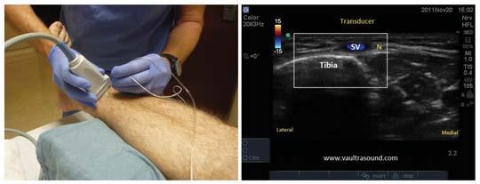
10 Sciatic Sciatic Sciatic nerve arises from L4-5 and S1-3 Indicated for surgical procedures of the hip, thigh, posterior knee, lower leg and foot. Anatomical knowledge is paramount; locating needle with landmark or US may be difficult. Sciatic Popliteal Gluteal contraction indicates the needle must be advanced further Complications include: Intraneural injection Intravasuclar injection Targets the sciatic nerve slightly above the knee Provides anesthesia for procedures involving the foot and ankle In the popliteal fossa, the sciatic is bordered superiorly and medially by the semi-tendinosus and semi-membranosus muscles and superiorly and laterally by the biceps femoris muscle


.")

11 Popliteal Anatomy Ultrasound Technique Prone, supine, or lateral position High frequency linear array transducer Transducer is placed in the popliteal crease Short-axis, in-plane or out-of-plane The sciatic nerve is superior to the popliteal artery and vein, then bifurcates in the tibial (larger), and peroneal (more lateral). 5 or 10 cm needle up to 30 cc s of local anesthetic injected incremental with negative aspiration Popliteal Pre-Procedure Scan Prone Supine


12 Popliteal Popliteal Ultrasound greatly reduces the traditional complications of intravascular and intra-neural injections When using nerve stimulation, dorsal or plantar flexion is acceptable Complications include: Intravascular injection Intraneural injection Popliteal Scan the proximal and distal to appreciate the anatomy The transducer may have to be angled toward to the foot to better image the nerves Circumferential spread around the nerve ensures a dense block Ankle Five nerves are blocked in the ankle Femoral saphenous Sciatic tibial sural deep peroneal superficial peroneal


13 Ankle Anatomy Ankle Ultrasound Technique Posterior Tibial Supine with foot elevated or extended past the end of the stretcher High frequency linear array transducer Short-axis, in-plane or out-of-plane Corresponding vascular structure identified first 5 cm B bevel needle 3-5 ml of local anesthetic injected following negative aspiration to achieve circumferential spread

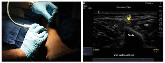
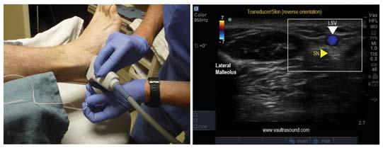
14 Deep Peroneal Superficial Peroneal Sural Saphenous
15 Ankle Blocks Questions? Aggressive injections of large volumes of local anesthetic can cause hydrostatic damage to small nerves such as the deep peroneal nerve because it is enclosed in ligamentous spaces References Chan V., & Pollard B.; An Introductory Cirriculum for Ultrasound- Guided Regional Anesthesia; 2009, University of Toronto Press. Chan, Vincent; Ultrasound Imaging for Regional Anesthesia: A Practical Guide; 3rd Edition; 2010, Toronto Printing Company. Gray, Andrew; Atlas of Ultrasound-Guided Regional Anesthesia; 2007, Saunders/Elsevier. Hadzic, Admir; Textbook of Regional Anesthesia and Acute Pain Management; 2007, McGraw-Hill Medical. Morgan, G., & Mikhail, M.; Clinical Anesthesiology; 4 th Edition; 2006, McGraw-Hill Medical. Sites, B., & Spence, B.; Ultrasound Guidance in Regional Anesthesia: Techniques for Upper-Extremity and Lower-Extremity Nerve Blocks; 2008, McMahon Publishing. Zwiebel, W., & Pellerito, J.; Introduction to Vascular Ultrasonography; 5th Edition; 2005, Elsevier Saunders.
USRA OF THE UPPER EXTREMITY
 USRA OF THE UPPER EXTREMITY Christian R. Falyar, DNAP, CRNA Department of Nurse Anesthesia Virginia Commonwealth University Disclosure Statement of Financial Interest I, Christian Falyar, DO NOT have a
Ultrasound Guided Lower Extremity Blocks
 Ultrasound Guided Lower Extremity Blocks CONTENTS: 1. Femoral Nerve Block 2. Popliteal Nerve Block Updated December 2017 1 1. Femoral Nerve Block Indications Surgery involving the knee, anterior thigh,
Surface Anatomy and Sonoanatomy for the Occasional Regional Anesthesiologist
 Surface Anatomy and Sonoanatomy for the Occasional Regional Anesthesiologist Edward R. Mariano, M.D., M.A.S. Professor of Anesthesiology, Perioperative & Pain Medicine Stanford University School of Medicine
Lower Extremity Ultrasound-Guided Regional Anesthesia. Stephanie Duffy, CRNA Regional Anesthesia Faculty Acute Pain Service NMCSD
 Lower Extremity Ultrasound-Guided Regional Anesthesia Stephanie Duffy, CRNA Regional Anesthesia Faculty Acute Pain Service NMCSD Objectives Review anatomy of lumbosacral plexus Lumbar plexus blocks Psoas
Regional Anaesthesia
 Regional Anaesthesia Lower limb anatomy and blocks Hip and Knee Joint Hip Joint: Nerve supply Lumbar plexus Femoral nerve through the nerve to the Rectus Femoris Ant division of the Obturator nerve The
HUMAN BODY COURSE LOWER LIMB NERVES AND VESSELS
 HUMAN BODY COURSE LOWER LIMB NERVES AND VESSELS October 22, 2010 D. LOWER LIMB MUSCLES 2. Lower limb compartments ANTERIOR THIGH COMPARTMENT General lfunction: Hip flexion, knee extension, other motions
ANATYOMY OF The thigh
 ANATYOMY OF The thigh 1- Lateral cutaneous nerve of the thigh Ι) Skin of the thigh Anterior view 2- Femoral branch of the genitofemoral nerve 5- Intermediate cutaneous nerve of the thigh 1, 2 and 3 are
ANATYOMY OF The thigh
 ANATYOMY OF The thigh 1- Lateral cutaneous nerve of the thigh Ι) Skin of the thigh Anterior view 2- Femoral branch of the genitofemoral nerve 5- Intermediate cutaneous nerve of the thigh 1, 2 and 3 are
ANATYOMY OF The thigh
 ANATYOMY OF The thigh 1- Lateral cutaneous nerve of the thigh Ι) Skin of the thigh Anterior view 2- Femoral branch of the genitofemoral nerve 1, 2 and 3 are From the lumber plexus 5- Intermediate cutaneous
Quillen College of Medicine
 Ea s t T e n n e s s e e St a t e Un i v e r s i t y Quillen College of Medicine Failing to prepare is preparing to fail. John Wooden, UCL A Dr. Tom Kwasigroch Associate Dean Director, Medical Human Gross
Sign up to receive ATOTW weekly -
 ULTRASOUND GUIDED ADDUCTOR CANAL BLOCK (SAPHENOUS NERVE BLOCK) ANAESTHESIA TUTORIAL OF THE WEEK 301 13 TH JANUARY 2014 Dr Daniel Quemby, Specialist Trainee Anaesthesia Dr Andrew McEwen, Consultant Anaesthetist
The Lower Limb II. Anatomy RHS 241 Lecture 3 Dr. Einas Al-Eisa
 The Lower Limb II Anatomy RHS 241 Lecture 3 Dr. Einas Al-Eisa Tibia The larger & medial bone of the leg Functions: Attachment of muscles Transfer of weight from femur to skeleton of the foot Articulations
The thigh. Prof. Oluwadiya KS
 The thigh Prof. Oluwadiya KS www.oluwadiya.com The Thigh: Boundaries The thigh is the region of the lower limb that is approximately between the hip and knee joints Anteriorly, it is separated from the
Mohammad Ashraf. Abdulrahman Al-Hanbali. Ahmad Salman. 1 P a g e
 - 7 Mohammad Ashraf Abdulrahman Al-Hanbali Ahmad Salman 1 P a g e Structures under the cover of Gluteus Maximus: 1-Bones: Ileum, Femur (Head, greater trochanter and gluteal tuberosity), Ischium (ischial
FASCIAL PLANE BLOCKS TOM BARIBEAULT MSN, CRNA
 FASCIAL PLANE BLOCKS TOM BARIBEAULT MSN, CRNA TECHNIQUES Abdominal Wall TAP Rectus Sheath Quadratus Lumborum Erector Spinae Chest PECS I & II Erector Spinae TECHNIQUES Knee Ipack/LIA Hip Fascia Iliaca
rotation of the hip Flexion of the knee Iliac fossa of iliac Lesser trochanter Femoral nerve Flexion of the thigh at the hip shaft of tibia
 Anatomy of the lower limb Anterior & medial compartments of the thigh Dr. Hayder The fascia lata encloses the entire thigh like a sleeve/stocking. Three intramuscular fascial septa (lateral, medial, and
Lecture 09. Popliteal Fossa. BY Dr Farooq Khan Aurakzai
 Lecture 09 Popliteal Fossa BY Dr Farooq Khan Aurakzai Dated: 14.02.2018 What is popliteus? Introduction Anything relating to, or near the part of the leg behind the knee. From New Latin popliteus the muscle
1-Muscles: 2-Blood supply: Branches of the profunda femoris artery. 3-Nerve supply: Sciatic nerve
 1-Muscles: B i c e p s f e m o r i s S e m i t e n d i n o s u s S e m i m e m b r a n o s u s a small part of the adductor magnus (h a m s t r i n g p a r t o r i s c h i a l p a r t ) 2-Blood supply:
Anterior and Medial compartments of the thigh. Dr. Heba Kalbouneh Associate Professor of Anatomy and Histology
 Anterior and Medial compartments of the thigh Dr. Heba Kalbouneh Associate Professor of Anatomy and Histology Terms Related to Movements Movement Flexion Extension Abduction Adduction Medial (internal)
Clinical Protocols of the Anesthesiology Department at the Dartmouth-Hitchcock Medical Center: Techniques for lower extremity nerve blocks.
 Clinical Protocols of the Anesthesiology Department at the Dartmouth-Hitchcock Medical Center: Techniques for lower extremity nerve blocks. Authors from DHMC: Brian D. Sites, MD. Assistant Professor of
Muscles of the lower extremities. Dr. Nabil khouri MD, MSc, Ph.D
 Muscles of the lower extremities Dr. Nabil khouri MD, MSc, Ph.D Posterior leg Popliteal fossa Boundaries Biceps femoris (superior-lateral) Semitendinosis and semimembranosis (superior-medial) Gastrocnemius
inerve Guide to Nerves 2009
 inerve Guide to Nerves 2009 A guide to self learning and self assessment Context: The following guide is intended to help interpret the sono-anatomy and follow a systematic stepwise approach to the practice
Lower Limb Nerves. Clinical Anatomy
 Lower Limb Nerves Clinical Anatomy Lumbar Plexus Ventral rami L1 L4 Supplies: Abdominal wall External genitalia Anteromedial thigh Major nerves.. Lumbar Plexus Nerves relation to psoas m. : Obturator n.
DISSECTION SCHEDULE. Session I - Hip (Front) & Thigh (Superficial)
 & Thigh (Superficial)") DISSECTION SCHEDULE Session I - Hip (Front) & Thigh (Superficial) Surface anatomy Inguinal region Gluteal region Thigh Leg Foot bones Hip bone Femur Superficial fascia Great saphenous vein Superficial
DISSECTION SCHEDULE Session I - Hip (Front) & Thigh (Superficial) Surface anatomy Inguinal region Gluteal region Thigh Leg Foot bones Hip bone Femur Superficial fascia Great saphenous vein Superficial
Where should you palpate the pulse of different arteries in the lower limb?
 Where should you palpate the pulse of different arteries in the lower limb? The femoral artery In the femoral triangle, its pulse is easily felt just inferior to the inguinal ligament midway between the
musculoskeletal system anatomy nerves of the lower limb 2 done by: Dina sawadha & mohammad abukabeer
 musculoskeletal system anatomy nerves of the lower limb 2 done by: Dina sawadha & mohammad abukabeer #Sacral plexus : emerges from the ventral rami of the spinal segments L4 - S4 and provides motor and
( 3-in-1 Technique according to Winnie, Femoral Nerve Block)
") Lower Limb 111 ( 3-in-1 Technique according to Winnie, Femoral Nerve Block) 9.1 Anatomical Overview The femoral nerve arises within the psoas muscle, usually from the anterior divisions of the four large
Lower Limb 111 ( 3-in-1 Technique according to Winnie, Femoral Nerve Block) 9.1 Anatomical Overview The femoral nerve arises within the psoas muscle, usually from the anterior divisions of the four large
Muscles of the Hip 1. Tensor Fasciae Latae O: iliac crest I: lateral femoral condyle Action: abducts the thigh Nerve: gluteal nerve
 Muscles of the Hip 1. Tensor Fasciae Latae O: iliac crest I: lateral femoral condyle Action: abducts the thigh Nerve: gluteal nerve 2. Gluteus Maximus O: ilium I: femur Action: abduct the thigh Nerve:
Human Anatomy Biology 351
 Human Anatomy Biology 351 Lower Limb Please place your name on the back of the last page of this exam. You must answer all questions on this exam. Because statistics demonstrate that, on average, between
Surgery Under Regional Anesthesia
 Surgery Under Regional Anesthesia Jean Daniel Eloy, MD Assistant Professor Residency Program Director Rutgers-New Jersey Medical School Rutgers The State University of New Jersey Peripheral Nerve Block
The Hip (Iliofemoral) Joint. Presented by: Rob, Rachel, Alina and Lisa
 Joint. Presented by: Rob, Rachel, Alina and Lisa") The Hip (Iliofemoral) Joint Presented by: Rob, Rachel, Alina and Lisa Surface Anatomy: Posterior Surface Anatomy: Anterior Bones: Os Coxae Consists of 3 Portions: Ilium Ischium Pubis Bones: Pubis Portion
The Hip (Iliofemoral) Joint Presented by: Rob, Rachel, Alina and Lisa Surface Anatomy: Posterior Surface Anatomy: Anterior Bones: Os Coxae Consists of 3 Portions: Ilium Ischium Pubis Bones: Pubis Portion
Year 2004 Paper one: Questions supplied by Megan
 QUESTION 47 A 58yo man is noted to have a right foot drop three days following a right total hip replacement. On examination there is weakness of right ankle dorsiflexion and toe extension (grade 4/5).
Contents of the Posterior Fascial Compartment of the Thigh
 Contents of the Posterior Fascial Compartment of the Thigh 1-Muscles: B i c e p s f e m o r i s S e m i t e n d i n o s u s S e m i m e m b r a n o s u s a small part of the adductor magnus (h a m s t
Identify the muscles associated with the medial compartment of the thigh. Identify the attachment points of the medial thigh muscles.
 L 8 A B O R A T O R Y Thigh MEDIAL THIGH Identify the muscles associated with the medial compartment of the thigh. Identify the attachment points of the medial thigh muscles. Identify the actions of these
CHAPTER 5 Femoral Nerve Block. Arun Nagdev, MD Mike Mallin, MD, RDCS, RDMS
 CHAPTER 5 Femoral Nerve Block Arun Nagdev, MD Mike Mallin, MD, RDCS, RDMS SECTION 1 Introduction An ultrasound-guided femoral nerve block (USFNB) can be a rapid and definitive tool for pain control for
Peripheral Nervous System: Lower Body
 Peripheral Nervous System: Lower Body MSTN121 - Neurophysiology Session 11 Department of Myotherapy Lumbar Plexus Iliohypogastric nerve (T12-L1) Motor: Transverse abdominis and internal obliques Sensory:
Baraa Ayed حسام أبو عوض. Ahmad Salman. 1 P a g e
 4 Baraa Ayed حسام أبو عوض Ahmad Salman 1 P a g e Today we are going to cover these concepts: Iliotibial tract Anterior compartment of the thigh and the hip Medial compartment of the thigh Femoral triangle
MUSCULOSKELETAL LOWER LIMB
 MUSCULOSKELETAL LOWER LIMB Spinal Cord Lumbar and Sacral Regions Spinal cord Dorsal root ganglion Conus medullaris Cauda equina Dorsal root ganglion of the fifth lumbar nerve End of subarachnoid space
Lecture 08 THIGH MUSCLES ANTERIOR COMPARTMENT. Dr Farooq Khan Aurakzai. Dated:
 Lecture 08 THIGH MUSCLES ANTERIOR COMPARTMENT BY Dr Farooq Khan Aurakzai Dated: 11.02.2017 INTRODUCTION to the thigh Muscles. The musculature of the thigh can be split into three sections by intermuscular
The Muscular System. Chapter 10 Part D. PowerPoint Lecture Slides prepared by Karen Dunbar Kareiva Ivy Tech Community College
 Chapter 10 Part D The Muscular System Annie Leibovitz/Contact Press Images PowerPoint Lecture Slides prepared by Karen Dunbar Kareiva Ivy Tech Community College Table 10.14: Muscles Crossing the Hip and
Brachial plexus blockade within the interscalene groove involves local anesthetic
 Interscalene Brachial Plexus Block- How I do it. Part 1 of a 2 part discussion on technique. Stuart Grant Professor of Anesthesiology Duke University Medical Center Durham NC Brachial plexus blockade within
Muscles of the Thigh. 6.1 Identify, describe the attachments of and deduce the actions of the muscles of the thigh: Anterior group
 Muscles of the Thigh 6.1 Identify, describe the attachments of and deduce the actions of the muscles of the thigh: Anterior group Sartorius: This is a long strap like muscle with flattened tendons at each
Gluteal region DR. GITANJALI KHORWAL
 Gluteal region DR. GITANJALI KHORWAL Gluteal region The transitional area between the trunk and the lower extremity. The gluteal region includes the rounded, posterior buttocks and the laterally placed
The Hay is in the Barn
 Anatomy 1 Practical 1 Review Made by Forrest Allen (nerd) Edited by TJ Williamson (not nerd) The Hay is in the Barn 2019 Thunderbringers Too much to handle https://www.youtube.com/watch?v=glii-kaza d8
Ultrasound-guided Sciatic Nerve Blocks: Higher and Popliteal Approaches
 10.5005/jp-journals-10027-1026 K Kondov, S Fransis REVIEW ARTICLE Ultrasound-guided Sciatic Nerve Blocks: Higher and Popliteal Approaches K Kondov, S Fransis ABSTRACT Background and objective: In modern
lower limb Anterior Compartment: lecture 3 The deep fascia ( fascia lata) divides the thigh into 3 compartments:
 divides the thigh into 3 compartments:") lower limb lecture 3 The deep fascia ( fascia lata) divides the thigh into 3 compartments: 1. Anterior Extensor compartment 2. Medial Adductor compartment 3. Posterior Flexor compartment Anterior Compartment:
lower limb lecture 3 The deep fascia ( fascia lata) divides the thigh into 3 compartments: 1. Anterior Extensor compartment 2. Medial Adductor compartment 3. Posterior Flexor compartment Anterior Compartment:
PERIPHERAL REGIONAL BLOCKS. by Mike DeBroeck, DNP, CRNA
 PERIPHERAL REGIONAL BLOCKS by Mike DeBroeck, DNP, CRNA Why am I bothering with this topic at all? Do CRNAs REALLY even do peripheral regional anesthetics? YES!!!!!!! TOPICS GENERAL INFO SUCCESS RATES
LOWER EXTREMITY VENOUS COMPRESSION ULTRASOUND. CPT Stacey Good, DO Emergency Medicine Ultrasound Fellow Madigan Army Medical Center
 LOWER EXTREMITY VENOUS COMPRESSION ULTRASOUND CPT Stacey Good, DO Emergency Medicine Ultrasound Fellow Madigan Army Medical Center Learning Objectives Setup and patient positioning for optimizing success
Femoral Artery. Its entrance to the thigh Position Midway between ASIS and pubic symphysis
 Lower Limb Vessels Lecture Objectives Describe the major arteries of the lower limb. Describe the deep and superficial veins of the lower limb. Describe the topographical relationships of the arteries
Leg. Dr. Heba Kalbouneh Associate Professor of Anatomy and Histology
 Leg Dr. Heba Kalbouneh Associate Professor of Anatomy and Histology Skin of the Leg Cutaneous Nerves Medially: The saphenous nerve, a branch of the femoral nerve supplies the skin on the medial surface
Master lower extremity blocks
 Master lower extremity blocks Anasuya Vasudevan MD FRCA Director- Regional anesthesia and Acute pain service Director- Anesthesia Quality Improvement Clinical Associate Professor Geisinger Commonwealth
Organization of the Lower Limb
 Organization of the Lower Limb Limb Development Lower limb develops in an aterolateral position at the level of the L2 to S3 trunk segments Great toe positioned cephalic direction with the soles of the
Human Anatomy Biology 351
 Human Anatomy Biology 351 Lower Limb Please place your name on the back of the last page of this exam. You must answer all questions on this exam. Because statistics demonstrate that, on average, between
Lower Limb Dr. Robin Paudel
 Lower Limb n What is a limb? n Skeleton n Joints n Pelvis or limb girdle n Hip/Hip Muscles n Lumber and sacral plexus getting spinal nerves out onto limb n Muscles anterior and posterior compartments n
Abdominal muscles. Subinguinal hiatus and ingiunal canal. Femoral and adductor canals. Neurovascular system of the lower limb. Sándor Katz M.D.,Ph.D.
 Abdominal muscles. Subinguinal hiatus and ingiunal canal. Femoral and adductor canals. Neurovascular system of the lower limb. Sándor Katz M.D.,Ph.D. External oblique muscle Origin: outer surface of the
musculoskeletal system anatomy nerves of the lower limb 1 done by: dina sawadha & mohammad abukabeer
 musculoskeletal system anatomy nerves of the lower limb 1 done by: dina sawadha & mohammad abukabeer What is the importance of plexuses? plexuses provides us the advantage of a phenomenon called convergence
Anatomy MCQs Week 13
 Anatomy MCQs Week 13 1. Posterior to the medial malleolus of the ankle: The neurovascular bundle lies between Tibialis Posterior and Flexor Digitorum Longus The tendon of Tibialis Posterior inserts into
Lumbar Plexus. Ventral rami L1 L4 Supplies: Major nerves.. Abdominal wall External genitalia Anteromedial thigh
 Lower Limb Nerves Lectures Objectives Describe the structure and relationships of the plexuses of the lower limb. Describe the course, relationships and structures supplied for the major nerves of the
Adductor canal (Subsartorial) or Hunter s canal
 or Hunter s canal") Adductor canal (Subsartorial) or Hunter s canal John Hunter described the exposure and ligation of the femoral artery in this canal for aneurysm of the popliteal artery; this method has the advantage that
Adductor canal (Subsartorial) or Hunter s canal John Hunter described the exposure and ligation of the femoral artery in this canal for aneurysm of the popliteal artery; this method has the advantage that
Ultrasound Guided Regional Nerve Blocks
 Ultrasound Guided Regional Nerve Blocks In the country of the blind the one eyed man is King -Deciderius Erasmus (1466-1536) Objectives Benefits of Regional Anesthesia Benefits of US guidance Role of ultrasound
5.1 Identify, describe the attachments of and deduce the actions of the muscles of the thigh:
 5.1 Identify, describe the attachments of and deduce the actions of the muscles of the thigh: Anterior group Proximal attachment Distal attachment Sartorius ASIS» Upper part of shaft tibia (middle surface)»
Organization of the Lower Limb
 Organization of the Lower Limb Most illustrations from: Thieme Atlas of Anatomy: Musculoskeletal System. M Schuenke, et al, 2006. Anatomy: A Regional Atlas of the Human Body. Carmine Clemente, 4th edition.
Distal Femoral Resection
 Distal Femoral Resection Annie Arteau, Bruno Fuchs Introduction This text is a general description of a distal femoral resection. Focus is on anatomical structures and muscle resection. Each femoral resection
Anatomage Table Instructors Guide- Lower Limb
 The Lower Limb Anatomage Table Instructors Guide- Lower Limb Table of Contents Lower Limb 1- The Skeletal System...3 1: Hip Bone...3 2: Hip Joint and Femur...4 3: Patella and Knee Joint...7 4: Tibia, Fibula,
Anatomy and principles of the fascia iliaca block
 Anatomy and principles of the fascia iliaca block Dr Ganesh Kumar 23 rd November 2016 Courtesy Dr Fred Sage Objectives Why do peripheral nerves blocks work? Why choose FIB over FNB? How does it work? How
Gross Anatomy Coloring Book Series. Lower Extremity Arteries
 Gross Anatomy Coloring Book Series Lower Extremity Arteries 1 Femoral Artery and Associated Branches For the life of the flesh is in the blood. Leviticus 17:11 Femoral Artery and Associated Branches After
Human Anatomy Biology 255
 Human Anatomy Biology 255 Exam #4 Please place your name and I.D. number on the back of the last page of this exam. You must answer all questions on this exam. Because statistics demonstrate that, on average,
This qualitative systematic review will summarize the existing
 REVIEW ARTICLE Ultrasound and Review of Evidence for Lower Extremity Peripheral Nerve Blocks Francis V. Salinas, MD Abstract: This qualitative systematic review summarizes existing evidence from randomized
lesser trochanter of femur lesser trochanter of femur iliotibial tract (connective tissue) medial surface of proximal tibia
 medial surface of proximal tibia") LOWER LIMB MUSCLES OF THE APPENDICULAR SKELETON The muscles that act on the lower limb fall into three groups: those that move the thigh, those that move the lower leg, and those that move the ankle, foot,
LOWER LIMB MUSCLES OF THE APPENDICULAR SKELETON The muscles that act on the lower limb fall into three groups: those that move the thigh, those that move the lower leg, and those that move the ankle, foot,
The Knee. Clarification of Terms. Osteology of the Knee 7/28/2013. The knee consists of: The tibiofemoral joint Patellofemoral joint
 The Knee Clarification of Terms The knee consists of: The tibiofemoral joint Patellofemoral joint Mansfield, p273 Osteology of the Knee Distal Femur Proximal tibia and fibula Patella 1 Osteology of the
Venous drainage of the lower limb
 Venous drainage of the lower limb INTRODUCTION It is of immense clinical and surgical importance. The venous blood against gravity. FACTORS HELPING THE VENOUS DRAINAGE OF THE LOWER LIMB The contraction
Dr Kelly Jones Anesthesiologist at Northwest Orthopedics
 Dr Kelly Jones Anesthesiologist at Northwest Orthopedics Decrease narcotic use in the immediate post operative period. Better Pain Control Less side effects then General Anesthesia Sedation Post operative
Case Report Use of Peripheral Nerve Blocks with Sedation for Total Knee Arthroplasty in a Patient with Contraindication for General Anesthesia
 Case Reports in Anesthesiology Volume 2015, Article ID 950872, 4 pages http://dx.doi.org/10.1155/2015/950872 Case Report Use of Peripheral Nerve Blocks with Sedation for Total Knee Arthroplasty in a Patient
Disclaimer. Why Regional anesthesia? Peripheral Nerve Blocks: Upper/Lower Extremity & TAP BLOCKS
 Peripheral Nerve Blocks: Upper/Lower Extremity & TAP BLOCKS Presented by: Nathan Merritt, MD Director of Regional Anesthesia and Acute Pain Parkhill Medical Center Fort Worth Co authored by: Mark Zimmerman,
Femoral Triangle and Adductor Canal. Dr. Heba Kalbouneh Associate Professor of Anatomy and Histology
 Femoral Triangle and Adductor Canal Dr. Heba Kalbouneh Associate Professor of Anatomy and Histology Femoral Triangle and Adductor Canal Femoral triangle Is a triangular depressed area located in the upper
Lectures of Human Anatomy
 Lectures of Human Anatomy Lower Limb Gluteal Region and Hip Joint By DR. ABDEL-MONEM AWAD HEGAZY M.B. with honor 1983, Dipl."Gynecology and Obstetrics "1989, Master "Anatomy and Embryology" 1994, M.D.
Ali Yaghi. Omar Eyad. Ahmad Salman. 1 P a g e
 5 Ali Yaghi Omar Eyad Ahmad Salman 1 P a g e **There are two types of groin hernia; the femoral hernia and the inguinal hernia. But how can we differentiate between the inguinal hernia and the femoral
Muscle Testing of Knee Extensors. Yasser Moh. Aneis, PhD, MSc., PT. Lecturer of Physical Therapy Basic Sciences Department
 Muscle Testing of Knee Extensors Yasser Moh. Aneis, PhD, MSc., PT. Lecturer of Physical Therapy Basic Sciences Department Muscle Testing of Knee Extensors othe Primary muscle Quadriceps Femoris -Rectus
ULTRASOUND GUIDED NERVE BLOCKS
 2 ULTRASOUND GUIDED NERVE BLOCKS Elizabeth Kwan, MD UCSF High Risk Emergency Medicine 2014 1 Instructors Kristin Berona Reza Danesh Sally Graglia Daniel Kievlan Starr Knight Allison Mulcahy Carmen Partida
Lower limb summary. Anterior compartment of the thigh. Done By: Laith Qashou. Doctor_2016
 Lower limb summary Done By: Laith Qashou Doctor_2016 Anterior compartment of the thigh Sartorius Anterior superior iliac spine Upper medial surface of shaft of tibia 1. Flexes, abducts, laterally rotates
MUSCLES OF THE LOWER LIMBS
 MUSCLES OF THE LOWER LIMBS Naming, location and general function Dr. Nabil khouri ROLES THAT SHOULD NOT BE FORGOTTEN Most anterior compartment muscles of the hip and thigh Flexor of the femur at the hip
Organization of the Lower Limb Audrone Biknevicius, Ph.D. Dept. Biomedical Sciences, OU HCOM at Dublin Clinical Anatomy Immersion 2014
 Organization of the Lower Limb Audrone Biknevicius, Ph.D. Dept. Biomedical Sciences, OU HCOM at Dublin Clinical Anatomy Immersion 2014 www.thestudio1.co.za LIMB FUNCTION choco-locate.com blog.coolibar.com
The psoas minor is medial to the psoas major. The iliacus is a fan-shaped muscle that when contracted helps bring the swinging leg forward in walking
 1 p.177 2 3 The psoas minor is medial to the psoas major. The iliacus is a fan-shaped muscle that when contracted helps bring the swinging leg forward in walking and running. The iliopsoas and adductor
Sports Medicine 15. Unit I: Anatomy. The knee, Thigh, Hip and Groin. Part 4 Anatomies of the Lower Limbs
 Sports Medicine 15 Unit I: Anatomy Part 4 Anatomies of the Lower Limbs The knee, Thigh, Hip and Groin Anatomy of the lower limbs In Part 3 of this section we focused upon 11 of the 12 extrinsic muscles
VENOUS DRAINAGE OF THE LOWER LIMB
 Anatomy of the lower limb Superficial veins & nerve injuries Dr. Hayder VENOUS DRAINAGE OF THE LOWER LIMB The venous drainage of the lower limb is of huge clinical & surgical importance. Since the venous
ULTRASOUND GUIDED TECHNIQUES FOR PERIOPERATIVE PAIN MANAGEMENT IN TOTAL KNEE ARTHOPLASTY
 No. 11 28 July 2017 ULTRASOUND GUIDED TECHNIQUES FOR PERIOPERATIVE PAIN MANAGEMENT IN TOTAL KNEE ARTHOPLASTY S Bobaker Moderator: Dr Y Hookamchand School of Clinical Medicine Discipline of Anaesthesiology
ANATOMY TEAM GLUTEAL REGION & BACK OF THIGH
 ANATOMY TEAM GLUTEAL REGION & BACK OF THIGH OBJECTIVES By the end of this lecture, the student should be able to identify and discuss: Contents of gluteal region: Groups of Glutei muscles and small muscles
In which arm muscle are intramuscular injections most often given? (not in text)
") AP1 Lab 9 - Muscles of the Arms and Legs Locate the following muscles on the models and on yourself. Recall anatomical position. Directional terms such as anterior, posterior, lateral, etc. all assume
AP1 Lab 9 - Muscles of the Arms and Legs Locate the following muscles on the models and on yourself. Recall anatomical position. Directional terms such as anterior, posterior, lateral, etc. all assume
BOGOMOLETS NATIONAL MEDICAL UNIVERSITY. Department of Human Anatomy GUIDELINES. The theme of the lesson The vessels of the lower limb.
 BOGOMOLETS NATIONAL MEDICAL UNIVERSITY Department of Human Anatomy GUIDELINES Academic discipline HUMAN ANATOMY Module 2 The theme of the lesson The vessels of the lower limb. Course Faculties І Medical
Muscles to know. Lab 21. Muscles of the Pelvis and Lower Limbs. Muscles that Position the Lower Limbs. Generally. Muscles that Move the Thigh
 Muscles to know Lab 21 Muscles of the Pelvis, Leg and Foot psoas major iliacus gluteus maximus gluteus medius sartorius quadriceps femoris (4) gracilus adductor longus biceps femoris semitendinosis semimembranosus
CHAPTER 8: THE BIOMECHANICS OF THE HUMAN LOWER EXTREMITY
 CHAPTER 8: THE BIOMECHANICS OF THE HUMAN LOWER EXTREMITY _ 1. The hip joint is the articulation between the and the. A. femur, acetabulum B. femur, spine C. femur, tibia _ 2. Which of the following is
Human Anatomy Lab #7: Muscles of the Cadaver
 Human Anatomy Lab #7: Muscles of the Cadaver Table of Contents: Expected Learning Outcomes.... 1 Introduction...... 1 Identifying Muscles on Yourself.... 2 Muscles of the Anterior Trunk and Arm.. 2 Muscles
LOWER LIMB. As we know the bony part of the body is divided into Axial and Appendicular (upper and lower Limbs)
") LOWER LIMB As we know the bony part of the body is divided into Axial and Appendicular (upper and lower Limbs) Bones of the Lower limb: 1-Pelvic Girdle: composed of: 1. Right hip bone : is formed by 3
LOWER LIMB As we know the bony part of the body is divided into Axial and Appendicular (upper and lower Limbs) Bones of the Lower limb: 1-Pelvic Girdle: composed of: 1. Right hip bone : is formed by 3
Regional Anesthesia. Fatiş Altındaş Dept. of Anesthesiology
 Regional Anesthesia Fatiş Altındaş Dept. of Anesthesiology Regional anesthesia - Definition Renders a specific area of the body, e.g. foot, arm, lower extremities insensating to stimulus of surgery or
THE LOWER LIMB NERVES VESSELS
 THE LOWER LIMB NERVES VESSELS LOWER LIMB: FEMORAL TRIANGLE FEMORAL TRIANGLE LOWER LIMB: FEMORAL TRIANGLE FEMORAL TRIANGLE is a triangular landmark useful in dissection and in understanding relationships
57b Deep Tissue: Technique Demo and Practice - Anterior Lower Body!
 57b Deep Tissue: Technique Demo and Practice - Anterior Lower Body! 57b Deep Tissue: Technique Demo and Practice - Anterior Lower Body! Class Outline" 5 minutes" "Attendance, Breath of Arrival, and Reminders
Muscles of the Gluteal Region
 Muscles of the Gluteal Region 1 Some of the most powerful in the body Extend the thigh during forceful extension Stabilize the iliotibial band and thoracolumbar fascia Related to shoulders and arms because
Ultrasound Guided Genicular Nerve Block-A Motor Sparing Technique for the Treatment of Acute and Chronic Knee Pain
 International Journal of Anesthesiology Research, 2015, 3, 37-43 37 Ultrasound Guided Genicular Nerve Block-A Motor Sparing Technique for the Treatment of Acute and Chronic Knee Pain Michael Meng 1, Reid
ON-Q * Pain Relief System ORTHOPEDIC SURGERY TECHNIQUES & CLINICAL EVIDENCE
 ON-Q * Pain Relief System ORTHOPEDIC SURGERY TECHNIQUES & CLINICAL EVIDENCE BETTER OUTCOMES. SATISFIED PATIENTS. DISCLAIMERS The disclaimers contained herein pertain to all information included in this
Lumbar and Sacral Plexuses. Dr. Heba Kalbouneh Associate Professor of Anatomy and Histology
 Lumbar and Sacral Plexuses Dr. Heba Kalbouneh Associate Professor of Anatomy and Histology Structure of Spinal Nerves: Somatic Pathways dorsal root CNS interneuron spinal nerve dorsal ramus somatic sensory