Skeleton of the lower limb
|
|
|
- Charles Craig
- 5 years ago
- Views:
Transcription
1 Skeleton of the lower limb (ossa membri inferioris) RNDr. Michaela Račanská, Ph.D. Lecture 3 DENTISTRY Autumn 2016
2 Skeleton of the lower limb support the weight of the body and transfers that support to the axial skeleton across the hip and sacro-iliac joints Stability and balance thanks to the hip and knee joints lock, long standing in anatomical position less mobility but more stability than the pectoral girdle of the upper limb thanks to anchoring to the axial skeleton by the pelvic girdle
3 Skeleton of the pelvic (hip) girdle: the hip bone os coxae (the ilium os ilium, the ischium os ischii, the pubis os pubis) Skeleton of the free part of the lower limb: The thigh bone femur The knee cap patella The shinbone tibia Fibula The tarsus ossa tarsi The metatarsus ossa metatarsi Bones of digits (ossa digitorum pedis phalanges)

4 Bones of the pelvic girdle Attaches the lower limbs to the axial skeleton Transmits the weight of the upper body to the lower limbs Supports the visceral organs of the pelvis Formed by a pair of hip bones called coxal bones, and the sacrum

5 Os coxae (os ilium, os pubis, os ischii) Each os coxae has an: Ilium: The large upper portion, often called your hip bone. os ilium corpus, ala Ishium: The posterior/inferior bone, often called your seat bone. os ischii corpus, ramus Pubis: The anterior/inferior bone, which connects in front at the pubic symphysis. os pubis corpus, ramus sup. et inf. Acetabulum: point of fusion of all three, also called your hip socket. facies lunata fossa acetabuli incisura acetabuli foramen obturatum

6 corpus ossis ilii Os ilium ala ossis ilii fossa iliaca facies auricularis tuberositas iliaca linea arcuata sulcus praeauricularis tuberculum m. piriformis crista iliaca labium externum linea intermedia labium internum spinae iliacae spina iliaca anterior superior et inferior spina iliaca posterior superior et inferior linea glutaea posterior, anterior et inferior LATERAL VIEW MEDIAL VIEW
7 Os ischii corpus ossis ischii ramus ossis ischii tuber ischiadicum spina ischiadica incisura ischiadica major Incisura ischiadica minor LATERAL VIEW MEDIAL VIEW
8 LATERAL VIEW MEDIAL VIEW Os pubis corpus ossis pubis ramus superior ossis pubis eminentia iliopubica pecten ossis pubis tuberculum pubicum sulcus obturatorius ramus inferior ossis pubis facies symphysialis crista phallica
 Distal")
9 Proximal end head Femur the tigh bone corpus (body) Proximal end caput femoris fovea capitis femoris Neck of the femur (collum femoris) Distal end
10 Femur corpus femoris trochanter major et minor linea intertrochanterica crista intertrochanterica fossa trochanterica linea aspera labium mediale (linea pectinea) labium laterale (tuberositas glutaea) Trochanter tertius


11 Distal end condylus medialis condylus lateralis epicondylus medialis epicondylus lateralis facies patellaris fossa intercondylaris linea intercondylaris facies poplitea (planum popliteum)
12
 et lateralis (larger) Facies anterior (the rounded, convex anterior surface) PATELLA (knee")
13 sesamoid bone encased in the patellar tendon Basis Apex Facies posterior (smooth articular surface) facies art. medialis (smaller) et lateralis (larger) Facies anterior (the rounded, convex anterior surface) PATELLA (knee cap)


14 Tibia (shin bone) Proximal end, body, distal end Proximal end condylus medialis et lateralis facies articularis superior medialis, lateralis eminentia intercondylaris tuberculum intercondylare mediale tuberculum intercondylare laterale area intercondylaris anterior et posterior facies articularis fibularis
15 Tibia (shin bone) Corpus (body) tuberositas tibiae linea musculi solei margo medialis margo anterior margo interosseus


16 Tibia (shin bone) Distal end malleolus medialis facies articularis malleoli medialis incisura fibularis tibiae facies articularis inferior tibiae sulcus malleolaris

17 Fibula Proximal end caput fibulae facies articularis capitis fibulae apex capitis fibulae


 fossa malleoli lateralis")
18 Fibula Corpus (body, shaft) margo anterior et posterior margo medialis et interosseus Distal end malleolus lateralis facies articularis malleoli lateralis sulcus malleolaris (mm.fibulares) fossa malleoli lateralis (lig. talofibulare post.)

19

20 FOOT Ossa pedis Ossa digitorum pedis Ossa metatarsi Ossa tarsi

21

 dx Processus lateralis tali Processus posterior tali (tuberculum laterale,")
22 Talus (ankle bone) Caput tali (facies articularis navicularis) Collum tali Corpus tali Trochlea tali - facies articularis superior Facies articularis malleolaris lateralis et medialis Facies articularis calcanea posterior, media, anterior Sulcus tali (sinus tarsi - lig. talocalcaneare interosseum) dx Processus lateralis tali Processus posterior tali (tuberculum laterale, mediale) Sulcus tendinis musculi flexoris hallucis longi

 (sulcus tendinis musculi fibularis longi) facies articularis")
23 Calcaneus (heel bone) tuber calcanei (processus medialis et lateralis) facies articularis talaris posterior, media et anterior sulcus calcanei (sulcus calcanei + sulcus tali = sinus tarsi) sustentaculum tali (medial) (sulcus tendinis musculi flexoris hallucis longi) trochlea fibularis (lateral) (sulcus tendinis musculi fibularis longi) facies articularis cuboidea
 sulcus tendinis")

24 Os naviculare (navicular) tuberositas ossis navicularis Os cuboideum (cuboid) sulcus tendinis musculi fibularis longi Ossa cuneiformia (cuneiforms) mediale, intermedium et laterale Source: Netter s clinical anatomy
 basis (base) (tuberositas ossis metatarsi I.")
25 Ossa metatarsi I. V. (metatarsals) basis (base) (tuberositas ossis metatarsi I. et V.) I. V. Corpus (body) caput (head)
26 Ossa digitorum pedis, phalanges Hallux (big toe) phalanx proximalis et distalis II. V. digit phalanx proximalis, media et distalis basis phalangis (base) corpus phalangis (body) caput/trochlea (head, trochlea) tuberositas phalangis distalis Ossa sesamoidea
27 X-rays

28

29 crista iliaca Pelvis sagittal projektion spina iliaca post. sup. fossa iliaca art. sacroiliaca linea arcuata os sacrum foramina sacralia spina iliaca ant. sup. spina iliaca post. inf. os coxae acetabulum symphysis foramen obturatum tuber ischiadicum os pubis femur

30 X-ray of the pelvis af an child

31 Hip joint sagital projection os coxae acetabulum caput femoris fossa acetabuli fossa trochanterica trochanter major collum femoris crista intertrochanterica foramen obturatum femur trochanter minor
32

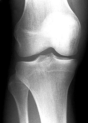
33 Knee Sagittal projection femur patella fossa intercondylaris condylus lat. femoris tuberculum intercondylare lat. condylus lat. tibiae condylus med. femoris tuberculum intercondylare mediale condylus medialis tibiae fibula tibia

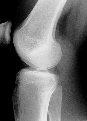
34 Knee Lateral projection femur base patella apex condylus lateralis condylus medialis eminentia intercondylaris apex capitis fibulae caput fibulae tuberositas tibiae fibula tibia

35
36

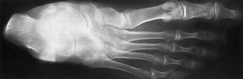
37 FOOT dorsoplantar projection phalanges sesamoids metatarsals tarsals navicular talus 5 caput base corpus calcaneus trochlea corpus base 1., 2., 3. = ossa cuneiformia mediale, intermedia et laterale tibia fibula

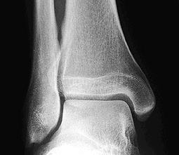
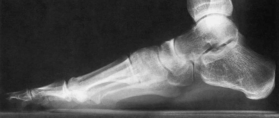
38 Foot Lateral projection sinus tarsi talus ossa cuneiformia calcaneus
39 The pictures used in this lectures were taken from following sources (if it was not written under the picture else): Atlas der Anatomie des Menschen/Sobotta. Putz,R., und Pabst,R. 20. Auflage. München:Urban & Schwarzenberg, 1993 Netter: Interactive Atlas of Human Anatomy. John T. Hansen Netter s clinical anatomy, Elsevier, 2014 Drake et al: Gray s Anatomy for Students Naňka, Elišková: Přehled anatomie. Galén, Praha Čihák: Anatomie I, II, III. Own archiv of the lecturer, archiv of Department of Anatomy, LF MU BRNO

40 Thank you for your attention
Skeleton of lower extremity Ossa membri inferioris
 Skeleton of lower extremity Ossa membri inferioris Skeleton of lower extremity consists of lower extremity girdle (cingulum membri inferioris) created by pelvis bone (os coxae) and bones of free lower
BOGOMOLETS NATIONAL MEDICAL UNIVERSITY. Department of human anatomy GUIDELINES
 BOGOMOLETS NATIONAL MEDICAL UNIVERSITY Department of human anatomy GUIDELINES Academic discipline HUMAN ANATOMY Module 1 Content module 2 The topic of the lesson Bones of the lower limb Course 1 st Number
The Lower Limb. Sevda LAFCI FAHRİOĞLU, MD.PhD.
 The Lower Limb Sevda LAFCI FAHRİOĞLU, MD.PhD. The Lower Limb The bones of the lower limb form the inferior part of the appendicular skeleton the organ of locomotion for bearing the weight of body stronger
Figure 7: Bones of the lower limb
 BONES OF THE APPENDICULAR SKELETON The appendicular skeleton is composed of the 126 bones of the appendages and the pectoral and pelvic girdles, which attach the limbs to the axial skeleton. Although the
The Appendicular Skeleton
 8 The Appendicular Skeleton PowerPoint Lecture Presentations prepared by Jason LaPres Lone Star College North Harris 8-1 The Pectoral Girdle The Pectoral Girdle Also called shoulder girdle Connects the
SKELETAL SYSTEM 206. AXIAL SKELETON 80 APPENDICULAR SKELETON 126 (see Figure 6.1) Clavicle. Clavicle. Pectoral girdles. Scapula. Scapula.
 Clavicle. Clavicle. Pectoral girdles. Scapula. Scapula.") SKELETAL SYSTEM 206 AXIAL SKELETON 80 APPENDICULAR SKELETON 126 (see Figure 6.1) Pectoral girdles 4 Clavicle Scapula 2 2 Clavicle Scapula Humerus 2 Humerus Upper limbs 60 Radius 2 Ulna Carpal bones Metacarpal
SKELETAL SYSTEM 206 AXIAL SKELETON 80 APPENDICULAR SKELETON 126 (see Figure 6.1) Pectoral girdles 4 Clavicle Scapula 2 2 Clavicle Scapula Humerus 2 Humerus Upper limbs 60 Radius 2 Ulna Carpal bones Metacarpal
Biology 218 Human Anatomy
 Chapter 8 Adapted from Tortora 10 th ed. LECTURE OUTLINE A. Introduction (p. 203) 1. The appendicular skeleton contains 126 bones that form: i. two pectoral (shoulder) girdles two upper limbs i one pelvic
Biology 218 Human Anatomy. Adapted from Martini Human Anatomy 7th ed. Chapter 7 The Skeletal System Appendicular Division
 Adapted from Martini Human Anatomy 7th ed. Chapter 7 The Skeletal System Appendicular Division Introduction The appendicular skeleton includes: Pectoral girdle Shoulder bones Upper limbs Pelvic girdle
SKELETON OF THE UPPER LIMB
 SKELETON OF THE UPPER LIMB L E C T U R E 2 D E N T I S T R Y 2016 R N D R. M I C H A E L A R A Č A N S K Á, P H. D. Skeleton of the upper limb (ossa membri superioris) Thirty-four bones form the skeletal
Pectoral (Shoulder) Girdle
 Girdle") Chapter 8 Skeletal System: Appendicular Skeleton Pectoral girdle Pelvic girdle Upper limbs Lower limbs 8-1 Pectoral (Shoulder) Girdle Consists of scapula and clavicle Clavicle articulates with sternum
Chapter 8 Skeletal System: Appendicular Skeleton Pectoral girdle Pelvic girdle Upper limbs Lower limbs 8-1 Pectoral (Shoulder) Girdle Consists of scapula and clavicle Clavicle articulates with sternum
Anatomy & Physiology. Muscles of the Lower Limbs.
 Anatomy & Physiology Muscles of the Lower Limbs http://www.ishapeup.com/musclecharts.html Muscles of the Lower Limbs Among the strongest muscles in the body. Because pelvic girdle is composed of heavy,
10/12/2010. Upper Extremity. Pectoral (Shoulder) Girdle. Clavicle (collarbone) Skeletal System: Appendicular Skeleton
 Girdle. Clavicle (collarbone) Skeletal System: Appendicular Skeleton") Skeletal System: Appendicular Skeleton Pectoral girdle Pelvic girdle Upper limbs Lower limbs 8-1 Pectoral (Shoulder) Girdle Consists of scapula and clavicle Clavicle articulates with sternum (Sternoclavicular
Skeletal System: Appendicular Skeleton Pectoral girdle Pelvic girdle Upper limbs Lower limbs 8-1 Pectoral (Shoulder) Girdle Consists of scapula and clavicle Clavicle articulates with sternum (Sternoclavicular
Bones of the Lower Limb Bone Structure Description Notes. border of the superior ramus. inferolaterally from the pubic symphysis
 Bones of the Lower Limb Bone Structure Description Notes pubis an angulated bone the forms the anterior part of the pelvis one of three bones that form the os coxae: ilium, ischium, pubis; its forms 1/5
Chapter 8. The Appendicular Skeleton. Lecture Presentation by Lee Ann Frederick University of Texas at Arlington Pearson Education, Inc.
 Chapter 8 The Appendicular Skeleton Lecture Presentation by Lee Ann Frederick University of Texas at Arlington An Introduction to the Appendicular Skeleton The Appendicular Skeleton 126 bones Allows us
Chapter 8B. The Skeletal System: Appendicular Skeleton. The Appendicular Skeleton. Clavicle. Pectoral (Shoulder) Girdle
 Girdle") The Appendicular Skeleton Chapter 8B The Skeletal System: Appendicular Skeleton 126 bones Pectoral (shoulder) girdle Pelvic (hip) girdle Upper limbs Lower limbs Functions primarily to facilitate movement
The Appendicular Skeleton Chapter 8B The Skeletal System: Appendicular Skeleton 126 bones Pectoral (shoulder) girdle Pelvic (hip) girdle Upper limbs Lower limbs Functions primarily to facilitate movement
Lab Activity 9. Appendicular Skeleton Martini Chapter 8. Portland Community College BI 231
 Lab Activity 9 Appendicular Skeleton Martini Chapter 8 Portland Community College BI 231 Appendicular Skeleton Upper & Lower extremities Shoulder Girdle Pelvic Girdle 2 Humerus 3 Humerus: Proximal End
Exercise 11. The Appendicular Skeleton
 Exercise 11 The Appendicular Skeleton The Appendicular Skeleton The appendicular skeleton contains 126 bones. Consists of the upper and lower limbs, the pectoral girdles, and the pelvic girdles. The pectoral
Bones of Lower Limb. Dr. Heba Kalbouneh Associate Professor of Anatomy and Histology
 Bones of Lower Limb Dr. Heba Kalbouneh Associate Professor of Anatomy and Histology Bones of the lower limb Hip Bone Made up of 3 bones: 1) Ilium (flat), superior in position 2) Ischium (L), postero-inferior
Anatomage Table Instructors Guide- Lower Limb
 The Lower Limb Anatomage Table Instructors Guide- Lower Limb Table of Contents Lower Limb 1- The Skeletal System...3 1: Hip Bone...3 2: Hip Joint and Femur...4 3: Patella and Knee Joint...7 4: Tibia, Fibula,
Dr.Israa H. Mohsen. Lecture 5. The vertebral column
 Anatomy Lecture 5 Dr.Israa H. Mohsen The vertebral column The vertebral column a flexible structure consisting of 33 vertebrae holds the head and torso upright, serves as an attachment point for the legs,
Principles of Anatomy and Physiology
 Principles of Anatomy and Physiology 14 th Edition CHAPTER 8 The Skeletal System: The Appendicular Skeleton The Appendicular Skeleton The 126 bones of the appendicular skeleton are primarily concerned
The Appendicular Skeleton
 8 The Appendicular Skeleton PowerPoint Lecture Presentations prepared by Jason LaPres Lone Star College North Harris An Introduction to the Appendicular Skeleton Learning Outcomes 8-1 Identify the bones
Biology 152 Appendicular Skeleton Anatomy Objectives
 Biology 152 Appendicular Skeleton Anatomy Objectives We will learn proper bone names, left/right/medial, and the parts of bones in this exercise. Start by learning the names of the bones. As you gain comfort
DORSAL MUSCLES OF THE HINDLIMB (ca)
") Fascia thoracolumbalis Fascia glutea Fascia lata Lamina superficialis Lamina profunda Fascia cruris DORSAL MUSCLES OF THE HINDLIMB (ca) M. gluteus superficialis o Origin: sacrum and first caudal vertebrae,
Fascia thoracolumbalis Fascia glutea Fascia lata Lamina superficialis Lamina profunda Fascia cruris DORSAL MUSCLES OF THE HINDLIMB (ca) M. gluteus superficialis o Origin: sacrum and first caudal vertebrae,
!"#$%&'(")*' '('+",' -.#/-.0*% #/-%#-#$"1% *'.+"2%
*' '('+,' -.#/-.0*% #/-%#-#$1% *'.+2%") !"#$%&'#(%( ) *%+,&-.) /%.#0#"12# 3-2)4(%( 5)62+ 7. 89-1+$12#!"#$%&'(")*' '('+",' -.#/-.0*% #/-%#-#$"1% *'.+"2% 5+2(+&12-.#1%&(-0#:- *%+,&-., 2012 University of Belgrade School of Medicine Du!ko V. Spasovski
!"#$%&'#(%( ) *%+,&-.) /%.#0#"12# 3-2)4(%( 5)62+ 7. 89-1+$12#!"#$%&'(")*' '('+",' -.#/-.0*% #/-%#-#$"1% *'.+"2% 5+2(+&12-.#1%&(-0#:- *%+,&-., 2012 University of Belgrade School of Medicine Du!ko V. Spasovski
OSTEOLOGY ОСТЕОЛОГИЯ
 КАЗАНСКИЙ ФЕДЕРАЛЬНЫЙ УНИВЕРСИТЕТ ИНСТИТУТ ФУНДАМЕНТАЛЬНОЙ МЕДИЦИНЫ И БИОЛОГИИ Кафедра морфологии и общей патологии А.А. Гумерова, С.Р. Абдулхаков, А.П. Киясов, Д.И. Андреева OSTEOLOGY ОСТЕОЛОГИЯ Учебно-методическое
Amy Warenda Czura, Ph.D. 1 SCCC BIO130 Lab 7 Appendicular Skeleton & Articulations
 The Skeletal System II: Appendicular Skeleton and Articulations Exercises 11, 13 (begins: page 145 in 9 th and 10 th editions) Exercises 10, 11 (begins: page 147 in 11 th edition, page 149 in 12 th edition)
Chapter 8 Outline. Pectoral Girdle Upper Limb Pelvic Girdle Lower Limb Aging of the Appendicular Skeleton Development of the Appendicular Skeleton
 Chapter 8 Outline Pectoral Girdle Upper Limb Pelvic Girdle Lower Limb Aging of the Appendicular Skeleton Development of the Appendicular Skeleton Figure 8.1 Appendicular Skeleton Pectoral Girdle Clavicle
Copyright 2003 Pearson Education, Inc. publishing as Benjamin Cummings. Dr. Nabil Khouri MD, MSc, Ph.D
 Dr. Nabil Khouri MD, MSc, Ph.D Pelvic Girdle (Hip) Organization of the Lower Limb It is divided into: The Gluteal region The thigh The knee The leg The ankle The foot The thigh and the leg have compartments
C. Bones of the Pelvic Girdle
 C. Bones of the Pelvic Girdle 1. 2 coxal bones (a.k.a hip bones): -bony pelvis is made up of hip bones, sacrum, & coccyx -pelvic bones are large & heavy & attach to the axial skeleton via sacrum/coccyx
Chapter 8 The Skeletal System: The Appendicular Skeleton. Copyright 2009 John Wiley & Sons, Inc.
 Chapter 8 The Skeletal System: The Appendicular Skeleton Appendicular Skeleton It includes bones of the upper and lower limbs Girdles attach the limbs to the axial skeleton The pectoral girdle consists
PRE-LAB EXERCISES. Before we get started, look up the definitions of these common bone marking terms: Canal: Condyle: Facet: Fissure:
 1 PRE-LAB EXERCISES When studying the skeletal system, the bones are often sorted into two broad categories: the axial skeleton and the appendicular skeleton. This lab focuses on the appendicular skeleton,
Chapter 8 The Skeletal System: The Appendicular Skeleton. Copyright 2009 John Wiley & Sons, Inc.
 Chapter 8 The Skeletal System: The Appendicular Skeleton Appendicular Skeleton The primary function is movement It includes bones of the upper and lower limbs Girdles attach the limbs to the axial skeleton
A. Incorrect! The appendicular skeleton includes bones of the shoulder, arm, hand, pelvis, leg and foot.
 Anatomy and Physiology - Problem Drill 08: The Skeletal System III No. 1 of 10 1. Which of the following statements about the appendicular skeleton is correct? A. The appendicular skeleton includes bones
Muscles of Lesson Five. Muscular Nomenclature and Kinesiology - Two. Muscles of Lesson Five, cont. Chapter 16
 Chapter 16 Muscular Nomenclature and Kinesiology - Two Lessons 5-6 Muscles of Lesson Five Iliopsoas (psoas major, iliacus) Hip outward rotators (piriformis, gemellus superior, gemellus inferior, obturator
Lower limb summary. Anterior compartment of the thigh. Done By: Laith Qashou. Doctor_2016
 Lower limb summary Done By: Laith Qashou Doctor_2016 Anterior compartment of the thigh Sartorius Anterior superior iliac spine Upper medial surface of shaft of tibia 1. Flexes, abducts, laterally rotates
Topic 7: Hip and pelvis. Parts of the hip. Parts of the femur
 Topic 7: Hip and pelvis Parts of the hip Parts of the femur Classifying the hip joint Ball and socket Synovial Multiaxial Movements of the hip: Abduction/adduction Flexion/extension Medial/lateral rotation
Anatomy of the lower limb
 Anatomy of the lower limb 1. Bones of the lower limb Pelvis Hip bone/coxal bone Acetabulum o Acetabular margin o Acetabular fossa o Acetabular notch o Lunate surface Ischiopubic ramus Obturator foramen
lesser trochanter of femur lesser trochanter of femur iliotibial tract (connective tissue) medial surface of proximal tibia
 medial surface of proximal tibia") LOWER LIMB MUSCLES OF THE APPENDICULAR SKELETON The muscles that act on the lower limb fall into three groups: those that move the thigh, those that move the lower leg, and those that move the ankle, foot,
LOWER LIMB MUSCLES OF THE APPENDICULAR SKELETON The muscles that act on the lower limb fall into three groups: those that move the thigh, those that move the lower leg, and those that move the ankle, foot,
Bone Flashcards for 10a
 Bone Flashcards for 0a CLAVICLE (collar bone). Sternal extremity (end) flat end. Acromial extremity (end) rounded end. SCAPULA (shoulder blade). Right or left scapula?. Superior border (superior margin).
Muscles of the lower extremities. Dr. Nabil khouri MD, MSc, Ph.D
 Muscles of the lower extremities Dr. Nabil khouri MD, MSc, Ph.D Posterior leg Popliteal fossa Boundaries Biceps femoris (superior-lateral) Semitendinosis and semimembranosis (superior-medial) Gastrocnemius
Appendicular Skeleton. Prof. Abdulameer Al-Nuaimi
 Appendicular Skeleton Prof. Abdulameer Al-Nuaimi a.alnuaimi@sheffield.ac.uk abdulameerh@yahoo.com Hi Prof, It is great to hear from you, I really enjoyed your teaching last year. You taught me the hardest
Human Anatomy, First Edition McKinley & O'Loughlin
 Human Anatomy, First Edition McKinley & O'Loughlin Chapter 8 : Appendicular Skeleton 8-1 Appendicular Skeleton Includes the bones of the upper and lower limbs. The girdles of bones that attach the upper
Copyright 2003 Pearson Education, Inc. publishing as Benjamin Cummings. Dr. Nabil khouri
 Dr. Nabil khouri Appendicular Skeleton The appendicular skeleton is made up of the bones of the upper and lower limbs and their girdles Two girdles: Pectoral girdles attach the upper limbs to the body
Practical 1 Worksheet
 Practical 1 Worksheet ANATOMICAL TERMS 1. Use the word bank to fill in the missing words. reference side stand body arms palms anatomical forward All anatomical terms have a(n) point which is called the
Riverside Community College Anatomy & Physiology 2B SPRING 2012 EXAM #1-ABC (Nervous System)
") Riverside Community College Anatomy & Physiology 2B SPRING 2012 EXAM #1-ABC (Nervous System) Name: 1) This vertebra is an example of a(n). 1) A) thoracic B) axis C) atlas D) lumbar E) sacral 1 2) W hich
Riverside Community College Anatomy & Physiology 2B SPRING 2012 EXAM #1-ABC (Nervous System) Name: 1) This vertebra is an example of a(n). 1) A) thoracic B) axis C) atlas D) lumbar E) sacral 1 2) W hich
Chapter 7 Part C The Skeleton
 Chapter 7 Part C The Skeleton Part 2 The Appendicular Skeleton Consists of bones of the limbs and their girdles Pectoral girdle Attaches upper limbs to body trunk Pelvic girdle Attaches lower limbs to
Human Anatomy Biology 351
 Human Anatomy Biology 351 Lower Limb Please place your name on the back of the last page of this exam. You must answer all questions on this exam. Because statistics demonstrate that, on average, between
It is formed by fusion of 3 bones: I. Ilium (superior bone). II. Pubis (antero-inferior bone). III. Ischium (postero-inferior bone).
. II. Pubis (antero-inferior bone). III. Ischium (postero-inferior bone).") It is formed by fusion of 3 bones: I. Ilium (superior bone). II. Pubis (antero-inferior bone). III. Ischium (postero-inferior bone). Pubis Acetabulum Ana (242 ) The three constituent of bones of the hip
It is formed by fusion of 3 bones: I. Ilium (superior bone). II. Pubis (antero-inferior bone). III. Ischium (postero-inferior bone). Pubis Acetabulum Ana (242 ) The three constituent of bones of the hip
Chapter 7: Skeletal System: Gross Anatomy
 Chapter 7: Skeletal System: Gross Anatomy I. General Considerations A. How many bones in an average adult skeleton? B. Anatomic features of bones are based on II. Axial Skeleton A. Skull 1. Functionally
The Lower Limb VII: The Ankle & Foot. Anatomy RHS 241 Lecture 7 Dr. Einas Al-Eisa
 The Lower Limb VII: The Ankle & Foot Anatomy RHS 241 Lecture 7 Dr. Einas Al-Eisa Ankle joint Synovial, hinge joint Allow movement of the foot in the sagittal plane only (1 degree of freedom): dorsiflexion:
Pelvic cavity. Gross anatomy of the lower limb. Walking. Sándor Katz M.D.,Ph.D.
 Pelvic cavity. Gross anatomy of the lower limb. Walking. Sándor Katz M.D.,Ph.D. Lower limb Pelvic girdle Free lower extremity Hip bone Definitive fusion of the Y- shaped growth plate occurs 16th -18th
Joints and muscles of the foot. Architecture of the foot. Sándor Katz M.D.,Ph.D.
 Joints and muscles of the foot. Architecture of the foot. Sándor Katz M.D.,Ph.D. Ankle (talocrural) joint type: hinge Talocrural joint - medial collateral ligament Medial collateral = deltoid ligament
BONES JOINTS MUSCLES OF THE LOWER LIMB
 BONES JOINTS MUSCLES OF THE LOWER LIMB LOWER LIMB: BONES LOWER LIMB GLUTEAL REGION consists of 6 major segments: FEMORAL REGION (THIGH) KNEE REGION LEG REGION TALOCRURAL REGION (ANKLE) FOOT REGION LOWER
11/25/2012. Chapter 7 Part 2: Bones! Skeletal Organization. The Skull. Skull Bones to Know Cranium
 Chapter 7 Part 2: Bones! 5) Distinguish between the axial and appendicular skeletons and name the major parts of each 6) Locate and identify the bones and the major features of the bones that compose the
The Skeletal System THE APPENDICULAR SKELETON
 The Skeletal System THE APPENDICULAR SKELETON The appendicular skeleton consists of the girdles and the skeleton of the limbs. The upper (anterior) limbs are attached to the pectoral (shoulder) girdle
BLUE SKY SCHOOL OF PROFESSIONAL MASSAGE AND THERAPEUTIC BODYWORK Musculoskeletal Anatomy & Kinesiology KNEE & ANKLE MUSCLES
 BLUE SKY SCHOOL OF PROFESSIONAL MASSAGE AND THERAPEUTIC BODYWORK Musculoskeletal Anatomy & Kinesiology KNEE & ANKLE MUSCLES MSAK201-I Session 3 1) REVIEW a) THIGH, LEG, ANKLE & FOOT i) Tibia Medial Malleolus
Exercise Science Section 2: The Skeletal System
 Exercise Science Section 2: The Skeletal System An Introduction to Health and Physical Education Ted Temertzoglou Paul Challen ISBN 1-55077-132-9 Role of the Skeleton Protection Framework Attachments for
Connection of the lower limb (juncturae ossium extremitatis inferioris)
") Connection of the lower limb (juncturae ossium extremitatis inferioris) Connection of the pelvic girdle (juncturae ossium cinguli extremitatis inferioris) 1. Sacroiliac joint (articulatio sacroiliaca)
Connection of the lower limb (juncturae ossium extremitatis inferioris) Connection of the pelvic girdle (juncturae ossium cinguli extremitatis inferioris) 1. Sacroiliac joint (articulatio sacroiliaca)
5.1 Identify, describe the attachments of and deduce the actions of the muscles of the thigh:
 5.1 Identify, describe the attachments of and deduce the actions of the muscles of the thigh: Anterior group Proximal attachment Distal attachment Sartorius ASIS» Upper part of shaft tibia (middle surface)»
Axial skeleton bones and markings
 Axial skeleton bones and markings Skull Cranial bones Frontal x 1 Supraorbital foramen Occipital x 1 Foramen magnum Occipital condyles Superior nuchal line Inferior nuchal line Anterior cranial fossa External
Contents of the Posterior Fascial Compartment of the Thigh
 Contents of the Posterior Fascial Compartment of the Thigh 1-Muscles: B i c e p s f e m o r i s S e m i t e n d i n o s u s S e m i m e m b r a n o s u s a small part of the adductor magnus (h a m s t
the Skeletal System provided by Academic Web Services Grand Canyon University
 Anatomy Resource Center Study Guides the Skeletal System HEAD & NECK REGIONAL VIEW SKULL BONES CRANIUM FACE SKULL LANDMARKS ANTERIOR SIDE SUPERIOR/INFERIOR VERTEBRAL COLUMN VERTEBRAL REGIONS CERVICAL C1
TEST YOURSELF- Chapter 7
 TEST YOURSELF- Chapter 7 Cranial Bones 1. Give the name of the bone for each of the following markings. Some of the markings are found on more than one bone. List all that apply. Cranium a. Frontal squama:
Perpendicular Plate Zygomatic Bone. Mental Foramen Mandible
 Glabella Frontal Middle Nasal Concha Nasal Lacrimal Perpendicular Plate Zygomatic Inferior Nasal Concha Maxilla Mental Mandible Skull (anterior view) Squamosal Suture Coronal Suture Frontal Parietal Nasal
BIOLOGY 113 LABORATORY Skeletal System
 BIOLOGY 113 LABORATORY Skeletal System Objectives Distinguish between the axial and appendicular skeleton. Distinguish between the cranium and facial skeleton. Locate and name the bones of the skull and
Located more distal and anterior together with Trapezoid, anterior to scaphoid Trapezium rarely to be fractured.
 The hand The hand consists of the 3 groups: Proximal part: carpals bones (8) Middle part: metacarpal bones (5) Distal part: fingers or phalanges bones (3 for each finger except for the thumb just 2 bones).
Lab Exercise #04 The Skeletal System Student Performance Objectives
 Lab Exercise #04 The Skeletal System Student Performance Objectives The material that you are required to learn in this exercise can be found in either the lecture text or the supplemental materials provided
Biology 2401 The Skeletal System
 Biology 2401 The Skeletal System Purpose: The lab will describe the microscopic and gross anatomy of bone, identify bones of the body, and identify important bone markings. I. Overview of the Skeleton
CHAPTER 8 LECTURE OUTLINE
 CHAPTER 8 LECTURE OUTLINE I. INTRODUCTION A. The appendicular skeleton includes the bones of the upper and lower extremities and the shoulder and hip girdles. B. The appendicular skeleton functions primarily
Chapter 8: The Appendicular Skeleton
 Chapter 8: The Appendicular Skeleton In Chapter 8, we complete our tour of the skeleton that began in Chapter 7. As with Chapter 7, much of this material is best learned in lab, but we will outline some
Important Parts of Bones
 Important Parts of Bones For 2015 Know: Humerus (posterior) Clavical Femur (Anterior) Foot Hand Mandible Os Coxa Scapula Skull (Anterior, Inferior, Lateral) Sternum Humerus (posterior) A. olecranon fossa
Human Anatomy Biology 351
 Human Anatomy Biology 351 Lower Limb Please place your name on the back of the last page of this exam. You must answer all questions on this exam. Because statistics demonstrate that, on average, between
bio4165 lab quiz 1 Posterior View Anterior View Lateral View Anterior View bio fall.quarter lab.quiz.1...page.1 of 6
 B A Posterior View D C E Lateral View bio.4165...fall.quarter.2005...lab.quiz.1...page.1 of 6 F I G 35 Posterior View H bio.4165...fall.quarter.2005...lab.quiz.1...page.2 of 6 J Posterior View L K Inferior
The Dance Hall by Vincent van Gogh,1888
 The Dance Hall by Vincent van Gogh,1888 Articulations of the pelvic girdle Lumbosacral joints, sacroiliac joints & pubic symphysis The remaining joints of the lower limb Hip joint Knee joint Tibiofibular
The Hip (Iliofemoral) Joint. Presented by: Rob, Rachel, Alina and Lisa
 Joint. Presented by: Rob, Rachel, Alina and Lisa") The Hip (Iliofemoral) Joint Presented by: Rob, Rachel, Alina and Lisa Surface Anatomy: Posterior Surface Anatomy: Anterior Bones: Os Coxae Consists of 3 Portions: Ilium Ischium Pubis Bones: Pubis Portion
The Hip (Iliofemoral) Joint Presented by: Rob, Rachel, Alina and Lisa Surface Anatomy: Posterior Surface Anatomy: Anterior Bones: Os Coxae Consists of 3 Portions: Ilium Ischium Pubis Bones: Pubis Portion
Spring Written By: J. E. Sutton. Contents: I. Overview of the Skeleton: II. Appendicular Skeleton III. Axial Skeleton IV.
 Spring 2012 Written By: J. E. Sutton Contents: I. Overview of the Skeleton: II. Appendicular Skeleton III. Axial Skeleton IV. Articulations Overview of the Skeleton: I. Orientation to Human Skeleton: a.
BLUE SKY SCHOOL OF PROFESSIONAL MASSAGE AND THERAPEUTIC BODYWORK. Musculoskeletal Anatomy & Kinesiology I TERMINOLOGY, STRUCTURES, & SKELETAL OVERVIEW
 BLUE SKY SCHOOL OF PROFESSIONAL MASSAGE AND THERAPEUTIC BODYWORK Musculoskeletal Anatomy & Kinesiology I TERMINOLOGY, STRUCTURES, & SKELETAL OVERVIEW MSAK101-I Session 1 Learning Objectives: 1. Define
Anatomy & Physiology Skeletal System Worksheet
 1. Name the five functions of the skeleton. c) d) e) Anatomy & Physiology Skeletal System Worksheet 2. The term for the shaft of a bone is:. 3. The bony struts found in spongy bone are called. 4. In ossification,
Lower Limb Dr. Robin Paudel
 Lower Limb n What is a limb? n Skeleton n Joints n Pelvis or limb girdle n Hip/Hip Muscles n Lumber and sacral plexus getting spinal nerves out onto limb n Muscles anterior and posterior compartments n
Bone List Anatomy
 1 Frontal Bone Skull 2 Parietal Bone Skull 3 Occipital Bone Skull 4 Temporal Bone Skull 5 Coronal Suture Skull 6 Sagittal Suture Skull 7 Squamous suture Skull 8 Lambdoid Suture Skull 9 Surpaorbital Ridge
Bones of Thorax (Rib Cage)
") Musculoskeletal System (Part A-2) Module 7 -Chapter 10 Overview Muscles Attachments Bones Bone types Surface features of bones Divisions of the skeletal system Joints or Articulations Susie Turner, M.D.
Musculoskeletal System (Part A-2) Module 7 -Chapter 10 Overview Muscles Attachments Bones Bone types Surface features of bones Divisions of the skeletal system Joints or Articulations Susie Turner, M.D.
Bone Composition. Bone is very strong for its relatively light weight The major components of bone are:
 Human Bones Bone Composition Bone is very strong for its relatively light weight The major components of bone are: Calcium carbonate Calcium phosphate Collagen Water Cortical Bone Spongy Bone Medullary
The Lower Limb. Anatomy RHS 241 Lecture 2 Dr. Einas Al-Eisa
 The Lower Limb Anatomy RHS 241 Lecture 2 Dr. Einas Al-Eisa The bony pelvis Protective osseofibrous ring for the pelvic viscera Transfer of forces to: acetabulum & head of femur (when standing) ischial
Hip joint and pelvic girdle. Lower Extremity. Pelvic Girdle 6/5/2017
 Hip joint and pelvic girdle Lower Extremity The relationship between the pelvic girdle and hip is similar to that between the shoulder girdle and shoulder joint. The lower limbs are attached to the axial
Dr Nabil khouri MD. MSc. Ph.D
 Dr Nabil khouri MD. MSc. Ph.D Foot Anatomy The foot consists of 26 bones: 14 phalangeal, 5 metatarsal, and 7 tarsal. Toes are used to balance the body. Metatarsal Bones gives elasticity to the foot in
LAB Notes#1. Ahmad Ar'ar. Eslam
 LAB Notes#1 Ahmad Ar'ar Eslam 1 P a g e Anatomy lab Notes Lower limb bones :- Pelvic girdle: It's the connection between the axial skeleton and the lower limb; it's made up of one bone called the HIP BONE
Balanced Body Movement Principles
 Balanced Body Movement Principles How the Body Works and How to Train it. Module 3: Lower Body Strength and Power Developing Strength, Endurance and Power The lower body is our primary source of strength,
Ch. 5 - Skeletal System
 Ch. 5 - Skeletal System Bones are living, ever-changing structures. This allows them grow and adapt to new situations that the body encounters. The functions of the skeletal system: 1) support bones are
Radiographic Positioning Summary (Basic Projections RAD 222)
") Lower Extremity Radiographic Positioning Summary (Basic Projections RAD 222) AP Pelvis AP Hip (Unilateral) (L or R) AP Femur Mid and distal AP Knee Lateral Knee Pt lies supine on table Align MSP to Center
Lower Extremity Radiographic Positioning Summary (Basic Projections RAD 222) AP Pelvis AP Hip (Unilateral) (L or R) AP Femur Mid and distal AP Knee Lateral Knee Pt lies supine on table Align MSP to Center
Appendicular Skeleton. Dr. Carmen E. Rexach Anatomy 35 Mt. San Antonio College
 Appendicular Skeleton Dr. Carmen E. Rexach Anatomy 35 Mt. San Antonio College Pectoral girdle clavicle scapula Upper limb brachium antebrachium carpus manus Pelvic girdle oscoxae Lower limb femoral region
Pelvic Girdle
 ARTICULATIONS OF LOWER EXTREMITY Pages 429-437 Pelvic Girdle formed by connection of the hip bones and the sacrum Sacroiliac Joints compound joints synovial joint - anterior, between the auricular surfaces
The Lower Limb II. Anatomy RHS 241 Lecture 3 Dr. Einas Al-Eisa
 The Lower Limb II Anatomy RHS 241 Lecture 3 Dr. Einas Al-Eisa Tibia The larger & medial bone of the leg Functions: Attachment of muscles Transfer of weight from femur to skeleton of the foot Articulations
TABLE OF MUSCLES OF LOWER EXTREMITY 2018zillmusom ANTERIOR THIGH
 TABLE OF MUSCLES OF LOWER EXTREMITY 2018zillmusom ANTERIOR THIGH MUSCLE ORIGIN INSERTION ACTION NERVE Iliopsoas Ilium, vertebra Femur Flex hip joint Femoral nerve (T12-L5) Pectineus Pubis Femur Flex hip
In-Depth Foundations: Anatomy Terms to Know
 Be familiar with / able to identify and define all the following parts. The Spine Cranium Vertebrae Cervical, Thoracic, Lumbar Sacrum Coccyx Bones of Upper Body Cranium Mastoid process; Occipital condyle,
First practical session. Bones of the gluteal region
 First practical session 2017 Bones of the gluteal region The Hip bone The hip bone is made of: 1 The ilium: superior in position 2 The ischium:postero-inferior in position 3 The pubis: antero-inferior
radiologymasterclass.co.uk
 http://radiologymasterclass.co.uk Hip X-ray anatomy - Normal AP (anterior-posterior) Shenton's line is formed by the medial edge of the femoral neck and the inferior edge of the superior pubic ramus Loss
topographical anatomy
 Kaan Yücel M.D., Ph.D. 30. September 2014 Tuesday topographical anatomy organization of the human body as major parts or segments Head Neck Trunk thorax, abdomen, back, & pelvis/perineum Upper limbs &
INDIVIDUAL TYPES OF VERTEBRAE
 THE VERTEBRAL COLUMN The vertebral column (columna vertebralis) or the spine has a metameric structure (a feature connecting the vertebrates with the earliest invertebrates) and consists of separate bone
~, /' ~::'~ EXTENSOR HALLUCIS LONGUS. Leg-anterolateral :.:~ / ~\,
 TIBIALIS ANTERIOR Lateral condyle of tibia, upper half of lateral surface of tibia, interosseous membrane Medial side and plantar surface of medial cuneiform bone, and base of first metatarsal bone Dorsiflexes