Appendix chapter 3: Pterosaurs from the Lower Cretaceous of Brazil in the Stuttgart Collection (and supplement)
|
|
|
- Griselda Lamb
- 5 years ago
- Views:
Transcription
1 Appendix chapter 3: Pterosaurs from the Lower Cretaceous of Brazil in the Stuttgart Collection (and supplement) Appendix chapter 3 203
2 3.5. Appendix Measurements Length, as preserved 385 Length, reconstructed 400 Height at last alveolus 23.3 Width at symphysis 34.0 Largest width of rami 98.4 Depth dentary sagittal crest, as preserved 35 Length dentary sagittal crest 118 Width at second pair of teeth 21.0 Width at third pair of teeth 22.3 Width at fourth pair of teeth 21.8 Width at fifth pair of teeth 21.7 Table 3.1. Measurements of the mandible, SMNS in mm. SMNS Length, as preserved 138 Diameter shaft 17* SMNS Length 243 Width shaft, dorsal ventral plane 28.4 Width shaft, anterior posterior plane 42.9 Width proximal aspect, dorsal ventral plane 37.3 Width proximal aspect, anterior posterior plane 56.6 Width distal aspect, dorsal ventral plane 43.3 Width distal aspect, anterior posterior plane 67.7 SMNS Length, as preserved 154 Width shaft, dorsal ventral plane (beginning deltopectoral crest) 23.5 Width shaft : maximum height deltopectoral crest, as preserved 56.9 Table 3.2. Measurements of isolated humeri in mm. SMNS ulna Length, as preserved 255 Diameter shaft (anterior) 19.7 Width proximal aspect, as preserved 33 Width proximal aspect, as preserved 33 SMNS ulna Length, as preserved 150* Width proximal aspect, as preserved 27 x 28* Diameter shaft 19 SMNS radius Length, as preserved 150* Width proximal aspect, as preserved 27 x 11* Diameter shaft 8.3 SMNS ulna Length 395 Diameter shaft (anterior) 27 Width proximal aspect 66* Width distal aspect 69 SMNS ulna Length, as preserved 117 Width proximal aspect 29* Diameter shaft 12 x 18 Table 3.3. Measurements of ulnae and radius in mm. *: approximate. Appendix chapter 3 204

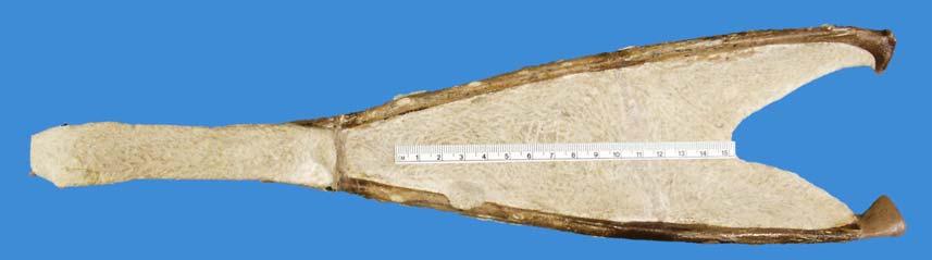
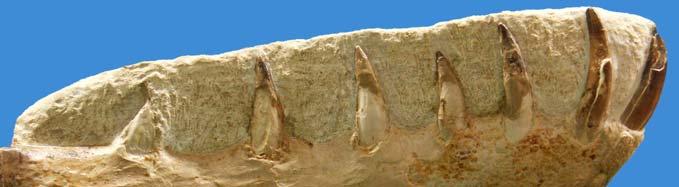
3 Diameter ulna Diameter radius Ratio radius:ulna Santadactylus pricei* (BSP 1980 I 122) :1.8 Santadactylus pricei (AMNH 22552) :1.9 SMNS :2.3 Coloborhynchus spielbergi** :2.6 Table 3.4. Ratios of the diameter of ulna/radius. *: measurements from Wellnhofer (1985: 134); **: for comparative reasons a large ulna and radius (410 and 401 mm respectively). Length Diameter Ratios Humerus Diameters radius:ulna 1:2.3 Ulna Diameter : length humerus 1:10.9 Radius 260 5* Lengths humerus : ulna 1:1.4 Table 3.5. Measurements of humerus and ulna/radius, SMNS in mm. *: approximate. SMNS Length, as preserved 40 Width shaft (anterior posterior) 13.6* Width proximal aspect 41.6* Width distal aspect (matrix) 22.4* SMNS Length 446 Width shaft (anterior posterior) 18.1 Width proximal aspect 41.2 Width distal aspect (matrix) 29.3 Table 3.6. Measurements of phalanges in mm. *: approximate Length, as preserved Diameter Ulna Radius First phalanx fourth digit 158* 5.5x124/6.2 x 9.5** Second phalanx fourth digit /10.7** Fourth phalanx fourth digit /7.3** Table 3.7. Measurements of front extremities in mm. *: approximate; **: proximal and distal measurements respectively Figures and plates Figure 3.1. Mandible of cf. Cr. mesembrinus (SMNS 56994) in various aspects. A (left): anterior. Below, next page B: left lateral; C: detail crest, left; D: dorsal; E: right lateral; F: detail crest, right. Photographs by E. Endenburg and the author. Courtesy of SMS, Stuttgart. Appendix chapter 3 205


4 B C D E F Appendix chapter 3 206
5 Figure 3.2. Mandible of cf. Cr. mesembrinus (SMNS 56994) in various aspects. A: anterior; B: left lateral; C: dorsal; D: posterior (right side); E: right lateral; F: ventral. Scale bars = 50 mm. Drawings by the author. Appendix chapter 3 207


6 Figure 3.3A (left) and 3.4A (right). Isolated humerus SMNS Photographs and drawings by E. Endenburg and the author. Courtesy of SMS, Stuttgart. Figure 3.3B (left) and 3.4B (right). Isolated humeri SMNS Photographs and drawings by E. Endenburg and the author. Courtesy of SMS, Stuttgart. Appendix chapter 3 208
 in (from top to bottom, left to right) proximal, posterior, ventral, anterior, dorsal and distal")
. Isolated humerus. Co.")
7 C Figure 3.3C (above). Isolated humerus. Co. araripensis (SMNS 55409) in (from top to bottom, left to right) proximal, posterior, ventral, anterior, dorsal and distal views; Photographs by E. Endenburg and the author. Courtesy of SMS, Stuttgart. Figure 3.4C (left). Isolated humerus. Co. araripensis (SMNS 55409) in (from top to bottom, left to right) proximal, posterior, ventral, dorsal and distal views. Scale bar = 50 mm. Drawings by the author. Courtesy of SMS, Stuttgart. Appendix chapter 3 209
.")


.")
: ulna of cf. S.")
: ulna/radius of cf. S.")
8 D Figure 3.4D (left) and 3.5D (right). Isolated humerus. Cf. S. pricei (SMNS 55883). Scale bar = 50 mm. Photographs and drawings by E. Endenburg and the author. Courtesy of SMS, Stuttgart. A B Figure 3.5 (top left and right) and 3.6 (bottom left and right). Isolated ulnae and radii. A (top left, bottom left): ulna of cf. S. pricei (SMNS 55410); B (top right, bottom right): ulna/radius of cf. S. pricei (SMNS 55411). Scale bar = 50 mm. Photographs and drawings by E. Endenburg and the author. Courtesy of SMS, Stuttgart. Appendix chapter 3 210
. Figure 3.6D (right): ulna SMNS 82001. Scale bar = 50 mm. Photographs and drawings by E.")
. Scale bar = 50 mm. Photographs and drawings by E. Endenburg and the author.")
9 C Figure 3.5 (left) and 3.6 (middle and right). Isolated ulnae and radii. Figure 3.5C (left and middles): ulna of cf. C. spielbergi (SMNS 55413). Figure 3.6D (right): ulna SMNS Scale bar = 50 mm. Photographs and drawings by E. Endenburg and the author. Courtesy of SMS, Stuttgart. Figure 3.7 (left) and 3.8 (right). Associated humerus and ulna/radius (SMNS 81976). Scale bar = 50 mm. Photographs and drawings by E. Endenburg and the author. Courtesy of SMS, Stuttgart. Appendix chapter 3 211
; B: SMNS 55415. Photographs by E.")
; B: SMNS 55415.")
10 A B Figure 3.9. Phalanges of wing finger. A: SMNS (obverse and reverse); B: SMNS Photographs by E. Endenburg and the author. Courtesy of SMS, Stuttgart. Figure Phalanges of wing finger. A: SMNS (obverse and reverse); B: SMNS Scale bars = 50 mm. Drawings by the author. Appendix chapter 3 212
.")
. Scale bar = 50 mm.")

11 Figure 3.11 (left) and 3.12 (right). Partial front extremity (SMNS 80437). Scale bar = 50 mm. Photographs and drawings by E. Endenburg and the author. Courtesy of SMS, Stuttgart. Appendix chapter 3 213
Appendix chapter 4: Description of two pterosaurs (Pterodactyloidea) mandibles from the Lower Cretaceous Santana Formation, Brazil
 mandibles from the Lower Cretaceous Santana Formation, Brazil") Appendix chapter 4: Description of two pterosaurs (Pterodactyloidea) mandibles from the Lower Cretaceous Santana Formation, Brazil Appendix chapter 4 215 4.5. Appendix 4.5.1. Measurements Anterior part
Appendix chapter 4: Description of two pterosaurs (Pterodactyloidea) mandibles from the Lower Cretaceous Santana Formation, Brazil Appendix chapter 4 215 4.5. Appendix 4.5.1. Measurements Anterior part
Appendix chapter 7: Updating the toothed taxa
 Appendix chapter 7: Updating the toothed taxa Appendix chapter 7 25 7.. Appendix 7..1. Figures and plates Figure 7.1. Type specimen of Brasileodactylus and holotype of B. araripensis (MN 484-V). Scale
Introduction to Human Osteology Chapter 3: Hands and Feet
 Introduction to Human Osteology Chapter 3: Hands and Feet Roberta Hall Kenneth Beals Holm Neumann Georg Neumann Gwyn Madden Revised in 1978, 1984, and 2008 Bones of the Hand Eight carpal bones, in two
Perpendicular Plate Zygomatic Bone. Mental Foramen Mandible
 Glabella Frontal Middle Nasal Concha Nasal Lacrimal Perpendicular Plate Zygomatic Inferior Nasal Concha Maxilla Mental Mandible Skull (anterior view) Squamosal Suture Coronal Suture Frontal Parietal Nasal
the study of the body s physical structures
 Chapter 1: Page 10 anatomy anterior (ventral) coronal (frontal) plane distal the study of the body s physical structures directional term meaning "toward the front" plane of reference which divides the
Lab Activity 11: Group II
 Lab Activity 11: Group II Muscles Martini Chapter 11 Portland Community College BI 231 Origin and Insertion Origin: The place where the fixed end attaches to a bone, cartilage, or connective tissue. Insertion:
Country Health SA Medical Imaging
 Country Health SA Medical Imaging REMOTE OPERATORS POSITIONING GUIDE Contents Image Evaluation Page 4 Positioning Guides Section 1 - THORAX 1.1 Chest Page 5 1.2 Bedside Chest Page 7 1.3 Ribs Page 8 Section
RADIOGRAPHY OF THE HAND, FINGERS & THUMB
 RADIOGRAPHY OF THE HAND, FINGERS & THUMB FINGERS (2nd 5th) - PA Projection Patient Position: Seated; hand ; elbow on IR table top Part Position: Fingers centered to IR unless protocol is Central Ray: Perpendicular
Forensic Archaeology & Forensic Anthropology. ADJ14 Advanced Criminal Investigations
 Forensic Archaeology & Forensic Anthropology ADJ14 Advanced Criminal Investigations Anthropology & Archaeology Anthropology is the study of the biological and cultural aspects of all humans in all places
Bone Flashcards for 10a
 Bone Flashcards for 0a CLAVICLE (collar bone). Sternal extremity (end) flat end. Acromial extremity (end) rounded end. SCAPULA (shoulder blade). Right or left scapula?. Superior border (superior margin).
Radiographic Positioning Summary (Basic Projections RAD 222)
") Lower Extremity Radiographic Positioning Summary (Basic Projections RAD 222) AP Pelvis AP Hip (Unilateral) (L or R) AP Femur Mid and distal AP Knee Lateral Knee Pt lies supine on table Align MSP to Center
Lower Extremity Radiographic Positioning Summary (Basic Projections RAD 222) AP Pelvis AP Hip (Unilateral) (L or R) AP Femur Mid and distal AP Knee Lateral Knee Pt lies supine on table Align MSP to Center
SD School Anatomy Program 1: Bones QuikNotes. Student Notes
 QuikNotes The transverse plane runs from right to left and divides the body into superior (upper) and inferior (lower) sections. Student Notes The frontal plane lies vertically along the body from head
Muscular Nomenclature and Kinesiology - One
 Chapter 16 Muscular Nomenclature and Kinesiology - One Lessons 1-3 (with lesson 4) 1 Introduction 122 major muscles covered in this chapter Chapter divided into nine lessons Kinesiology study of human
Exercise Science Section 2: The Skeletal System
 Exercise Science Section 2: The Skeletal System An Introduction to Health and Physical Education Ted Temertzoglou Paul Challen ISBN 1-55077-132-9 Role of the Skeleton Protection Framework Attachments for
Hand Anatomy A Patient's Guide to Hand Anatomy
 Hand Anatomy A Patient's Guide to Hand Anatomy Introduction Few structures of the human anatomy are as unique as the hand. The hand needs to be mobile in order to position the fingers and thumb. Adequate
Types of Body Movements
 Types of Body Movements Bởi: OpenStaxCollege Synovial joints allow the body a tremendous range of movements. Each movement at a synovial joint results from the contraction or relaxation of the muscles
Medical Terminology Concepts - Excerpt -
 Medical Terminology Concepts - Excerpt - Foundational Curriculum: Cluster 2: Clinical Process Module 2: Clinical Practice and Documentation Unit 6: Medical Terminology Concepts project has received funding
Muscles of the Upper Limb
 Muscles of the Upper Limb anterior surface of ribs 3 5 coracoid process Pectoralis minor pectoral nerves protracts / depresses scapula Serratus anterior Subclavius ribs 1-8 long thoracic nerve rib 1 ----------------
SKELETAL SYSTEM 206. AXIAL SKELETON 80 APPENDICULAR SKELETON 126 (see Figure 6.1) Clavicle. Clavicle. Pectoral girdles. Scapula. Scapula.
 Clavicle. Clavicle. Pectoral girdles. Scapula. Scapula.") SKELETAL SYSTEM 206 AXIAL SKELETON 80 APPENDICULAR SKELETON 126 (see Figure 6.1) Pectoral girdles 4 Clavicle Scapula 2 2 Clavicle Scapula Humerus 2 Humerus Upper limbs 60 Radius 2 Ulna Carpal bones Metacarpal
SKELETAL SYSTEM 206 AXIAL SKELETON 80 APPENDICULAR SKELETON 126 (see Figure 6.1) Pectoral girdles 4 Clavicle Scapula 2 2 Clavicle Scapula Humerus 2 Humerus Upper limbs 60 Radius 2 Ulna Carpal bones Metacarpal
Upper Limb Imaging Requirements
 Imaging Requirements Upper Limb Imaging Requirements Instructions for Measurement Radiography and CT Scans Please read before commencing radiography Stanmore Implants 210 Centennial Avenue Centennial Park
COURSE TITLE: Skeletal Anatomy and Fractures of the Lower Arm, Wrist, and Hand
 COURSE DESCRIPTION Few parts of the human body are required to pivot, rotate, abduct, and adduct like the wrist and hand. The intricate and complicated movements of the arm, wrist, and hand exist partly
Bollettino della Società Paleontologica Italiana, 56 (3), Modena
, Modena") ' PA E O N TO O G I C A S. P. I. SOCI ETA I TA I A N A Bollettino della Società Paleontologica Italiana, 56 (3), 2017. Modena econstructing the life appearance of a Pleistocene giant: size, shape, sexual
' PA E O N TO O G I C A S. P. I. SOCI ETA I TA I A N A Bollettino della Società Paleontologica Italiana, 56 (3), 2017. Modena econstructing the life appearance of a Pleistocene giant: size, shape, sexual
New insights into the lifestyle of Allosaurus (Dinosauria: Theropoda) based on
 based on") Supplementary information New insights into the lifestyle of Allosaurus (Dinosauria: Theropoda) based on another specimen with multiple pathologies Christian Foth 1,2,3, Serjoscha W. Evers 3,4, Ben Pabst
Supplementary information New insights into the lifestyle of Allosaurus (Dinosauria: Theropoda) based on another specimen with multiple pathologies Christian Foth 1,2,3, Serjoscha W. Evers 3,4, Ben Pabst
Lecture 2. Statics & Dynamics of Rigid Bodies: Human body 30 August 2018
 Lecture 2. Statics & Dynamics of Rigid Bodies: Human body 30 August 2018 Wannapong Triampo, Ph.D. Static forces of Human Body Equilibrium and Stability Stability of bodies. Equilibrium and Stability Fulcrum
10/10/2014. Structure and Function of the Hand. The Hand. Osteology of the Hand
 Structure and Function of the Hand 19 bones and 19 joints are necessary to produce all the motions of the hand The Hand Dorsal aspect Palmar aspect The digits are numbered 1-5 Thumb = #1 Little finger
Lab Exercise #04 The Skeletal System Student Performance Objectives
 Lab Exercise #04 The Skeletal System Student Performance Objectives The material that you are required to learn in this exercise can be found in either the lecture text or the supplemental materials provided
Wrist and Hand Anatomy
 Wrist and Hand Anatomy Bone Anatomy Scapoid Lunate Triquetrium Pisiform Trapeziod Trapezium Capitate Hamate Wrist Articulations Radiocarpal Joint Proximal portion Distal portion Most surface contact found
11/25/2012. Chapter 7 Part 2: Bones! Skeletal Organization. The Skull. Skull Bones to Know Cranium
 Chapter 7 Part 2: Bones! 5) Distinguish between the axial and appendicular skeletons and name the major parts of each 6) Locate and identify the bones and the major features of the bones that compose the
A NEW DINOSAUR FROM THE LANCE
 SMITHSONIAN MISCELLANEOUS COLLECTIONS VOLUME 61. NUMBER 5 A NEW DINOSAUR FROM THE LANCE FORMATION OF WYOMING BY CHARLES W. GILMORE (Publication 2184) CITY OF WASHINGTON PUBLISHED BY THE SMITHSONIAN INSTITUTION
BLUE SKY SCHOOL OF PROFESSIONAL MASSAGE AND THERAPEUTIC BODYWORK. Musculoskeletal Anatomy & Kinesiology I TERMINOLOGY, STRUCTURES, & SKELETAL OVERVIEW
 BLUE SKY SCHOOL OF PROFESSIONAL MASSAGE AND THERAPEUTIC BODYWORK Musculoskeletal Anatomy & Kinesiology I TERMINOLOGY, STRUCTURES, & SKELETAL OVERVIEW MSAK101-I Session 1 Learning Objectives: 1. Define
Introduction. The wrist contains eight small carpal bones, which as a group act as a flexible spacer between the forearm and hand.
 Wrist Introduction The wrist contains eight small carpal bones, which as a group act as a flexible spacer between the forearm and hand. Distal forearm Distal forearm 4 Distal end of the radius A. anterior
1. Define Anatomy and Physiology-
 Name Date Anatomy and Physiology Midterm Study Guide Directions: This packet contains an extensive study guide that will help you prepare for the upcoming Midterm Exam. Pace yourself and be prepared to
TERMINOLOGY AS IT APPLIES TO TICA BREED STANDARDS. Interpretation by Marge Hanna
 TERMINOLOGY AS IT APPLIES TO TICA BREED STANDARDS Interpretation by Marge Hanna 1. Nose: The area, with its underlying cartilage, from the top edge of the nose leather up to the bottom of the bridge of
Flower Opening Wedge Plate
 Flower Opening Wedge Plate PROCEDURE GUIDE www.flowerortho.com The Flower Foot & Ankle Application NC FUSION PLATE 2-HOLE COMPRESSION PLATE TMT FUSION PLATE LAPIDUS FUSION PLATE COMPRESSION T-PLATE, OBLIQUE
Lab no 1 Structural organization of the human body
 Physiology Lab Manual Page 1 of 6 Lab no 1 Structural organization of the human body Physiology is the science which deals with functions of the body parts, and how they work. Since function cannot be
Tetrapod Limb Development
 Biology 4361 Developmental Biology Tetrapod Limb Development July 29, 2009 Tetrapod Limbs Merlin D. Tuttle Vicki Lockard and Paul Barry Father Alejandro Sanchez Anne Fischer Limb Development - Overview
Kinesiology of The Wrist and Hand. Cuneyt Mirzanli Istanbul Gelisim University
 Kinesiology of The Wrist and Hand Cuneyt Mirzanli Istanbul Gelisim University Bones The wrist and hand contain 29 bones including the radius and ulna. There are eight carpal bones in two rows of four to
The Forearm 2. Extensor & lateral Compartments of the Forearm
 The Forearm 2 Extensor & lateral Compartments of the Forearm 1-Lateral Fascial Compartment (at the lateral side of the forearm ) *Some books mention the lateral compartment contain just the Brachioradialis
Sports Medicine Part I : ANATOMY OF THE SPINE, ABDOMEN AND SHOULDER COMPLEX
 Sports Medicine 25 1.1 Part I : ANATOMY OF THE SPINE, ABDOMEN AND SHOULDER COMPLEX c.w.p. Wagner High School, Sports Medicine, A. Morgan, T. Morgan 2008 Anatomy of the Upper Body In this section of the
Lecture 04 Osteology Of Hand
 Lecture 04 Osteology Of Hand By: A. Prof. Dr Farooq A. Khan PMC Date: 09 th Jan. 2018 HAND Distal to wrist joint Divided into three parts: Carpus Metacarpus Digits Bones Carpal bones o Prox row S L Tq
Important Parts of Bones
 Important Parts of Bones For 2015 Know: Humerus (posterior) Clavical Femur (Anterior) Foot Hand Mandible Os Coxa Scapula Skull (Anterior, Inferior, Lateral) Sternum Humerus (posterior) A. olecranon fossa
Chapter 1: Introduction to the Human Body Test Bank
 Chapter 1: Introduction to the Human Body Test Bank MULTIPLE CHOICE 1. What is the branch of science that studies how the body functions? a. Anatomy b. Histology c. Pathology d. Physiology 2. Which word
Tetrapod Limb Development
 Biology 4361 Developmental Biology Tetrapod Limb Development July 29, 2009 Tetrapod Limbs Merlin D. Tuttle Vicki Lockard and Paul Barry Father Alejandro Sanchez Anne Fischer Limb Development - Overview
ARM Brachium Musculature
 ARM Brachium Musculature Coracobrachialis coracoid process of the scapula medial shaft of the humerus at about its middle 1. flexes the humerus 2. assists to adduct the humerus Blood: muscular branches
External Acoustic Meatus. Mastoid Process. Zygomatic Process. Temporal Bone
 Bone lab review 1. Frontal Bone 2. Supra-Orbital Foramen 3. Orbit (Orbital Cavity) 4. Superior Orbital Fissure 5. Inferior Orbital Fissure 6. Zygomatic Bone 7. Infra-Orbital Foramen 8. Maxilla 9. Mandible
Objectives. You will understand: Human Remains
 Objectives You will understand: How anthropologists can use bones to determine: Whether remains are human Gender Age Sometimes race Estimated height When the death occurred. 2 Objectives, continued You
Human Anatomy, First Edition McKinley & O'Loughlin
 Human Anatomy, First Edition McKinley & O'Loughlin Chapter 8 : Appendicular Skeleton 8-1 Appendicular Skeleton Includes the bones of the upper and lower limbs. The girdles of bones that attach the upper
Pleistocene Peccary Platygonus Compressus Leconte from Sandusky County, Ohio
 The Ohio State University Knowledge Bank kb.osu.edu Ohio Journal of Science (Ohio Academy of Science) Ohio Journal of Science: Volume 64, Issue 3 (May, 1964) 1964-05 Pleistocene Peccary Platygonus Compressus
Robert J. Terry Anatomical Skeletal Collection Postcranial Osteometric Database
 Robert J. Terry Anatomical Skeletal Collection Postcranial Osteometric Database Daniel DiMichele & David R. Hunt This database is a set of postcranial osteometric data collected from the Robert J. Terry
Exercise 13. Articulations and Body Movements
 Exercise 13 Articulations and Body Movements Articulations Articulations, or joints, are points where a bone is connected to one or more other bones. Articulations hold the skeleton together. Articulations
Biceps Brachii. Muscles of the Arm and Hand 4/4/2017 MR. S. KELLY
 Muscles of the Arm and Hand PSK 4U MR. S. KELLY NORTH GRENVILLE DHS Biceps Brachii Origin: scapula Insertion: radius, fascia of forearm (bicipital aponeurosis) Action: supination and elbow flexion Innervation:
Body Organizations Flashcards
 1. What are the two main regions of the body? 2. What three structures are in the Axial Region? 1. Axial Region (Goes down midline of the body) 2. Appendicular Region (limbs) 3. Axial Region (Goes down
Bone List Anatomy
 1 Frontal Bone Skull 2 Parietal Bone Skull 3 Occipital Bone Skull 4 Temporal Bone Skull 5 Coronal Suture Skull 6 Sagittal Suture Skull 7 Squamous suture Skull 8 Lambdoid Suture Skull 9 Surpaorbital Ridge
Myologia Part II Objective: Students will examine the muscles of a canine in order to identify the musculature of the body.
 Okay Anatomy Anatomy I: Lesson 11 Myologia Part II Objective: Students will examine the muscles of a canine in order to identify the musculature of the body. Practical Tasks: 6) carpal flexors, pronators
The skeleton consists of: Bones: special connective tissue, hard. Cartilage: special connective tissue, less hard than bones. Joints: joint is the
 The skeleton consists of: Bones: special connective tissue, hard. Cartilage: special connective tissue, less hard than bones. Joints: joint is the location at witch two bones make contact, whereas ligaments
Abrasion, finger, infected (915.1) Abrasion, forearm (913.0) Abrasion, shoulder/arm, infected (912.1) Abrasion, forearm, infected (913.
 Abrasion, forearm (913.0) Abrasion, shoulder/arm, infected (912.1) Abrasion, forearm, infected (913.") Abrasion, finger, infected (915.1) Abrasion, forearm (913.0) Abrasion, shoulder/arm, infected (912.1) Abrasion, forearm, infected (913.1) Abrasion, hand (914.0) Free, official coding info for 2018 ICD-10-CM
Abrasion, finger, infected (915.1) Abrasion, forearm (913.0) Abrasion, shoulder/arm, infected (912.1) Abrasion, forearm, infected (913.1) Abrasion, hand (914.0) Free, official coding info for 2018 ICD-10-CM
10/15/2014. Wrist. Clarification of Terms. Clarification of Terms cont
 Wrist Clarification of Terms Palmar is synonymous with anterior aspect of the wrist and hand Ventral is also synonymous with anterior aspect of the wrist and hand Dorsal refers to the posterior aspect
10/12/2010. Upper Extremity. Pectoral (Shoulder) Girdle. Clavicle (collarbone) Skeletal System: Appendicular Skeleton
 Girdle. Clavicle (collarbone) Skeletal System: Appendicular Skeleton") Skeletal System: Appendicular Skeleton Pectoral girdle Pelvic girdle Upper limbs Lower limbs 8-1 Pectoral (Shoulder) Girdle Consists of scapula and clavicle Clavicle articulates with sternum (Sternoclavicular
Skeletal System: Appendicular Skeleton Pectoral girdle Pelvic girdle Upper limbs Lower limbs 8-1 Pectoral (Shoulder) Girdle Consists of scapula and clavicle Clavicle articulates with sternum (Sternoclavicular
divided by the bones ( redius and ulna ) and interosseous membrane into :
 and interosseous membrane into :") fossa Cubital Has: * floor. * roof : - Skin - superficial fasica - deep fascia ( include bicipital aponeurosis ) Structures within the roof : -cephalic and basilic veins -and between them median cubital
fossa Cubital Has: * floor. * roof : - Skin - superficial fasica - deep fascia ( include bicipital aponeurosis ) Structures within the roof : -cephalic and basilic veins -and between them median cubital
RADIOGRAPHY OF THE WRIST
 RADIOGRAPHY OF THE WRIST Patient Position: WRIST PA Projection, elbow in same plane Part Position: Hand ; fingers centered to IR Central Ray: Structures Shown: NOTE: Optional AP projection best demonstrates
Anatomy. Anatomy deals with the structure of the human body, and includes a precise language on body positions and relationships between body parts.
 Anatomy deals with the structure of the human body, and includes a precise language on body positions and relationships between body parts. Proper instruction on safe and efficient exercise technique requires
Copyright 2003 Pearson Education, Inc. publishing as Benjamin Cummings. Dr. Nabil khouri
 Dr. Nabil khouri Appendicular Skeleton The appendicular skeleton is made up of the bones of the upper and lower limbs and their girdles Two girdles: Pectoral girdles attach the upper limbs to the body
8/25/2014. Radiocarpal Joint. Midcarpal Joint. Osteology of the Wrist
 Structure and Function of the Wrist 2 joints and 10 different bones Combine to create wrist motion Anatomical Terms: Wrist/Hand Palmar = anterior aspect of the wrist and hand Dorsal = posterior aspect
Principles of Anatomy and Physiology
 Principles of Anatomy and Physiology 14 th Edition CHAPTER 8 The Skeletal System: The Appendicular Skeleton The Appendicular Skeleton The 126 bones of the appendicular skeleton are primarily concerned
Structure and Function of the Hand
 Structure and Function of the Hand Some say it takes a village to raise a child, but it takes 19 bones and 19 joints in the hand for it to function smoothly. The Hand Dorsal aspect 2 3 4 The digits are
Body Planes & Positions
 Learning Objectives Objective 1: Identify and utilize anatomical positions, planes, and directional terms. Demonstrate what anatomical position is and how it is used to reference the body. Distinguish
Figure 1: Bones of the upper limb
 BONES OF THE APPENDICULAR SKELETON The appendicular skeleton is composed of the 126 bones of the appendages and the pectoral and pelvic girdles, which attach the limbs to the axial skeleton. Although the
Flower Medium Headless & Cannulated Screws
 Flower Medium Headless & Cannulated Screws PROCEDURE GUIDE www.flowerortho.com The Flower Foot & Ankle Application NC FUSION PLATE 2-HOLE COMPRESSION PLATE TMT FUSION PLATE LAPIDUS FUSION PLATE COMPRESSION
The Elbow and Radioulnar Joints Kinesiology. Dr Cüneyt Mirzanli Istanbul Gelisim University
 The Elbow and Radioulnar Joints Kinesiology Dr Cüneyt Mirzanli Istanbul Gelisim University 1 The Elbow & Radioulnar Joints Most upper extremity movements involve the elbow & radioulnar joints. Usually
Dr.Israa H. Mohsen. Lecture 5. The vertebral column
 Anatomy Lecture 5 Dr.Israa H. Mohsen The vertebral column The vertebral column a flexible structure consisting of 33 vertebrae holds the head and torso upright, serves as an attachment point for the legs,
Fibrous Joints * OpenStax
 OpenStax-CNX module: m46403 1 Fibrous Joints * OpenStax This work is produced by OpenStax-CNX and licensed under the Creative Commons Attribution License 3.0 By the end of this section, you will be able
Muscles in the Shoulder, Chest, Arm, Stomach, and Back
 Muscles in the Shoulder, Chest, Arm, Stomach, and Back Shoulder Muscles Deltoid Supraspinatus Infraspinatus Teres Major Teres Minor Subscapularis Deltoid (Delts) Function: Raises the upper arm Origin:
Chapter 8 The Skeletal System: The Appendicular Skeleton. Copyright 2009 John Wiley & Sons, Inc.
 Chapter 8 The Skeletal System: The Appendicular Skeleton Appendicular Skeleton It includes bones of the upper and lower limbs Girdles attach the limbs to the axial skeleton The pectoral girdle consists
WARD S Sherlock Bones: Identification of Skeletal Activity Lab Activity Student Study Guide
 WARD S Sherlock Bones: Identification of Skeletal Activity Lab Activity Student Study Guide BACKGROUND Imagine that you are hiking in the woods when suddenly you stumble upon what appears to be a human
Positional signalling and the development of the humerus in the chick limb bud
 Development 100, 333-338 (1987) Printed in Great Britain The Company of Biologists Limited 1987 333 Positional signalling and the development of the humerus in the chick limb bud L. WOLPERT and AMATA HORNBRUCH
Biology 218 Human Anatomy. Adapted from Martini Human Anatomy 7th ed. Chapter 7 The Skeletal System Appendicular Division
 Adapted from Martini Human Anatomy 7th ed. Chapter 7 The Skeletal System Appendicular Division Introduction The appendicular skeleton includes: Pectoral girdle Shoulder bones Upper limbs Pelvic girdle
Human Remains from La Florida, Quito, Ecuador
 SMITHSONIAN CONTRIBUTIONS TO ANTHROPOLOGY NUMBER 4 Human Remains from La Florida, Quito, Ecuador Douglas H. Ubelaker Smithsonian Institution Press Washington, D.C. ABSTRACT Ubelaker, Douglas H. Human Remains
CLASSIFICATION OF JOINTS STRUCTURAL VS FUNCTIONAL
 CHAPTER 8 JOINTS CLASSIFICATION OF JOINTS STRUCTURAL VS FUNCTIONAL The most moveable type of joint is a 1) Synarthrosis 2) Amphiarthrosis 3) Diarthrosis FIBROUS JOINTS Figure 8.1 Fibrous joints. (a) Suture
Tetrapod Limb Development
 IBS 8102 Cell, Molecular and Developmental Biology Tetrapod Limb Development February 11, 2008 Tetrapod Limbs Merlin D. Tuttle Vicki Lockard and Paul Barry Father Alejandro Sanchez Anne Fischer Limb Patterning
bio4165 lab quiz 1 Posterior View Anterior View Lateral View Anterior View bio fall.quarter lab.quiz.1...page.1 of 6
 B A Posterior View D C E Lateral View bio.4165...fall.quarter.2005...lab.quiz.1...page.1 of 6 F I G 35 Posterior View H bio.4165...fall.quarter.2005...lab.quiz.1...page.2 of 6 J Posterior View L K Inferior
Forensic Anthropology
 Forensic Anthropology a type of applied anthropology that specializes in the changes and variations in the human skeleton for the purpose of legal inquiry A forensic anthropologist may provide basic identification
Chapter 8. The Appendicular Skeleton. Lecture Presentation by Lee Ann Frederick University of Texas at Arlington Pearson Education, Inc.
 Chapter 8 The Appendicular Skeleton Lecture Presentation by Lee Ann Frederick University of Texas at Arlington An Introduction to the Appendicular Skeleton The Appendicular Skeleton 126 bones Allows us
Medical Terminology. Unit 2
 Medical Terminology Unit 2 Students will apply medical terminology. Objective 1: Identify and utilize anatomical positions, planes, and directional terms. Demonstrate what anatomical position is and how
Ascension. Silicone MCP surgical technique. surgical technique Ascension Silicone MCP
 Ascension Silicone MCP surgical technique WW 2 Introduction This manual describes the sequence of techniques and instruments used to implant the Ascension Silicone MCP (FIGURE 1A). Successful use of this
Names: Block: Date: Building an Articulated Model of the Hand
 Names: Block: Date: Building an Articulated Model of the Hand Background : Musculoskeletal movement Humans are large and complex organisms that require muscular and skeletal systems for support and locomotion.
7 Grip aperture and target shape
 7 Grip aperture and target shape Based on: Verheij R, Brenner E, Smeets JBJ. The influence of target object shape on maximum grip aperture in human grasping movements. Exp Brain Res, In revision 103 Introduction
ANATOMICAL DISPOSITION OF CARPAL BONES OF GOLDEN RETRIEVER DOG BY X-RAY EXPOSURE
 Explor. Anim. Exploratory Med. Res., Vol.2, Animal Issue and - 1, Medical 2012, p. Research, 76-80 Vol.2, Issue -1, July, 2012 ISSN 2277-470X ANATOMICAL DISPOSITION OF CARPAL BONES OF GOLDEN RETRIEVER
Biology 218 Human Anatomy
 Chapter 8 Adapted from Tortora 10 th ed. LECTURE OUTLINE A. Introduction (p. 203) 1. The appendicular skeleton contains 126 bones that form: i. two pectoral (shoulder) girdles two upper limbs i one pelvic
The Appendicular Skeleton
 8 The Appendicular Skeleton PowerPoint Lecture Presentations prepared by Jason LaPres Lone Star College North Harris 8-1 The Pectoral Girdle The Pectoral Girdle Also called shoulder girdle Connects the
Objectives. You will understand: Human Remains
 Human Remains Objectives You will understand: How anthropologists can use bones to determine: Whether remains are human Gender Age Sometimes race Estimated height When the death occurred. 2 Objectives,
9/26/2012. Basic Terminology. Basic Terminology continued. Kinesiology Terminology. Kinesiology = The study of movement
 Kinesiology Terminology Basic Terminology Kinesiology = The study of movement This definition is so broad. What other fields of study come together to create kinesiology? Yes!! And it relates them all
~, /' ~::'~ EXTENSOR HALLUCIS LONGUS. Leg-anterolateral :.:~ / ~\,
 TIBIALIS ANTERIOR Lateral condyle of tibia, upper half of lateral surface of tibia, interosseous membrane Medial side and plantar surface of medial cuneiform bone, and base of first metatarsal bone Dorsiflexes
Cubital fossa and forearm
 Cubital fossa and forearm Cubital fossa is the triangular space in front of elbow joint. - The Cubital fossa has boundaries: apex, base, roof and floor and it has contents. The base: an imaginary horizontal
Pectoral (Shoulder) Girdle
 Girdle") Chapter 8 Skeletal System: Appendicular Skeleton Pectoral girdle Pelvic girdle Upper limbs Lower limbs 8-1 Pectoral (Shoulder) Girdle Consists of scapula and clavicle Clavicle articulates with sternum
Chapter 8 Skeletal System: Appendicular Skeleton Pectoral girdle Pelvic girdle Upper limbs Lower limbs 8-1 Pectoral (Shoulder) Girdle Consists of scapula and clavicle Clavicle articulates with sternum
MSK CT Extremities: Positioning and Reformations
 MSK CT Extremities: Positioning and Reformations Hand: Patient lying in prone position, with affected arm extended above head. Place body off centered in effort to set affected hand in isocenter. Hand
Biology 2401 The Skeletal System
 Biology 2401 The Skeletal System Purpose: The lab will describe the microscopic and gross anatomy of bone, identify bones of the body, and identify important bone markings. I. Overview of the Skeleton
Biology 152 Appendicular Skeleton Anatomy Objectives
 Biology 152 Appendicular Skeleton Anatomy Objectives We will learn proper bone names, left/right/medial, and the parts of bones in this exercise. Start by learning the names of the bones. As you gain comfort
Anatomy of the Shoulder Girdle. Prof Oluwadiya Kehinde FMCS (Orthop)
") Anatomy of the Shoulder Girdle Prof Oluwadiya Kehinde FMCS (Orthop) www.oluwadiya.com Bony Anatomy Shoulder Complex: Sternum(manubrium) Clavicle Scapula Proximal humerus Manubrium Sterni Upper part of
Anatomy of the Shoulder Girdle Prof Oluwadiya Kehinde FMCS (Orthop) www.oluwadiya.com Bony Anatomy Shoulder Complex: Sternum(manubrium) Clavicle Scapula Proximal humerus Manubrium Sterni Upper part of
Evidence-Based Examination of the Foot Presented by Alexis Wright, PT, PhD, DPT, FAAOMPT Practice Sessions/Skill Check-offs
 Evidence-Based Examination of the Foot Presented by Alexis Wright, PT, PhD, DPT, FAAOMPT Practice Sessions/Skill Check-offs Module Five: Movement Assessment of the Foot/Ankle (1 hour CEU Time) Skilled
medial half of clavicle; Sternum; upper six costal cartilages External surfaces of ribs 3-5
 MUSCLE ORIGIN INSERTION ACTION NERVE Pectoralis Major medial half of clavicle; Sternum; upper six costal cartilages Lateral lip of intertubercular groove of horizontal adduction Medial and lateral pectoral