5/21/2018. Objectives
|
|
|
- Eugenia Byrd
- 5 years ago
- Views:
Transcription
1 2018 Pathology CME Dermatopathology Symposium Napa CA May 19 th Pediatric Histiocytoses What s new in the WHO World? Alejandro A. Gru, M.D. Assistant Professor of Pathology & Dermatology Dermatopathology Division and Fellowship Director University of Virginia Charlottesville, VA Conflicts of Interest disclosure: - Seattle Genetics: consultant, advisory board in CTCL, lecturer (SOLAR program) - Bristol-Meyer Squibb: consultant, advisory board Objectives Understand common clinical and histopathologic findings in common cutaneous histiocytoses Introduce prospective changes to the WHO classification of skin histiocytic neoplasms Understand common clinical and histopathologic findings in rare types of histiocytoses Use of molecular and ancillary techniques that can help in the differential between reactive histiocytic infiltrates and neoplastic ones 1

![(A) L group: Histology of LCH (skin [i-ii] and bone [iii]) and of ECD (perirenal [iv-v]). Jean-François Emile et al.](/docs-images/90/101584252/images/2-1.jpg "Blood 2016;127:2672-2681 2016 by American Society of Hematology True Histiocytic malignancy: a vanishing diagnosis Original Diagnosis Histiocytic lymphoma, nodular and diffuse Histiocytic medular")
2 Cutaneous Histiocytoses Langerhans cell histiocytosis Langerhans cell sarcoma Indeterminate dendritic cell tumor Erdheim-Chester disease Histiocytic sarcoma Juvenile and Adult Xanthogranuloma Necrobiotic xanthogranuloma Rosai-Dorfman disease Hemophagocytic lymphohistiocytoses Histology and somatic mutations of histiocytoses of group L, C, R, M, and H. (A) L group: Histology of LCH (skin [i-ii] and bone [iii]) and of ECD (perirenal [iv-v]). Jean-François Emile et al. Blood 2016;127: by American Society of Hematology True Histiocytic malignancy: a vanishing diagnosis Original Diagnosis Histiocytic lymphoma, nodular and diffuse Histiocytic medular reticulosis Malignant histiocytosis Regressing atypical histiocytosis Intestinal malignant histiocytosis Histiocytic cytophagic panniculitis Current Consideration DLBCL FL, grade 3 PTCL Histiocytic rich variants of DLBCL, PTCL, and CHL ALCL Hemophagocytic syndromes Hemophagocytic syndromes ALCL CD30+ LPD EATL SPTCL WHO
 LN Skin Lungs Aggressive")


3 L Group Langerhans cell histiocytosis Clonal neoplastic proliferation of Langerhans cells that express CD1a, langerin, S100 and show Birbeck granules on EM Incidence 5/1,000,000 Male to female 3.7:1 Primary LCH of the lung is a disease of smokers Clinical features: unilesional, mutifocal within a system, or disseminated and multisystemic Bone and soft tissue (+++) LN Skin Lungs Aggressive clinical course if <2 years-old Langerhans cell histiocytosis Clinical Findings Cutaneous involvement is most common in multisystem disease in infants and children Erythematous and maculopapular or nodular with ulceration at times Lesions often involve the scalp, intertriginous areas, including the perineal and perianal sites and the trunk ~ seborrheic dermatitis or eczema; crusting, scaling and alopecia often accompany the scalp lesions Erosive intertrigo of the axilla, perianal region, and other flexural folds is welldescribed, and may mimic diaper rash in infants Isolated skin involvement by LCH is infrequent and occurs in <5% of all cases Skin-limited LCH occurs in the form of one or multiple, often asymptomatic but occasionally erosive, papules or nodules with surrounding erythema or rarely forming masses with or without ulceration Oral and genital involvement in 20% of cases 3


.")
,")
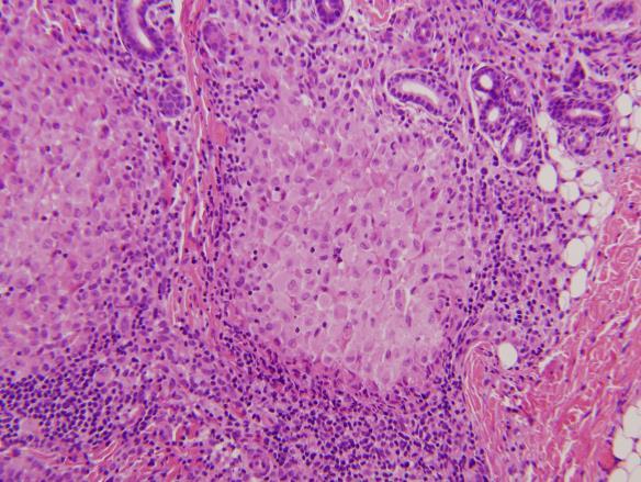
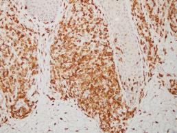
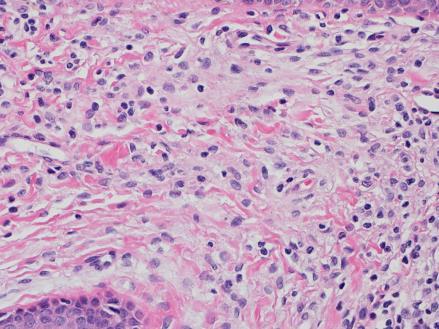
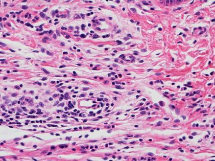
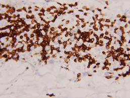
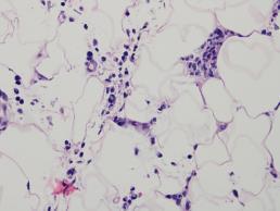
4 Langerhans cell histiocytosis Clinical Findings Langerhans cell histiocytosis Histopathologic Findings Demonstration of LCs in aggregates in a background of eosinophils, macrophages, lymphocytes, neutrophils and nonneoplastic multinucleated giant cells medium to large in size, exhibit abundant eosinophilic cytoplasm and have a coffee bean shaped grooved nuclei Small collections of cells at the dermoepidermal interface with variable basal vacuolar degeneration or as dense infiltrates throughout the dermis with or without the formation of eosinophilic abscesses Epidermotropism of Langerhans cells into the epidermis is common Positive staining of Langerhans cells for S-100 protein and CD1a (more specific, commonly used). CD207 (langerin) stains Langerhans cell specifically but is a newer stain not yet as commonly used; PD-L1+, CD68+, Vimentin+, CD30- Molecular: BRAFV600E (50%), MAP2K1 (25%) 30% with IGH, IGK or TCR clonality Langerhans cell histiocytosis Histopathologic Findings 4


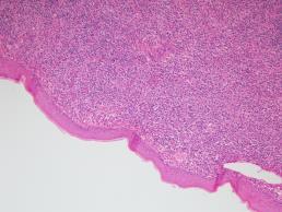
5 Langerhans cell histiocytosis Histopathologic Findings Langerhans cell histiocytosis Histopathologic Findings Langerhans cell histiocytosis Histopathologic Findings 5


6 Langerhans cell histiocytosis Histopathologic Findings Langerhans cell histiocytosis Histopathologic Findings CD1a 6


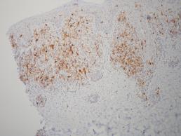
7 Langerin Langerhans cell histiocytosis Histopathologic Findings Langerhans cell histiocytosis Histopathologic Findings 7


8 Congenital self-healing LCH A.k.a. Hashimoto-Pritzker disease Lesions typically involve the deep dermis in a nodular fashion, unlike conventional LCH lesions where lesions are mainly in upper dermis, extending to epidermis Similar staining with CD1a, S100 and langerin. Langerhans cells of CSH-LCH may show Birbeck granules similar to conventional LCH cells by electron microscopy They can be separated ultrastructurally by the presence of unique cytoplasmic dense bodies containing concentrically arranged laminar structures 8


9 9


10 CD68 CD1a 10


11 Langerhans cell histiocytosis Diffferential Diagnosis LCH ECD ICH LCS Non-LCH JXG Reticulohistiocytosis RDD Histiocytic sarcoma Reactive langerhans cell proliferations LCH? LCH? 11


12 LCH? CD1a Langerin 12


13 Fibrous Papule with reactive langerhans cell hyperplasia LCH? LCH? 13


14 LCH? CD3 CD1a Reactive langerhans cell proliferation in a T-cell lymphoma 14


15 LCH? LCH? CD1a - LCH? 15

, bone (B, hematoxylin and eosin) and lymph node (C; hematoxylin and eosin) with cyclin D1 expression within CD1a+ lesional cells demonstrated by a cyclin")

 with cyclin D1 (brown)/cd1a (red) double stain (B) demonstrating lack of cyclin D1 expression in CD1a+ cells.")
 double stain (D). E, Normal skin also showing lack of cyclin D1 (brown) expression in epidermal Langerhans cells highlighted by CD1a (red).")
16 CD30 ALCL pyogenic variant Cyclin D1 and LCH Cyclin D1 Is Expressed in Neoplastic Cells of Langerhans Cell Histiocytosis but Not Reactive Langerhans Cell Proliferations. Shanmugam, Vignesh; Craig, Jeffrey; MD, PhD; Hornick, Jason; MD, PhD; Morgan, Elizabeth; Pinkus, Geraldine; Pozdnyakova, Olga; MD, PhD American Journal of Surgical Pathology. 41(10): , October DOI: /PAS FIGURE 1. Cyclin D1 expression in LCH lesional cells involving a variety of sites. LCH involving skin (A; hematoxylin and eosin), bone (B, hematoxylin and eosin) and lymph node (C; hematoxylin and eosin) with cyclin D1 expression within CD1a+ lesional cells demonstrated by a cyclin D1 (brown)/cd1a (pink) double stain (D and E) or cyclin D1 single stain showing strong expression in the multinucleated giant cell (F). Copyright 2017 Wolters Kluwer Health, Inc. All rights reserved. Published by Lippincott Williams & Wilkins, Inc. 2 Cyclin D1 and LCH Cyclin D1 Is Expressed in Neoplastic Cells of Langerhans Cell Histiocytosis but Not Reactive Langerhans Cell Proliferations. Shanmugam, Vignesh; Craig, Jeffrey; MD, PhD; Hornick, Jason; MD, PhD; Morgan, Elizabeth; Pinkus, Geraldine; Pozdnyakova, Olga; MD, PhD American Journal of Surgical Pathology. 41(10): , October DOI: /PAS FIGURE 3. Reactive/normal Langerhans cells do not show cyclin D1 expression. A and B, Florid dermatopathic lymphadenopathy (A; hematoxylin and eosin) with cyclin D1 (brown)/cd1a (red) double stain (B) demonstrating lack of cyclin D1 expression in CD1a+ cells. C and D, Spongiotic dermatitis with Langerhans cell microabscess formation (C; hematoxylin and eosin); the Langerhans cells are negative for cyclin D1 expression as shown by a cyclin D1 (brown)/cd1a (red) double stain (D). E, Normal skin also showing lack of cyclin D1 (brown) expression in epidermal Langerhans cells highlighted by CD1a (red). Note cyclin D1 expression in parabasal cells of the epidermis (internal control). Copyright 2017 Wolters Kluwer Health, Inc. All rights reserved. Published by Lippincott Williams & Wilkins, Inc. 2 16
:726-730, October 2017. DOI: 10.1097/DAD.")
 and with LC hyperplasia (C).")

17 p53 and LCH p53 Is a Helpful Marker in Distinguishing Langerhans Cell Histiocytosis From Langerhans Cell Hyperplasia. Grace, Shane; Sutton, Angela; Armbrecht, Eric; Vidal, Claudia; MD, PhD; Rosman, Ilana; Hurley, Maria American Journal of Dermatopathology. 39(10): , October DOI: /DAD FIGURE 3. p53. A, p53 staining was significantly higher in the epidermis and dermis in LCH (Group 2) compared with other inflammatory dermatoses (Group 1). B, Representative photomicrographs showing absent staining in the epidermis and dermis of an inflammatory dermatosis without LC hyperplasia (B) and with LC hyperplasia (C). D, Representative photomicrograph of LCH showing prominent staining of the dermal inflammatory infiltrate. Original magnifications x200. Copyright 2017 Wolters Kluwer Health, Inc. All rights reserved. Published by Lippincott Williams & Wilkins, Inc. 2 Indeterminate dendritic cell tumor Also known as indeterminate cell histiocytosis is a neoplastic proliferation with phenotypic features similar to those of normal indeterminate cells, the alleged precursor of Langerhans cells One or multiple generalized papules, nodules, or plaques on the skin No systemic symptoms Dermal based histiocytic proliferation with +/- extension to the adipose tissue Diffuse infiltration similar to LCH, irregular nuclear grooves and clefts, abundant eosinophilic cytoplasm Multinucleated giant cells Spindle cells IHC: S100+, CD1a+, Langerin-, CD30-, CD21-,CD23-,CD35- Variable clinical presentation: spontaneous regression or rapid progression Indeterminate cell histiocytosis 17


18 Indeterminate cell histiocytosis Indeterminate cell histiocytosis Indeterminate cell histiocytosis 18


19 Indeterminate cell histiocytosis Indeterminate cell histiocytosis Indeterminate cell histiocytosis 19


20 Indeterminate cell histiocytosis Indeterminate cell histiocytosis CD163 20


21 CD1a CD1a S100 21

 (9%), and spleen (2%) identified in 1 of 6 positive; positive; associated malignancies cases (O Malley); 3 22%")
 (Brown) 3.")
22 BRAF V600E FISH targeting the ETV3-NCOA2 gene fusion in ICH. Red probe for ETV3 in chromosome 1 and green probe for NCOA2 in chromosome 8. Ryanne A. Brown et al. Blood 2015;126: by American Society of Hematology Indeterminate cell histiocytosis Indeterminate cell histiocytosis Langerhans cell histiocytosis Juvenile xanthogranuloma Typical age Isolated skin Most common sites of Molecular Findings Immunophenotype Association with other of onset involvement involvement CD1a Langerin S-100 CD68 malignancies 45 years 88% Skin (93%), lymph nodes BRAF mutations Positive Negative 78% 87% 13% of patients had one or more (median) (9%), and spleen (2%) identified in 1 of 6 positive; positive; associated malignancies cases (O Malley); 3 22% focal, 13% focal, including: of 3 cases weak, or weak, or FL (4 cases); AML (3 cases); CLL demonstrated an negative negative (2 cases); AITCL (2 cases); BALL ETV3-NCOA2 (1 case); Acute mast cell translocation leukemia (1 case) (Brown) <5% Bone (77%), skin (39%), Approximately 50% Positive Positive Positive Positive Associated with lymphomas, years and lymph nodes (19%) of cases harbor (virtually leukemias, and other solid BRAF mutations 100%) tumors. 3.5% of patients had an associated malignancy in one study of 116 patients. (Egeler) 5 months 74% (Dehner Skin (approximately 2/3 Associated with Negative Negative Rarely Positive Associated with JMML (Janssen) 2003) cases present as solitary neurofibromatosis positive cutaneous lesions) > type I subcutaneous soft tissue > deep soft tissue; <5% have visceral-systemic involvement Multicentric Typically Skin and joints reticulohistiocytosis years involves skin and joints Dendritic cell Predomina Rare Lymph nodes; skin sarcomas ntly adults involvement has been in 4 th and reported but is 5 th decades uncommon No welldocumented positive Negative Negative Rarely molecular findings No welldocumented negative positive FDCS: Negative Typically molecular findings IDCS: negative Positive Typically positive Associated with malignancy in up to 30% of cases (includes solid tumors as well as hematologic malignancies) N/A INDCS: positive 22


23 Erdheim-Chester Disease Now part of the L group of histiocytosis 75% of cases have BRAFV600E mutations Symmetric diaphyseal and metaphyseal osteosclerosis of long bones as well as cardiac, retroperitoneal, vascular, and cerebral involvement. Clinical presentations are diverse and range from bone pain to diabetes insipidus, and neurological and constitutional symptoms 30% cutaneous manifestations are the first sign of the disease XLL 83% Other patches, papulonodular lesions, rare erythroderma Histologically diffuse dermal infiltrate of xanthomatized histiocytes and Toutontype giant cells IHC: CD68+, CD163+, Factor XIIIa+, BRAFV600E+, CD1a-, Langerin-, S100- Mixed LCH/ECD show histopathologic and IHC of LCH J Am Acad Dermatol 2016 Erdheim-Chester Disease Erdheim-Chester Disease 23
.")


24 Erdheim-Chester Disease FIGURE 3. Xanthelasma-like tumors as cutaneous ECD manifestations in case 2. A, Large xanthelasma-like tumor around the eyes. B, Diffuse accumulation of foamy cells, fibrosis of the connective tissue (hematoxylin and eosin x40). C, Multiple Touton giant cell and hemosiderin deposits in the dermis (hematoxylin and eosin x400). Histopathological and Clinical Findings in Cutaneous Manifestation of Erdheim-Chester Disease and Langerhans Cell Histiocytosis Overlap Syndrome Associated With the BRAFV600E Mutation. Liersch, Julia; Carlson, J; Andrew MD, FRCPC; Schaller, Jorg American Journal of Dermatopathology. 2 39(7): , July DOI: /DAD Langerhans cell sarcoma Malignant neoplasm with Langerhans cell phenotype Very rare All cases reported in adults Median age: 41 Some cases in association with Merkell cell polyomavirus Rare association with FL Clinical Skin and soft tissue most cases; hepatosplenomegaly; pancytopenia Histopathology Malignant neoplasm with marked pleomorphism Nuclear grooves variable High mitotic rare: >50 mitoses / 10 HPF + Birbeck granules IHC: CD1a+, S100+, Langerin+ One case with BRAF V600E mutation; possible TCR and IGH rearrangements WHO


25 Langerhans cell sarcoma Langerhans cell sarcoma Langerhans cell sarcoma 25


26 Langerhans cell sarcoma Langerhans cell sarcoma Langerhans cell sarcoma CD1a LANGERIN 26


27 Langerhans cell sarcoma FASCIN Ki67 Juvenile Xanthogranuloma and Adult Xanthogranuloma Group C for cutaneous limited forms and group L for the systemic variants 0.5% of pediatric tumors +++ infants and young children, but adolescents and adults also Solitary, well demarcated, firm nodule Head and neck, trunk and upper limbs 70-75% of cases Keratotic, pedunculated, linear, flat, or plaque-like usually 5 to 10 mm in diameter but can measure up to a few centimeters Initially, the lesion is raised and pink to erythematous in color but over time they become yellow-brown and may flatten Regression may occur over a period of months to years with residual hyperpigmentation, atrophy or anetoderma Lesions are multiple in 10% of cases Juvenile Xanthogranuloma and Adult Xanthogranuloma Extracutaneous sites with our WITHOUT skin involvement (15%) Soft tissue Multifocal visceral and osseous sites are involved in about 5% of cases and may include the oropharynx, nasopharynx, gastrointestinal tract, liver, lung, testis, spleen, lymph nodes, central nervous system and kidney Systemic cases occur in young children under the age of two years Association with NF-1 and JMML mutations in the GTPase neurofibromin, which is present in NF1, leads to RAS dysfunction Systemic presentations of JXG with CNS involvement have shown BRAF V600E mutations should those cases be qualified as ECD/L group lesions that haven t presented themselves with systemic findings (i.e brain only), or there is a truly separate group of JXG/BRAF V600E positive tumors? 27


28 JXG - Histopathology Dense mononuclear histiocytic infiltrate in the dermis extending to the interface with the epidermis Limited to the dermis, but in large lesions extends to the subcutis Flattening of the epidermis, and sometimes collarette Epidermis and adnexae are spared Epithelioid histiocytes, xanthomatized cells and multinucleated Toutontype giant cells Giant cells might be absent, particularly in extracutaneous lesions Lymphocytes, eosinophils, plasma cells, neutrophils, and mast cells may be scattered throughout the infiltrate or altogether absent The morphology of the histiocytes can vary depending on the "age" of the lesion Early: epithelioid histiocytes without vacuolization Mature: histiocytes with lipidized or xanthomatized cytoplasm Scalloped histiocytes with an angulated or jagged border and spindled histiocytes may also be seen some lesions show a predominance of spindle cell component Mitotic figures are common, but NOT atypical mitoses IHC: CD68+, CD163+, CD4+, CD14+, factor XIIIa+, HLA-DR+, fascin+, vimentin+, and lysozyme+ and are negative for CD1a, and langerin S100 stains rich background in dendritic cells (JXG cells NEGATIVE) JXG JXG 28


29 JXG JXG JXG 29


30 JXG spindle cell variant JXG family variants Benign cephalic histiocytosis Early form of JXG affecting young children Multiple tan-pink papules on the cheek, neck, and upper trunk are the clinical manifestations with sparing of the palms and soles, mucous membranes, and viscera Self-limiting disorder Histologically JXG-like: diffuse dermal infiltrates of mononuclear histiocytes with ill-defined pale cytoplasm Generalized eruptive histiocytosis Both children and adults Recurrent crops of small, reddish papules symmetrically distributed on the trunk and extensor surfaces of the extremities without visceral involvement Lesions develop rapidly and tend to resolve spontaneously with no residual findings or at most slight hyperpigmentation In adults frequently associated with underlying leukemia / lymphoma Numerous mononuclear histiocytes, including spindled forms, are present within the upper to mid dermis and a perivascular distribution may be present. Scattered lymphocytes may be seen but foamy cells or giant cells are generally absent Progressive nodular histiocytosis Adults 40 to 60 years of age and is characterized by the presence of two types of skin lesions: multiple small, superficial yellow-brown papules and larger, deeper subcutaneous nodules with a predilection for the trunk A dermal infiltrate of histiocytes, predominantly spindle-shaped forms, and foamy cells (including Touton cells) are the histologic feature Benign cephalic histiocytosis 30

31 Benign cephalic histiocytosis JXG Differential diagnosis L Group: LCH, ICH, ECD Non L NXG Reticulohistiocytoma RDD BCH Histiocytic sarcoma Spitz nevi Dermatofibroma Lymphomatoid papulosis Xanthoma/Xanthelasma Xanthomatized Sweet s syndrome Reticulohistiocytoma There is a circumscribed dermal nodule, often with overlying epidermal thinning. As with multicentric reticulohistiocytosis, there is an irregular admixture of oncocytic mononuclear histiocytes, multinucleate giant cells with a ground-glass appearance, and inflammatory cells. Phagocytosis of leukocytes is sometimes seen. A few Touton giant cells may be present and these may contain lipid. Reticulin fibers are increased and surround individual cells There is usually more of a neutrophilic infiltrate than in multicentric reticulohistiocytosis, and a further point of distinction is the greater propensity for the stroma in reticulohistiocytoma to have many spindle-shaped cells and for there to be xanthomatized cells Nowadays, reticulohistiocytoma are considered VARIANTS of JXG More typically in adults than children: hands 31


32 Reticulohistiocytoma Reticulohistiocytoma Reticulohistiocytoma CD68 RETICULIN 32


33 Dermatofibroma Dermatofibroma Lymphomatoid papulosis 33


34 Lymphomatoid papulosis Lymphomatoid papulosis CD30 CD3 Spitz Nevus 34


35 Xanthoma / Xanthelasma Foam cells in dermis (positive for lipid with special stains such as oil-red-o) Touton giant cells sometimes Small numbers of lymphocytes or neutrophils in younger lesions, especially in eruptive xanthomas Fibrosis or cholesterol clefts in older lesions Xanthelasma: eyelids, associated with hypercholesterolemia in 50% of cases. Xanthelasma Xanthelasma 35


36 Lipophagic polyp Usually solitary or few polypoid lesions that mimic papillomas Variable age Retention mastitis? Sheets of benign lipidized histiocytes in the absence of multinucleated giant cells. IHC: CD68+,CD163+,CD1a - Am J Dermatopathol 2015 Lipophagic polyp Lipophagic polyp 36


 MDS, AML, CMML, CML Solid")


37 CD68 Histiocytoid Sweet s Syndrome Tender erythematous plaques and nodules on the extremities and trunk accompanied by systemic symptoms such as fever and arthralgia Hematologic malignancies were present in 8 patients (24%) MDS, AML, CMML, CML Solid tumors Histopathology spared epidermis moderate to intense edema in the papillary dermis no evidence of vasculitis dense, bandlike, inflammatory infiltrate involving the superficial and mid dermis predominantly composed of mononuclear cells IHC: MPO+,CD163+ JAMA Dermatol 2017 Histiocytoid Sweet s Syndrome 37


38 Histiocytoid Sweet s Syndrome Histiocytoid Sweet s Syndrome Histiocytoid Sweet s Syndrome CD163 MPO 38
39 Cutaneous RDD Uncommon histiocytosis with less than 600 cases reported 80% present in first 2 decades Cutaneous dissemination in SRDD occurs in 1/3 of cases while pure CRDD is extraordinary rare SRDD young adult men and children CRDD adult white women Etiologic factors: HHV-6 Parvovirus B19 ALPS (autoimmune lymphoproliferative syndrome) IgG4 autoimmune disorders Cutaneous RDD Clinical presentation Benign and self-limited Exacerbations and remissions Fever, massive, non-tender, bilateral lymph node enlargement LAD can be focal 40% extranodal involvement Skin, soft tissues, bone Intrathoracic manifestations: airway, pleural effusion, ILD (GOOD prognosis) Lower respiratory tract, liver and kidney (POOR prognosis) Cutaneous manifestations: Eyelids, malar region Multiple, focal, erythematousbrown, xanthomatous papules, pustules, nodules and plaques Histopathologic findings LNs: dilated sinuses containing PMNs, lymphocytes, plasma cells, and histiocytes Skin: histiocytes, PMNs, plasma cells, lymphocytes in the dermis Histiocytes are polygonal, with abundant foamy, eosinophilic cytoplasm, feathery borders, and large vesicular nuclei Emperipolesis Multinucleated histiocytes (touton-type giant cells); plasma cells with Russell bodies, xanthoma cells IHC: CD68+,CD163+,S100+,CD1A-,IgG4+PCs Cutaneous RDD - Molecular RDD pattern has been associated with autoimmune hemolytic anemia, systemic lupus erythematosus, and juvenile idiopathic arthritis Always evaluate serum levels of IgG4 overlap with IgG4 syndrome H syndrome is an autosomal genetic condition characterized by hyperpigmentation, hypertrichosis, hepatosplenomegaly, hearing loss, heart anomalies, hypogonadism, low height (short stature), hyperglycemia/diabetes mellitus, and hallux valgus/flexion contractures mutation within the SLC29A3 gene RDD lesions in 25% of cases Patients with ALPS type Ia have RDD lesions in 41% of cases TNFRSF6 heterozygous germ line mutations affecting the gene encoding Fas PLOS Genetics


40 RDD RDD RDD 40


41 RDD Emperipolesis RDD S100 41


42 RDD - IHC CD163 CD68 RDD RDD 42


43 RDD RDD RDD 43


44 Cutaneous Plasmacytosis Rare entity (<100 cases reported) Middle age Slight male predominance Affects individual of Asian descent +++ Japanese Closely related to plasma cell variant of Castleman disease IgG4-related disorders Clinical: red to brown macules, plaques and papules Face, trunk and extremities Superficial LAD 58% Hypergammaglobulinemia POLYCLONAL Interstitial nephritis, ILD Cutaneous plasmacytosis Histopathology Dermal infiltrate of plasma cells, with admixed lymphocytes and histiocytes PC infiltrate is periadnexal and perivascular, but also diffuse Heavier in superficial to mid dermis, +/- deep involvement Follicles and GC can be present Lack cytologic atypia POLYCLONAL INFILTRATE IgG4+ 44


45 Cutaneous plasmacytosis Cutaneous plasmacytosis Cutaneous IgG4-related disease LN, skin, lung and pancreatic involvement Autoimmune pancreatitis Sclerosing cholangitis Sclerosing sialadenitis LN often show Castlemanlike changes >15 PC/hpf IgG4/IgG ratio >40% Fibrosis, phlebitis Other diseases linked to IgG4: pemphigus vulgaris Hum Pathol


 LAD, hyperproteinemia,")

, plasma cell (10%),")
46 Cutaneous IgG4-related disease IgG4 Cutaneous Castleman Disease Most cases of CD present in HIV- patients 5-10% are HIV+ and typically associated with HHV-8 infection Cutaneous involvement in CD is very rare (<15 cases reported) LAD, hyperproteinemia, hypergammaglobulinemia, systemic symptoms, POEMS Skin: multiple erythematous to brownish nodules Histopathology: hyaline vascular type (90%), plasma cell (10%), mixed Cutaneous Castleman Disease 46
.")
47 Cutaneous Castleman Disease Cytophagic histiocytic panniculitis (CHP) H group Originally described by Crotty and Winkelmann (JAAD 1981) as multiple subcutaneous nodules experiencing a terminal hemorrhagic diathesis with variable clinical course (6 months 10 years). Clinical course is variable: systemic symptoms include fever, weight loss, hepatosplenomegaly, cytopenias, serosal effusions, coagulation abnormalities; etc Some cases are associated with subcutaneous panniculitis-like T-cell lymphoma or cutaneous γδ T-cell lymphoma HLH can also occur in association with infectious processes, or specific gene mutations Histopathology Mixed septal and lobular panniculitis with a variable infiltrate of histiocytes and lymphocytes Erythrophagocytosis ( bean bag cells ) and cytophagocytosis IHC can help: CD163, CD68 Always look for coexistence of LPD: SPTCL or PGDTCL Clonality studies are always recommended If cytopenias present: systemic work-up (bone marrow bx, visceral organ bx) Other diagnostic considerations: Weber-Christian, connective tissue disorders, etc 47


48 Clinical Presentation 14 2 Clinical Presentation Clinical Presentation 48
 14")
 49")

49 Cytophagic histiocytic panniculitis (CHP) 14 5 Cytophagic histiocytic panniculitis (CHP) Cytophagic histiocytic panniculitis (CHP) 49


50 Cytophagic histiocytic panniculitis (CHP) Histopathology Histopathology 50


51 CD163 Bone marrow aspirate Melkerson-Rosenthal Syndrome Marked dermal edema and perivascular inflammation with lymphs, plasma cells, eos, histiocytes Loose granulomas, tuberculoid, small naked collection of epithelioid cells Sometimes Schaumann bodies are present Lymphatics are widely dilated and cotain collections of inflammatory cells Older lesions show dermal fibrosis Cutaneous histiocytic lymphangitis and angioendotheliomatosis in the spectrum (also RA related), intravascular histiocytosis 51


52 Melkerson-Rosenthal Syndrome Melkerson-Rosenthal Syndrome Melkerson-Rosenthal Syndrome 52


53 Melkerson-Rosenthal Syndrome Melkerson-Rosenthal Syndrome Thank you for your attention! 53

54 54
Spectrum of clinical presentations
 Spectrum of clinical presentations Case History A 7-day-old male patient born full-term via uncomplicated vaginal delivery was seen for multiple erythematous red-brown purpuric lesions that were present
الفتوي الاصفر الحبيبوم = Xanthogranuloma_Juvenile JUVENILE XANTHOGRANULOMA 1 / 9
 JUVENILE XANTHOGRANULOMA 1 / 9 Clinical Findings CUTANEOUS LESIONS JXG is a benign, self-healing disorder that is characterized by asymptomatic yellowish papulonodular lesions of the skin and other organs
Primary Cutaneous CD30-Positive T-cell Lymphoproliferative Disorders
 Primary Cutaneous CD30-Positive T-cell Lymphoproliferative Disorders Definition A spectrum of related conditions originating from transformed or activated CD30-positive T-lymphocytes May coexist in individual
3/24/2017 DENDRITIC CELL NEOPLASMS: HISTOLOGY, IMMUNOHISTOCHEMISTRY, AND MOLECULAR GENETICS. Disclosure of Relevant Financial Relationships
 DENDRITIC CELL NEOPLASMS: HISTOLOGY, IMMUNOHISTOCHEMISTRY, AND MOLECULAR GENETICS Jason L. Hornick, M.D., Ph.D. Director of Surgical Pathology and Immunohistochemistry Brigham and Women s Hospital Professor
المركب النموذج--- سبيتز وحمة = Type Spitz's Nevus, Compound SPITZ NEVUS 1 / 7
 SPITZ NEVUS 1 / 7 Epidemiology An annual incidence rate of 1.4 cases of Spitz nevus per 100,000 individuals has been estimated in Australia, compared with 25.4 per 100,000 individuals for cutaneous melanoma
1/14/2018. Objectives
 2018 Pathology CME Cutaneous Hematopathology Maui, HI Jan 18 th 26 th Pseudolymphomas Alejandro A. Gru, M.D. Assistant Professor of Pathology & Dermatology Dermatopathology Division and Fellowship Director
PHYSIOLOGY AND MANAGEMENT OF HISTIOCYTIC DISEASE. Brant Ward, MD, PhD Division of Rheumatology, Allergy, and Immunology
 PHYSIOLOGY AND MANAGEMENT OF HISTIOCYTIC DISEASE Brant Ward, MD, PhD Division of Rheumatology, Allergy, and Immunology What do histiocytes do? Apoptotic body removal Phagocytosis Antigen presentation Types
21/07/2017. Hobnail endothelial cells are not the same as epithelioid endothelial cells
 UPDATE IN CUTANEOUS VASCULAR S DERMATOPATHOLOGY SESSION BELFAST PATHOLOGY JUNE 21/2017 Dr E Calonje St John s Institute of Dermatology, London, United Kingdom THE FAMILY OF VASCULAR S WITH EPITHELIOID
Dermatopathology. Dr. Rafael Botella Estrada. Hospital La Fe de Valencia
 Dermatopathology Dr. Rafael Botella Estrada. Hospital La Fe de Valencia Melanoma and mimics Dr. Martin Mihm Malignant lesions result from the accumulation of mutations Class I lesions (benign) Class II
SWISS SOCIETY OF NEONATOLOGY. A newborn with a papulonodular rash at birth
 SWISS SOCIETY OF NEONATOLOGY A newborn with a papulonodular rash at birth June 2001 2 Glanzmann R, Neonatal Intensive Care Unit, UKKB Basel, Switzerland Swiss Society of Neonatology, Thomas M Berger, Webmaster
Overview of Cutaneous Lymphomas: Diagnosis and Staging. Lauren C. Pinter-Brown MD, FACP Health Sciences Professor of Medicine and Dermatology
 Overview of Cutaneous Lymphomas: Diagnosis and Staging Lauren C. Pinter-Brown MD, FACP Health Sciences Professor of Medicine and Dermatology Definition of Lymphoma A cancer or malignancy that comes from
Dermatopathology. Dr. Rafael Botella Estrada. Hospital La Fe de Valencia
 Dermatopathology Dr. Rafael Botella Estrada. Hospital La Fe de Valencia DERMATOPATHOLOGY CASE CHALLENGE: RECOGNIZING MIMIS AND MASQUERADERS Rosalie Elenitsas. University of Pennsylvania Spectrum Lupus
Differential diagnosis of hematolymphoid tumors composed of medium-sized cells. Brian Skinnider B.C. Cancer Agency, Vancouver General Hospital
 Differential diagnosis of hematolymphoid tumors composed of medium-sized cells Brian Skinnider B.C. Cancer Agency, Vancouver General Hospital Lymphoma classification Lymphoma diagnosis starts with morphologic
Important Decisions in Dermatopathology: The Clinico- Pathologic Correlation. Dermatopathology Specialists Needed. Changing Trends
 Important Decisions in Dermatopathology: The Clinico- Pathologic Correlation Uma Sundram, MD, PhD Departments of Pathology and Dermatology Stanford University May 29, 2008 Dermatopathology Specialists
Arthritis and Rosai-Dorfman Disease of the Skin: A Diagnostic Dilemma
 Arthritis and Rosai-Dorfman Disease of the Skin: A Diagnostic Dilemma Introduction Pages with reference to book, From 280 To 282 Irshad Nabi Soomro ( Department of Pathology, The Aga Khan University Hospital,
Lymphoma and Pseudolymphoma
 Lymphoma and Pseudolymphoma Laura B. Pincus, MD Co-Director, Cutaneous Lymphoma Clinic Associate Professor Dermatology and Pathology University of California, San Francisco I HAVE NO RELEVANT RELATIONSHIPS
ACCME/Disclosures ALK FUSION-POSITIVE MESENCHYMAL TUMORS. Tumor types with ALK rearrangements. Anaplastic Lymphoma Kinase. Jason L.
 Companion Meeting of the International Society of Bone and Soft Tissue Pathology The Evolving Concept of Mesenchymal Tumors ALK FUSION-POSITIVE MESENCHYMAL TUMORS Jason L. Hornick, MD, PhD March 13, 2016
أملس عضلي غرن = Leiomyosarcoma. Leiomyosarcoma 1 / 5
 Leiomyosarcoma 1 / 5 EPIDEMIOLOGY Exact incidence is unknown, but older studies suggest that leiomyosarcomas comprise approximately 3 percent of soft-tissue sarcomas. Superficial leiomyosarcoma occurs
Malignant Peripheral Nerve Sheath Tumor
 C H A P T E R 120 Malignant Peripheral Nerve Sheath Tumor Currently, malignant peripheral nerve sheath tumor (MPNST) is the most commonly used generic name for the neoplasms known in the past as neurosarcoma,
Contents. vii. Preface... Acknowledgments... v xiii
 Contents Preface... Acknowledgments... v xiii SECTION I 1. Introduction... 3 Knowledge-Based Diagnosis... 4 Systematic Examination of the Lymph Node... 7 Cell Type Identification... 9 Cell Size and Cellularity...
Case year female. Routine Pap smear
 Case 1 57 year female Routine Pap smear Diagnosis? 1. Atypical glandular cells of unknown significance (AGUS) 2. Endocervical AIS 3. Endocervical adenocarcinoma 4. Endometrial adenocarcinoma 5. Adenocarcinoma
Burkitt lymphoma. Sporadic Endemic in Africa associated with EBV Translocations involving MYC gene on chromosome 8
 Heme 8 Burkitt lymphoma Sporadic Endemic in Africa associated with EBV Translocations involving MYC gene on chromosome 8 Most common is t(8;14) Believed to be the fastest growing tumor in humans!!!! Morphology
Primer of Immunohistochemistry (Leukocytic)
") Primer of Immunohistochemistry (Leukocytic) Paul K. Shitabata, M.D. Dermatopathology Institute Torrance, CA BENIGN LYMPHOID SKIN LESIONS CAPABLE OF SIMULATING LYMPHOMA -Jessner s lymphoid infiltrate -Dermal-subcutaneous
Primer of Immunohistochemistry (Leukocytic) Paul K. Shitabata, M.D. Dermatopathology Institute Torrance, CA BENIGN LYMPHOID SKIN LESIONS CAPABLE OF SIMULATING LYMPHOMA -Jessner s lymphoid infiltrate -Dermal-subcutaneous
Case Presentation. Maha Akkawi, MD, Fatima Obeidat, MD, Tariq Aladily, MD. Department of Pathology Jordan University Hospital Amman, Jordan
 Case Presentation Maha Akkawi, MD, Fatima Obeidat, MD, Tariq Aladily, MD Department of Pathology Jordan University Hospital Amman, Jordan The 25th Annual Congress of the ADIAP The 8/11/2013 1 5th International
Lymphomatoid Papulosis 3 Case Reports
 IOSR Journal of Dental and Medical Sciences (IOSR-JDMS) e-issn: 2279-0853, p-issn: 2279-0861.Volume 14, Issue 7 Ver. III (July. 2015), PP 31-35 www.iosrjournals.org Lymphomatoid Papulosis 3 Case Reports
MOSTLY SEPTAL PANNICULITIS
 Margot S. Peters, M.D. Professor of Dermatology and Laboratory Medicine & Pathology, Mayo Clinic PANNICULITIS C017 Essential Dermatopathology, What You Need to Know for Clinical Practice: Inflammatory.
Plasma cell myeloma (multiple myeloma)
") Plasma cell myeloma (multiple myeloma) Common lymphoid neoplasm, present at old age (70 years average) Remember: plasma cells are terminally differentiated B-lymphocytes that produces antibodies. B-cells
Plasma cell myeloma (multiple myeloma) Common lymphoid neoplasm, present at old age (70 years average) Remember: plasma cells are terminally differentiated B-lymphocytes that produces antibodies. B-cells
Michi Shinohara MD Associate Professor University of Washington/Seattle Cancer Care Alliance Dermatology, Dermatopathology
 Michi Shinohara MD Associate Professor University of Washington/Seattle Cancer Care Alliance Dermatology, Dermatopathology Agenda Overview of cutaneous T and B- cell lymphomas Diagnosis, Staging, Prognosis
Dermatopathology: The tumor is composed of keratinocytes which show atypia, increase mitoses and abnormal mitoses.
 Squamous cell carcinoma (SCC): A common malignant tumor of keratinocytes arising in the epidermis, usually from a precancerous condition: 1- UV induced actinic keratosis, usually of low grade malignancy.
FOLLICULARITY in LYMPHOMA
 FOLLICULARITY in LYMPHOMA Reactive Follicular Hyperplasia Follicular Hyperplasia irregular follicles Follicular Hyperplasia dark and light zones Light Zone Dark Zone Follicular hyperplasia MIB1 Follicular
Benign and malignant epithelial lesions: Seborrheic keratosis: A common benign pigmented epidermal tumor occur in middle-aged or older persons more
 Benign and malignant epithelial lesions: Seborrheic keratosis: A common benign pigmented epidermal tumor occur in middle-aged or older persons more common on the trunk; but extremities, head and neck are
ACCME/Disclosures. USCAP Specialty Evening Conference: Cytopathology New and recent Developments in Cytopathology: A case based approach
 USCAP Specialty Evening Conference: Cytopathology New and recent Developments in Cytopathology: A case based approach Sara E. Monaco, MD Associate Professor Program Director, UPMC Cytopathology Fellowship
Adult Orbital Xanthogranulomatous Disease
 Adult Orbital Xanthogranulomatous Disease Evening Specialty Conference: Ophthalmic Pathology Sunday, March 22, 2015 Lynn Schoenfield Associate Professor, Ohio State University Wexner Medical Center No
Selected Pseudomalignant Soft Tissue Tumors of the Skin and Subcutis
 Selected Pseudomalignant Soft Tissue Tumors of the Skin and Subcutis Andrew L. Folpe, M.D. Professor of Laboratory Medicine and Pathology Mayo Clinic, Rochester, MN folpe.andrew@mayo.edu 2016 MFMER slide-1
Desmoplastic Melanoma R/O BCC. Clinical Information. 74 y.o. man with lesion on left side of neck r/o BCC
 R/O BCC Sabine Kohler, M.D. Professor of Pathology and Dermatology Dermatopathology Service Stanford University School of Medicine Clinical Information 74 y.o. man with lesion on left side of neck r/o
Cellular Neurothekeoma
 Cellular Neurothekeoma Scott W Binder, MD Pritzker Professor of Pathology & Dermatology Sr. Vice Chair Director, Pathology Clinical Services Chief, Dermatopathology Geffen/UCLA School of Medicine Clinical
BSD Self Assessment Workshop 7 th July 2013 CASE 27 RAC6123
 BSD Self Assessment Workshop 7 th July 2013 CASE 27 RAC6123 M55. 4/7 tender lesions on knee, legs and arms. Also iritis/ weight loss/headache, synovitis.?vasculitis. Sarcoidosis. Biopsy from left elbow
Anaplastic Large Cell Lymphoma (of T cell lineage)
") Anaplastic Large Cell Lymphoma (of T cell lineage) Definition T-cell lymphoma comprised of large cells with abundant cytoplasm and pleomorphic, often horseshoe-shaped nuclei CD30+ Most express cytotoxic
Anaplastic Large Cell Lymphoma (of T cell lineage) Definition T-cell lymphoma comprised of large cells with abundant cytoplasm and pleomorphic, often horseshoe-shaped nuclei CD30+ Most express cytotoxic
MULTISYSTEMIC LANGERHANS CELL HISTIOCYTOSIS IN ADULT AN UNCOMMON INCIDENCE POSING A DIAGNOSTIC CHALLENGE. Monal Trisal
 Pathology Case Report International Journal of Clinical And Diagnostic Research ISSN 2395-3403 Volume 4, Issue 4, July-Aug 2016. Glorigin Lifesciences Private Limited. MULTISYSTEMIC LANGERHANS CELL HISTIOCYTOSIS
CPC. Chutika Srisuttiyakorn, M.D. Kobkul Aunhachoke, M.D. Phramongkutklao Hospital Bangkok, Thailand
 CPC Chutika Srisuttiyakorn, M.D. Kobkul Aunhachoke, M.D. Phramongkutklao Hospital Bangkok, Thailand A 53 year-old woman with fever, facial swelling and rashes on face, trunk and upper extremities for 3
Normal thyroid tissue
 Thyroid Pathology Overview Normal thyroid tissue Normal thyroid tissue with follicles filled with colloid. Thyroid cells form follicles, spheres of epithelial cells (always single layered in health, usually
Diseases of the breast (1 of 2)
") Diseases of the breast (1 of 2) Introduction A histology introduction Normal ducts and lobules of the breast are lined by two layers of cells a layer of luminal cells overlying a second layer of myoepithelial
Diseases of the breast (1 of 2) Introduction A histology introduction Normal ducts and lobules of the breast are lined by two layers of cells a layer of luminal cells overlying a second layer of myoepithelial
Synonyms. Mast cell disease Mast cell proliferative disease
 Mastocytosis Definition Mastocytosis is a proliferation of mast cells and their subsequent accumulation in one or more organ systems. Mast cells are derived from hematopoietic progenitors, thus mastocytosis
A middle-aged man with self-healing papulonecrotic lesions over the trunk and proximal limbs
 Hong Kong J. Dermatol. Venereol. (2011) 19, 30-34 Case Report A middle-aged man with self-healing papulonecrotic lesions over the trunk and proximal limbs JC Chan, N Trendell-Smith, CK Yeung Lymphomatoid
Isolated Juvenile Xanthogranuloma in Thoracic Spine: Intraoperative Cytological Diagnosis of a Rare Presentation
 J Interdiscipl Histopathol 2014; 2(3): 158-162 ISSN: 2146-8362 Case Report Isolated Juvenile Xanthogranuloma in Thoracic Spine: Intraoperative Cytological Diagnosis of a Rare Presentation Shashi Singhvi,
7 Omar Abu Reesh. Dr. Ahmad Mansour Dr. Ahmad Mansour
 7 Omar Abu Reesh Dr. Ahmad Mansour Dr. Ahmad Mansour -Leukemia: neoplastic leukocytes circulating in the peripheral bloodstream. -Lymphoma: a neoplastic process in the lymph nodes, spleen or other lymphatic
Citation The Journal of Dermatology, 37(8), available at
, available at") NAOSITE: Nagasaki University's Ac Title Two cases of blaschkitis with promi Author(s) Utani, Atsushi Citation The Journal of Dermatology, 37(8), Issue Date 2010-08 URL Right http://hdl.handle.net/10069/25634
NAOSITE: Nagasaki University's Ac Title Two cases of blaschkitis with promi Author(s) Utani, Atsushi Citation The Journal of Dermatology, 37(8), Issue Date 2010-08 URL Right http://hdl.handle.net/10069/25634
The Relevance of Cytologic Atypia in Cutaneous Neural Tumors
 The Relevance of Cytologic Atypia in Cutaneous Neural Tumors Recent Findings - New Developments New Problems Zsolt B. Argenyi, M.D. Professor of Pathology & Dermatology Director of Dermatopathology Department
SESSION 1: GENERAL (BASIC) PATHOLOGY CONCEPTS Thursday, October 16, :30am - 11:30am FACULTY COPY
 PATHOLOGY CONCEPTS Thursday, October 16, :30am - 11:30am FACULTY COPY") SESSION 1: GENERAL (BASIC) PATHOLOGY CONCEPTS Thursday, October 16, 2008 9:30am - 11:30am FACULTY COPY GOAL: Describe the basic morphologic (structural) changes which occur in various pathologic conditions.
SESSION 1: GENERAL (BASIC) PATHOLOGY CONCEPTS Thursday, October 16, 2008 9:30am - 11:30am FACULTY COPY GOAL: Describe the basic morphologic (structural) changes which occur in various pathologic conditions.
22 year old QH mare with regionally extensive alopecia and scaling on one front limb and ventral chest (Figure 1 and 2).
.") 22 year old QH mare with regionally extensive alopecia and scaling on one front limb and ventral chest (Figure 1 and 2). Which of the following is the most likely disease? a. Sterile granuloma complex
22 year old QH mare with regionally extensive alopecia and scaling on one front limb and ventral chest (Figure 1 and 2). Which of the following is the most likely disease? a. Sterile granuloma complex
PET Imaging in Langerhans Cell Histiocytosis
 PET Imaging in Langerhans Cell Histiocytosis Christiane Franzius Bremen, Germany Histiocytosis Histiocytosis Idiopathic proliferation of histiocytes Two types of histiocytes macrophages: antigen processing
Non-Hodgkin lymphomas (NHLs) Hodgkin lymphoma )HL)
 Hodgkin lymphoma )HL)") Non-Hodgkin lymphomas (NHLs) Hodgkin lymphoma )HL) Lymphoid Neoplasms: 1- non-hodgkin lymphomas (NHLs) 2- Hodgkin lymphoma 3- plasma cell neoplasms Non-Hodgkin lymphomas (NHLs) Acute Lymphoblastic Leukemia/Lymphoma
Non-Hodgkin lymphomas (NHLs) Hodgkin lymphoma )HL) Lymphoid Neoplasms: 1- non-hodgkin lymphomas (NHLs) 2- Hodgkin lymphoma 3- plasma cell neoplasms Non-Hodgkin lymphomas (NHLs) Acute Lymphoblastic Leukemia/Lymphoma
Disclosures. Advisory Board. Consultant. Investigator. MiRagen, Actelion, Celgene, Therakos. Mindera
 Cutaneous Lymphomas Christiane Querfeld, MD, PhD Director, Cutaneous Lymphoma Program City of Hope ~ How the Experts Treat Hematologic Malignancies Symposium March 10 13, 2017 Disclosures Advisory Board
Histiocytic Neoplasms of the Dog and Cat
 Histiocytic Neoplasms of the Dog and Cat V.E. Valli DVM Histiocytic and Dendritic Cell Populations Both lineages are bone marrow derived. Macrophages are part of the innate immune system that are phagocytic
Juvenile Xanthogranulomas in the First Two Decades of Life
 The American Journal of Surgical Pathology 27(5): 579 593, 2003 2003 Lippincott Williams & Wilkins, Inc., Philadelphia Juvenile Xanthogranulomas in the First Two Decades of Life A Clinicopathologic Study
Diplomate of the American Board of Pathology in Anatomic and Clinical Pathology
 A 33-year-old male with a left lower leg mass. Contributed by Shaoxiong Chen, MD, PhD Assistant Professor Indiana University School of Medicine/ IU Health Partners Department of Pathology and Laboratory
SMOOTH MUSCLE TUMOURS
 SMOOTH MUSCLE TUMOURS NORMAL SMOOTH MUSCLE Cytology Immunohistochemistry Ultrastructure Masson Trichrome Smooth Muscle Ultrastructure Many myofilaments running parallel to the long axis of the smooth
Salivary Glands 3/7/2017
 Salivary Glands 3/7/2017 Goals and objectives Focus on the entities unique to H&N Common board type facts Information for your future practice Salivary Glands Salivary Glands Major gland. Paratid. Submandibular.
Evening specialty conference: Liver
 Evening specialty conference: Liver Joseph Misdraji, M.D. Disclosure of Relevant Financial Relationships Disclosure of Relevant Financial Relationships USCAP requires that all planners (Education Committee)
Hematolymphoid lesions of the skin Part II Myeloid neoplastic proliferations Houston Society of Clinical Pathologists Symposium April 14, 2018
 Hematolymphoid lesions of the skin Part II Myeloid neoplastic proliferations Houston Society of Clinical Pathologists Symposium April 14, 2018 Carlos A. Torres-Cabala, MD Associate Professor Chief, Dermatopathology
العصوي الوعاي ي الورام = angiomatosis Bacillary
 1 / 7 BACILLARY ANGIOMATOSIS Epidemiology BA is most commonly seen in patients with acquired immunodeficiency syndrome (AIDS) and a CD4 count less than 50 cells/mm 3, with an incidence of 1.2 cases per
Melanoma Update: 8th Edition of AJCC Staging System
 Melanoma Update: 8th Edition of AJCC Staging System Rosalie Elenitsas, M.D. Professor of Dermatology Director, Dermatopathology University of Pennsylvania DISCLOSURE OF RELATIONSHIPS WITH INDUSTRY None
Challenging Cases in Dermatopathology. Rosalie Elenitsas, M.D. Professor of Dermatology Director, Dermatopathology University of Pennsylvania
 Challenging Cases in Dermatopathology Rosalie Elenitsas, M.D. Professor of Dermatology Director, Dermatopathology University of Pennsylvania DISCLOSURE OF RELATIONSHIPS WITH INDUSTRY Rosalie Elenitsas
Among the benign intraepithelial melanocytic proliferations, Inflamed Conjunctival Nevi. Histopathological Criteria. Resident Short Reviews
 Resident Short Reviews Inflamed conjunctival nevi (ICN) may suggest malignancy because of their rapid growth and atypical histology. The objective of this study was to characterize the diagnostic features
Osteosclerotic Myeloma (POEMS Syndrome)
") Osteosclerotic Myeloma (POEMS Syndrome) Osteosclerotic Myeloma (POEMS Syndrome) Synonyms Crow-Fukase syndrome Multicentric Castleman disease Takatsuki syndrome Acronym coined by Bardwick POEMS Scheinker,
Osteosclerotic Myeloma (POEMS Syndrome) Osteosclerotic Myeloma (POEMS Syndrome) Synonyms Crow-Fukase syndrome Multicentric Castleman disease Takatsuki syndrome Acronym coined by Bardwick POEMS Scheinker,
Incidence. Bimodal age incidence 15-40, >55 years Childhood form (0-14) more common in developing countries M:F=1.5:1; in all subtypes except NS
 more common in developing countries M:F=1.5:1; in all subtypes except NS") Hodgkin Lymphoma Hodgkin Lymphoma 30% of all lymphomas Absolute incidence unchanged Arise in lymph node, cervical region Neoplastic tissues usually contain a small number of tumor cells Incidence Bimodal
Hodgkin Lymphoma Hodgkin Lymphoma 30% of all lymphomas Absolute incidence unchanged Arise in lymph node, cervical region Neoplastic tissues usually contain a small number of tumor cells Incidence Bimodal
, , 2011 HODGKIN LYMPHOMA
 European Federation of Cytology Societies 4tu Annual Tutorial in Cytopathology Trieste, June 6-10, 2011 HODGKIN LYMPHOMA Classification The World Health Organization Classification of Lymphomas (2001)
Lymphocytoma Cutis. Cynthia M. Magro MD. Director of Dermatopathology Weill Medical College of Cornell University New York, New York
 Lymphocytoma Cutis Cynthia M. Magro MD Professor of Pathology Director of Dermatopathology Weill Medical College of Cornell University New York, New York Lymphocytoma Cutis Falls under other designations
Case Report Xanthoma Disseminatum with Tumor-Like Lesion on Face
 Case Reports in Dermatological Medicine, Article ID 621798, 4 pages http://dx.doi.org/10.1155/2014/621798 Case Report Xanthoma Disseminatum with Tumor-Like Lesion on Face Habib Ansarin, 1 Hoda Berenji
From Morphology to Molecular Pathology: A Practical Approach for Cytopathologists Part 1-Cytomorphology. Songlin Zhang, MD, PhD LSUHSC-Shreveport
 From Morphology to Molecular Pathology: A Practical Approach for Cytopathologists Part 1-Cytomorphology Songlin Zhang, MD, PhD LSUHSC-Shreveport I have no Conflict of Interest. FNA on Lymphoproliferative
Update on Spitzoid and Blue nevus-like melanocytic lesions Emphasis on molecular studies informing diagnosis, prognosis and therapy
 Update on Spitzoid and Blue nevus-like melanocytic lesions Emphasis on molecular studies informing diagnosis, prognosis and therapy Michael T. Tetzlaff MD, PhD Associate Professor Department of Pathology,
Degos Disease: A Case Report and Review of Literature
 Degos Disease: A Case Report and Review of Literature Monira waked Egyptian Dermatology Online Journal 4 (1): 5, June 2008 Al Houd Al Marsod Hospital Submitted for publication: May 25 th, 2008 Accepted
Salivary Gland Cytology
 Salivary Gland Cytology Diagnostic challenges and potential pitfalls Tarik M. Elsheikh, MD Professor and Medical Director Anatomic Pathology Cleveland Clinic FNA Salivary Gland Lesions Indications Distinguish
Self assessment case. Dr Saleem Taibjee Dorset County Hospital, Dorchester
 Self assessment case Dr Saleem Taibjee saleemtaibjee@gmail.com Dorset County Hospital, Dorchester Clinical details 34-year-old man: Shave excision Skin tag / papilloma left thigh The best diagnosis is:
Diploma Examination. Dermatopathology: First paper. Tuesday 20 March Candidates must answer FOUR questions. Time allowed: 3 hours
 Dermatopathology: First paper Tuesday 20 March 2018 Candidates must answer FOUR questions Time allowed: 3 hours 1. Give an account of the genetic aberrations encountered in Spitzoid neoplasms and how these
SLIDE SEMINAR NON NEOPLASTIC LYMPH NODE DISORDERS DR SHEILA NAIR CMC, VELLORE
 SLIDE SEMINAR NON NEOPLASTIC LYMPH NODE DISORDERS DR SHEILA NAIR CMC, VELLORE Case 1 34 year old male, mass right cervical region, for 4 years. No other significant findings. Grossly, the mass was well
New Haven, Connecticut
 New Haven, Connecticut Yale University Main Campus Yale mascot: Handsome Dan Cutaneous Lymphomas Tony Subtil, MD, MBA Associate Professor Yale University Cutaneous Lymphomas: 1. Intro 2. CTCL/NK 3. CBCL
SEBACEOUS NEOPLASMS. Dr. Prachi Saraogi Clinical Fellow in Dermatology
 SEBACEOUS NEOPLASMS Dr. Prachi Saraogi Clinical Fellow in Dermatology Sebaceous neoplasms Sebaceous adenoma (Benign) Sebaceous carcinoma (Malignant) SEBACEOUS ADENOMA Benign tumours composed of incompletely
BAP-oma & BEYOND MICHAEL A NOWAK, MD
 BAP-oma & BEYOND MICHAEL A NOWAK, MD CONFLICTS No conflicts with the content of this lecture BAP-oma Wiesner 2011: Families with multiple tan dome-shaped papules of head, neck, trunk, and extremities.
MECHANISMS OF HUMAN DISEASE: LABORATORY SESSIONS LYMPHOMA. April 16, 2008
 MECHANISMS OF HUMAN DISEASE: LABORATORY SESSIONS LYMPHOMA April 16, 2008 FACULTY COPY GOAL: Learn the appearance of normal peripheral blood elements and lymph nodes. Recognize abnormal peripheral blood
Glistening, Skin-Colored Nodule
 To Print: Click your browser's PRINT button. NOTE: To view the article with Web enhancements, go to: http://www.medscape.com/viewarticle/436334 Medscape Dermatology Clinic Glistening, Skin-Colored Nodule
Contents. Part I Genodermatoses
 Contents Part I Genodermatoses 1 Hyperkeratotic Palms and Soles with Periorificial Keratosis............... 3 2 Indurated, Dark, Hairy Plaques, with Arthritis and Deafness.............. 9 3 Cleft Palate,
Brief Report. Shivanand Gundalli 1, Smita Kadadavar 1, Somil Singhania 1, Rutuja Kolekar 2 INTRODUCTION. Melanocytic Nevus
 Our Dermatology Online Histopathological spectrum of benign melanocytic nevi our experience in a tertiary care centre Shivanand Gundalli 1, Smita Kadadavar 1, Somil Singhania 1, Rutuja Kolekar 2 1 Department
2. Sézary syndrome (SS)
") Go Back to the Top To Order, Visit the Purchasing Page for Details Clinical images are available in hardcopy only. Clinical images are available in Clinical images are available in d e f g h i j Fig..36-2
Go Back to the Top To Order, Visit the Purchasing Page for Details Clinical images are available in hardcopy only. Clinical images are available in Clinical images are available in d e f g h i j Fig..36-2
Canine Histiocytic Disorders DR. MEREDITH GAUTHIER, DVM DACVIM (ONCOLOGY) OCTOBER 29, 2015
 OCTOBER 29, 2015") Canine Histiocytic Disorders DR. MEREDITH GAUTHIER, DVM DACVIM (ONCOLOGY) OCTOBER 29, 2015 Canine Histiocytes! Cells derived from CD34+ stem cells and blood monocytes! Macrophages! Dendritic cells (DC)!
Canine Histiocytic Disorders DR. MEREDITH GAUTHIER, DVM DACVIM (ONCOLOGY) OCTOBER 29, 2015 Canine Histiocytes! Cells derived from CD34+ stem cells and blood monocytes! Macrophages! Dendritic cells (DC)!
Case year old Chinese female. Radiological echo-distortion in the right breast at o clock. Core biopsy of the o clock lesion.
 Case 3 64 year old Chinese female. Radiological echo-distortion in the right breast at 10-12 o clock. Core biopsy of the 11-12 o clock lesion. Division of Pathology Courtesty of Dr Lester Leong ill-defined,
What is New in the 2015 WHO Lung Cancer Classification? Zhaolin Xu, MD, FRCPC, FCAP
 What is New in the 2015 WHO Lung Cancer Classification? Zhaolin Xu, MD, FRCPC, FCAP Professor, Dept of Pathology, Dalhousie University, Canada Pulmonary Pathologist and Cytopathologist, QEII HSC Senior
CASE year old male with a PET avid nodule in the left adrenal gland
 CASE 1 55 year old male with a PET avid nodule in the left adrenal gland Case 1 Adrenal gland parenchyma partly replaced by a spindle cell tumour with mild nuclear pleomorphism Atypical mitoses present
Lymphoma: What You Need to Know. Richard van der Jagt MD, FRCPC
 Lymphoma: What You Need to Know Richard van der Jagt MD, FRCPC Overview Concepts, classification, biology Epidemiology Clinical presentation Diagnosis Staging Three important types of lymphoma Conceptualizing
88-year-old Female with Lymphadenopathy. Faizi Ali, MD
 88-year-old Female with Lymphadenopathy Faizi Ali, MD Clinical History A 88-year-old caucasian female presented to our hospital with the complaints of nausea, vomiting,diarrhea, shortness of breath and
Simulators of melanoma
 Simulators of melanoma Philip E. LeBoit, M.D. Depts. of Pathology and Dermatology University of California, San Francisco Simulators of melanoma Simulators of melanoma in situ Melanocytic Non-melanocytic
11/8/2018 DISCLOSURES. I have NO Conflicts of Interest to Disclose. UTILTY OF DETECTING PATTERNS
 Bharat N. Nathwani, M.D. City of Hope Medical Center Professor, Director of Pathology Consultation Services, 1500 East Duarte Road, Duarte, California, 91010 DISCLOSURES -------------------------------------------------------
Erdheim-Chester disease and Skin issues
 Erdheim-Chester disease and Skin issues STÉPHANE BARETE, MD, PHD UNIT OF DERMATOLOGY PITIÉ -SALPÊTRIÈRE HOSPITAL PARIS stephane.barete@aphp.fr Introduction Erdheim-Chester (ECD) is an orphan disease included
Financial disclosures
 Mesenchymal Neoplasms with Melanocytic Differentiation By Konstantinos Linos MD, FCAP, FASDP Bone, Soft Tissue and Dermatopathology Assistant Professor of Pathology Dartmouth-Hitchcock Medical Center Geisel
Pathology of the Thyroid
 Pathology of the Thyroid Thyroid Carcinoma Arising from Follicular Cells 2015-01-19 Prof. Dr. med. Katharina Glatz Pathologie Carcinomas Arising from Follicular Cells Differentiated Carcinoma Papillary
IN THE NAME OF GOD Dr. Kheirandish Oral and maxillofacial pathology
 IN THE NAME OF GOD Dr. Kheirandish Oral and maxillofacial pathology ORAL FOCAL MUCINOSIS Uncommon Tumorlike Cutaneous myxoid cyst Overproduction of hyaluronic acid by firoblasts Young adults Female Gingiva
Histopathology: skin pathology
 Histopathology: skin pathology These presentations are to help you identify, and to test yourself on identifying, basic histopathological features. They do not contain the additional factual information
manifestations are uncommon. Initial descriptions of the disease (Rosai and Dorfman, 1969) specifically
 specifically") Postgraduate Medical Journal (July 1980) 56, 521-525 Diffuse cutaneous involvement and sinus histiocytosis with massive lymphadenopathy A. A. WOODCOCK B.Sc., M.B., Ch.B., M.R.C.P. Summary Severe skin involvement
Postgraduate Medical Journal (July 1980) 56, 521-525 Diffuse cutaneous involvement and sinus histiocytosis with massive lymphadenopathy A. A. WOODCOCK B.Sc., M.B., Ch.B., M.R.C.P. Summary Severe skin involvement
Diagnostic Approach for Eosinophilia and Mastocytosis. Curtis A. Hanson, M.D.
 Diagnostic Approach for Eosinophilia and Mastocytosis Curtis A. Hanson, M.D. 2014 MFMER slide-1 DISCLOSURES: Relevant Financial Relationship(s) None Off Label Usage None 2014 MFMER slide-2 Molecular Classification
An Overview of Cutaneous Vascular Neoplasms
 An Overview of Cutaneous Vascular Neoplasms By Konstantinos Linos MD, FCAP, FASDP Bone, Soft Tissue and Dermatopathology Assistant Professor of Pathology Dartmouth-Hitchcock Medical Center Geisel School