An Overview of Cutaneous Vascular Neoplasms
|
|
|
- Derick McKinney
- 5 years ago
- Views:
Transcription
1 An Overview of Cutaneous Vascular Neoplasms By Konstantinos Linos MD, FCAP, FASDP Bone, Soft Tissue and Dermatopathology Assistant Professor of Pathology Dartmouth-Hitchcock Medical Center Geisel School of Medicine at Dartmouth Hanover, NH, USA

2 Financial disclosures Book Royalties
3 Benign Vascular Tumors
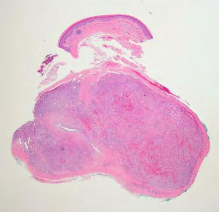
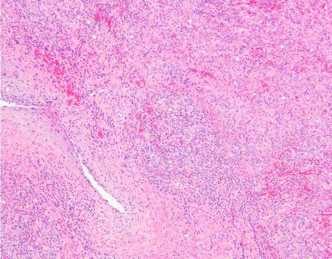
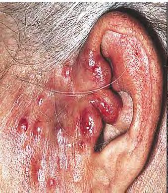
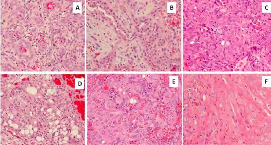
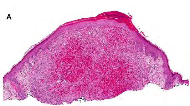
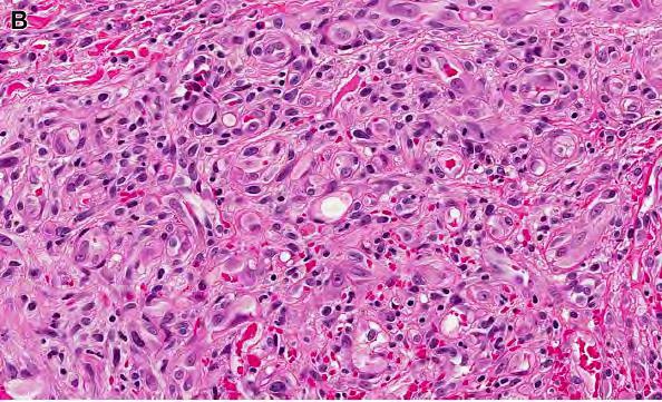
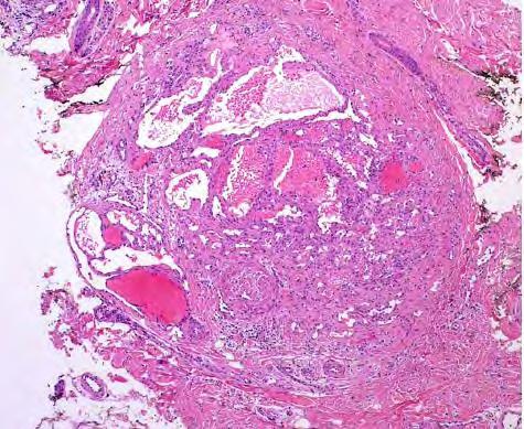
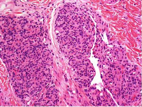
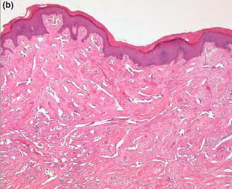
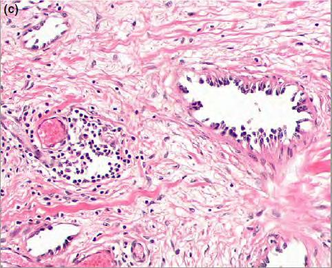
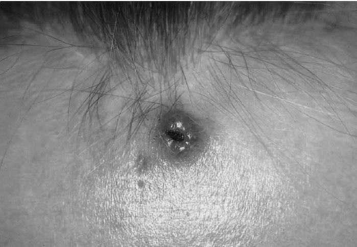
4 Epithelioid Hemangioma Most commonly present on the Head & Neck area Often periauricular Middle-aged adults, female predominance However wide range of cutaneous location Rarely intravascular Also affects bone Can be multifocal in 25%
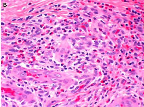
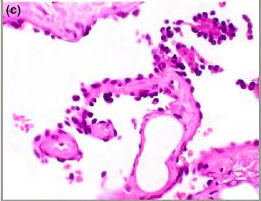
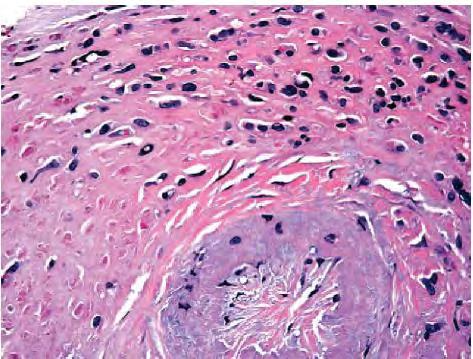
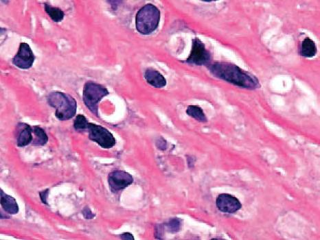
5 Microscopic features Circumscribed dermal or subcutaneous lobular proliferations of well-formed capillaries Usually surrounds a large vessel Inflammatory infiltrate Lymphocytes (sometimes germinal centers), eosinophils, histiocytes and plasma cells Involved capillaries retain lumina Lined by epithelioid cells Variably hobnail Mitoses may be seen but not atypical
6
7
8
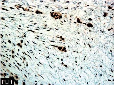
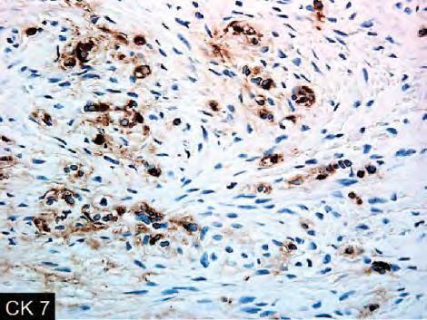
9 IHC Positive for vascular markers CD31, CD34, ERG Also positive for D2-40 Pitfall!! Immunoreactivity for EMA and keratins may be seen
10
11
12
13
14
15
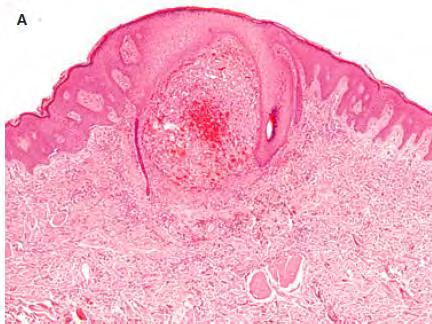
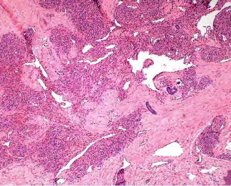
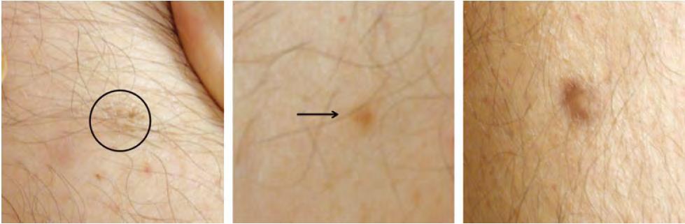
16 Cutaneous Epithelioid Angiomatous Nodule Most cases solitary as small violaceous superficial nodules Occasional multiple or eruptive forms Unilobular dermal and circumscribed Large solid sheets of epithelioid endothelial cells Intracytoplasmic vacuoles No significant pleomorphism Conspicuous mitotic activity but no atypical
17

18
19
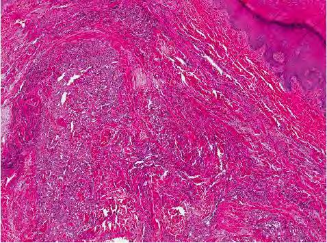
20 Symplastic Hemangioma Degenerative-type pleomorphism of vascular smooth muscle and interstitial cells Pre-existing long-standing vascular neoplasm Probably due to inflammation or hypoxia Extremities or face of young to elderly adults No sex predilection Characteristically bizarre pleomorphic cells within the walls of vascular channels Occasional mitotic figures are seen

21
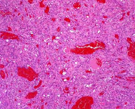
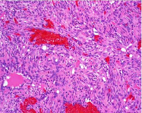
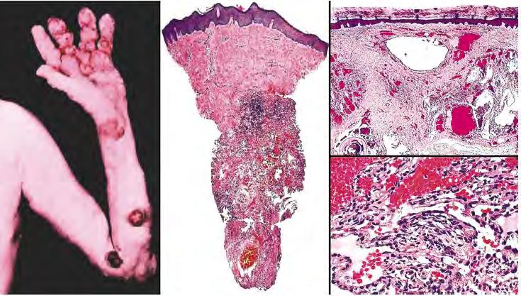
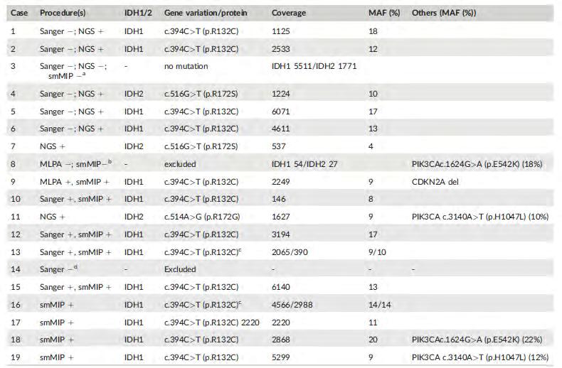
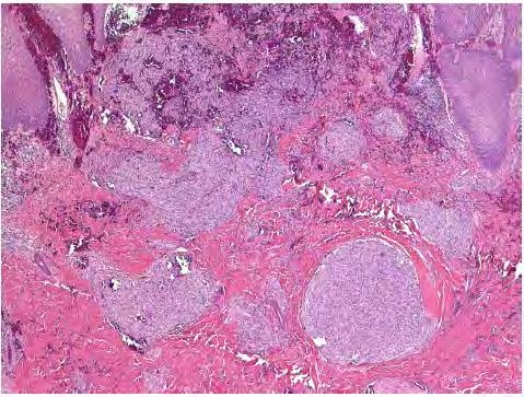
22 Spindle Cell Hemangioma Superficial subcutaneous or dermal erythematous to violaceous nodule Predilection for distal extremities of young adults, especially hands Sometimes multiple small nodules Subset of cases a/w Maffucci Syndrome Multiple spindle cell hemangiomas Enchondromas Increased risk for chondrosarcoma A/w IDH mutations
23 Microscopic Features Circumscribed subcutaneous or dermal tumors Half partially intravascular Biphasic composition Cavernous vascular spaces Solid spindle cell areas Cavernous vessels may contain phleboliths Spindle cell areas with slit-like spaces and intracytoplasmic lumina Mitotic activity and atypia low
24
25
26

27
28
29 Acquired Tufted Angioma Aka Angioblastoma of Nakagawa Most frequently in early childhood as progressive enlarging macule in head & neck area Cases with Kasabach-Merritt Syndrome have been described Less frequently than kaposiform hemangioendothelioma
30 Microscopic features In dermis or subcutis with so-called cannonball pattern of spherical welldermacated nodules Dilated lymphatic channels at the periphery Single endothelial cell layer surrounded by SMA positive pericytes Pericytes can be prominent and have a spindled appearance D2-40 positive in lymphatics and partially in capillary network
31
32
33 Vascular Tumors of Intermediate Malignancy
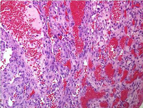
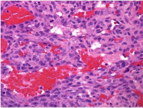
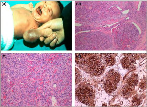
34 Kaposiform Hemangioendothelioma Vary rare tumor of infancy and childhood Most frequently as multinodular lesions in peripheral soft tissue or skin Followed by retroperitoneum Subset of cases a/w lymphangiomatosis Most patients in the first two years Has been reported in adults
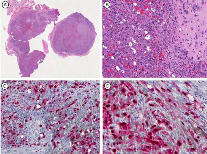
35 Microscopic features Plexiform mass of lobules separated by fibrous septa Capillary hemangioma-like areas alternating with spindle cells Spindle cells slit-like lumina with mild atypia and few mitoses Glomeruloid structures characteristic Small vessels surrounded by pericytes and hyaline globules At the periphery striking lymphatic proliferation with ectatic vascular spaces
36 Immunohistochemistry Endothelial cells express CD31, CD34 and ERG SMA highlights the pericytes Lymphatic markers (VEFG3, D2-40, Prox1) are positive both in the lymphatic channels at the lobule periphery and spindle cells GLUT1 is negative
37 Prognosis May show regional perinodal soft tissue involvement Not been reported to produce distal metastases Can be infiltrative and retroperitoneal cases can extensively infiltrate surrounding organs Can induce Kasabach-Merritt Syndrome Thrombocytopenia and consumption coagulopathy Related to tumor size and location 10% lethal secondary to KMS complications

38
39

40
41 D2-40
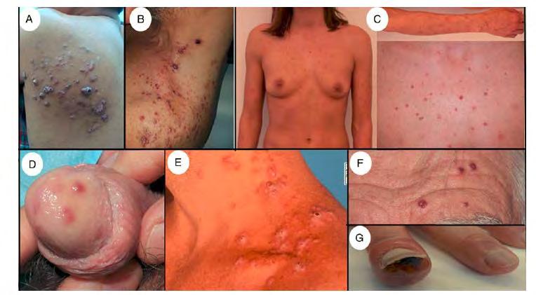
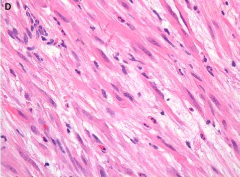
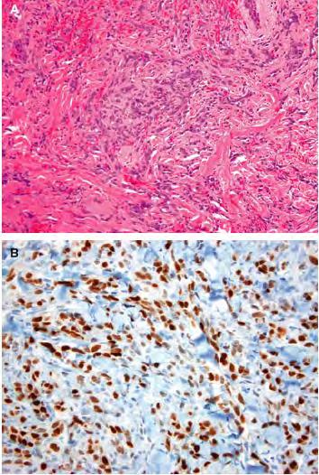
42 Papillary Intralymphatic Hemangioendothelioma (PILA) Aka Dabska tumor Closely related to retiform hemangioendothelioma Most in infants and children Some cases congenital Up to 25% in adults Single slowly growing nodule or plaque Most cases skin and superficial tissue of head and neck, trunk or extremities
43 Microscopic features Numerous and dilated, thin-walled lymphatic like spaces line by bland hobnail endothelial cells Intravascular papillae lined by hobnail endothelial cells as well Papillae containing a collagenous core Mononuclear inflammatory component may be present Usually not prominent
44 IHC Immunophenotype that resembles normal lymphatic endothelium CD31, CD34, ERG VEGFR3, podoplanin Lack of actin-positive cuff of pericytes
45
46
47 Retiform Hemangioendothelioma Superficially located, mainly occurs in adults Elongated-shaped vessels resembling rete testis Similarly to PILA lined by hobnail endothelial cells and a/w lymphocytes Intravascular papillary projections absent or very few Closely related to PILA Hobnail Hemangioendotheliomas
48
49 Epithelioid sarcoma-like/pseudomyogenic hemangioendothelioma Typically young adults with marked male (4:1) predilection Rare soft tissue of intermediate biological potential Propensity for local recurrence or frequent (and characteristic) development of additional nodules in the same region Metastasis is rare Conservative management is the mainstay of therapy

50

51 Radiologic findings Diagnostic Pathology: Soft Tissue Tumors 2nd Ed 2015, Elsevier

52 Microscopic findings Histopathology 2016 May;68:776-95
53
54
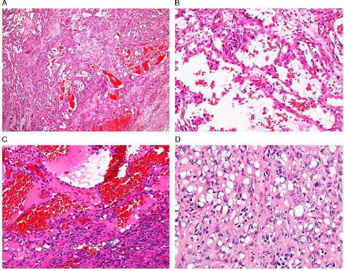
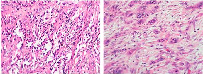
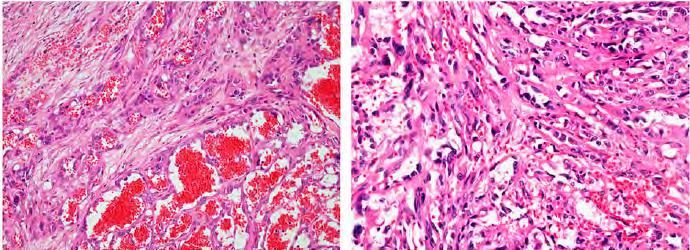
 is retained AE1/AE3 INI1")
55 Immunohistochemistry POSITIVE CKAE1/3 ERG CD31 (variable) AE1/AE3 NEGATIVE CD34 INI-1 (SMRCB1) is retained AE1/AE3 INI1 CD31 ERG
56 Other Differential Diagnosis Spindle cell squamous cell carcinoma Cellular benign fibrous histiocytoma Smooth muscle neoplasms Epithelioid Hemangioendothelioma
57 Molecular Recurrent balanced translocation t(7;19)(q22;q13) Fusion of SERPINE1 and FOSB genes To date this translocation has not been identified in other soft tissue tumors

58

59
60
61
62 WHO 2013 definition Locally aggressive, rarely metastasizing vascular neoplasm, containing an admixture of histologically distinct components Chiefly in adults Composite Hemangioendothelioma Very rare pediatric or congenital cases Predominantly skin and superficial soft tissues High rate of local recurrence (50%) Low risk of lymph node (6%) or distant metastases(1%)
63
64
65
66
67
68 CD31 Synaptophysin
69
70 D2-40 Synaptophysin
71
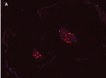
72 Malignant Vascular Tumors
73 Epithelioid Hemangioendothelioma (EHE) and CAMTA1 Rare low-grade, malignant vascular neoplasm that shows endothelial differentiation Less aggressive than angiosarcoma Risk of metastasis in ~ 20-30% of cases Death in approximately 15% of cases
74
75 Clinical Features Affects patients of all ages but rare during childhood Typically solitary lesion on the extremities Can involve larger preexisting vessels Multiple cutaneous nodules!!!!metastasizing deep soft tissue or osseous EHE should be ruled out!!!!


76 Microscopic findings Diagnostic Pathology: Vascular 2016 Elsevier

77

78

79 Immunohistochemistry POSITIVE CD31 CD34 ERG AE1/AE3 in 25% of cases NEGATIVE S100 protein Desmin EMA CD31
80 Molecular Recurrent translocation t(1;3)(p36;q25) involving WWTR1 (3q25) and CAMTA1 (1p36) Approximately 90% of EHE with classic morphology and not identified in histologic mimics A subset shown to harbor YAP1-TFE3

81
82

83 CAMTA1
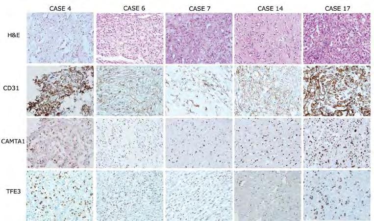
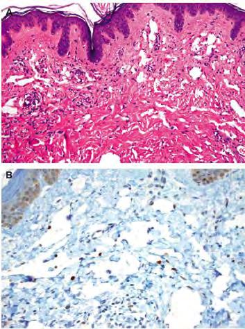
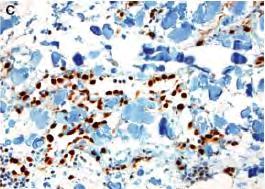
84
85
86 TFE3 TFE3 is a member of the microphthalmia (MiT) family of transcription factors, which includes MiTF, TFEB, TFEC, and TFE3 Although TFE3 is ubiquitously expressed in humans, native TFE3 protein is usually not detected by routine immunohistochemical methods
87 TFE3 Alveolar soft part sarcoma Xp11 translocation renal cell carcinoma melanotic Xp11 translocation renal cell carcinoma A subset of PEComas Epithelioid hemangioendotheliomas
88

89 Antonescu et al, Genes, Chromosomes and Cancer 52, , 2013

90 CD31 TFE3 CAMTA1
91
92 Angiosarcoma Angiosarcoma arises in four typical clinical settings: Chronically sun-damaged skin, particularly the scalp or face Sporadic visceral angiosarcoma In the setting of chronic lymphedema (eg, after mastectomy in Stewart-Treves syndrome) In areas of prior therapeutic radiation, such as for the management of breast carcinoma.
93 Atypical vascular proliferations, which have been described under various nomenclature designations, occur in areas of prior radiation In some cases may be difficult to distinguish from vasoformative angiosarcoma.
94 MYC MYC proto-oncogene is a transcription factor located on the long arm of chromosome 8 (8q24.21) Nuclear expression of MYC occurs in the vast majority of secondary angiosarcomas Only very rarely seen in primary angiosarcoma Not detected in atypical or benign vascular lesions occurring in irradiated skin.
95
96
97
98 Angiosarcoma FISH MYC

99 Angiosarcomas Case 1 Case 2

100 Atypical Vascular lesion (AVL)
101
102 Conclusion MYC immunohistochemistry is therefore useful in differentiating atypical or benign vascular lesions occurring in irradiated skin from secondary postradiation angiosarcomas.

103
104
105
106 CD31 ERG


107 KonstantinosLin
21/07/2017. Hobnail endothelial cells are not the same as epithelioid endothelial cells
 UPDATE IN CUTANEOUS VASCULAR S DERMATOPATHOLOGY SESSION BELFAST PATHOLOGY JUNE 21/2017 Dr E Calonje St John s Institute of Dermatology, London, United Kingdom THE FAMILY OF VASCULAR S WITH EPITHELIOID
Financial disclosures
 Mesenchymal Neoplasms with Melanocytic Differentiation By Konstantinos Linos MD, FCAP, FASDP Bone, Soft Tissue and Dermatopathology Assistant Professor of Pathology Dartmouth-Hitchcock Medical Center Geisel
Vascular Tumors in Children and Adults. Thuy Phung, MD, PhD Houston Methodist Hospital Texas Children s Hospital Baylor College of Medicine
 Vascular Tumors in Children and Adults Thuy Phung, MD, PhD Houston Methodist Hospital Texas Children s Hospital Baylor College of Medicine What are these lesions? (Marcelo Hochman, MD) What are these lesions?
Financial disclosures
 Cutaneous Mesenchymal Neoplasms with EWSR1 Rearrangement By Konstantinos Linos MD, FCAP, FASDP Bone, Soft Tissue and Dermatopathology Assistant Professor of Pathology Dartmouth-Hitchc Geisel School of
Cutaneous Mesenchymal Neoplasms with EWSR1 Rearrangement
 Cutaneous Mesenchymal Neoplasms with EWSR1 Rearrangement By Konstantinos Linos MD, FCAP, FASDP Bone, Soft Tissue and Dermatopathology Assistant Professor of Pathology Dartmouth-Hitchcock Medical Center
Disclosure of Relevant Financial Relationships
 Surgical Pathology Companion Meeting Case 5: Locally Recurrent Chest wall Mass Cristina Antonescu, MD Department of Pathology, Memorial Sloan Kettering Cancer Center Disclosure of Relevant Financial Relationships
An Overview of Genital Stromal Tumors
 An Overview of Genital Stromal Tumors By Konstantinos Linos MD, FCAP, FASDP Bone, Soft Tissue and Dermatopathology Assistant Professor of Pathology Dartmouth-Hitchcock Medical Center Geisel School of Medicine
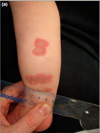
Vascular Tumors. Case 1: Papillary endothelial hyperplasia
 Vascular Tumors Case 1: Papillary endothelial hyperplasia Papillary endothelial hyperplasia (PEH) essentially represents a stage in the recanalization of a thrombosed blood vessel. It may occur in any
SOFT TISSUE TUMOR PATHOLOGY: AN UPDATE
 SOFT TISSUE TUMOR PATHOLOGY: AN UPDATE Jason L. Hornick, MD, PhD July 18, 2013 Department of Pathology Brigham and Women s Hospital Harvard Medical School Boston, MA, USA I have no disclosures. New Soft
Evening Specialty Conference Bone and Soft Tissue Pathology. Diagnostic pitfalls in bone and soft tissue pathology
 Evening Specialty Conference Bone and Soft Tissue Pathology. Case 1 Elizabeth G Demicco, MD, PhD Mount Sinai Hospital, New York Disclosure of Relevant Financial Relationships USCAP requires that all planners
Selected Pseudomalignant Soft Tissue Tumors of the Skin and Subcutis
 Selected Pseudomalignant Soft Tissue Tumors of the Skin and Subcutis Andrew L. Folpe, M.D. Professor of Laboratory Medicine and Pathology Mayo Clinic, Rochester, MN folpe.andrew@mayo.edu 2016 MFMER slide-1
USCAP 2011: ASDP companion meeting. Steven D. Billings 1
 USCAP 2011: ASDP companion meeting. Steven D. Billings (billins@ccf.org) 1 Spindle cell tumors that make you say, Oh $*&%! This lecture will focus on examples of cutaneous tumors that present particular
Hemangiomas and Other Vascular Tumors
 facebook.com/cincykidsrad Hemangiomas and Other Vascular Tumors Disclosures No relevant financial disclosures Bernadette L. Koch, M.D. Departments of Radiology and Pediatrics Cincinnati Children s Hospital
Enterprise Interest Nothing to declare
 Enterprise Interest Nothing to declare Diagnoses one would not like to miss in soft tissue pathology early in your career Marta Sbaraglia, MD Department of Pathology Hospital of Treviso University of Padua
Epithelioid Hemangioendothelioma. Ibtehal Al-Harbi Medical Intern
 Epithelioid Hemangioendothelioma Ibtehal Al-Harbi Medical Intern Hemangioendothelioma Hemangioendothelioma (HE) is the term used to describe a diverse group of vascular cancers. Very rare, accounting for
Dermatopathology. Dr. Rafael Botella Estrada. Hospital La Fe de Valencia
 Dermatopathology Dr. Rafael Botella Estrada. Hospital La Fe de Valencia DERMATOPATHOLOGY CASE CHALLENGE: RECOGNIZING MIMIS AND MASQUERADERS Rosalie Elenitsas. University of Pennsylvania Spectrum Lupus
أملس عضلي غرن = Leiomyosarcoma. Leiomyosarcoma 1 / 5
 Leiomyosarcoma 1 / 5 EPIDEMIOLOGY Exact incidence is unknown, but older studies suggest that leiomyosarcomas comprise approximately 3 percent of soft-tissue sarcomas. Superficial leiomyosarcoma occurs
Desmoplastic Melanoma R/O BCC. Clinical Information. 74 y.o. man with lesion on left side of neck r/o BCC
 R/O BCC Sabine Kohler, M.D. Professor of Pathology and Dermatology Dermatopathology Service Stanford University School of Medicine Clinical Information 74 y.o. man with lesion on left side of neck r/o
Update on Cutaneous Mesenchymal Tumors. Thomas Brenn
 Update on Cutaneous Mesenchymal Tumors Thomas Brenn Cutaneous Mesenchymal Tumours Wide morphological and biological spectrum Myofibroblastic, smooth muscle, neural, vascular, apidocytic, undifferentiated;
Disclosure. Relevant Financial Relationship(s) None. Off Label Usage None MFMER slide-1
 None. Off Label Usage None MFMER slide-1") Disclosure Relevant Financial Relationship(s) None Off Label Usage None 2013 MFMER slide-1 Case Presentation A 43 year old male, with partial nephrectomy for a right kidney mass 2013 MFMER slide-2 2013
Disclosure Relevant Financial Relationship(s) None Off Label Usage None 2013 MFMER slide-1 Case Presentation A 43 year old male, with partial nephrectomy for a right kidney mass 2013 MFMER slide-2 2013
Financial disclosures
 An update on immunohistochemical markers in mesenchymal neoplasms By Konstantinos Linos MD, FCAP, FASDP Assistant Professor of Pathology Geisel School of Medicine at Dartmouth Dartmouth-Hitchcock Medical
1/10/2018. Soft Tissue Tumors Showing Melanocytic Differentiation. Overview. Desmoplastic/ Spindle Cell Melanoma
 2016 MFMER slide-1 2016 MFMER slide-2 2016 MFMER slide-3 Soft Tissue Tumors Showing Melanocytic Differentiation Andrew L. Folpe, M.D. Professor of Laboratory Medicine and Pathology Mayo Clinic, Rochester,
ACCME/Disclosures ALK FUSION-POSITIVE MESENCHYMAL TUMORS. Tumor types with ALK rearrangements. Anaplastic Lymphoma Kinase. Jason L.
 Companion Meeting of the International Society of Bone and Soft Tissue Pathology The Evolving Concept of Mesenchymal Tumors ALK FUSION-POSITIVE MESENCHYMAL TUMORS Jason L. Hornick, MD, PhD March 13, 2016
Malignant Peripheral Nerve Sheath Tumor
 C H A P T E R 120 Malignant Peripheral Nerve Sheath Tumor Currently, malignant peripheral nerve sheath tumor (MPNST) is the most commonly used generic name for the neoplasms known in the past as neurosarcoma,
SMOOTH MUSCLE TUMOURS
 SMOOTH MUSCLE TUMOURS NORMAL SMOOTH MUSCLE Cytology Immunohistochemistry Ultrastructure Masson Trichrome Smooth Muscle Ultrastructure Many myofilaments running parallel to the long axis of the smooth
Newer soft tissue entities
 Newer soft tissue entities Examples among fibroblastic tumors Turku, May 6, 2010 Markku Miettinen, M.D. AFIP, Washington, DC Fibroblastic neoplasms Solitary fibrous tumor /Hemangiopericytoma Low-grade
3/27/2017. Disclosure of Relevant Financial Relationships
 Ophthalmic Pathology Evening Specialty Conference USCAP 2017 5 th March, 2017 Mukul K. Divatia, MD Assistant Professor Department of Pathology & Genomic Medicine Weill Cornell Medical College Houston Methodist
Special slide seminar
 Special slide seminar Tomáš Rozkoš The Fingerland Department of Pathology Charles University Medical Faculty and Faculty Hospital in Hradec Králové Czech Republic Case history, 33 years old resistance
Case 27 Male 42. Painless, static, well-circumscribed, subcutaneous nodule right lower leg,?lipoma. The best diagnosis is:
 Case 27 Male 42. Painless, static, well-circumscribed, subcutaneous nodule right lower leg,?lipoma. The best diagnosis is: A. Angiosarcoma B. Haemangiopericytoma C.Myopericytoma D.Myofibroma E. Angioleiomyoma
Myxo-inflammatory Fibroblastic sarcoma
 AKA Myxo-inflammatory Fibroblastic sarcoma Acral Myxoinflammatory fibroblastic sarcomaam.j.surg.path1998; 22; 911-924 Inflammatory myxoid tumour of soft parts with bizarre giant cells [Pathol.Res.Pract.
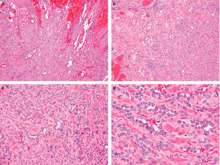
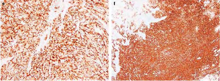
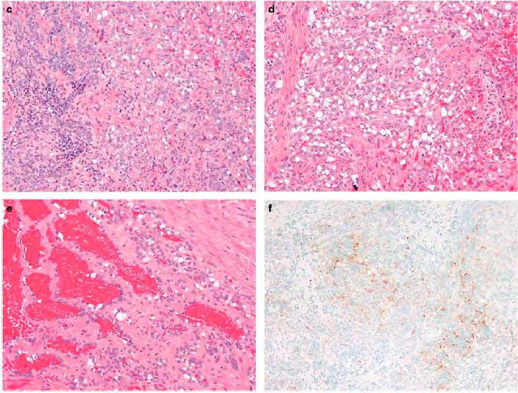
Hemangioendothelioma with a Prominent Lymphoid Infiltrate Mimicking Follicular Dendritic Cell Tumor: Report of a Case
 Journal of Cancer Research Updates, 2013, 2, 135-139 135 Hemangioendothelioma with a Prominent Lymphoid Infiltrate Mimicking Follicular Dendritic Cell Tumor: Report of a Case Justin Kerstetter 1, Mia Perez
Diplomate of the American Board of Pathology in Anatomic and Clinical Pathology
 A 33-year-old male with a left lower leg mass. Contributed by Shaoxiong Chen, MD, PhD Assistant Professor Indiana University School of Medicine/ IU Health Partners Department of Pathology and Laboratory
Vascular Tumors of Intermediate Malignancy: A Review and Update
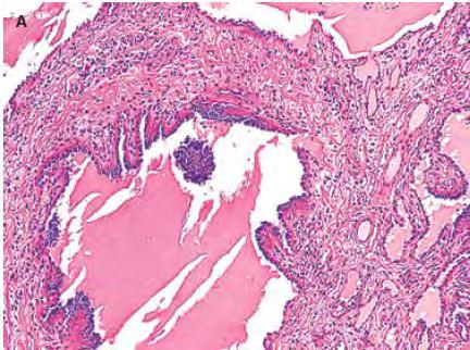
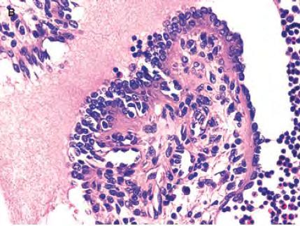
 Review Vascular Tumors of Intermediate Malignancy: A Review and Update Jason S. Stratton 1 Steven D. Billings 1, 2 Vascular tumors of intermediate malignancy encompass a broad range of histologic entities.
Advances in the clinicopathological and molecular classification of cutaneous mesenchymal neoplasms
 Histopathology 2016 DOI: 10.1111/his.12930 REVIEW Advances in the clinicopathological and molecular classification of cutaneous mesenchymal neoplasms Danielle C Costigan & Leona A Doyle 1 Department of
Case: The patient is a 24 year- old female who was found to have multiple mural nodules within the antrum. Solid and cystic components were noted on
 Case: The patient is a 24 year- old female who was found to have multiple mural nodules within the antrum. Solid and cystic components were noted on imaging. There is no significant past medical history.
Self assessment case. Dr Saleem Taibjee Dorset County Hospital, Dorchester
 Self assessment case Dr Saleem Taibjee saleemtaibjee@gmail.com Dorset County Hospital, Dorchester Clinical details 34-year-old man: Shave excision Skin tag / papilloma left thigh The best diagnosis is:
Pathology Mystery and Surprise
 Pathology Mystery and Surprise Tim Smith, MD Director Anatomic Pathology Medical University of South Carolina Disclosures No conflicts to declare Some problem cases Kidney tumor Scalp tumor Bladder tumor
59 yo male with past medical history of prostate carcinoma, presented with upper abdominal pain
 December 2016 59 yo male with past medical history of prostate carcinoma, presented with upper abdominal pain Contributed by: Divya Sharma, MD. Fellow, Gastrointestinal Pathology, Department of Pathology
57th Annual HSCP Spring Symposium 4/16/2016
 An Unusual Malignant Spindle Cell Lesion to Involve the Breast Erinn Downs-Kelly, D.O. Associate Professor of Pathology University of Utah & ARUP Laboratories No disclosures Case 39 y/o female with no
SEBACEOUS NEOPLASMS. Dr. Prachi Saraogi Clinical Fellow in Dermatology
 SEBACEOUS NEOPLASMS Dr. Prachi Saraogi Clinical Fellow in Dermatology Sebaceous neoplasms Sebaceous adenoma (Benign) Sebaceous carcinoma (Malignant) SEBACEOUS ADENOMA Benign tumours composed of incompletely
Fun with Fat. General Rules. Case
 Fun with Fat General Rules Imaging: location (deep vs. superficial) Superficial lesions are seldom liposarcomas Deep lesions may be benign or malignant Myxoid stroma is common in benign and malignant lesions
IN THE NAME OF GOD Dr. Kheirandish Oral and maxillofacial pathology
 IN THE NAME OF GOD Dr. Kheirandish Oral and maxillofacial pathology ORAL FOCAL MUCINOSIS Uncommon Tumorlike Cutaneous myxoid cyst Overproduction of hyaluronic acid by firoblasts Young adults Female Gingiva
Benign and malignant epithelial lesions: Seborrheic keratosis: A common benign pigmented epidermal tumor occur in middle-aged or older persons more
 Benign and malignant epithelial lesions: Seborrheic keratosis: A common benign pigmented epidermal tumor occur in middle-aged or older persons more common on the trunk; but extremities, head and neck are
Lung Tumor Cases: Common Problems and Helpful Hints
 Lung Tumor Cases: Common Problems and Helpful Hints Brandon T. Larsen, MD, PhD Senior Associate Consultant Department of Laboratory Medicine and Pathology Mayo Clinic Arizona Arizona Society of Pathologists
Sonography of soft-tissue vascular lesions
 Sonography of soft-tissue vascular lesions Oscar M. Navarro Associate Professor, University of Toronto Dept. of Diagnostic Imaging, The Hospital for Sick Children Toronto, Canada Declaration of Disclosure
Spindle Cell Lesions Of The Breast. Emad Rakha Professor of Breast Pathology and Consultant Pathologist
 Spindle Cell Lesions Of The Breast Emad Rakha Professor of Breast Pathology and Consultant Pathologist * SCLs comprise a wide spectrum of diseases, ranging from reactive processes to aggressive malignant
Ultrasound imaging of vascular anomalies: pearls and pitfalls
 Ultrasound imaging of vascular anomalies: pearls and pitfalls Oscar Navarro, MD Dept. of Medical Imaging, University of Toronto Dept. of Diagnostic Imaging, The Hospital for Sick Children Declaration of
Primary Cutaneous CD30-Positive T-cell Lymphoproliferative Disorders
 Primary Cutaneous CD30-Positive T-cell Lymphoproliferative Disorders Definition A spectrum of related conditions originating from transformed or activated CD30-positive T-lymphocytes May coexist in individual
What is New in the 2015 WHO Lung Cancer Classification? Zhaolin Xu, MD, FRCPC, FCAP
 What is New in the 2015 WHO Lung Cancer Classification? Zhaolin Xu, MD, FRCPC, FCAP Professor, Dept of Pathology, Dalhousie University, Canada Pulmonary Pathologist and Cytopathologist, QEII HSC Senior
POORLY DIFFERENTIATED, HIGH GRADE AND ANAPLASTIC CARCINOMAS: WHAT IS EVERYONE TALKING ABOUT?
 POORLY DIFFERENTIATED, HIGH GRADE AND ANAPLASTIC CARCINOMAS: WHAT IS EVERYONE TALKING ABOUT? AGGRESSIVE THYROID CANCERS PAPILLARY CARCINOMA CERTAIN SUBTYPES POORLY DIFFERENTIATED CARCINOMA HIGH GRADE DIFFERENTIATED
SESSION 1: GENERAL (BASIC) PATHOLOGY CONCEPTS Thursday, October 16, :30am - 11:30am FACULTY COPY
 PATHOLOGY CONCEPTS Thursday, October 16, :30am - 11:30am FACULTY COPY") SESSION 1: GENERAL (BASIC) PATHOLOGY CONCEPTS Thursday, October 16, 2008 9:30am - 11:30am FACULTY COPY GOAL: Describe the basic morphologic (structural) changes which occur in various pathologic conditions.
SESSION 1: GENERAL (BASIC) PATHOLOGY CONCEPTS Thursday, October 16, 2008 9:30am - 11:30am FACULTY COPY GOAL: Describe the basic morphologic (structural) changes which occur in various pathologic conditions.
Dermatopathology. Dr. Rafael Botella Estrada. Hospital La Fe de Valencia
 Dermatopathology Dr. Rafael Botella Estrada. Hospital La Fe de Valencia Melanoma and mimics Dr. Martin Mihm Malignant lesions result from the accumulation of mutations Class I lesions (benign) Class II
USCAP Pediatrics Evening Subspecialty Conference 2015
 USCAP Pediatrics Evening Subspecialty Conference 2015 Sunday 22 March 2015 Alexander Lazar MD/PhD Department of Pathology S Section of Bone Soft TIssue Pathology Sarcoma Research Center The Case Patient
Papillary Lesions of the Breast A Practical Approach to Diagnosis. (Arch Pathol Lab Med. 2016;140: ; doi: /arpa.
 Papillary Lesions of the Breast A Practical Approach to Diagnosis (Arch Pathol Lab Med. 2016;140:1052 1059; doi: 10.5858/arpa.2016-0219-RA) Papillary lesions of the breast Span the spectrum of benign,
Surgical Pathology Evening Specialty Conference USCAP 2015
 Surgical Pathology Evening Specialty Conference USCAP 2015 John R. Goldblum, M.D. Chairman, Department of Pathology, Cleveland Clinic Professor of Pathology, Cleveland Clinic Lerner College of Medicine
Cellular Neurothekeoma
 Cellular Neurothekeoma Scott W Binder, MD Pritzker Professor of Pathology & Dermatology Sr. Vice Chair Director, Pathology Clinical Services Chief, Dermatopathology Geffen/UCLA School of Medicine Clinical
Update on Pediatric Vascular Tumors
 Update on Pediatric Vascular Tumors Jim Treat, MD Associate Professor of Clinical Pediatrics and Dermatology No Relevant Conflicts of Interest PHOTOGRAPHY & VIDEOTAPING ARE STRICTLY PROHIBITED IN ALL EDUCATIONAL
Synonyms. Nephrogenic metaplasia Mesonephric adenoma
 Nephrogenic Adenoma Synonyms Nephrogenic metaplasia Mesonephric adenoma Definition Benign epithelial lesion of urinary tract with tubular, glandular, papillary growth pattern Most frequently in the urinary
CHAPTER 7. Vascular Tumours
 HPTER 7 Vascular Tumours enign vascular tumours are very common and most frequently occur in the skin (see WHO classification of skin tumours). t all sites, it is often difficult to determine whether benign
A 42-year-old woman with a liver mass
 April 2016 Case of the Month A 42-year-old woman with a liver mass Contributed by: Natalia I. Rush, MD, Resident Physician, Indiana University School of Medicine, Department of Pathology and Laboratory
THE EVOLVING CONCEPT OF HEMANGIOENDOTHELIOMA
 THE EVOLVING CONCEPT OF HEMANGIOENDOTHELIOMA CHRISTOPHER D.M. FLETCHER, M.D., FRCPath Department of Pathology Harvard Medical School & Brigham and Women s Hospital 75 Francis Street Boston, MA 02115 Introduction
المركب النموذج--- سبيتز وحمة = Type Spitz's Nevus, Compound SPITZ NEVUS 1 / 7
 SPITZ NEVUS 1 / 7 Epidemiology An annual incidence rate of 1.4 cases of Spitz nevus per 100,000 individuals has been estimated in Australia, compared with 25.4 per 100,000 individuals for cutaneous melanoma
AGGRESSIVE VARIANTS OF PAPILLARY THYROID CARCINOMA DIAGNOSIS AND PROGNOSIS
 AGGRESSIVE VARIANTS OF PAPILLARY THYROID CARCINOMA DIAGNOSIS AND PROGNOSIS PAPILLARY THYROID CARCINOMA Clinical Any age Microscopic to large Female: Male= 2-4:1 Radiation history Lymph nodes Prognosis
04/10/2018. Intraductal Papillary Neoplasms Of Breast INTRADUCTAL PAPILLOMA
 Intraductal Papillary Neoplasms Of Breast Savitri Krishnamurthy MD Professor of Pathology Deputy Division Head The University of Texas MD Anderson Cancer Center 25 th Annual Seminar in Pathology Pittsburgh,
Pathology of the skin. 2nd Department of Pathology, Semmelweis University
 Pathology of the skin 2nd Department of Pathology, Semmelweis University Histology of the skin Epidermis: Stratum corneum Stratum granulosum Stratum spinosum Stratum basale Dermis: papillary and reticular
Dermatopathology: The tumor is composed of keratinocytes which show atypia, increase mitoses and abnormal mitoses.
 Squamous cell carcinoma (SCC): A common malignant tumor of keratinocytes arising in the epidermis, usually from a precancerous condition: 1- UV induced actinic keratosis, usually of low grade malignancy.
Pathology of Sarcoma ELEANOR CHEN, MD, PHD, ASSISTANT PROFESSOR DEPARTMENT OF PATHOLOGY UNIVERSITY OF WASHINGTON
 Pathology of Sarcoma ELEANOR CHEN, MD, PHD, ASSISTANT PROFESSOR DEPARTMENT OF PATHOLOGY UNIVERSITY OF WASHINGTON Presentation outline Background and epidemiology of sarcomas Sarcoma classification Sarcoma
Immunohistochemistry in Bone and Soft Tissue Tumors. Sahar Rassi Zankoul, MD
 Immunohistochemistry in Bone and Soft Tissue Tumors Sahar Rassi Zankoul, MD Introduction Bone tumors represent a wide variety of tumors of various origins and malignant potentials. These different tumor
A 25 year old female with a palpable mass in the right lower quadrant of her abdomen
 May 2016 A 25 year old female with a palpable mass in the right lower quadrant of her abdomen Contributed by: Paul Ndekwe, MD, Resident Physician, Indiana University School of Department of Pathology and
5/10. Pathology Soft tissue tumors. Farah Bhani. Mohammed Alorjani
 5/10 Pathology Soft tissue tumors Mohammed Alorjani Farah Bhani Slides are included in this sheet. Objectives: Soft tissue tumors 1. Describe soft tissue tumors. 2. Understand the classification of soft
Atypical Palisaded Myofibroblastoma of Lymph Node: Report of a rare case.
 ISPUB.COM The Internet Journal of Pathology Volume 10 Number 1 Atypical Palisaded Myofibroblastoma of Lymph Node: Report of a rare case. V Kinnera, R Nandyala, M Yootla, K Mandyam Citation V Kinnera, R
- Selected Tumors of the Skin Appendages - Primary vs. Metastasis
 - Selected Tumors of the Skin Appendages - Primary vs. Metastasis Napa Valley 2018 Victor G. Prieto, MD, PhD Chair of Pathology UT MD Anderson Cancer Center vprieto@mdanderson.org Napa Valley in May Introduction
The Relevance of Cytologic Atypia in Cutaneous Neural Tumors
 The Relevance of Cytologic Atypia in Cutaneous Neural Tumors Recent Findings - New Developments New Problems Zsolt B. Argenyi, M.D. Professor of Pathology & Dermatology Director of Dermatopathology Department
The Genetics of Myoepithelial Tumors: salivary glands, soft tissue and bone
 The Genetics of Myoepithelial Tumors: salivary glands, soft tissue and bone Cristina Antonescu, MD Memorial Sloan-Kettering Cancer Center, New York Nothing to declare Disclosure Spectrum of Myoepithelial
Self assesment Case 21
 17-18 MAY 2018 London Dermatopathology Symposium 2018 Self assesment Case 21 MARC HASPESLAGH CASE 21 1802-50585 48 year old lady with eczematous lesions at ear helix and red patch on nose bridge since
Oncocytic-Appearing Salivary Gland Tumors. Oncocytic, Cystic, Mucinous, and High Grade Salivary Gland Tumors SALIVARY GLAND FNA: PART II
 William C. Faquin, MD, PhD Professor of Pathology Harvard Medical School Director of Head and Neck Pathology Massachusetts Eye and Ear Massachusetts General Hospital SALIVARY GLAND FNA: PART II Oncocytic,
Every Vascular Tumor is NOT a Hemangioma What the Hematologist/Oncologist needs to know about Rare Vascular Tumors
 Welcome! To join the call dial (866) 740-1260, passcode 3754894#. All participants are placed on mute for the duration of the webinar. If you have questions, type them in the chat box at the bottom left
Case: The patient is a 62 year old woman with a history of renal cell carcinoma that was removed years ago. A 2.4 cm liver mass was found on CT
 Case: The patient is a 62 year old woman with a history of renal cell carcinoma that was removed years ago. A 2.4 cm liver mass was found on CT during follow- up. ALT, AST, Alk Phos and bilirubin were
Basement membrane in lobule.
 Bahram Memar, MD Basement membrane in lobule. Normal lobule-luteal phase Normal lobule-follicular phase Lactating breast Greater than 95% are adenocarcinomas in situ carcinomas and invasive carcinomas.
Note: The cause of testicular neoplasms remains unknown
 - In the 15- to 34-year-old age group, they are the most common tumors of men. - Tumors of the testis are a heterogeneous group of neoplasms that include: I. Germ cell tumors : 95%; all are malignant.
Rhabdomyomas and Rhabdomyosarcomas (RMS) David M. Parham, MD Chief of Anatomic Pathology
 David M. Parham, MD Chief of Anatomic Pathology") Rhabdomyomas and Rhabdomyosarcomas (RMS) David M. Parham, MD Chief of Anatomic Pathology Tumors of skeletal muscle: Rhabdomyomas and rhabdomyosarcomas Embryonal muscle 2 3 4 5 6 7 8 Rhabdomyoma Benign
Rhabdomyomas and Rhabdomyosarcomas (RMS) David M. Parham, MD Chief of Anatomic Pathology Tumors of skeletal muscle: Rhabdomyomas and rhabdomyosarcomas Embryonal muscle 2 3 4 5 6 7 8 Rhabdomyoma Benign
Normal thyroid tissue
 Thyroid Pathology Overview Normal thyroid tissue Normal thyroid tissue with follicles filled with colloid. Thyroid cells form follicles, spheres of epithelial cells (always single layered in health, usually
Breast pathology. 2nd Department of Pathology Semmelweis University
 Breast pathology 2nd Department of Pathology Semmelweis University Breast pathology - Summary - Benign lesions - Acute mastitis - Plasma cell mastitis / duct ectasia - Fat necrosis - Fibrocystic change/
Diagnostically Challenging Cases in Gynecologic Pathology
 Diagnostically Challenging Cases in Gynecologic Pathology Eric C. Huang, M.D., Ph.D. Department of Pathology and Laboratory Medicine University of California, Davis Medical Center Case 1 Presentation 38
Case Presentation. Maha Akkawi, MD, Fatima Obeidat, MD, Tariq Aladily, MD. Department of Pathology Jordan University Hospital Amman, Jordan
 Case Presentation Maha Akkawi, MD, Fatima Obeidat, MD, Tariq Aladily, MD Department of Pathology Jordan University Hospital Amman, Jordan The 25th Annual Congress of the ADIAP The 8/11/2013 1 5th International
Various hereditary, acquired and neoplastic conditions can lead to cyst formation in the kidney.
 Dr. Fatima AlAl-Hashimi Hashimi,, MD, FRCPath Salmaniya Medical Complex, Bahrain Various hereditary, acquired and neoplastic conditions can lead to cyst formation in the kidney. The most frequently encountered
The last 10 years have seen the description of many
 An Update on the Application of Newly Described Immunohistochemical Markers in Soft Tissue Pathology George Lin, MD, PhD; Leona A. Doyle, MD Context. During the last 5 to 10 years, significant progress
Primary bone tumors > metastases from other sites Primary bone tumors widely range -from benign to malignant. Classified according to the normal cell
 Primary bone tumors > metastases from other sites Primary bone tumors widely range -from benign to malignant. Classified according to the normal cell counterpart and line of differentiation. Among the
Gross appearance of nodular hyperplasia in material obtained from suprapubic prostatectomy. Note the multinodular appearance and the admixture of
 Tiền liệt tuyến Tiền liệt tuyến Gross appearance of nodular hyperplasia in material obtained from suprapubic prostatectomy. Note the multinodular appearance and the admixture of solid and microcystic areas.
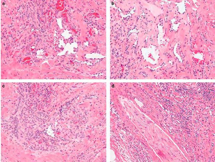
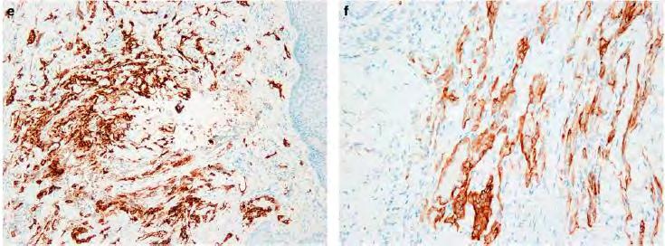
Case Report A case of retiform-hemangioendothelioma with unusual presentation and aggressive clinical features
 Int J Clin Exp Pathol 2010;3(5):528-533 www.ijcep.com /IJCEP1004002 Case Report A case of retiform-hemangioendothelioma with unusual presentation and aggressive clinical features Guiying Zhang 1, Qianjin
What s New in Pathology of Genitourinary Tumors. Jiaoti Huang, MD, PhD Department of Pathology Duke University School of Medicine
 What s New in Pathology of Genitourinary Tumors Jiaoti Huang, MD, PhD Department of Pathology Duke University School of Medicine Kidney Tumors Multilocular cystic renal neoplasm of low malignant potential
Conflict of Interest 9/2/2014. Pathogenesis and Comparison of Atypical Spitz Nevi vs Benign Spitz, and Childhood Melanoma
 Pathogenesis and Comparison of Atypical Spitz Nevi vs Benign Spitz, and Childhood Melanoma Martin C. Mihm Jr., M.D., F.A.C.P. Harvard Medical School Brigham and Women s Hospital Dana Farber Cancer Center
3/24/2017 DENDRITIC CELL NEOPLASMS: HISTOLOGY, IMMUNOHISTOCHEMISTRY, AND MOLECULAR GENETICS. Disclosure of Relevant Financial Relationships
 DENDRITIC CELL NEOPLASMS: HISTOLOGY, IMMUNOHISTOCHEMISTRY, AND MOLECULAR GENETICS Jason L. Hornick, M.D., Ph.D. Director of Surgical Pathology and Immunohistochemistry Brigham and Women s Hospital Professor
Slide seminar. Asist. Prof. Jože Pižem, MD, PhD Institute of Pathology Medical Faculty, University of Ljubljana
 Slide seminar Asist. Prof. Jože Pižem, MD, PhD Institute of Pathology Medical Faculty, University of Ljubljana Case 5 A 57-year-old man with a dermal/subcutaneous lesion on the scalp, which was interpreted
Cover Page. The handle holds various files of this Leiden University dissertation.
 Cover Page The handle http://hdl.handle.net/17/3271 holds various files of this Leiden University dissertation. Author: Verbeke, Sofie Lieve Jozef Title: Primary vascular tumours of bone: towards a new
Diseases of the breast (1 of 2)
") Diseases of the breast (1 of 2) Introduction A histology introduction Normal ducts and lobules of the breast are lined by two layers of cells a layer of luminal cells overlying a second layer of myoepithelial
Diseases of the breast (1 of 2) Introduction A histology introduction Normal ducts and lobules of the breast are lined by two layers of cells a layer of luminal cells overlying a second layer of myoepithelial
Keywords solitary fibrous tumor, dedifferentiation, dedifferentiated solitary fibrous tumor, STAT6, GRIA2, cytokeratin, rhabdomyosarcomatous
 758452IJSXXX10.1177/1066896918758452International Journal of Surgical PathologyCreytens et al research-article2018 Pitfalls in Pathology Multifocal Cytokeratin Expression in a Dedifferentiated Solitary
Mammary analogue secretory carcinoma of salivary gland A case report of new entity
 Case Report Mammary analogue secretory carcinoma of salivary gland A case report of new entity Vaibhav Bhika Bari 1*, Sandhya Unmesh Bholay 2 1 Assistant Professor, 2 Associate Professor Rajiv Gandhi Medical
Differential diagnosis of hematolymphoid tumors composed of medium-sized cells. Brian Skinnider B.C. Cancer Agency, Vancouver General Hospital
 Differential diagnosis of hematolymphoid tumors composed of medium-sized cells Brian Skinnider B.C. Cancer Agency, Vancouver General Hospital Lymphoma classification Lymphoma diagnosis starts with morphologic