Case Based Fetal Lung Masses
|
|
|
- Cory Jenkins
- 6 years ago
- Views:
Transcription

1 Case Based Fetal Lung Masses Advances in Fetal and Neonatal Imaging Course Orlando, Florida, January 28, 2017 Leann E. Linam, MD Associate Professor Radiology University of Arkansas for Medical Sciences/ Arkansas Children s Hospital
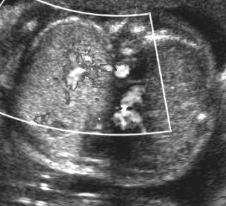
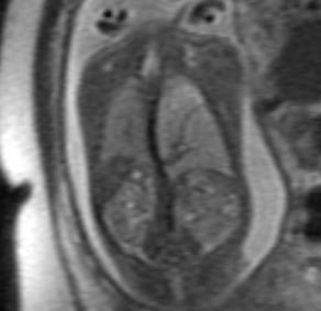
2 Prenatal Imaging of fetal lung masses US primary imaging modality Lungs homogeneous; hyperechoic to liver and increasing echogenicity with gestational age Focal increase in echogenicity, mediastinal shift, cysts can indicate mass MRI can help further delineate lung mass Added clinical value debated Levine, et al 2003, MRI changed management in only 8% of cases with fetal thoracic abnormalities; additional information in 38%
3 Fetal Chest: KEY POINTS CLASSIFICATION IS DIFFICULT BRONCHIAL ATRESIA IS LIKELY UNDERLYING CAUSE OF MOST LESIONS BIG IS BAD HYDROPS IS VERY VERY BAD
4 Fetal Lung Masses Bronchopulmonary malformations: Bronchial Atresia Congenital Lobar Overinflation Foregut Duplication Cyst Congenital Pulmonary Airway Malformation Bronchopulmonary Sequestration Hybrid Lesions Other Masses: Pleuropulmonary Blastoma Mediastinal Teratoma Congenital High Airway Obstruction
5 Fetal Lung Masses Bronchopulmonary malformations: Bronchial Atresia Congenital Lobar Overinflation Foregut Duplication Cyst Congenital Pulmonary Airway Malformation Bronchopulmonary Sequestration Hybrid Lesions Other Masses: Pleuropulmonary Blastoma Mediastinal Teratoma Congenital High Airway Obstruction
6 Bronchial Atresia Proximal bronchial atresia with mucoid impacted central bronchi and normal distal lung architecture. Most common: apicoposterior segment of left upper lobe > RUL > RML. Likely occurs between gestational weeks 5 and 15, may be due to loss of arterial supply. Associations: CLE, BPS, CPAM ( bronchial obstruction sequence )

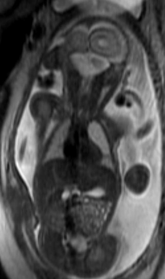
7 Bronchial Atresia

8 Bronchial Atresia
9 Fetal Lung Masses Bronchopulmonary malformations: Bronchial Atresia Congenital Lobar Overinflation Foregut Duplication Cyst Congenital Pulmonary Airway Malformation Bronchopulmonary Sequestration Hybrid Lesions Other Masses: Pleuropulmonary Blastoma Mediastinal Teratoma Congenital High Airway Obstruction
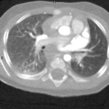
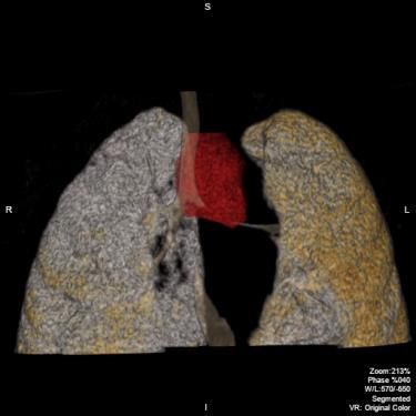
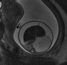
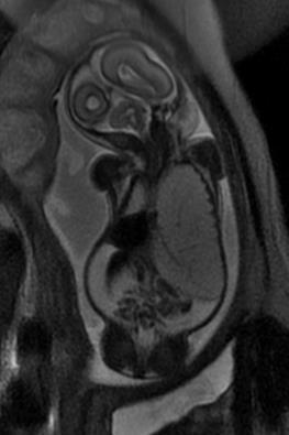
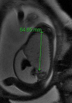
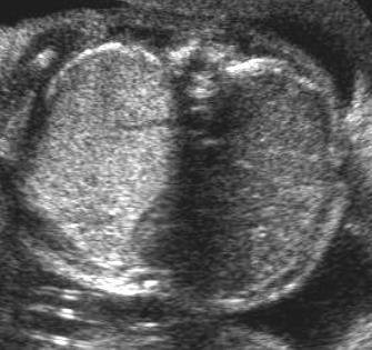
10 Congenital Lobar Overinflation Progressive over distention from obstruction in at least one segment or lobe. Obstruction can be intrinsic or external Congenital heart disease 15-40%.
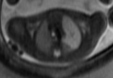
11 Congenital Lobar Overinflation
12 Congenital Lobar Overinflation
13 Fetal Lung Masses Bronchopulmonary malformations: Bronchial Atresia Congenital Lobar Overinflation Foregut Duplication Cyst Congenital Pulmonary Airway Malformation Bronchopulmonary Sequestration Hybrid Lesions Other Masses: Pleuropulmonary Blastoma Mediastinal Teratoma Congenital High Airway Obstruction
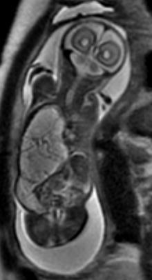
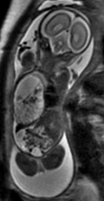
14 Foregut Duplication Cyst Bronchogenic cyst, enteric cyst, neuroenteric cyst. Abnormal ventral budding of the tracheobronchial tree. Well defined mass, usually middle mediastinum Homogeneous, brighter than CSF in T2 and STIR.

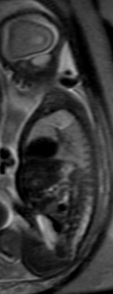
15 Foregut Duplication Cyst

16 Foregut Duplication Cyst
17 Fetal Lung Masses Bronchopulmonary malformations: Bronchial Atresia Congenital Lobar Overinflation Foregut Duplication Cyst Congenital Pulmonary Airway Malformation Bronchopulmonary Sequestration Hybrid Lesions Other Masses: Pleuropulmonary Blastoma Mediastinal Teratoma Congenital High Airway Obstruction
18 Congenital Pulmonary Airway Malformation Stocker Classification: Original, 1977, 3 types Current 4 types Controversial, not comprehensive Langston Classification: more descriptive, based on pathogenesis

19 Congenital Pulmonary Airway Malformation
20 Congenital Pulmonary Airway Malformation Type 0 Acinar dysplasia Exceedingly rare Usually bilateral Incompatible with life


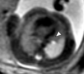
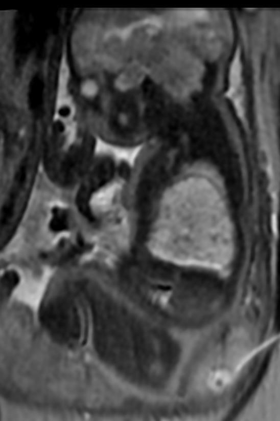
21 Congenital Pulmonary Airway Malformation Type 1 Large cyst type 65% Associated with airway obstruction in utero Hamartoma vs. neoplasm
22


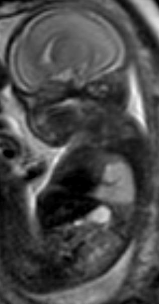
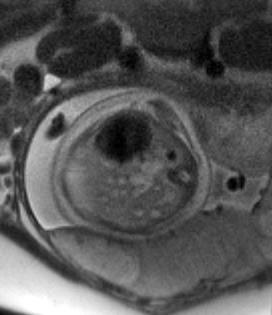
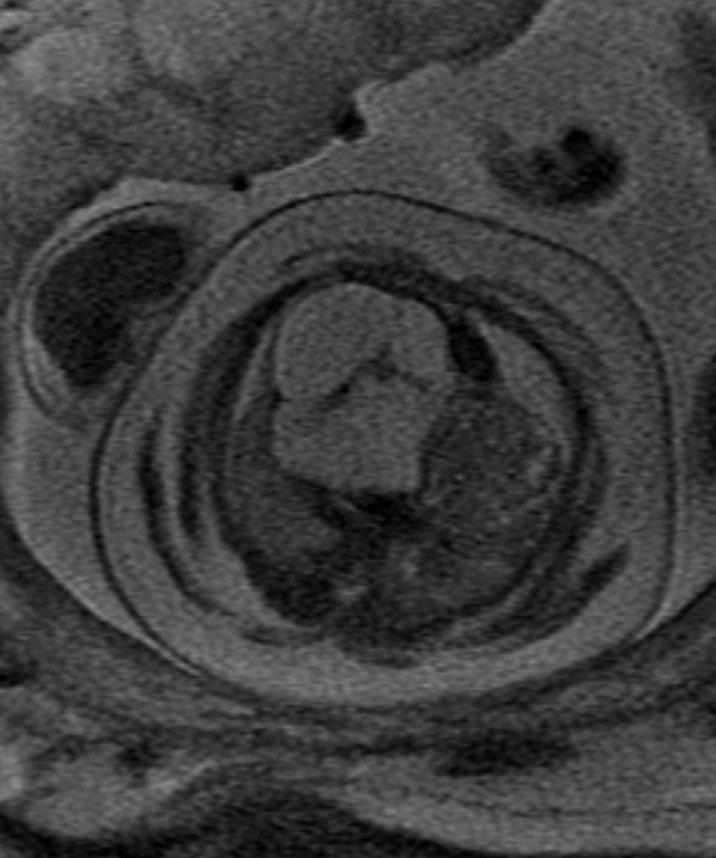
23 Congenital Pulmonary Airway Malformation Type 2 medium cysts Bronchial atresia with obstruction 10-15% Seen in ELS and ILS
24


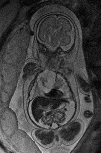
25 Congenital Pulmonary Airway Malformation Type 3 very small cysts Pulmonary hyperplasia, solid/adenomatoid type 5-8% Hamartoma vs. hyperplasia
26
27 Congenital Pulmonary Airway Malformation Type 4 Peripheral cyst type Pleuropulmonary blastoma: type 1 PPB? Regressed type 1 adenomatoid malformation 10-15% neoplasm
28 Fetal Lung Masses Bronchopulmonary malformations: Bronchial Atresia Congenital Lobar Overinflation Foregut Duplication Cyst Congenital Pulmonary Airway Malformation Bronchopulmonary Sequestration Hybrid Lesions Other Masses: Pleuropulmonary Blastoma Mediastinal Teratoma Congenital High Airway Obstruction
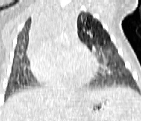
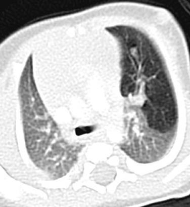
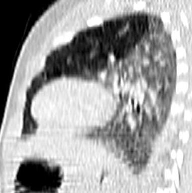
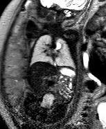
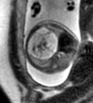
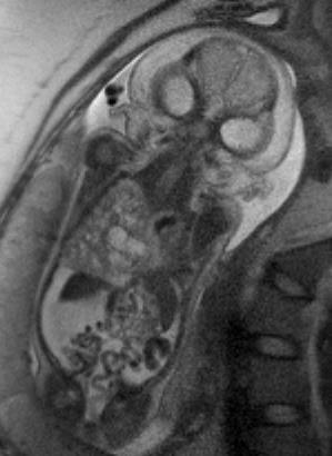
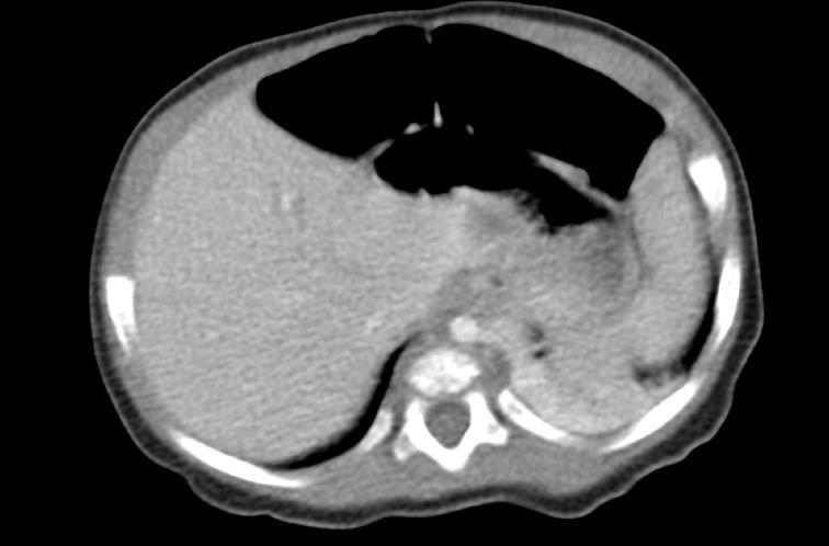
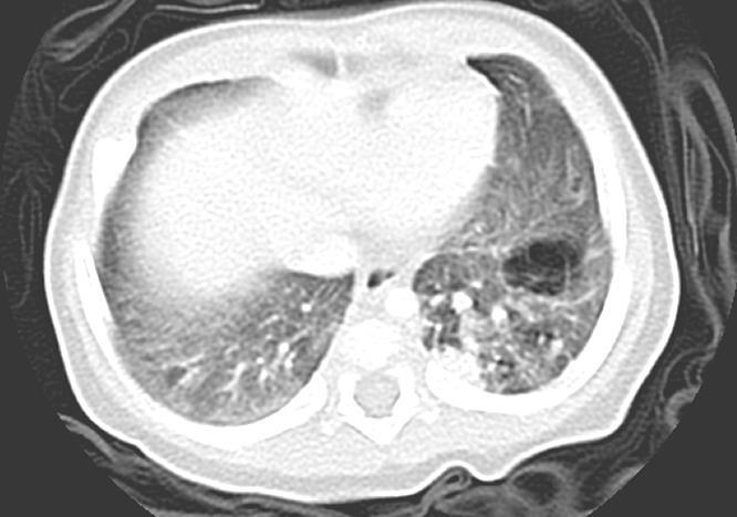
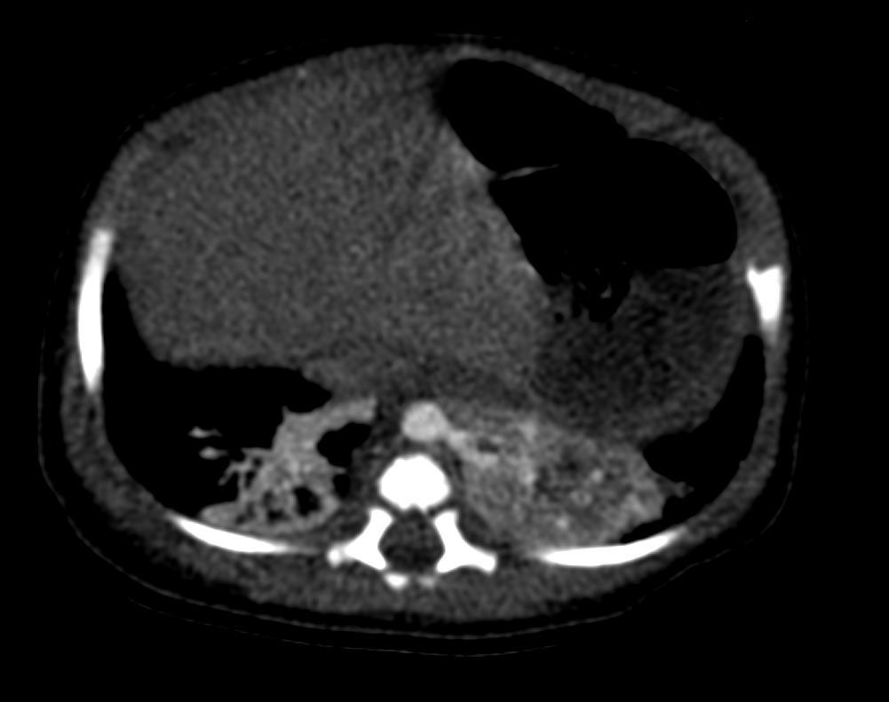
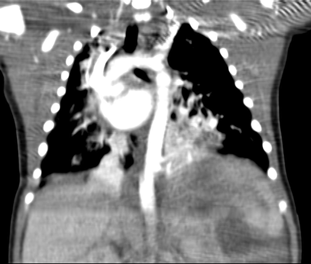
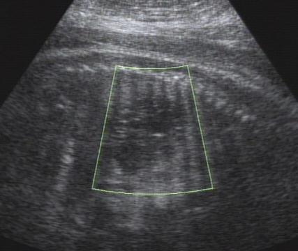
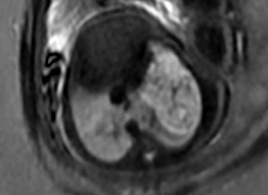
29 Bronchopulmonary Sequestration Bronchial atresia with systemic vascular connection Nonfunctioning pulmonary tissue with no connection to the tracheobronchial tree or pulmonary arteries. Well defined wedge shaped mass. Most common in posterior segment of left lower lobe. LLL > RLL. 90% above and 10% below the diaphragm. Homogeneous and solid if above diaphragm. Often cystic if below the diaphragm- can be mistaken for an adrenal mass.
30 Bronchopulmonary Sequestration

31
32
33 Fetal Lung Masses Bronchopulmonary malformations: Bronchial Atresia Congenital Lobar Overinflation Foregut Duplication Cyst Congenital Pulmonary Airway Malformation Bronchopulmonary Sequestration Hybrid Lesions Other Masses: Pleuropulmonary Blastoma Mediastinal Teratoma Congenital High Airway Obstruction


34 BPS/CPAM hybrid lesions Systemic vascular supply with cystic components bronchial atresia malformation sequence

35 BPS/CPAM hybrid lesions
36 Fetal Lung Masses Bronchopulmonary malformations: Bronchial Atresia Congenital Lobar Overinflation Foregut Duplication Cyst Congenital Pulmonary Airway Malformation Bronchopulmonary Sequestration Hybrid Lesions Other Masses: Pleuropulmonary Blastoma Mediastinal Teratoma Congenital High Airway Obstruction
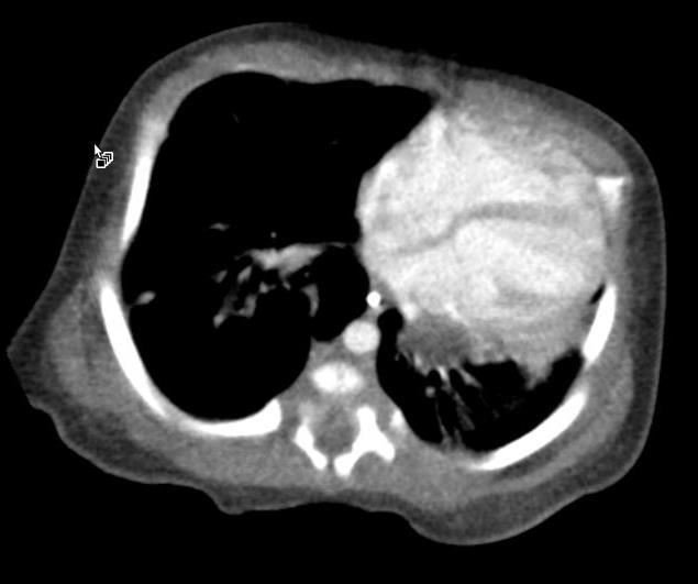
37 Pleuropulmonary Blastoma Malignant embryonal tumor 3 types: cystic, mixed cystic and solid, solid Type 1 occurs only in infants Current thinking is that Stocker Type 4 CPAM and PPB can be easily mistaken Possible regressed or undersampled
38 Pleuropulmonary Blastoma
39 Fetal Lung Masses Bronchopulmonary malformations: Bronchial Atresia Congenital Lobar Overinflation Foregut Duplication Cyst Congenital Pulmonary Airway Malformation Bronchopulmonary Sequestration Hybrid Lesions Other Masses: Pleuropulmonary Blastoma Mediastinal Teratoma Congenital High Airway Obstruction
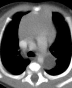
40 Mediastinal Teratoma Most common mediastinal germ cell tumor. Arise from primitive germ cell rests.
41 Mediastinal Teratoma
42 Fetal Lung Masses Bronchopulmonary malformations: Bronchial Atresia Congenital Lobar Overinflation Foregut Duplication Cyst Congenital Pulmonary Airway Malformation Bronchopulmonary Sequestration Hybrid Lesions Other Masses: Pleuropulmonary Blastoma Mediastinal Teratoma Congenital High Airway Obstruction


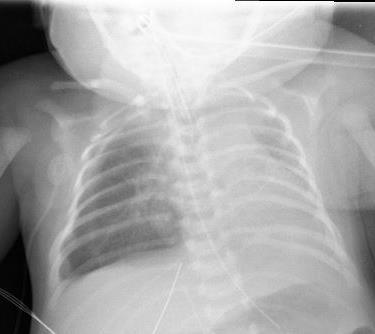
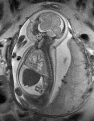
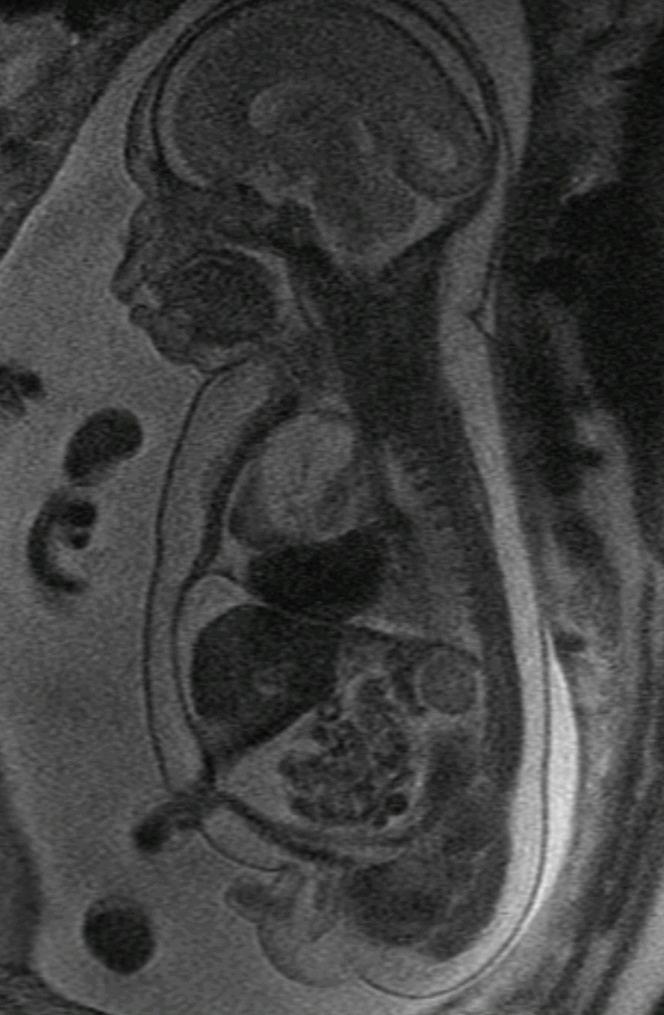
43 Congenital High Airway Obstruction Syndrome Pulmonary hyperplasia resulting from laryngeal atresia Hyperechogenic lungs

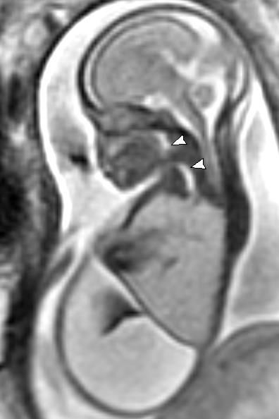
44 Congenital High Airway Obstruction Syndrome


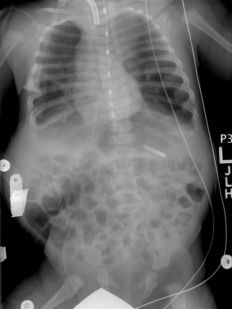
45 CHAOS: day 1 day 2 3 months
46 Fetal Lung Masses Bronchopulmonary malformations: Bronchial Atresia Congenital Lobar Overinflation Foregut Duplication Cyst Congenital Pulmonary Airway Malformation Bronchopulmonary Sequestration Hybrid Lesions Other Masses: Pleuropulmonary Blastoma Mediastinal Teratoma Congenital High Airway Obstruction
47 Fetal Chest: KEY POINTS CLASSIFICATION IS DIFFICULT BRONCHIAL ATRESIA IS LIKELY UNDERLYING CAUSE OF MOST LESIONS BIG IS BAD HYDROPS IS VERY VERY BAD
48 References: Pacharn P, et al. Congenital lung lesions: prenatal MRI and postnatal findings. Pediatr Radiol (2013) 43: Zucker EJ, et al. Perinatal ThoracicMassLesions:Pre-and Postnatal Imaging. Semin Ultrasound CT MRI 36: Fowler DJ, SJ Gould. The Pathology of congenital Lung Lesions. Semin Ped Surg 24 (2015)

49 Thank You!!
Congenital Lung Malformations: Radiologic-Pathologic Correlation
 Acta Radiológica Portuguesa, Vol.XVIII, nº 70, pág. 51-60, Abr.-Jun., 2006 Congenital Lung Malformations: Radiologic-Pathologic Correlation Marilyn J. Siegel Mallinckrodt Institute of Radiology, Washington
HOW TO IMAGE AND DESCRIBE CONGENITAL LUNG MALFORMATIONS
 HOW TO IMAGE AND DESCRIBE CONGENITAL LUNG MALFORMATIONS Paul Thacker, MD Assistant Professor Departments of Radiology and Pediatrics Medical University of South Carolina DISCLOSURES I have no relevant
Case Report Coexistent Congenital Diaphragmatic Hernia with Extrapulmonary Sequestration
 Canadian Respiratory Journal Volume 2016, Article ID 1460480, 4 pages http://dx.doi.org/10.1155/2016/1460480 Case Report Coexistent Congenital Diaphragmatic Hernia with Extrapulmonary Sequestration Nao
It s Rare So Be Aware: Pleuropulmonary Blastoma Mimicking Congenital Pulmonary Airway Malformation
 e10 Case Report: Thoracic THIEME It s Rare So Be Aware: Pleuropulmonary Blastoma Mimicking Congenital Pulmonary Airway Malformation Fayza Haider 1 Khulood Al Saad 2 Fatima Al-Hashimi 3 Hakima Al-Hashimi
ISUOG Basic Training. Assessing the Neck & Chest Gihad Chalouhi, Lebanon
 ISUOG Basic Training Assessing the Neck & Chest Gihad Chalouhi, Lebanon Learning objectives 9 & 10 At the end of the lecture you will be able to: recognise the differences between the normal & most common
Congenital bronchopulmonary malformation: CT histopathological correlation
 Congenital bronchopulmonary malformation: CT histopathological correlation Martin Kyncl a, Martin Koci a, Lea Ptackova a, Ludmila Hornofova b, Ondrej Fabian b, Jiri Snajdauf c, Miloslav Rocek a, Marcela
Imaging of the Lung in Children
 Imaging of the Lung in Children Imaging methods X-Ray of the Lung (Anteroposterior, ) CT, HRCT MRI USG Congenital developmental defects of the lungs Agenesis, aplasia, hypoplasia Tension pulmonary anomalies
Lung sequestration and Scimitar syndrome
 Lung sequestration and Scimitar syndrome Imaging approaches M. Mearadji International Foundation for Pediatric Imaging Aid Rotterdam, The Netherlands Pulmonary sequestration Pulmonary sequestration (PS)
Perinatal Imaging in Congenital Thoracic Cystic Malformations.
 Perinatal Imaging in Congenital Thoracic Cystic Malformations. Poster No.: C-0898 Congress: ECR 2013 Type: Educational Exhibit Authors: R. Llorens, G. Montoliu, A. Moreno, F. Menor; Valencia/ES Keywords:
Congenital Pulmonary Airways Malformation: an update
 Congenital Pulmonary Airways Malformation: an update Poster No.: P-0111 Congress: ESTI 2014 Type: Educational Poster Authors: S. M. Mak, B. Annan, S. P. G. Padley, A. G. Nicholson; London/UK Keywords:
Congenital Pulmonary Airways Malformation: an update
 Congenital Pulmonary Airways Malformation: an update Poster No.: P-0111 Congress: ESTI 2014 Type: Educational Poster Authors: S. M. Mak, B. Annan, S. P. G. Padley, A. G. Nicholson; London/UK Keywords:
Congenital lung anomalies: can we postpone resection?
 Journal of Pediatric Surgery (2012) 47, 87 92 www.elsevier.com/locate/jpedsurg Congenital lung anomalies: can we postpone resection? Nadja Colon a, Cameron Schlegel a, John Pietsch a, Dai H. Chung a,b,
24. An infant with recurrent pneumonia underwent a frontal chest radiograph (Fig 24-A) followed by
 followed by") 24. An infant with recurrent pneumonia underwent a frontal chest radiograph (Fig 24-A) followed by diagnosis? ndings, what is the most likely A. Pulmonary sequestration B. Congenital pulmonary airway malformation
24. An infant with recurrent pneumonia underwent a frontal chest radiograph (Fig 24-A) followed by diagnosis? ndings, what is the most likely A. Pulmonary sequestration B. Congenital pulmonary airway malformation
Bronchopulmonary foregut malformation: A pictorial review.
 Bronchopulmonary foregut malformation: A pictorial review. Poster No.: C-1676 Congress: ECR 2013 Type: Educational Exhibit Authors: N. L. Eun, C. S. Yoon, M.-J. Lee, M.-J. Kim ; Rep. of KOREA/ 1 2 2 2
Pulmonary Sequestration
 July 26, 2004 Pulmonary Sequestration Jonathan Shaw, Harvard Medical School Year IV What do these two patients have in common? Patient 1: 50 y.o. non-smoking female with several months cough and hemoptysis;
Imaging in pediatric lung diseases The roles of CT and pathology in diagnosing inherited and developmental lung diseases
 Imaging in pediatric lung diseases The roles of CT and pathology in diagnosing inherited and developmental lung diseases Dr Alistair D Calder Consultant Radiologist We re not so different, you and I. Invasiveness
Fetal MRI as an extended arm to ultrasound in evaluation of chest anomalies
 Fetal MRI as an extended arm to ultrasound in evaluation of chest anomalies Poster No.: C-1441 Congress: ECR 2014 Type: Educational Exhibit Authors: E. C. nandury, R. Jyothi, A. SRIRAMBHATLA ; Hyderabad/
CONGENITAL LUNG LESION Round Table. Objectives. Congenital Lung Lesions: Anatomy and Physiology Leah Barefoot, DNP, CPNP-PC
 CONGENITAL LUNG LESION Round Table L. Barefoot, E. Paton, C. Schultz, R. Caskey, M. O Day Objectives Review the anatomy and pathophysiology of congenital lung lesions List the preoperative evaluation of
Case report Esophageal lung: a rare case of communicating bronchopulmonary foregut malformation
 Case report Esophageal lung: a rare case of communicating bronchopulmonary foregut malformation 1 Dr.Varsha Rathi, 2 Dr. Saurabh Deshpande*, 3 Dr.Almas Nazim, 4 Dr.Shilpa Domkundwar 1 Professor, Department
Cystic adenomatoid malformation in adults: radiological findings and pathologic correlation
 Cystic adenomatoid malformation in adults: radiological findings and pathologic correlation Poster No.: C-1734 Congress: ECR 2014 Type: Educational Exhibit Authors: C. Batz Colvée, M. Vidal, M. Ruiz Tolón,
The Foetal Lung. Educational Exhibit Authors:
 The Foetal Lung Poster No.: C-1649 Congress: ECR 2015 Type: Educational Exhibit Authors: V. B. Pai, R. N. Chaubal, N. G. Chaubal, B. Pai ; Navi Mumbai/ 1 2 2 2 3 1 3 IN, Thane/IN, Mumbai/IN Keywords: Foetal
Monitor Images for Respiratory System Dissection
 Monitor Images for Respiratory System Dissection **This document includes extra images of the radiology of the bronchopulmonary segments. These imaged are an excellent way to review the three-dimensional
Heart and Lungs. LUNG Coronal section demonstrates relationship of pulmonary parenchyma to heart and chest wall.
 Heart and Lungs Normal Sonographic Anatomy THORAX Axial and coronal sections demonstrate integrity of thorax, fetal breathing movements, and overall size and shape. LUNG Coronal section demonstrates relationship
SWISS SOCIETY OF NEONATOLOGY. Bilateral pulmonary sequestration in a neonate
 SWISS SOCIETY OF NEONATOLOGY Bilateral pulmonary sequestration in a neonate February 2008 2 Woerner A, Schwendener K, Casaulta C, Raio L, Wolf R, Zachariou Z, Nelle M, Division of Neonatology, (WA, SK,
Index. Note: Page numbers of article titles are in boldface type.
 Index Note: Page numbers of article titles are in boldface type. A Abdominal wall defects of, 375 385 gastroschisis, 379 382 omphalocele, 375 379 muscle flaps from, for diaphragmatic hernia repair, 368
Boy 8 months TPRC. 21 Sep 06 CXR. Flat and. CLE findings. BPD findings a. Left opacity
 CLE in BPD lung Boy 8 months 17 Sep 06 21 Sep 06 CXR Flat and low position of the diaphragm ICD insertion, right; ET tube slightly shift to the left RUL atelectasis RML hyperinflation, herniating across
Obstetrics Content Outline Obstetrics - Fetal Abnormalities
 Obstetrics Content Outline Obstetrics - Fetal Abnormalities Effective February 2007 10 16% renal agenesis complete absence of the kidneys occurs when ureteric buds fail to develop Or degenerate before
PRESENCE OF LOWER ACCESSORY LOBES IN THE LUNGS
 Int. J. Pharm. Med. & Bio. Sc. 2013 Hemanth Kommuru et al., 2013 Research Paper ISSN 2278 5221 www.ijpmbs.com Vol. 2, No. 3, July 2013 2013 IJPMBS. All Rights Reserved PRESENCE OF LOWER ACCESSORY LOBES
Anti-Reflux Surgery in Cerebral Palsy Patients
 Anti-Reflux Surgery in Cerebral Palsy Patients Cecostomy for Bowel Management Surgery for Prenatally Identified Congenital Lung Lesions Dr. Mike Giacomantonio IWK Health Centre, Halifax, NS G. E. Reflux
Thoracoscopic treatment of congenital malformation of the lung
 Jemis, 1 2013 Thoracoscopic treatment of congenital malformation of the lung Preliminary experience with preoperative 3D virtual rendering F. Destro M. Maffi T. Gargano G. Ruggeri L. Soler M. Lima Table
Chest Radiology Interpretation: Findings of Tuberculosis
 Chest Radiology Interpretation: Findings of Tuberculosis Get out your laptops, smart phones or other devices pollev.com/chestradiology Case #1 1 Plombage Pneumonia Cancer 2 Reading the TB CXR Be systematic!
CONGENITAL LUNG MALFORMATIONS, UPDATE AND TREATMENT
 CONGENITAL LUNG MALFORMATIONS, UPDATE AND TREATMENT STEVEN ROTHENBERG MD.(1) 1. Department of Pediatrics. The Rocky Mountain Hospital For Children. Hospital for Children, Denver, Colorado, USA. steverberg@aol.com
Fetal Magnetic Resonance Imaging of Congenital Chest Malformations: A Pictorial Review
 J Radiol Sci 2013; 38: 119-127 Fetal Magnetic Resonance Imaging of Congenital Chest Malformations: A Pictorial Review Yu-Peng Liu 1,3 Yi-Lan Lin 1,3 Su-Chiu Chen 2,3 Department of Radiology 1, Mackay Memorial
Case Report Pulmonary Sequestration with Renal Aplasia and Elevated SUV Level in PET/CT
 Case Reports in Pulmonology Volume 2012, Article ID 276012, 4 pages doi:10.1155/2012/276012 Case Report Pulmonary Sequestration with Renal Aplasia and Elevated SUV Level in PET/CT Serdar Şen, 1 Nilgün
Pulmonary vascular anatomy & anatomical variants
 Review Article Pulmonary vascular anatomy & anatomical variants Asha Kandathil, Murthy Chamarthy Department of Radiology, University of Texas Southwestern Medical Center, Dallas, TX, USA Contributions:
TB Radiology for Nurses Garold O. Minns, MD
 TB Nurse Case Management Salina, Kansas March 31-April 1, 2010 TB Radiology for Nurses Garold O. Minns, MD April 1, 2010 TB Radiology for Nurses Highway Patrol Training Center Salina, KS April 1, 2010
Basic Data. Sex:Male 31 years old Occupation: 搬家工人
 Basic Data Sex:Male 31 years old Occupation: 搬家工人 Chief Complaint Intermittent chest pain with shortness of breath for 2-3 months. Present Illness 4 months ago, he started having occasional chest pain
Congenital anomalies of the lungs. Atelectasis. Acute lung injury
 Congenital anomalies of the lungs Atelectasis Acute lung injury Gábor Smuk M.D. Developmental lung diseases I.a. Bronchogenic cyst: abnormal budding of the tracheobronchial primordium of the primitive
Fetal tracheolaryngeal airway obstruction: prenatal evaluation by sonography and MRI
 Pediatr Radiol (2010) 40:1800 1805 DOI 10.1007/s00247-010-1800-x PICTORIAL ESSAY Fetal tracheolaryngeal airway obstruction: prenatal evaluation by sonography and MRI Jesse Courtier & Liina Poder & Zhen
Surgical indications: Non-malignant pulmonary diseases. Punnarerk Thongcharoen
 Surgical indications: Non-malignant pulmonary diseases Punnarerk Thongcharoen Non-malignant Malignant as a pathological term: Cancer Non-malignant = not cancer Malignant as an adjective: Disposed to cause
The Fetal Care Center at NewYork-Presbyterian/ Weill Cornell Medicine
 The Fetal Care Center at NewYork-Presbyterian/ Weill Cornell Medicine Prompt and Personalized Care for Women with Complex Pregnancies A Team of Experts additional training in maternal and fetal complications
Evaluation of Clinical Outcomes in Neonates Undergoing Lung Resection for Congenital Lesions
 e4 Original Article THIEME Evaluation of Clinical Outcomes in Neonates Undergoing Lung Resection for Congenital Lesions Hemonta Kumar Dutta 1 Madhuchanda Bora 2 Diganta Saikia 2 1 Department of Pediatric
Chest and cardiovascular
 Module 1 Chest and cardiovascular A. Doss and M. J. Bull 1. Regarding the imaging modalities of the chest: High resolution computed tomography (HRCT) uses a slice thickness of 4 6 mm to identify mass lesions
Management of antenatally diagnosed pulmonary sequestration associated with congenital cystic adenomatoid malformation
 Thorax 1999;54:701 706 701 Wessex Regional Center for Pediatric Surgery, Southampton General Hospital, Southampton SO16 6YD, UK M Samuel D M Burge Correspondence to: Dr M Samuel, Department of Pediatric
SWISS SOCIETY OF NEONATOLOGY. Peripartal management of a prenatally diagnosed large oral cyst
 SWISS SOCIETY OF NEONATOLOGY Peripartal management of a prenatally diagnosed large oral cyst May 2007 2 Fontana M, Berger TM, Winiker H, Jöhr M, Nagel H, Neonatal and Pediatric Intensive Care Unit (FM,
Gastrointestinal tract
 Chapter 7 Gastrointestinal tract NORMAL SONOGRAPHIC ANATOMY Sonographically, the fetal stomach is visible from 9 weeks of gestation as a sonolucent cystic structure in the upper left quadrant of the abdomen.
Situs inversus. Dr praveena pulmonology- final year post graduate
 Situs inversus Dr praveena pulmonology- final year post graduate Definiton History Types Cause Clinical features Diagnosis Treatment Definition The term situs inversus is a short form of the latin phrase
Pediatric High-Resolution Chest CT
 Pediatric High-Resolution Chest CT Alan S. Brody, MD Professor of Radiology and Pediatrics Chief, Thoracic Imaging Cincinnati Children s s Hospital Cincinnati, Ohio, USA Pediatric High-Resolution CT Short
SPR 2017 General Pediatric Radiology Categorical Course: Chest I May 16, 2017 SAM References
 SPR 2017 General Pediatric Radiology Categorical Course: Chest I May 16, 2017 SAM Plain Film Cardiac Dx I: Heart Size and Shape Laureen M. Sena, MD 1. Which of the following systemic to pulmonary shunts
TB Intensive Houston, Texas
 TB Intensive Houston, Texas October 15-17, 17 2013 Diagnosis of TB: Radiology Rosa M Estrada-Y-Martin, MD MSc FCCP October 16, 2013 Rosa M Estrada-Y-Martin, MD MSc FCCP, has the following disclosures to
Chest X-ray Interpretation
 Chest X-ray Interpretation Introduction Routinely obtained Pulmonary specialist consultation Inherent physical exam limitations Chest x-ray limitations Physical exam and chest x-ray provide compliment
Pulmonary Sequestration Causing Severe Cardiac Failure Requiring Lobectomy in an Extreme Preterm Infant
 Scholarship@Western Surgery Publications Surgery Department 10-2015 Pulmonary Sequestration Causing Severe Cardiac Failure Requiring Lobectomy in an Extreme Preterm Infant Rohit Nagar Andreana Bütter,
Function of Breathing. Jeanine D Armiento, M.D., Ph.D. Respiratory Portion. Conducting Portion. Critical to the Development of the Lung
 Function of Breathing Jeanine D Armiento, M.D., Ph.D. Associate Professor Department of Medicine P&S 9-449 5-3745 jmd12@columbia.edu Air Sacs (alveoli) Ventilation-air conduction Moving gas in and out
PET/CT in lung cancer
 PET/CT in lung cancer Andrei Šamarin North Estonia Medical Centre 3 rd Baltic Congress of Radiology 08.10.2010 Imaging in lung cancer Why do we need PET/CT? CT is routine imaging modality for staging of
There are four general types of congenital lung disorders:
 Pediatric Pulmonology Conditions Evaluated and Treated As a parent, watching a child suffer from a respiratory disorder can be frightening and worrisome. Our respiratory specialists provide compassionate
Congenital lung anomalies (CLA) with prenatal diagnosis multicentric database
 with prenatal diagnosis multicentric database") Congenital lung anomalies (CLA) with prenatal diagnosis multicentric database Pediatric Surgery Unit, HUG and CHUV Dr. Isabelle Andrieu Vidal Prof. Barbara Wildhaber Pediatric Pneumology Unit, HUG Dr.
Congenital lung anomalies (CLA) with prenatal diagnosis multicentric database Pediatric Surgery Unit, HUG and CHUV Dr. Isabelle Andrieu Vidal Prof. Barbara Wildhaber Pediatric Pneumology Unit, HUG Dr.
Bronchial syndrome. Atelectasis Draining bronchus Bronchiectasis
 Bronchial syndrome Atelectasis Draining bronchus Bronchiectasis Etienne Leroy Terquem Pierre L Her SPI / ISP Soutien Pneumologique International / International Support for Pulmonology Atelectasis Consequence
An Image Repository for Chest CT
 An Image Repository for Chest CT Francesco Frajoli for the Chest CT in Antibody Deficiency Group An Image Repository for Chest CT he Chest CT in Antibody Deficiency Group is an international and interdisciplinary
Genitourinary Radiology In-Training Test Questions for Diagnostic Radiology Residents
 Genitourinary Radiology In-Training Test Questions for Diagnostic Radiology Residents March, 2013 Sponsored by: Commission on Education Committee on Residency Training in Diagnostic Radiology 2013 by American
Excavated pulmonary nodule: steps to diagnosis?
 Excavated pulmonary nodule: steps to diagnosis? Poster No.: C-1044 Congress: ECR 2014 Type: Authors: Keywords: DOI: Educational Exhibit W. Mnari, M. MAATOUK, A. Zrig, B. Hmida, M. GOLLI; Monastir/ TN Metastases,
Causes of pleural effusion and its imaging approach in pediatrics. M. Mearadji International Foundation for Pediatric Imaging Aid
 Causes of pleural effusion and its imaging approach in pediatrics M. Mearadji International Foundation for Pediatric Imaging Aid Pleural fluid A tiny amount of fluid in the pleural cavity is physiological.
Diagnosis: R 2878/16. Microscopy. Macroscopy. Bronchogenic cyst. Bronchiogenic cyst. = congenital malformation of the respiratory tract;
 Paediatric Lung Lesions 14.45h 15.30h Cystic lung lesions and pneumothorax R 2878/16 Specimen: Thoracic Specimen Boy, 3,5 yrs; status post DILV (Double Inlet Left Ventricle) TGA (transposition of great
Cardiopulmonary Syndromes: Conditions With Concomitant Cardiac and Pulmonary Abnormalities
 Cardiopulmonary Syndromes: Conditions With Concomitant Cardiac and Pulmonary Abnormalities Carlos S. Restrepo M.D. Professor of Radiology The University of Texas HSC at San Antonio Cardiopulmonary Syndromes
Bronchogenic Cyst: Imaging Features with Clinical and Histopathologic Correlation 1
 Thoracic Imaging H. Page McAdams, MD Wanda M. Kirejczyk, MD 2 Melissa L. Rosado-de- Christenson, Col, USAF, MC Shigeru Matsumoto, MD Bronchogenic Cyst: Imaging Features with Clinical and Histopathologic
PULMONARY TUBERCULOSIS RADIOLOGY
 PULMONARY TUBERCULOSIS RADIOLOGY RADIOLOGICAL MODALITIES Medical radiophotography Radiography Fluoroscopy Linear (conventional) tomography Computed tomography Pulmonary angiography, bronchography Ultrasonography,
A Case of Pediatric Plasma Cell Granuloma
 August 2001 A Case of Pediatric Plasma Cell Granuloma Nii Tetteh, Harvard Medical School Year IV Our Patient 8 year old male with history of recurrent left lower lobe and lingular pneumonias since 1994.
Molla Teshome MD, Habtamu Belete MD Aurora Health Care Internal Medicine Residency Program
 Molla Teshome MD, Habtamu Belete MD Aurora Health Care Internal Medicine Residency Program History 32 year-old male who presented with a 4 days history of: Productive cough Right sided pleuritic chest
10/17/2016. Nuts and Bolts of Thoracic Radiology. Objectives. Techniques
 Nuts and Bolts of Thoracic Radiology October 20, 2016 Carleen Risaliti Objectives Understand the basics of chest radiograph Develop a system for interpreting chest radiographs Correctly identify thoracic
The Upper Airway. Trachea. The Human Airway. Nasopharynx Oropharynx Larynx
 The Human Airway (with thanks to David N. Hager, MD, PhD Johns Hopkins University) The Upper Airway Nasopharynx Oropharynx Larynx voice airflow Gray, Anatomy of the Human Body Trachea Length: 9-15 cm Internal
Signs in Chest Radiology
 Signs in Chest Radiology Jonathan H. Chung, MD Disclosures No pertinent disclosures Jonathan H. Chung, MD Assistant Professor Institute t of fadvanced d Biomedical Imaging National Jewish Health Denver,
IMAGING IN FAMILY MEDICINE
 IMAGING IN FAMILY MEDICINE Imaging Methods Ultrasonography (USG) X-ray Computed tomography (CT) Magnetic resonance imaging (MRI) Scintigraphy Angiography SNC MEDIASTINUM LUNGS ABDOMEN PELVIS USG SNC -
Development of Respiratory System. Dr. Sanaa Alshaarawy& Dr. Saeed Vohra
 Development of Respiratory System Dr. Sanaa Alshaarawy& Dr. Saeed Vohra OBJECTIVES At the end of the lecture the students should be able to: Identify the development of the laryngeotracheal (respiratory)
May 2017 Imaging Case of the Month. Prasad M. Panse, MD and Michael B. Gotway, MD. Department of Radiology Mayo Clinic Arizona Scottsdale, Arizona USA
 May 2017 Imaging Case of the Month Prasad M. Panse, MD and Michael B. Gotway, MD Department of Radiology Mayo Clinic Arizona Scottsdale, Arizona USA Clinical History: A 32-year-old man presented for routine
Clinical History. 29 yo woman with polyhydramnios Cardiac mass at fetal ultrasound At 35 weeks, newborn died 30 minutes after delivery
 CASE 1 a Clinical History 29 yo woman with polyhydramnios Cardiac mass at fetal ultrasound At 35 weeks, newborn died 30 minutes after delivery Interface between tumor and normal myocardium Smaller well-demarcated
Regional Comparisons of CT Air Trapping and MRI Ventilation Defect Percent in Asthma
 Regional Comparisons of CT Air Trapping and MRI Ventilation Defect Percent in Asthma V. A. Zavaletta 1, D.G. Mummy 4, T. Lampkins 4, W. Zha 4, M. L. Schiebler 1, N. Jarjour 3, L. Denlinger 3, and S.B.
Pulmonary Imaging Abnormalities in an Adult Case of Congenital Lobar Emphysema
 Pulmonary Imaging Abnormalities in an Adult Case of Congenital Lobar Emphysema Damien Pike 1,2, Sindu Mohan 1,3, Weijing Ma 1, James F Lewis 3, Grace Parraga 1,2,4* 1. Imaging Research Laboratories, Robarts
Ultrasound of malignant testicular lesions. Arne Hørlyck Department of Radiology Aarhus University Hospital, Skejby
 Ultrasound of malignant testicular lesions Arne Hørlyck Department of Radiology Aarhus University Hospital, Skejby Testis Ultrasound is fantastic!! Scrotum Extratesticular mass: Benign Intratesticular
Clinical spectrum of congenital cystic disease of the lung in children 1
 European Journal of Cardio-thoracic Surgery 15 (1999) 11 17 Clinical spectrum of congenital cystic disease of the lung in children 1 Shin-ichi Takeda*, Shinichiro Miyoshi, Masayoshi Inoue, Ken-ichi Omori,
Imaging of congenital pulmonary malformations
 Acta Biomed 2016; Vol. 87, Supplement 3: 45-50 Mattioli 1885 Review Imaging of congenital pulmonary malformations Francesco Emanuele Praticò 1, Michele Corrado 1, Giovanni Della Casa 1, Raffaele Parziale
Rare diseases in pulmonology. Agnieszka Strzelak
 Rare diseases in pulmonology Agnieszka Strzelak Rare diseases: < 1/2000 genetic, infectious, autoimmune, rare neoplasms, of unknown origin in most cases severe, chronic and progressing present after birth,
Pediatric Ocular Sonography
 Pediatric Ocular Sonography Cicero J Torres A Silva, MD Associate Professor of Radiology 2016 SPR Pediatric Ultrasound Course Yale University School of Medicine None Disclosures Objectives of Presentation
Case Report Left Upper Lobectomy for Congenital Lobar Emphysema in a Low Weight Infant
 Case Reports in Surgery Volume 2016, Article ID 4182741, 4 pages http://dx.doi.org/10.1155/2016/4182741 Case Report Left Upper Lobectomy for Congenital Lobar Emphysema in a Low Weight Infant Meletios Kanakis,
B-I-2 CARDIAC AND VASCULAR RADIOLOGY
 (YEARS 1 3) CURRICULUM FOR RADIOLOGY 13 B-I-2 CARDIAC AND VASCULAR RADIOLOGY KNOWLEDGE To describe the normal anatomy of the heart and vessels including the lymphatic system as demonstrated by radiographs,
Ultrasound of soft-tissue vascular anomalies
 Ultrasound of soft-tissue vascular anomalies Oscar M. Navarro Associate Professor, University of Toronto Dept. of Diagnostic Imaging, The Hospital for Sick Children Toronto, Canada Declaration of Disclosure
RADPrimer Curriculum Breast Topics Covered Basic Intermediate 225
 Breast Anatomy & Normal Variants 11 Breast Imaging Modalities 13 BI RADS Lexicon 3 Mammography: Masses 9 Mammography: Calcifications 17 Mammography: Additional Findings 8 Ultrasound Features 10 Ultrasound
Clinical presentation and characteristics of 25 adult cases of pulmonary sequestration
 Original Article Clinical presentation and characteristics of 25 adult cases of pulmonary sequestration Mateusz Polaczek 1,2, Inga Baranska 3, Malgorzata Szolkowska 4, Jacek Zych 1, Piotr Rudzinski 5,
Financial Disclosure
 Benign Liver Masses Adil Abdalla, MBBS Creighton University-CHI Health August 25, 2018 Financial Disclosure Nothing to disclose Financial Disclosure 1 Objectives To assess patients with benign liver tumors
disease, bronchopulmonary dysplasia, pulmonary hypoplasia and congenital diaphragmatic hernia.
 Neonatal Chest Imaging - What the Nurse Should Know Expires Monday, April 30, 2018 Nursing Michael J. Diament, M.D. Objectives 1. Describe a good technique for positioning a neonate for the purpose of
Subject Index. Bacterial infection, see Suppurative lung disease, Tuberculosis
 Subject Index Abscess, virtual 107 Adenoidal hypertrophy, features 123 Airway bleeding, technique 49, 50 Airway stenosis, see Stenosis, airway Anaesthesia biopsy 47 complications 27, 28 flexible 23 26
Imaging of cardio-pulmonary treatment related damage. Radiotheraphy and Lung
 Imaging of cardio-pulmonary treatment related damage Dr. Andrea Borghesi Dr. Emanuele Gavazzi Department of Radiology 2 University of Brescia Radiotheraphy and Lung The goal of radiation therapy (RT) is
Prenatal and Neonatal MRI of Sacrococcygeal Teratoma With Surgical Correlation
 Prenatal and Neonatal MRI of Sacrococcygeal Teratoma With Surgical Correlation Ali Mahmood, M.D., and Nadia F. Mahmood, M.D. Citation: Mahmood A, Mahmood NF. Prenatal and Neonatal MRI of Sacrococcygeal
ULTRASOUND OF THE FETAL HEART
 ULTRASOUND OF THE FETAL HEART Cameron A. Manbeian, MD Disclosure Statement Today s faculty: Cameron Manbeian, MD does not have any relevant financial relationships with commercial interests or affiliations
Available online at
 Annals of Clinical & Laboratory Science, vol. 45, no. 1, 2015 Available online at www.annclinlabsci.org Congenital Peribronchial Myofibroblastic Tumor: Case Report of an Asymptomatic Infant with a Rapidly
Financial Disclosures. Do we really need fetal MRI? Fetal airway compromise. Facial lesions
 Financial Disclosures In-utero imaging, Decision making and surgical planning Fetal Imaging No relevant financial relationships with any commercial interests. Mariana L. Meyers, MD Director of Fetal Imaging
UERMMMC Department of Radiology. Basic Chest Radiology
 UERMMMC Department of Radiology Basic Chest Radiology PHYSICS DENSITIES BONE SOFT TISSUES WATER FAT AIR TELEROENTGENOGRAM Criteria for an Ideal Chest Radiograph 1. Upright 2. Posteroanterior View 3. Full
Pediatric TB Intensive Houston, Texas October 14, 2013
 Pediatric TB Intensive Houston, Texas October 14, 2013 Radiologic Presentation of Childhood TB Susan D. John, MD, FACR October 14, 2013 Disclosures I have no disclosures or conflicts of interest to report
ACUTE PULMNARY INFECTIONS: UNDERSTANDING THE CHEST RADIOGRAPH. Leonard E. Swischuk, M.D. University of Texas Medical Branch
 ACUTE PULMNARY INFECTIONS: UNDERSTANDING THE CHEST RADIOGRAPH Leonard E. Swischuk, M.D. University of Texas Medical Branch AUTHOR HAS NOTHING TO DECLARE LEARNING OBJETIVES Understand the pathophysiology
Undergraduate Teaching
 Prof. James F Meaney Undergraduate Teaching Chest X-Ray Understanding the normal anatomical by reference to cross sectional imaging Radiology? It s FUN! Cryptic puzzle Sudoku (Minecraft?) It s completely