Pulmonary Sequestration
|
|
|
- Mary Wright
- 5 years ago
- Views:
Transcription
1 July 26, 2004 Pulmonary Sequestration Jonathan Shaw, Harvard Medical School Year IV
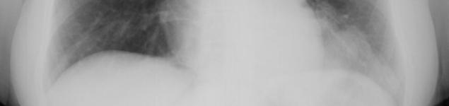
2 What do these two patients have in common? Patient 1: 50 y.o. non-smoking female with several months cough and hemoptysis; CXR: posterior left lower lung consolidation Patient 2: Asymptomatic neonate with incidental chest mass found on prenatal U/S; CT: posterior left lower lung mass


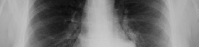
3 PACS, BIDMC 3 Jonathan Shaw, HMS IV Patient 1 50 y.o. female with several months of cough and hemoptysis
4 Patient 1 50 y.o. female with several months of cough and hemoptysis Findings: Ill-defined density in posterior LLL several rounded densities, some with air-fluid level PACS, BIDMC 4
5 Patient 1 50 y.o. female with several months of cough and hemoptysis Wide differential for this infiltrate with apparent cysts/cavitations includes: Bronchogenic carcinoma Metastatic Disease Abscess Infarction TB PCP Fungal infection Wegners Pulmonary sequestration and more PACS, BIDMC 5

6 Patient 2 CT of Neonate with L chest mass on prenatal U/S Courtesy of Dr. Fabio Komlos, Children s Hospital 6
7 Patient 2 CT of Neonate with L chest mass on prenatal U/S Findings: Well-circumbscribed heterogenous mass Posterobasal, above left hemi-diaphragm Arterial supply to mass from the aorta (not visible on this slice) Courtesy of Dr. Fabio Komlos, Children s Hospital 7
 Bronchogenic cyst Paraspinal lesion Courtesy of Dr.")
8 Patient 2 CT of Neonate with L chest mass on prenatal U/S Differential of this congenital pulmonary mass includes: Dipahragmatic hernia Pulmonary sequestration CCAM (Congenital cystic adenomatoid malformation) Bronchogenic cyst Paraspinal lesion Courtesy of Dr. Fabio Komlos, Children s Hospital 8
9 9 Jonathan Shaw, HMS IV What do the two patients have in common? Patient 1 and 2 share a similar diagnosis-- admittedly uncommon, but not rare The clue: both patient s lesions are in the posterior left lower lung
10 10 Jonathan Shaw, HMS IV What do the two patients have in common? One of these diagnosis is characteristically found in left lower lung Patient 1 DDx: Abscess Bronchogenic carcinoma Fungal infection PCP TB Metastatic Infarction Wegners Pulmonary sequestration Patient 2 DDx: Dipahragmatic hernia Pulmonary Sequestration CCAM (Congenital cystic adenomatoid malformation) Bronchogenic cyst Paraspinal lesion
11 11 Jonathan Shaw, HMS IV What do the two have in common? When you encounter a persistent lesion in the left lower lung, add this to your differential: Pulmonary Sequestration Patient 1 DDx: Abscess Bronchogenic carcinoma Fungal infection PCP TB Metastatic Infarction Wegners Pulmonary sequestration Patient 2 DDx: Dipahragmatic hernia Pulmonary Sequestration CCAM (Congenital cystic adenomatoid malformation) Bronchogenic cyst Paraspinal lesion
12 12 Jonathan Shaw, HMS IV Pulmonary Sequestration A Congenital lung malformation: A mass of abnormal, nonfunctioning, Pulmonary tissue No communication with the tracheobronchial tree Receives blood from an anomalous systemic artery (instead of pulmonary arterial system) Usually occurs in left lower lung
13 13 Jonathan Shaw, HMS IV Pulmonary Sequestration Pathophysiology: 1. Primitive foregut gives rise to accessory lung bud 2. Pluripotent tissue migrates caudally with the developing normal lung 3. Remains connected to aortic blood supply for the primitive foregut
14 14 Jonathan Shaw, HMS IV Two Types of Sequestration Intralobar (75%) = WITHIN visceral pleura of a pulmonary lobe Extralobar (25%) = Accessory lung tissue in its own pleura
15 Intralobar vs. Extralobar WITHIN visceral pleura of a pulmonary lobe Accessory lung": lung tissue in its own pleura Drawings from CIBA Netter Collection 15
16 Intralobar vs. Extralobar Venous Drainage into pulmonary veins (L L recirculation) Venous Drainage into systemic (hemiazygous) (L R shunt) Drawings from CIBA Netter Collection 16


17 Intralobar vs. Extralobar Cystic, frequently infected air gets in thru pores of Kohn between adjacent alveoli Less likely to get infected isolated from lung Drawings from CIBA Netter Collection 17
18 18 Jonathan Shaw, HMS IV Intralobar vs. Extralobar Diagnosis usually made in adolescence or adulthood Diagnosis usually made in neonates or infants Presenting with recurrent pneumonia Often asymptomatic, but discovered during evaluation of other anomalies If not diagnosed prenatally/neonatally, usual presents by 6 months with cyanosis or difficulty feeding
19 19 Jonathan Shaw, HMS IV Intralobar vs. Extralobar Not associated with other anomalies Often associated with: Diaphragmatic hernias Cardiac malformations Foregut anomalies


20 e.g. Neonate with Dextrocardia and Extralobar Sequestration Courtesy of Dr. Fabio Komlos, Children s Hospital 20
21 21 Jonathan Shaw, HMS IV Which type of Sequestration? Intralobar or Extralobar Patient 1: 50 y.o. female with several months cough and hemoptysis Patient 2: Asymptomatic neonate with mass found on prenatal U/S PACS, BIDMC Courtesy of Dr. Fabio Komlos, Children s Hospital
22 Patient 1 Intralobar Sequestration PACS, BIDMC 22

23 Patient 1 Intralobar Sequestration PACS, BIDMC 23
24 Patient 1 Intralobar Sequestration PACS, BIDMC 24
25 Patient 1 Intralobar Sequestration PACS, BIDMC 25
26 Patient 1 Intralobar Sequestration PACS, BIDMC 26

27 Patient 1 Intralobar Sequestration PACS, BIDMC 27

28 Patient 1 Intralobar Sequestration PACS, BIDMC 28
29 Patient 1 Intralobar Sequestration CT demonstrates typical findings of infected interlobar sequestration - Dense, heterogeneous opacity within the posterobasal segment of the LLL - Cystic appearance with multiple fluid-air levels PACS, BIDMC 29

30 Patient 1 Intralobar Sequestration 5 months later, after extended fluoroquinolone therapy -Resolution of fluid -persistant cystic spaces and consolidation PACS, BIDMC 30
31 31 Jonathan Shaw, HMS IV Treatment Surgical resection for both intralobar and extralobar sequestrations, to avoid/treat Chronic infection Symptoms from compression of normal lung
32 Intralobar vs. Extralobar Anatomy Intra vs. Extra? Intralobar Within visceral pleura Extralobar Has own pleura is an accesory lobe PACS, BIDMC Courtesy of Dr. Fabio Komlos, Children s Hospital 32
33 33 Jonathan Shaw, HMS IV Intralobar vs. Extralobar Anatomy Intra vs. Extra? Location Arterial Supply Intralobar Within visceral pleura in Left Lower Lobe (67%) Aorta (Abdominal/thoracic) Extralobar Has own pleura is an accesory lobe on Left Hemidiaphragm (95%) Aorta (usually abdominal)
34 Patient 1 CT with contrast (vessels) Twin anomalous arteries aorta sequestration PACS, BIDMC 34
35 Patient 1 CT with contrast (vessels) Twin anomalous arteries aorta sequestration PACS, BIDMC 35

36 Patient 1 CT with contrast (vessels) Twin anomalous arteries aorta sequestration PACS, BIDMC 36
37 Patient 1 CT with contrast (vessels) Twin anomalous arteries aorta sequestration PACS, BIDMC 37
38 38 Jonathan Shaw, HMS IV Intralobar vs. Extralobar Anatomy Intra vs. Extra? Location Arterial Supply Venous Return Intralobar Within visceral pleura in Left Lower Lobe (67%) Aorta (Abdominal/thoracic) Pulmonary Veins Extralobar Has own pleura is an accesory lobe on Left Hemidiaphragm (95%) Aorta (usually abdominal) Systemic Veins
39 Patient 1 CT with contrast (vessels) Venous drainage into pulmonary veins Characteristic of intralobar sequestration PACS, BIDMC 39
40 40 Jonathan Shaw, HMS IV Imaging Pulmonary Sequestrations Identifying anomalous vessels = key to definitive diagnosis Useful for planning surgical resection Variety of arterial paterns:»descending Thoracic aorta (usually)»infradiaphragmatic aorta (20%)»Dual arteries (10%)»Coronary arteries (rare reports)
41 Imaging Pulmonary Sequestrations Various techniques have been used Definitive Diagnosis traditionally by Invasive Arteriography Image from: T. Nakamura, MD 41
42 Imaging Pulmonary Imaging Pulmonary Sequestrations Aortagram: Anomalous vessel from infradiaphragmatic aorta to density at left lung base Venous phase (not shown): drainage to the hemiazygous. Typical for an extralobar sequestration Sequestrations Image from: T. Nakamura, MD 42

43 Imaging Pulmonary Sequestrations Conventional angiography is largely being replaced by non-invasive techniques: CT, U/S, and MRI Image from: T. Nakamura, MD 43
44 44 Jonathan Shaw, HMS IV CXR for Sequestrations Remains an important screening tool But doesn t show the vessels


45 45 Jonathan Shaw, HMS IV Radiographic Appearance of Intralobar Sequestrations CXR typically shows: Lower lobe paraspinal opacity Often cavitary or cystic DDx Pneumonia Lung abscess CCAM Courtesy of Dr. Fabio Komlos, Children s Hospital Intrapulmonary sequestration with large air-fluid level

46 Radiographic Appearance of Extralobar Sequestrations CXR typically shows: Solid retrocardiac opacity rounded or triangular supra or sub-diaphragmatic DDx (in neonate) Diaphragmatic hernia Loculated pleral effusion Esophageal duplication cyst Neuroblastoma Adrenal hemorrhage Triangular extralobar sequestration in a 13 month-old p/w 1 month of wheezing, coughing and rhinorrhea. Image from: T. Nakamura, MD 46
47 47 Jonathan Shaw, HMS IV CT Appearance of Sequestrations CT/CTA provides the best display of parenchyma Intralobar: mixed cystic/solid lesion or homogenous soft tissue. Extralobar: well circumscribed homogenous lesion. PACS, BIDMC Patient 1- Intralobar Sequestration

48 48 Jonathan Shaw, HMS IV CT Appearance of Sequestrations CT/CTA typically can show the anomalous systemic arterial supply PACS, BIDMC Patient 1 Arterial supply
49 49 Jonathan Shaw, HMS IV CT Appearance of Sequestrations Newer MultiDetector CT combined with 3D reconstruction can consistently identify both the arterial and venous anatomy
50 3D CT Reconstruction Patient 2 Neonate with extralobar sequestration (left antero-lateral perspective) Courtesy of Dr. Fabio Komlos, Children s Hospital 50

51 3D CT Reconstruction Patient 2 Neonate with extralobar sequestration Not easily seen on axial scan a 3-D reconstruction reveals: Typical arterial supply from the aorta Unusual systemic venous drainage via the internal mammary vein Courtesy of Dr. Fabio Komlos, Children s Hospital 51
 LA LV ILS Anomalous arterial supply from descending aorta Courtesy of Dr.")
52 3D CT Reconstruction A 6 month old girl with recurrent LLL PNA Intralobar Sequestration (not recognized on axial CT) Anomalous venous drainage to LA (posterior view) LA LV ILS Anomalous arterial supply from descending aorta Courtesy of Dr. Fabio Komlos, Children s Hospital 52

53 53 Jonathan Shaw, HMS IV Sonogram of Sequestration Aorta A Anomalous Vessel May show anomalous vessel to the sequestration Sequestration Courtesy of Dr. Fabio Komlos, Children s Hospital
. Cysts (arrows) are also visible.). from Raipal Dhingal et al.")
54 54 Jonathan Shaw, HMS IV Sonogram of Sequestration Can also image prenatally Axial sonogram of chest obtained at 22 weeks gestation in fetus with extralobar sequestration (asterisk). Cysts (arrows) are also visible.). from Raipal Dhingal et al. AJR 2003; 180:
. from Raipal Dhingal et al.")
55 55 Jonathan Shaw, HMS IV Sonogram of Sequestration Prenatal use of Doppler Distinguish sequestration from other congenital thoracic lesions Sagital sonogram of chest obtained at 32 weeks' gestation in fetus with extralobar sequestration (mass). from Raipal Dhingal et al. AJR 2003; 180:
. from Raipal Dhingal et al.")
56 56 Jonathan Shaw, HMS IV MRI of Sequestration MRI can also show precise anatomy of sequestrations (CT still better) Aorta Arterial supply MRI and U/S safe prenatally Accuracy still unknown Coronal T2-weighted MRI obtained at 23 weeks' gestation, showing extralobar sequestration (asterix). from Raipal Dhingal et al. AJR 2003; 180:
57 57 Jonathan Shaw, HMS IV Recap Intralobar 1. Adults/adolescents 2. Pulmonary veins 3. No anomalies Extralobar 1. Neonates/infants 2. Systemic veins 3. Often other anomalies
58 58 Jonathan Shaw, HMS IV Acknowledgements Many thanks for kind assistance to: Dr. Gillian Lieberman Dr. Fabio Komlos Dr. Raja Kyriakos Pamela Lepkowski Larry Barbaras
59 59 Jonathan Shaw, HMS IV References Dhingsa, R. et al: Prenatal Sonography and MR Imaging of Pulmonary Sequestration. AJR 180: , 2003 Johnson A. and Huubard A. Congenital Anomalies of the fetal/neonatal Chest, Seminars in Roentgenology 2004; 39 (2): Khan, A.N: Pulmonary Sequestration January 10, 2003 Lee, E. et al: Evaluation of Angioarchitecture of Pulmonary Sequestration in Pediatric Patients Using 3D MDCT Angiography. AJR 183: , 2004 Nakamura, C. Pulmonary Sequestration Radiology Cases in Pediatric Emergency Medicine, Vol 5, Case Pediatric Diagnostic Imaging, 10 th ed., Chapter 2: Anomalies of the Lung eds. Kuhn, Slovis, and Haller, 10ed., Philadelphia 2004 Schnapf, B.M. Pulmonary Sequestration, March 4, 2002 Scheung-Fat K et al: Noninvasive Imaging of Bronchopulmonary Sequestration. AJR 175: , 2000
Lung sequestration and Scimitar syndrome
 Lung sequestration and Scimitar syndrome Imaging approaches M. Mearadji International Foundation for Pediatric Imaging Aid Rotterdam, The Netherlands Pulmonary sequestration Pulmonary sequestration (PS)
Congenital Lung Malformations: Radiologic-Pathologic Correlation
 Acta Radiológica Portuguesa, Vol.XVIII, nº 70, pág. 51-60, Abr.-Jun., 2006 Congenital Lung Malformations: Radiologic-Pathologic Correlation Marilyn J. Siegel Mallinckrodt Institute of Radiology, Washington
Molla Teshome MD, Habtamu Belete MD Aurora Health Care Internal Medicine Residency Program
 Molla Teshome MD, Habtamu Belete MD Aurora Health Care Internal Medicine Residency Program History 32 year-old male who presented with a 4 days history of: Productive cough Right sided pleuritic chest
Case Report Coexistent Congenital Diaphragmatic Hernia with Extrapulmonary Sequestration
 Canadian Respiratory Journal Volume 2016, Article ID 1460480, 4 pages http://dx.doi.org/10.1155/2016/1460480 Case Report Coexistent Congenital Diaphragmatic Hernia with Extrapulmonary Sequestration Nao
24. An infant with recurrent pneumonia underwent a frontal chest radiograph (Fig 24-A) followed by
 followed by") 24. An infant with recurrent pneumonia underwent a frontal chest radiograph (Fig 24-A) followed by diagnosis? ndings, what is the most likely A. Pulmonary sequestration B. Congenital pulmonary airway malformation
24. An infant with recurrent pneumonia underwent a frontal chest radiograph (Fig 24-A) followed by diagnosis? ndings, what is the most likely A. Pulmonary sequestration B. Congenital pulmonary airway malformation
Case Based Fetal Lung Masses
 Case Based Fetal Lung Masses Advances in Fetal and Neonatal Imaging Course Orlando, Florida, January 28, 2017 Leann E. Linam, MD Associate Professor Radiology University of Arkansas for Medical Sciences/
Surgical indications: Non-malignant pulmonary diseases. Punnarerk Thongcharoen
 Surgical indications: Non-malignant pulmonary diseases Punnarerk Thongcharoen Non-malignant Malignant as a pathological term: Cancer Non-malignant = not cancer Malignant as an adjective: Disposed to cause
Imaging of the Lung in Children
 Imaging of the Lung in Children Imaging methods X-Ray of the Lung (Anteroposterior, ) CT, HRCT MRI USG Congenital developmental defects of the lungs Agenesis, aplasia, hypoplasia Tension pulmonary anomalies
Extralobar Pulmonary Sequestration with Hemorrhagic Infarction in a Child: Preoperative Imaging Diagnosis and Pathological Correlation
 Case Report Thoracic Imaging http://dx.doi.org/10.3348/kjr.2015.16.3.662 pissn 1229-6929 eissn 2005-8330 Korean J Radiol 2015;16(3):662-667 Extralobar Pulmonary Sequestration with Hemorrhagic Infarction
Thoracoscopic treatment of congenital malformation of the lung
 Jemis, 1 2013 Thoracoscopic treatment of congenital malformation of the lung Preliminary experience with preoperative 3D virtual rendering F. Destro M. Maffi T. Gargano G. Ruggeri L. Soler M. Lima Table
PULMONARY VENOLOBAR SYNDROME. Dr.C.Anandhi DNB Resident, Southern Railway Headquarters Hospital.
 PULMONARY VENOLOBAR SYNDROME Dr.C.Anandhi DNB Resident, Southern Railway Headquarters Hospital. Presenting complaint: 10 yrs old girl with recurrent episodes of lower respiratory tract infection from infancy.
Case Report Pulmonary Sequestration with Renal Aplasia and Elevated SUV Level in PET/CT
 Case Reports in Pulmonology Volume 2012, Article ID 276012, 4 pages doi:10.1155/2012/276012 Case Report Pulmonary Sequestration with Renal Aplasia and Elevated SUV Level in PET/CT Serdar Şen, 1 Nilgün
Imaging of pulmonary sequestration: what the radiologist needs to know
 Imaging of pulmonary sequestration: what the radiologist needs to know Poster No.: C-1478 Congress: ECR 2016 Type: Educational Exhibit Authors: S. Accogli, M. Gabelloni, L. Faggioni, D. Caramella; pisa/it
A Case of Pediatric Plasma Cell Granuloma
 August 2001 A Case of Pediatric Plasma Cell Granuloma Nii Tetteh, Harvard Medical School Year IV Our Patient 8 year old male with history of recurrent left lower lobe and lingular pneumonias since 1994.
HOW TO IMAGE AND DESCRIBE CONGENITAL LUNG MALFORMATIONS
 HOW TO IMAGE AND DESCRIBE CONGENITAL LUNG MALFORMATIONS Paul Thacker, MD Assistant Professor Departments of Radiology and Pediatrics Medical University of South Carolina DISCLOSURES I have no relevant
ISUOG Basic Training. Assessing the Neck & Chest Gihad Chalouhi, Lebanon
 ISUOG Basic Training Assessing the Neck & Chest Gihad Chalouhi, Lebanon Learning objectives 9 & 10 At the end of the lecture you will be able to: recognise the differences between the normal & most common
Situs inversus. Dr praveena pulmonology- final year post graduate
 Situs inversus Dr praveena pulmonology- final year post graduate Definiton History Types Cause Clinical features Diagnosis Treatment Definition The term situs inversus is a short form of the latin phrase
The Focal Hepatic Lesion: Radiologic Assessment
 The Focal Hepatic Lesion: Radiologic Assessment Kevin Kuo, Harvard Medical School Year III Our Patient: PS 67 y/o female w/ long history of alcohol use Drinking since age 18, up to one bottle of wine/day
Joseph Garland, HMS IV Gillian Lieberman, MD. Round Pneumonia. Joseph Garland, HMS IV Gillian Lieberman, MD
 Round Pneumonia Joseph Garland, HMS IV Case 1: Mr. H Mr. H is a 45-year-old man who presents with a 4 day history of full-body myalgias, headaches and fever to 103 F. He also complains of sharp leftsided
Pediatric Imaging Studies: Congenital and Acquired Diagnoses
 Pediatric Imaging Studies: Congenital and Acquired Diagnoses Robin Foster MD FAAP FACEP Division Chief Pediatric Emergency Medicine Children s Hospital of Richmond at Virginia Commonwealth University Health
Positron Emission Tomography in Lung Cancer
 May 19, 2003 Positron Emission Tomography in Lung Cancer Andrew Wang, HMS III Patient DD 53 y/o gentleman presented with worsening dyspnea on exertion for the past two months 30 pack-year smoking Hx and
Case report Esophageal lung: a rare case of communicating bronchopulmonary foregut malformation
 Case report Esophageal lung: a rare case of communicating bronchopulmonary foregut malformation 1 Dr.Varsha Rathi, 2 Dr. Saurabh Deshpande*, 3 Dr.Almas Nazim, 4 Dr.Shilpa Domkundwar 1 Professor, Department
Assignable revenue codes: Explanation of services:
 computed tomography Chest/Cardiac Assignable revenue codes: Explanation of services: 0350 CT Scan General Classification 0351 CT Scan Head Scan 0352 CT Scan Body Scan 0359 CT Scan Other CT Scans Known
Clinical presentation and characteristics of 25 adult cases of pulmonary sequestration
 Original Article Clinical presentation and characteristics of 25 adult cases of pulmonary sequestration Mateusz Polaczek 1,2, Inga Baranska 3, Malgorzata Szolkowska 4, Jacek Zych 1, Piotr Rudzinski 5,
Radiological Aspects of Pulmonary Tuberculosis in Immunocompetent Hosts
 Nov 2003 Radiological Aspects of Pulmonary Tuberculosis in Immunocompetent Hosts Josh Rempell, Harvard Medical School Year III Tuberculosis: the captain of all (wo)men of death Overall, one third of the
PRESENCE OF LOWER ACCESSORY LOBES IN THE LUNGS
 Int. J. Pharm. Med. & Bio. Sc. 2013 Hemanth Kommuru et al., 2013 Research Paper ISSN 2278 5221 www.ijpmbs.com Vol. 2, No. 3, July 2013 2013 IJPMBS. All Rights Reserved PRESENCE OF LOWER ACCESSORY LOBES
TB Radiology for Nurses Garold O. Minns, MD
 TB Nurse Case Management Salina, Kansas March 31-April 1, 2010 TB Radiology for Nurses Garold O. Minns, MD April 1, 2010 TB Radiology for Nurses Highway Patrol Training Center Salina, KS April 1, 2010
Heart and Lungs. LUNG Coronal section demonstrates relationship of pulmonary parenchyma to heart and chest wall.
 Heart and Lungs Normal Sonographic Anatomy THORAX Axial and coronal sections demonstrate integrity of thorax, fetal breathing movements, and overall size and shape. LUNG Coronal section demonstrates relationship
Chest and cardiovascular
 Module 1 Chest and cardiovascular A. Doss and M. J. Bull 1. Regarding the imaging modalities of the chest: High resolution computed tomography (HRCT) uses a slice thickness of 4 6 mm to identify mass lesions
An Unusual Presentation of Diaphragmatic Hernia
 January 2007 An Unusual Presentation of Diaphragmatic Hernia Daniel B. Horton Harvard Medical School Year III Patient LG: Clinical Presentation, Nov. 2004 52 year old woman presents with new nonproductive
Undergraduate Teaching
 Prof. James F Meaney Undergraduate Teaching Chest X-Ray Understanding the normal anatomical by reference to cross sectional imaging Radiology? It s FUN! Cryptic puzzle Sudoku (Minecraft?) It s completely
Basic Data. Sex:Male 31 years old Occupation: 搬家工人
 Basic Data Sex:Male 31 years old Occupation: 搬家工人 Chief Complaint Intermittent chest pain with shortness of breath for 2-3 months. Present Illness 4 months ago, he started having occasional chest pain
Excavated pulmonary nodule: steps to diagnosis?
 Excavated pulmonary nodule: steps to diagnosis? Poster No.: C-1044 Congress: ECR 2014 Type: Authors: Keywords: DOI: Educational Exhibit W. Mnari, M. MAATOUK, A. Zrig, B. Hmida, M. GOLLI; Monastir/ TN Metastases,
CONGENITAL LUNG MALFORMATIONS, UPDATE AND TREATMENT
 CONGENITAL LUNG MALFORMATIONS, UPDATE AND TREATMENT STEVEN ROTHENBERG MD.(1) 1. Department of Pediatrics. The Rocky Mountain Hospital For Children. Hospital for Children, Denver, Colorado, USA. steverberg@aol.com
CONGENITAL LUNG LESION Round Table. Objectives. Congenital Lung Lesions: Anatomy and Physiology Leah Barefoot, DNP, CPNP-PC
 CONGENITAL LUNG LESION Round Table L. Barefoot, E. Paton, C. Schultz, R. Caskey, M. O Day Objectives Review the anatomy and pathophysiology of congenital lung lesions List the preoperative evaluation of
Eun-Young Kang, M.D., Jae Wook Lee, M.D., Ji Yung Choo, M.D., Hwan Seok Yong, M.D., Ki Yeol Lee, M.D., Yu-Whan Oh, M.D.
 Eun-Young Kang, M.D., Jae Wook Lee, M.D., Ji Yung Choo, M.D., Hwan Seok Yong, M.D., Ki Yeol Lee, M.D., Yu-Whan Oh, M.D. Department of Radiology, Korea University Guro Hospital, College of Medicine, Korea
Chest XRay interpretation INTERPRETATIONS Identifications: Name & Date Technical evaluation Basic Interpretations
 Chest XRay interpretation INTERPRETATIONS Identifications: Name & Date Technical evaluation Basic Interpretations TECHNICAL EVALUATION 1. Projection: AP/PA view To differentiate between AP & PA films,
Pulmonary Aspergillosis
 May 2005 Pulmonary Aspergillosis Nancy Wei, Harvard Medical School, Year III Overview Pulmonary aspergillosis background information Patient presentations Common radiographic findings for each type of
Pulmonary vascular anatomy & anatomical variants
 Review Article Pulmonary vascular anatomy & anatomical variants Asha Kandathil, Murthy Chamarthy Department of Radiology, University of Texas Southwestern Medical Center, Dallas, TX, USA Contributions:
Pulmonary Arteriovenous Malformations Complicated with Paradoxical Embolic Stroke
 Archives of Clinical and Medical Case Reports doi: 10.26502/acmcr.96550032 Volume 2, Issue 4 Case Report Pulmonary Arteriovenous Malformations Complicated with Paradoxical Embolic Stroke Cheah Wai Hun
Boy 8 months TPRC. 21 Sep 06 CXR. Flat and. CLE findings. BPD findings a. Left opacity
 CLE in BPD lung Boy 8 months 17 Sep 06 21 Sep 06 CXR Flat and low position of the diaphragm ICD insertion, right; ET tube slightly shift to the left RUL atelectasis RML hyperinflation, herniating across
General Imaging. Imaging modalities. Incremental CT. Multislice CT Multislice CT [ MDCT ]
![General Imaging. Imaging modalities. Incremental CT. Multislice CT Multislice CT [ MDCT ]](/thumbs/76/74079340.jpg "General Imaging. Imaging modalities. Incremental CT. Multislice CT Multislice CT [ MDCT ]") General Imaging Imaging modalities Conventional X-rays Ultrasonography [ US ] Computed tomography [ CT ] Radionuclide imaging Magnetic resonance imaging [ MRI ] Angiography conventional, CT,MRI Interventional
General Imaging Imaging modalities Conventional X-rays Ultrasonography [ US ] Computed tomography [ CT ] Radionuclide imaging Magnetic resonance imaging [ MRI ] Angiography conventional, CT,MRI Interventional
TB Intensive Houston, Texas
 TB Intensive Houston, Texas October 15-17, 17 2013 Diagnosis of TB: Radiology Rosa M Estrada-Y-Martin, MD MSc FCCP October 16, 2013 Rosa M Estrada-Y-Martin, MD MSc FCCP, has the following disclosures to
Lung Perfusion Analysis New Pathways in Lung Imaging. Case Study Brochure PLA 309 Hospital
 Lung Perfusion Analysis New Pathways in Lung Imaging Case Study Brochure PLA 309 Hospital http://www.toshibamedicalsystems.com Toshiba Medical Systems Corporation 2012 all rights reserved. Design and specifications
Individual Pulmonary Vein Atresia in Adults: Report of Two Cases
 Case Report DOI: 10.3348/kjr.2011.12.3.395 pissn 1229-6929 eissn 2005-8330 Korean J Radiol 2011;12(3):395-399 Individual Pulmonary Vein Atresia in Adults: Report of Two Cases Hyoung Nam Lee, MD, Young
Assignable revenue codes: Explanation of services:
 COMPUTED TOMOGRAPHY Chest/Cardiac Assignable revenue codes: 0350 CT Scan General Classification 0351 CT Scan Head Scan 0352 CT Scan Body Scan 0359 CT Scan Other CT Scans Explanation of services: Known
10/17/2016. Nuts and Bolts of Thoracic Radiology. Objectives. Techniques
 Nuts and Bolts of Thoracic Radiology October 20, 2016 Carleen Risaliti Objectives Understand the basics of chest radiograph Develop a system for interpreting chest radiographs Correctly identify thoracic
Genitourinary Radiology In-Training Test Questions for Diagnostic Radiology Residents
 Genitourinary Radiology In-Training Test Questions for Diagnostic Radiology Residents March, 2013 Sponsored by: Commission on Education Committee on Residency Training in Diagnostic Radiology 2013 by American
B-I-2 CARDIAC AND VASCULAR RADIOLOGY
 (YEARS 1 3) CURRICULUM FOR RADIOLOGY 13 B-I-2 CARDIAC AND VASCULAR RADIOLOGY KNOWLEDGE To describe the normal anatomy of the heart and vessels including the lymphatic system as demonstrated by radiographs,
Chest X-ray Interpretation
 Chest X-ray Interpretation Introduction Routinely obtained Pulmonary specialist consultation Inherent physical exam limitations Chest x-ray limitations Physical exam and chest x-ray provide compliment
Five Views of Transitional Cell Carcinoma: One Man s Journey
 September 2006 Five Views of Transitional Cell Carcinoma: One Man s Journey Amsalu Dabela, Harvard Medical School III Outline Overview: Renal Anatomy Our Patient s Story Diagnostic Imaging Studies Appearance
Systemic lupus erythematosus (SLE): Pleuropulmonary Manifestations
: Pleuropulmonary Manifestations") 08/30/10 09/26/10 Systemic lupus erythematosus (SLE): Pleuropulmonary Manifestations Camila Downey S. Universidad de Chile, School of Medicine, Year VII Harvard University, School of Medicine Sept 17,
08/30/10 09/26/10 Systemic lupus erythematosus (SLE): Pleuropulmonary Manifestations Camila Downey S. Universidad de Chile, School of Medicine, Year VII Harvard University, School of Medicine Sept 17,
Alex Lam, HMS III. September The Acute Scrotum. Alex Lam, Harvard Medical School Year III Gillian Lieberman, MD. Gillian Lieberman, MD
 September 2002 The Acute Scrotum Alex Lam, Harvard Medical School Year III DDx: : Acute Scrotal Pain & Enlargement PAIN Inflammatory disorder Testicular torsion Testicular infarction Testicular abscess
UERMMMC Department of Radiology. Basic Chest Radiology
 UERMMMC Department of Radiology Basic Chest Radiology PHYSICS DENSITIES BONE SOFT TISSUES WATER FAT AIR TELEROENTGENOGRAM Criteria for an Ideal Chest Radiograph 1. Upright 2. Posteroanterior View 3. Full
Daniela Faivovich K., MS VII Universidad de Chile Gillian Lieberman, MD Harvard Medical School
 Daniela Faivovich K., MS VII Universidad de Chile Gillian Lieberman, MD Harvard Medical School May 21st, 2010 56 year old male patient History of hypertension, hyperlipidemia and insulin-resistance 2009:
Coronary Artery Anomalies from Birth to Adulthood; the Role of CT Coronary Angiography in Sudden Cardiac Death Screening
 Coronary Artery Anomalies from Birth to Adulthood; the Role of CT Coronary Angiography in Sudden Cardiac Death Screening E O Dwyer 1, C O Brien 1, B Loo 1, A Snow Hogan 1, O Buckley1 2, B 1. Department
in PAEDIATRIC CARDIOLOGY
 IMAGES in PAEDIATRIC CARDIOLOGY Morrison ML, 1 Sands AJ, 1 Paterson A. 2 Primitive hepatic venous plexus in a child with scimitar syndrome and pulmonary 1 Department of Paediatric Cardiology, Royal Belfast
Imaging of Thoracic Trauma: Tips and Traps. Arun C. Nachiappan, MD Associate Professor of Clinical Radiology University of Pennsylvania
 Imaging of Thoracic Trauma: Tips and Traps Arun C. Nachiappan, MD Associate Professor of Clinical Radiology University of Pennsylvania None Disclosures Objectives Describe blunt and penetrating traumatic
PULMONARY TUBERCULOSIS RADIOLOGY
 PULMONARY TUBERCULOSIS RADIOLOGY RADIOLOGICAL MODALITIES Medical radiophotography Radiography Fluoroscopy Linear (conventional) tomography Computed tomography Pulmonary angiography, bronchography Ultrasonography,
Pitfalls of the Pediatric Chest and Abdomen SPR 2017
 Pitfalls of the Pediatric Chest and Abdomen SPR 2017 Richard I. Markowitz, MD, FACR Children s Hospital of Philadelphia Perelman School of Medicine University of Pennsylvania No Disclosures Cognitive Perceptual
Vascular Imaging in the Pediatric Abdomen. Jonathan Swanson, MD
 Vascular Imaging in the Pediatric Abdomen Jonathan Swanson, MD Goals and Objectives To understand the imaging approach, appearance, and clinical manifestations of the common pediatric abdominal vascular
Looking Outside the Box: Incidental Extracardiac Finding in Echo
 Looking Outside the Box: Incidental Extracardiac Finding in Echo Dr. Aijaz Shah Head of Division, Adult Echocardiography Laboratory Prince Sultan Cardiac Centre Riyadh Case 1 17 year old boy presented
Pediatric Retroperitoneal Masses Radiologic-Pathologic Correlation
 Acta Radiológica Portuguesa, Vol.XVIII, nº 70, pág. 61-70, Abr.-Jun., 2006 Pediatric Retroperitoneal Masses Radiologic-Pathologic Correlation Marilyn J. Siegel Mallinckrodt Institute of Radiology, Washington
Imaging The Turkish Saddle. Russell Goodman, HMS III Dr. Gillian Lieberman
 Imaging The Turkish Saddle Russell Goodman, HMS III Dr. Gillian Lieberman Learning Objectives Review the anatomy of the sellar region Discuss the differential diagnosis of sellar masses Discuss typical
Cardiopulmonary Syndromes: Conditions With Concomitant Cardiac and Pulmonary Abnormalities
 Cardiopulmonary Syndromes: Conditions With Concomitant Cardiac and Pulmonary Abnormalities Carlos S. Restrepo M.D. Professor of Radiology The University of Texas HSC at San Antonio Cardiopulmonary Syndromes
Do you want to be an excellent Radiologist? - Focus on the thoracic aorta on lateral chest image!!!
 The lateral chest radiograph: Challenging area around the thoracic aorta!!! Do you want to be an excellent Radiologist? - Focus on the thoracic aorta on lateral chest image!!! Dong Yoon Han 1, So Youn
How to Analyse Difficult Chest CT
 How to Analyse Difficult Chest CT Complex diseases are:- - Large lesion - Unusual or atypical pattern - Multiple discordant findings Diffuse diseases are:- - Numerous findings in both sides 3 basic steps
Use of Integrated PET CT in the Clinical Staging of Non Small Cell Lung Cancer
 November 2010 Use of Integrated PET CT in the Clinical Staging of Non Small Cell Lung Cancer Laura Myers, Harvard Medical School, Year III Clinical Presentation 79yo woman with cough productive of green
SWISS SOCIETY OF NEONATOLOGY. Bilateral pulmonary sequestration in a neonate
 SWISS SOCIETY OF NEONATOLOGY Bilateral pulmonary sequestration in a neonate February 2008 2 Woerner A, Schwendener K, Casaulta C, Raio L, Wolf R, Zachariou Z, Nelle M, Division of Neonatology, (WA, SK,
ORIGINAL ARTICLE. Complete video-assisted thoracoscopic surgery for pulmonary sequestration
 ORIGINAL ARTICLE Complete video-assisted thoracoscopic surgery for pulmonary sequestration Jian-Fei Shen, Xiao-Xue Zhang, Shu-Ben Li, Zhi-Hua Guo, Zhi-Qiang Xu, Xiao-Sun Shi, Jian-Xing He Department of
The Role of PET / CT in Lung Cancer Staging
 July 2004 The Role of PET / CT in Lung Cancer Staging Vlad Vinarsky, Harvard Medical School Year IV Patient AM HPI: 81 yo F p/w hemoptysis x 1 month LLL lesion on CXR, not responsive to Abx 35 pack-year
Cardiac CT Techniques in Neonates (and infants)
") Cardiac CT Techniques in Neonates (and infants) Siddharth P. Jadhav, MD Director, Body CT and MRI Edward B. Singleton Department of Pediatric Radiology Texas Children s Hospital Disclosures None Objectives
Cardiac CT Techniques in Neonates (and infants) Siddharth P. Jadhav, MD Director, Body CT and MRI Edward B. Singleton Department of Pediatric Radiology Texas Children s Hospital Disclosures None Objectives
Management of antenatally diagnosed pulmonary sequestration associated with congenital cystic adenomatoid malformation
 Thorax 1999;54:701 706 701 Wessex Regional Center for Pediatric Surgery, Southampton General Hospital, Southampton SO16 6YD, UK M Samuel D M Burge Correspondence to: Dr M Samuel, Department of Pediatric
Bronchogenic Cyst: Imaging Features with Clinical and Histopathologic Correlation 1
 Thoracic Imaging H. Page McAdams, MD Wanda M. Kirejczyk, MD 2 Melissa L. Rosado-de- Christenson, Col, USAF, MC Shigeru Matsumoto, MD Bronchogenic Cyst: Imaging Features with Clinical and Histopathologic
Objectives. What is a Chest X Ray? CXR Workshop. Definition (diagnostic tool/internal PE) Types. Cost
 Types. Cost") Objectives CAPA 2011 Christy Wilson, PA C Georgia Lung Associates Identify the radiographic landmarks on a chest radiograph Recognize identifiers of poor quality on the chest radiograph Outline an approach
Objectives CAPA 2011 Christy Wilson, PA C Georgia Lung Associates Identify the radiographic landmarks on a chest radiograph Recognize identifiers of poor quality on the chest radiograph Outline an approach
Pulmonary vascular anomalies in adult; a pictorial review
 Pulmonary vascular anomalies in adult; a pictorial review Award: Magna Cum Laude Poster No.: C-0901 Congress: ECR 2015 Type: Educational Exhibit Authors: K. Tokunaga, T. Yamaoka, A. Hamada, T. Kubo, K.
Lung- and airway emergencies
 Lung- and airway emergencies Charlotte de Lange,MD,PhD Pediatric Radiology unit, Oslo University Hospital, Norway 5th Nordic course - Emergency Radiology Oslo 18-21.5.2015 clange@ous-hf.no How come pediatric
The Foetal Lung. Educational Exhibit Authors:
 The Foetal Lung Poster No.: C-1649 Congress: ECR 2015 Type: Educational Exhibit Authors: V. B. Pai, R. N. Chaubal, N. G. Chaubal, B. Pai ; Navi Mumbai/ 1 2 2 2 3 1 3 IN, Thane/IN, Mumbai/IN Keywords: Foetal
Case 1. A 35-year-old male presented with fever, cough, and purulent sputum for one week. This was his CXR (Fig. 1.1). What is the diagnosis?
. What is the diagnosis?") 1 Interpreting Chest X-Rays CASE 1 Fig. 1.1 Case 1. A 35-year-old male presented with fever, cough, and purulent sputum for one week. This was his CXR (Fig. 1.1). What is the diagnosis? CASE 1 Interpreting
1 Interpreting Chest X-Rays CASE 1 Fig. 1.1 Case 1. A 35-year-old male presented with fever, cough, and purulent sputum for one week. This was his CXR (Fig. 1.1). What is the diagnosis? CASE 1 Interpreting
100 Chest X Rays for Study Group. by Dr. Suneet Khurana
 100 Chest X Rays for Study Group by Dr. Suneet Khurana Approach to - Chest X Ray (shadow of the viscera on a photographic plate) Gas appears Black Fat appears Dark Grey Water Appears as Light Grey Bone
Radiologic Features of The Pulmonary Embolus
 January 2003 Radiologic Features of The Pulmonary Embolus Travis McGlothin HMSIII Mr. J is a 51 y.o. male who presented to the BIDMC ED w/ acute onset of: Lft. Hemiparesis slurred speech mild dyspnea mild
Update on RECIST and Staging of Common Pediatric Tumors Ethan A. Smith, MD
 Update on RECIST and Staging of Common Pediatric Tumors Ethan A. Smith, MD Section of Pediatric Radiology C.S. Mott Children s Hospital University of Michigan ethans@med.umich.edu Disclosures No relevant
An Introduction to Radiology for TB Nurses
 An Introduction to Radiology for TB Nurses Garold O. Minns, MD September 14, 2017 TB Nurse Case Management September 12 14, 2017 EXCELLENCE EXPERTISE INNOVATION Garold O. Minns, MD has the following disclosures
Pulmonary nodules, masses and infiltrates with specific semiology: when diagnosis is based on imaging
 Pulmonary nodules, masses and infiltrates with specific semiology: when diagnosis is based on imaging Poster No.: C-2014 Congress: ECR 2012 Type: Scientific Exhibit Authors: M. L. L. Rodriguez Rodriguez;
Pulmonary nodules, masses and infiltrates with specific semiology: when diagnosis is based on imaging
 Pulmonary nodules, masses and infiltrates with specific semiology: when diagnosis is based on imaging Poster No.: C-2014 Congress: ECR 2012 Type: Scientific Exhibit Authors: M. L. Rodriguez Rodriguez;
Chest Radiology Interpretation: Findings of Tuberculosis
 Chest Radiology Interpretation: Findings of Tuberculosis Get out your laptops, smart phones or other devices pollev.com/chestradiology Case #1 1 Plombage Pneumonia Cancer 2 Reading the TB CXR Be systematic!
9/8/2009 < 1 1,2 3,4 5,6 7,8 9,10 11,12 13,14 15,16 17,18 > 18. Tetralogy of Fallot. Complex Congenital Heart Disease.
 Current Indications for Pediatric CTA S Bruce Greenberg Professor of Radiology Arkansas Children s Hospital University of Arkansas for Medical Sciences greenbergsbruce@uams.edu 45 40 35 30 25 20 15 10
Early View Article: Online published version of an accepted article before publication in the final form.
 Early View Article: Online published version of an accepted article before publication in the final form. Journal Name: Edorium Journal of Anatomy and Embryology Type of Article: Case Report Title: Pulmonary
Rare diseases in pulmonology. Agnieszka Strzelak
 Rare diseases in pulmonology Agnieszka Strzelak Rare diseases: < 1/2000 genetic, infectious, autoimmune, rare neoplasms, of unknown origin in most cases severe, chronic and progressing present after birth,
Cardiac MRI in ACHD What We. ACHD Patients
 Cardiac MRI in ACHD What We Have Learned to Apply to ACHD Patients Faris Al Mousily, MBChB, FAAC, FACC Consultant, Pediatric Cardiology, KFSH&RC/Jeddah Adjunct Faculty, Division of Pediatric Cardiology
Pulmonary Embolism. Thoracic radiologist Helena Lauri
 Pulmonary Embolism Thoracic radiologist Helena Lauri 8.5.2017 Statistics 1-2 out of 1000 adults annually are diagnosed with deep vein thrombosis (DVT) and/or pulmonary embolism (PE) About half of patients
Introduction to Chest CT Interpretation. Objectives 8/28/2017
 Introduction to Chest CT Interpretation Deborah Stein ACNP BC, CCRN NP Education Specialist Department of Anesthesia and Critical Care Medicine August 28, 2017 Objectives Basic Principles Thoracic Anatomy
May 2017 Imaging Case of the Month. Prasad M. Panse, MD and Michael B. Gotway, MD. Department of Radiology Mayo Clinic Arizona Scottsdale, Arizona USA
 May 2017 Imaging Case of the Month Prasad M. Panse, MD and Michael B. Gotway, MD Department of Radiology Mayo Clinic Arizona Scottsdale, Arizona USA Clinical History: A 32-year-old man presented for routine
Case 9799 Stanford type A aortic dissection: US and CT findings
 Case 9799 Stanford type A aortic dissection: US and CT findings Accogli S, Aringhieri G, Scalise P, Angelini G, Pancrazi F, Bemi P, Bartolozzi C Department of Diagnostic and Interventional Radiology, University
CT - Brain Examination
 CT - Brain Examination Submitted by: Felemban 1 CT - Brain Examination The clinical indication of CT brain are: a) Chronic cases (e.g. headache - tumor - abscess) b) ER cases (e.g. trauma - RTA - child
Low-dose CT Lung Cancer Screening Guidelines for Pulmonary Nodules Management Version 2
 Low-dose CT Lung Cancer Screening Guidelines for Pulmonary Nodules Management Version 2 The Committee for Management of CT-screening-detected Pulmonary Nodules 2009-2011 The Japanese Society of CT Screening
Computed tomography of the chest: I. Basic principles
 BJA Education, 15 (6): 299 304 (2015) doi: 10.1093/bjaceaccp/mku063 Advance Access Publication Date: 2 February 2015 Matrix reference 1A03, 2A12 Computed tomography of the chest: I. Basic principles P
Pediatric TB Intensive Houston, Texas
 Pediatric TB Intensive Houston, Texas November 13, 2009 Radiographic Manifestations of Pediatric TB Susan D. John, MD, FACR November 13, 2009 Radiologic Presentation of Childhood TB Susan D. John, MD,