What do you mean by Respiration?
|
|
|
- Jerome Holland
- 5 years ago
- Views:
Transcription


1

2 Objective Anatomy & Physiology of Pulmonary System Who requires Ventilation? What is a Ventilator and how does it works? Type of Ventilation and Modes. Monitoring and Sensing. Alarms and Indications.

3 What do you mean by Respiration? Respiration is defined as the movements of gas molecules across the membrane. The blood distributes oxygen and nutrients to the many different cells in the body, carries CO2 generated by the cells to the lungs for exhalation This oxygen is used to provide energy for all the tissues and organs of the body. 1) External respiration Oxygen moves in to the bloodstream and carbon dioxide moves from bloodstream in to the alveoli. 2) Internal respiration At the cellular level, carbon dioxide moves from the cell in to the blood and oxygen moves from the blood in to the cell.
 Nasopharynx 2) Oropharynx 3) Laryngeal pharynx Larynx 1) Vocal cords 2) Thyroid Lower Airway Conducting Airways: Non-alveolar region Consists of series of tubes that divide like the")
4 Anatomy and Physiology Pulmonary System Upper Airway Nasal Passages Beyond the olfactory function, it warms, filters and moistens inspired air. Pharynx 1) Nasopharynx 2) Oropharynx 3) Laryngeal pharynx Larynx 1) Vocal cords 2) Thyroid Lower Airway Conducting Airways: Non-alveolar region Consists of series of tubes that divide like the branches of a tree. Its function are to conduct air and provide mucociliary defense. Respiratory Zone: Alveolar Region Important perform external gas exchange.


5 Upper Airway Nasal Cavity Function

6 Pharynx The throat provides a transport function for both food and air. When swallowing, food is prevented from entering the nasal cavity by closing upward movement of the soft palate in the roof of the mouth. Breathing Swallowing Soft palate Epiglottis


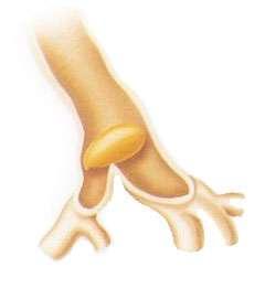
7 Larynx The larynx contains epiglottis is a leaf-shaped cartilaginous structure extending from the base of the tongue and attached to the thyroid cartilage by ligaments During swallowing the epiglottis flaps down to direct food into the esophagus. Vocal cords for phonation and is also an organ with sphincter functions that help preventing aspiration. Vocal cords are drawn apart during inspiration and relax towards midline during expiration.

8 Lower Airway A. Conducting airways: Nonalveolate region Consists of series of tubes that divide like the branches of a tree. Its function are to conduct air and provide mucociliary defense. The trachea is made up16 to 20 C-shaped rings of cartilage, lined by ciliated epithelium and mucous producing goblet cell.

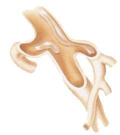
9 Trachea It is average 2 to 2.5 cm in diameter and cm in length. The trachea divides at carina into right and left main-stem bronchus, Right Main bronchus angle degrees from the midline. This position promotes incidence of aspiration and right main stem intubation. Left main bronchus short, narrow and deflects sharply at degree. Excessive pressure on this smooth muscle by the cuff of an artificial airway can lead to erosion and tracheoesophageal fistula.

10 The first 16 divisions of the tracheobronchial tree take no part in gas exchange. The volume is 150 ml approximately know as anatomical dead space. The large bronchi are supplied with sensory nerve receptors involved in cough reflex. There are 64,000 terminal bronchioles.

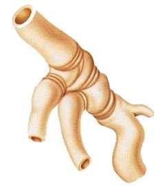
11 Lower Airway B. Respiratory Zone: Alveolate Region Lungs The lungs are paired, cone-shaped organs, average adult lung is 800g. The lungs are divided into lobes, the left has two and right has three. Lobes are divided into bronchopulmonary segments, there are 10 segments into right lung and 8 segments in left lung. This Segments divides into lobules, which are the lung smallest unit.

12 Pleura Each lung is enveloped in its pleural membrane, an inner and an outer which are in intimate contact with each other. The inner membrane closely envelope the lungs surface while the outer membrane covers the inside of the chest wall and the large respiratory muscles, the diaphragm. During inspiration the negative pressure is maintained by the rib cage, which pulls the parietal pleura outward, thus causing the lungs to expand and air to be sucked in. If air, blood or other fluid are introduced in to the plural space, the two plural surface can separate.

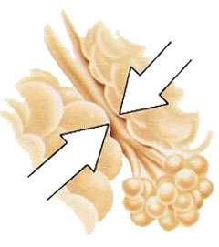
13 Transitional and Respiratory zone Acinus is the primary gas-exchanging unit consisting of the respiratory bronchiole, alveolar ducts, alveolar sacs and alveoli. There are approximately 300 million alveolar-capillary units in adult lung. The total surface area of lung 2 parenchyma is 50 to 100m, about the size of a tennis court. The alveoli are surrounded by capillaries. The alveolar cells are squamous epithelium one cell layer thick, to promote gas exchange and prevent fluid transudation into alveolus Surfactant prevent the alveoli and bronchioles from collapsing.

14 Thoracic cage The lungs are housed in the thoracic cage. Structure provide support and protection for the contents of thorax. Sternum is a dragger-shaped bony structure in the median line at the chest. In adult sternum averages 18cm in length. There are 12 pairs of ribs correspond to the twelve thoracic vertebrae and connect to the sternum via costal cartilages. 1-7 rib are connect directly to sternum rib are connect indirectly to sternum ribs has no connection to sternum, so called as floating ribs.

15 Respiratory Muscles The principle muscle of respiration is the diaphragm, made up of fibrous tissue. The resting position is dome shaped. On inspiration the diaphragm contracts, flattening the dome. The anterior-posterior dimension of the thoracic cavity elevate and increase during contraction of external intercostal muscles. During inspiration scalene muscles contract elevating first two rib. Sternomastoid assist in elevating the sternum. Oxygen plays an important role in the energy metabolism of living organisms.
16 Innervation of the Ventilatory Muscles C1 Accessory (XI)n, C2-C3 Sternomastoid Muscle Diaphragm Intercostal Muscles Intercostal n. T1-T12 C8 T1 Trapezius Muscle Scalene Muscle Abdominal Muscles T7-T12, L1 T12 L1

17 Physiology of Pulmonary System Regulation of breathing in the Brain stem. The volume and the frequency of ventilation is determined by impulses from the respiratory center in the Medulla oblongata to the respiratory muscles. The impulses from the central receptor depends on the CO2 in the blood, affect the fluid value of ph in the brain and spinal cord. In the peripheral receptors when PaO2 is lowered about 60 mmhg then the respiratory center is stimulated. Brain cells are extremely sensitive to oxygen deprivation and can begin to die within five minutes after oxygen supply has been cut off.
18 Impulses from receptors and regulation of breathing Receptors Signal to the respiratory centre Muscular activity The blood Central Low ph Hyperventilation PaCO2 Peripheral High ph Hypoventilation The regulation of breathing in a patient with chronic lung disease Signal to the respiratory centre Muscular activity The blood Receptors Low PaO2 Hyperventilation PaO2 Peripheral High PaO2 Hypoventilation

19 Muscular activity during Inspiration

20 Muscular activity during Expiration

21 Measurement of lung volumes and capacities with spirometer

22 Physiology Pulmonary System External Respiration Internal Respiration

23 Physiology Pulmonary System
24 Factores affecting ventilation a) Compliance Measurement of the distensibility of the respiratory tissue. Elasticity of the pulmonary tissues is primarily due to its interstitial makeup of elastin and collagen fibers. Total lung compliance = Change in Volume Change in Pressure Two forms of compliance Static compliance Truest measure of the compliance of the lung tissue. It is measured when there is no gas flowing into or out of the lung. (Normal value are 70 to 100 ml/cm H2O) Static compliance = Exhaled tidal Volume Plateau PEEP Dynamic compliance Measurement taken while gases are moving in the lung, therefore it measure the resistance to the gas flow too.(normal value are 50 to 80 ml/cm H2O) Dynamic compliance = Exhaled tidal volume Peak inspiratory pressure - Peep

25 High compliance Low pressure Low compliance High pressure
26 b) Resistance Measurement of the opposition to the flow of gases through the airways. There are two type of resistance. 1) Tissue Resistance caused by tissue friction during inspiration and expiration normally makes up about 20% of total resistance. 2) Airway resistance is opposition to the flow of gas caused by friction between the wall of the airway and gas molecules, as well as viscous friction between gas molecules themselves.(normal values 0.5 to 3.0 cmh2o/sec, at standard flow rate 0.5 L/sec) Airway resistance = Peak pressure Plateau pressure Flow Factors affect airway resistance includes airway length, radius and the flow rate.
27 Bronchospasm Pulmonary Edema Emphysema Foreign particles Excessive Secretions

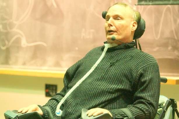
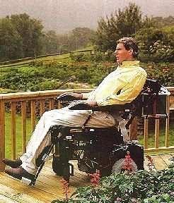
28 Ventilation and Purfusion Relationship "V" - Ventilation - the air which reaches the lungs. "Q" - Perfusion - the blood which reaches the lungs. Normal ventilation - perfusion balance The blood flow is normally greatest in the most dependent parts of the lungs, due to the effect of gravity. If the flow of blood is cut off from one lung, then there will be twice the normal flow in the other lung. Normal alveolar ventilation of 4 l/min and a pulmonary blood flow of 5 l/min, the ratio is 4/5=0.8. If the ratio is high, PaO2 rises and PaCO2 diminishes. If the ratio is low, PaO2 is markedly reduced and PaCO2 rises somewhat. An area with no ventilation (and thus a V/Q of zero) is termed "shunt."
.")
29 Impaired Ventilation Blood which supplies poorly ventilated alveoli (with low PaO2) cannot be adequately oxygenated. Therefore a certain amount of incompletely oxygenated blood enters the arterial circulation and thereby reduces PaO2 (physiological shunt). Compensatory changes in perfusion for impaired ventilation A low PaO2 and a raised PaCO2 initiates a blood vessel construction so that blood flow to under-ventilated alveoli diminishes and instead is redirected to well-ventilated alveoli.

30 Impaired Perfusion Good ventilation of poorly perfused sections of lung tissue, for example, in pulmonary embolus, gives rise to a large dead space. Cardiac output is then unevenly distributed within the lungs without a corresponding redistribution in ventilation. An area with no perfusion (and thus a V/Q of infinity) is termed dead space.

31 Arterial blood gases An arterial blood gas (ABG) test measures the ph and the levels of oxygen and carbon dioxide from the arterial blood. This test is used to check how well the lungs are able to move oxygen into the blood and remove carbon dioxide from the blood. Four-step guide to ABG analysis: 1. Is the ph normal, acidosis or alkalosis? 2. Are the PCO2 or HCO3 abnormal? Which one appears to influence the ph. 3. If both the PCO2 and HCO3 are abnormal, the one which deviates most from the normal is most likely causing an abnormal ph. 4. Check the PO2, is the patient hypoxic. Normal Arterial Blood Gas Values ph = PaCO2 = mm Hg PaO2 = mm Hg HCO3 = meq/l O2 Saturation = 95-99% BE = +/- 1
32 Why ABG Is Done Check for severe breathing problems and lung diseases, such as asthma, cystic fibrosis, or chronic obstructive pulmonary disease (COPD). See how well treatment for lung diseases is working. Find out if you need extra oxygen or help with breathing (mechanical ventilation). Find out if you are receiving the right amount of oxygen when you are using oxygen in the hospital. Measure the acid-base level in the blood of people who have heart failure, kidney failure, uncontrolled diabetes, sleep disorders, severe infections, or after a drug overdose. Respiratory Acidosis ph PaCO 2 HCO 3 Acute < 7.35 > 45 Normal Compensated Normal > 45 > 26 Respiratory Alkalosis Acute > 7.45 < 35 Normal Compensated Normal < 35 < 22 Metabolic Acidosis Acute < 7.35 Normal < 22 Compensated Normal < 35 < 22 Metabolic Alkalosis Acute > 7.45 Normal > 26 Compensated Normal > 45 > 26
33 Who requires ventilator? Post- Operative Major Abdominal Brain Cardiac surgery Respiratory Circulatory Neurological Trauma ARDS Pneumonia COPD Acute Asthma Interstitial Lung Disease Pleural Disease Disorder of sleep Aspiration Pneumonia Smoke inhalation Drowning Venous thromboembolisum Cardiac arrest Myocardial Infarct Left ventricular failure Low cardiac Output Stork Status epileptics Drug overdose Poisoning Respiratory Muscles failure Head injury Poly trauma Spinal cord injury Burns
34 Type of respiratory Failure Typical blood gases Type I PaO2 < 60 mmhg and PaCO2 < 45mmHg Acute Chronic PaO2 PaCO2 ph or HCO3 or PaO2 PaCO2 ph HCO3 Type II PaO2 < 80 mmhg and PaCO2 > 45mmHg Acute Chronic PaO2 PaCO2 ph HCO3 PaO2 PaCO2 ph or HCO3 Causes Asthma Pulmonary embolus Pulmonary oedema ARDS Pneumothorax Pneumonia Emphysema Lung fibrosis Lymphangitis carcinomatosa R L shunt Anaemia Severe acute asthma Acute epiglottis Inhaled foreign body Respiratory muscle paralysis Flail chest injury Sleep apnoea Brain-stem lesion Narcotic drugs COPD Primery alveolar hypoventilation Kyphoscoliosis Ankylosing spondylisis Therapy Treat underline cause; High-concentration O2; Mechanical ventilation Treat underline disorder; Long-term O2 Treat underline disorder; Controlled low-concentration O2; Mechanical ventilation or tracheostomy if necessary Treat underline disorder; Controlled long-term O2; Mechanical ventilation if necessary

35 Reduction in central drive Mechanical defect in chest wall Respiratory muscle fatigue


36 Christopher Reeve


37 Superman
38
39 What is a Ventilator and how does it works? Ventilator is machine designed to alter, transmit and direct applied energy in a predetermined manner to perform useful work. The principal benefits is to improved gas exchange and decreased work of breathing. All modern ventilators used in inpatient clinical settings are Positive Pressure Ventilators. Positive Pressure Ventilators uses a power source, known as the drive mechanism, to force air into the lungs during inspiration. Expiration occurs passively during Positive Pressure Ventilation. If the machine has to perform the respiratory cycle, it must be told. a) What the component phase of respiratory cycle. b) How to carry out each of the phases as determined by the setting of the phase variable. Ventilator has four basic phase that it must complete in providing a ventilatory cycle to the patients. 1. Inspiration 2. Inspiratory-expiratory changeover 3. Expiration 4. Expiratory-inspiratory changeover
 Changeover from Expiratory to inspiratory phase Pressure 1) Inspiratory Phase 3) Expiratory Phase")
40 Phase Variables of respiratory cycle in ventilator 2) Changeover from inspiratory phase to Expiratory phase 4) Changeover from Expiratory to inspiratory phase Pressure 1) Inspiratory Phase 3) Expiratory Phase Time(s)
41 1. Inspiratory Phase Positive pressure is generated in order to create a pressure gradient that leads to lung inflation and the thoracic cavity to expand. Pressure within the airway, alveoli and intrapleural spaces all become positive during inspiration. 2. Inspirator-Expiratory Changeover Ventilator are classified by the mechanism that cycles ventilation from the inspiratory phase to expiratory phase. The mechanisms that cycle the inspiratory-expiratory change over: a) Volume cycle ventilation. Ventilator cycle to end inspiration and begin expiration when a predetermined volume is delivered into the Pt s circuit. b) Time cycled ventilation Inspiration ends and expiration begins after a predetermined time interval has been reached. Cycling may be controlled by timing mechanism or I:E ratio. c) Pressure cycled ventilation Inspiration ends and expiration begins after a predetermined maximum airway pressure limit is reached.
42 3. Expiratory Phase Exhalation occurs passively because of the elastic recoil of the lung, but patient passively exhales to a controlled baseline pressure. The end-expiratory pressure may be equilibrium with atmospheric pressure or may be above, know as PEEP. 4. Expiratory-Inspiratory changeover Once the expiratory phase has been completed, the changeover to the next inspiration phase begins. This phase may be initiated by the patient or by the ventilator based on classified mode. The variable measured by the ventilator and that determines the initiation of a breath is know as Trigger variable:. a) Machine timed trigger The ventilator will trigger a breath after a preset time interval, determined by respiratory frequency. b) Pressure trigger The patient spontaneous respiratory effort decreases the pressure within the inspiratory circuit and inspiration begins. c) Flow trigger A patient effort draws flow from the circuit (continuous bias flow) and mechanical breaths are initiated when flow into the patient exceeds the set flow threshold.
 - 152.4} x 0.91+ 50 for male and 45.5 in female.")
43 Ventilator Settings O2 concentration or Fraction of Inspired Oxygen Concentration (FiO2) Depends on the patients disease condition and ABG report. Tidal Volume (Vt or TV) = 6 to 10 ml per body weight. Predicted Body Weight = {Height (cm) } x for male and 45.5 in female. Pressure Control level above PEEP Pressure Support above PEEP Respiratory Rate Peak End Expiratory Pressure (PEEP) Triggered Sensitivity 1. Pressure Triggered 2. Flow Triggered Inspiratory-to-Expiratory Ratio Inspiratory Pause Time Inspiratory Rise Time Inspiratory Cycle Off Trigger Time Out Breath Cycle Time
44 Ventilator Settings Positive End-Expiratory Pressure (PEEP) A method of ventilation in which airway pressure is maintained above atmospheric pressure at the end of exhalation by means of a mechanical impedance, usually a valve, within the circuit. The purpose of PEEP is to increase the volume of gas remaining in the lungs at the end of expiration. Inspiration Pressure (cmh2o) Expiration PEEP Time(s) FiO2/O PEEP Positive physiological effects 1. Improved gas exchange 2. Alveolar recruitment 3. Increased functional residual capacity 4. Redistribution of fluids in the alveolus Negative physiological effects 1. Decreased Cardiac output 2. Decreased venous return 3. Increased ICP 4. Barotrauma 5. Hypotension
45 Ventilator Settings Inspiratory-to-Expiratory Ratio (I:E) It is the duration of inspiration in comparison with expiration. Generaly the I:E ratio is set at 1:2; that is, 33% of the respiration cycle is spent in inspiration and 66% in the expiratory phase. Example: Set, RR = 15 b/min; I:E ratio is 1:2 Therefore, time for one cycle is 60/15 = 4sec Than 4/3 = 1.3sec is the time inspiration and 2.7 sec is expiration. Shorter inspiratory time contributes to dead space ventilation by over expanding the most compliant alveoli. Prolonged Expiration at 1:3, 1:4 Longer inspiratory time increases the Mean Airway Pressure (MAP), which may lead to hemodynamic instability. Inverse ratio at 2:1, 3:1 or 4:1 Pause time It is pause in between inspiration and expiration changeover to allowed the distribution of inhaled gases. During this phase there is no flow, both the inspiratory and Expiratory valves are closed. Monitoring the resistance and the compliance of the lungs.
46 Ventilator Settings Inspiratory Rise Time (%/sec) Is the time taken to reach peak inspiratory flow or pressure at the start of each breath. The flow and pressure can be adapted according to patient need. Example: Set RR = 15, so the time for each breath is 60/15 = 4sec If set Inspiratory rise time is 10% then, 4 x10/100= 0.4sec 100% Insp Rise Time P T V T
47 Ventilator Settings Inspiratory Cycle Off (%) Fraction of maximum flow at which inspiratory to switch to expiration. Is the point at which inspiration changes to expiration in spontaneous and support mode of ventilation to avoid hyperinflation of lung. 100% V 70% 10% 10% T 5%
48 Ventilator Settings Triggered Sensitivity Determines the level of patient effort needed to trigger the ventilator to start inspiration. It can be set either in Flow Triggering (Trigg.Flow) or Pressure Trigger (Trigg.Pressure). Flow triggered is preferable as this enables the patient to breath with less effort. When patient triggers T appears on the screen. Pressure Triggered The pressure triggering threshold is indicated by a negative pressure level below baseline. The purple color indication appears on the pressure curve. Sensitivity can be set within the range 0 to -20 cmh2o Flow Triggered When the difference between the inspiratory and the expiratory flow equals the preset flow trigger level, the ventilator will start a new inspiration. Threshold is indicated by an inspiratory flow above baseline bias flow (2L/min in Adult and 0.5L/min in Neonate) and purple color indication appears on the flow curve
49 Ventilator Settings Trigger Time Out Is the maximum allowed apnea time before controlled ventilation is activated. It initially adapts with a dynamic trigger timeout, increases successively during the first 10 breaths. Example: RR set= 20 b/min Time for each breath is 60/20 = 3sec Trigger timeout set = 7 sec then, 7-3 = 4s, divided in to 10 steps = 0.4 sec 0.4 s If the patient stop breathing after 4 breath, the ventilator delivers a controlled breath after 4.6s. After 10 triggered breath the ventilator waits 7s before providing a controlled breath.
50 Ventilator Settings Breath Cycle Time Is the length of the time allowed for total respiratory cycle for the mandatory breaths. Example: RR = 6 b/min If Breath cycle Time set is 3sec with I:E Ratio = 1:2 so1 sec Inspiration and 2 sec Expiration. Time for one cycle is 60/6 = 10sec So, the time for spontaneous breath is 10-3 = 7sec 3 sec 7sec Time for mandatory breath Time for spontaneous breath Time for mandatory breath
51 Type of Ventilation and Modes 1. Non-Invasive Ventilation (NIV) A flow of gas is delivered to the conscious patients airway via tight fitting face or nasal mask. NIV Presser Support (PS) NIV Presser Control (PC) Nasal CPAP (Infants) 2. Invasive Positive Pressure Ventilation (IPPV) A flow of gas is delivered to the patients airway via Endotracheal / Tracheostomy tube. Controlled modes Supported modes Combined modes VC PC PRVC PS VS Automode: VC-VS PC-PS PRVC-VS SIMV: VC +PS PC + PS PRVC + PS Bi-Vent

52 Non-Invasive Ventilation (NIV) Indications A. Acute respiratory failure 1. Hypercapnic acute respiratory failure Acute exacerbation of COPD Post extubation Weaning difficulties Post surgical respiratory failure Thoracic wall deformities Cystic fibrosis Status asthmatic Acute respiratory failure in Obesity hypoventilation Syndrome

53 2. Hypoxaemic acute respiratory failure Evidence is less convincing to show efficacy of NIV in hypoxaemic respiratory failure. The possible indications are: Cardiogenic pulmonary oedema Community acquired pneumonia Post traumatic respiratory failure ARDS Weaning difficulties B. Chronic Respiratory Failure C. Immunocompromised Patients D. Do Not Intubate Patients Modes for NIV PC - Pressure Control PS - Pressure Support Nasal CPAP for Infants - Continuous Positive Airway Pressure
54 Prerequisites for successful Non-Invasive support Patient is able to cooperate. Patient can control airway and secretions. Adequate cough reflex. Patient is able to co-ordinate breathing with ventilator. Patient can breathe unaided for several minutes. Haemodynamically stable. Blood ph>7.1 and PaCO2 <92 mmhg. Improvement in gas exchange, heart rate and respiratory rate within first two hours. Normal functioning gastrointestinal tract.
55 NIV Presser Support Provide adequate support for patients who do not have sufficient capacity. Provide ventilation in which every breath is patient triggered. Provide support during the patients inspiration according to the preset pressure support level. Maintain an adequate Positive End Expiratory Pressure PEEP. Provide a fast and flexible response to the patient s need. Provide an accurate monitoring and alarm function. Facilitate the weaning process. Settings PS (Pressure support) above PEEP. PEEP (cmh2o) Oxygen concentration (%) Inspiratory rise time (% of breath cycle time/sec) Inspiratory cycle off (%) NIV backup rate, (breaths/min) NIV backup Ti (time in sec) Trigger sensitivity cannot be set in NIV, Pt s either lowers the pressure to 1 cmh2o below PEEP during expiratory or an expiratory flow decrease of 6 ml during 100ms, breath is delivered.
56 NIV Presser Control The ventilator delivers a flow to maintain the preset pressure at a preset respiratory rate and during a preset inspiratory time. Provide a constant pressure level during the entire inspiration. Enable controlled respiratory rate. Provide a decelerating flow pattern. Provide ventilation if the breath is patient triggered. Maintain an adequate Positive End Expiratory Pressure PEEP. Provide an accurate monitoring and alarm function. Settings PC (Pressure Control) above PEEP. Respiratory rate (b/min) PEEP (cmh2o) Oxygen concentration (%) I:E ratio or Ti (insp. time in sec) Inspiratory rise time (% of breath cycle time/s) The bios flow is delivered with a constant flow of 7.5 l/min to detect the breathing effort of the patient.

57 Nasal CPAP CPAP help to recruit collapsed alveoli, improve oxygenation, stabilize the chest wall and inhibits paradoxical movements during inspiration and collapse during expiration. Regarded as alternative to intubation. Treatment of respiratory condition like RDS in premature infants. Easy to wean infants off mechanical ventilation. Also handle the problem of apnea of premature. Infants from 500g to 10 kg can be ventilated via nasal mask or prongs. Settings CPAP (Continuous Positive Airway Pressure ) Oxygen concentration (%) The maximum available flow in nasal CPAP is 33 l/min
 Dryness of nasal mucosa and mouth. Un synchronization.")
58 Complications of NIV Leak causing redness to the eyes. Pressure source to the bridge of the nose. Abdominal distention. Aspiration. Discomfort. Claustrophobia (Fear of confined space) Dryness of nasal mucosa and mouth. Un synchronization. Patients no able to sleep because of sound.

59 Invasive Positive Pressure Ventilation (IPPV) Control Modes a) Volume Control mode (VC) Preset tidal volume is delivered with a constant flow during the preset inspiratory time and the preset respiratory rate. Provide a controlled inspiratory time and pause time. During the pause time both inspiratory and expiratory valves are closed and no flow is delivered. Peak pressure can vary breath to breath if the pt s compliance and resistance changes. Patient makes an inspiratory effort during the expiratory phase an assisted breath is delivered. Extra flow is delivered if the patients decrease airway pressure by 3 cmh2o during the inspiratory phase. If the patient exhale during Inspiration the pressure increases more than 3cmH2O above set, expiratory valve opens and allows expiration.
60 VC Settings Tidal volume (ml) Respiratory Rate (b/min) PEEP (cmh2o) Oxygen concentration (%) I:E ratio Pause time (%/s) Inspiratory rise time(% breath cycle time/sec) Trigger sensitivity Pressure Flow Peak Pressure Plateau Pressure End Expiratory Flow End Inspiratory Flow End Expiratory Flow P resistance P compliance T T Alarm Settings Pressure (cmh2o) Minute Volume (cmh2o) Respiratory Rate End Exp Pressure Sound level Volume Inspiration Expiration Time(s)

61 b) Pressure Control mode (PC) Ventilator delivers a flow to maintain the preset pressure at a preset respiratory rate and preset inspiratory time. Pressure is constant during the inspiratory time and the flow is decelerating. The volume can vary from breath to breath if the patient s compliance and resistance change. Maximum available flow is 200l/min in Adult and 33 l/min in Infant. If for any reason, pressure decrease during inspiration, than the flow from the ventilator will immediately increase to maintain the set inspiratory pressure. If the pressure increases to the set upper pressure limit, than the expiratory valve opens and the ventilator switches to expiration.
 Minute Volume (cmh2o) Respiratory Rate End Exp Pressure Sound level Volume Inspiration Expiration")
62 PC Settings PC Pressure Control(cmH2O) Respiratory Rate (b/min) PEEP (cmh2o) Oxygen concentration (%) I:E ratio Inspiratory rise time(% breath cycle time/sec) Trigger sensitivity Pressure Flow T T Alarm Settings Pressure (cmh2o) Minute Volume (cmh2o) Respiratory Rate End Exp Pressure Sound level Volume Inspiration Expiration Time(s)
 breath until that preset tidal volume is achieved.")
63 c) Pressure Regulated Volume Control mode (PRVC) Controlled mode of ventilation which combines the volume controlled and pressure controlled ventilation. The physician presets a desired tidal volume, and the ventilator delivers a pressure-limited (controlled) breath until that preset tidal volume is achieved. The breath is like a conventional pressure-controlled ventilation breath, but the ventilator can guarantee a predetermined tidal volume. First breath delivered to the patient is a volume controlled. The plateau pressure is used as the pressure level for the next breath. The set tidal volume is achieved by automatic breath-by-breath pressure regulation. The ventilator adjusts the inspiratory PC level to the lowest possible level to guarantee the preset tidal volume. If the tidal volume increases/decreases below the preset volume than the pressure increases/ decreases by 3cmH2O. The maximum available pressure level is 5cmH2O below the preset upper pressure limit.
64 PRVC Settings Tidal volume (ml) Respiratory Rate (b/min) PEEP (cmh2o) Oxygen concentration (%) I:E ratio Pause time (%/s) Inspiratory rise time(% breath cycle time/sec) Trigger sensitivity Pressure Flow T T T Alarm Settings Pressure (cmh2o) Volume Time(s)

65 Supported Modes A spontaneous mode of ventilation where no mandatory breath are given. Patient trigger each breath and ventilator delivers support with the preset pressure support. The patient regulate the respiratory rate and the tidal volume with support from the ventilator. If the patient is not breathing and the set apnea limit is reached, the ventilator will switch to backup controlled ventilation. a) Pressure Support Mode (PS) b) Volume Support Mode (VS)
66 a) Pressure Support Mode (PS) Settings PS(Pressure support) above PEEP PEEP (cmh2o) Oxygen concentration (%) Inspiratory rise time(% breath cycle time/sec) Trigger sensitivity Inspiratory cycle off (%) Backup ventilation - PC above PEEP (cmh2o) Pressure Flow T T Volume Time(s)
67 b) Volume Support Mode (VS) A spontaneous mode of ventilation where no mandatory breath are given. Patient trigger each breath and ventilator delivers support with the preset pressure support. The patient regulate the respiratory rate with support from the ventilator. Start-up sequence is 4 breath, The first breath is given with a support of 10 cmh2o. The ventilator calculates and regulates the pressure needed to deliver the preset Tidal volume. During the remaining 3 test breath, the maximum pressure increased is 20cmH2O. After the start-up sequence,the pressure support is automatically adjusted according to the patient's lung compliance and resistance to deliver a preset tidal volume. The pre-set tidal volume is maintained by increasing or decreasing the pressure support by 3cmH2O. If the patient is not breathing and the set apnea limit is reached, the ventilator will switch to backup controlled ventilation.
68 b) Volume Support Mode (VS) The pre-set tidal volume is maintained by increasing or decreasing the pressure by 3cmH2O. Pressure Settings Tidal Volume (ml) PEEP (cmh2o) Oxygen concentration (%) Inspiratory rise time(% breath cycle time/sec) Trigger sensitivity Inspiratory cycle off (%) Flow T T Volume Time(s)
69 Combined Modes a) Automode Automatically controls the transition between controlled and support mode in accordance with patient s breathing efforts. It is interactive mode of ventilation. It allows the patients to go into support mode automatically if they trigger the ventilator. Better adapted to the patients efforts. It provide the best possible means of starting the weaning period. Three different coupling modes combining control and support: 1. Volume Control Volume Support 2. Pressure Control Pressure Support 3. PRVC Volume Support
70 Volume Control-Volume Support Mode Settings Tidal volume (ml) Respiratory Rate (b/min) PEEP (cmh2o) Oxygen concentration (%) Inspiratory Time (s) Pause time (%/s) Inspiratory rise time(% breath cycle time/sec) Trigger sensitivity Inspiratory cycle off (%) Trigger Timeout (s) Pressure Flow Volume T T Time(s)
71 Pressure Control-Pressure Support Mode Settings PC Pressure Control level (cmh2o) Respiratory rate (b/min) PEEP (cmh2o) Oxygen concentration (%) Time Inspiration (s) Inspiratory rise time(% breath cycle time/sec) Trigger sensitivity Inspiratory cycle off (%) Trigger Timeout (s) Pressure Supported above PEEP (cmh2o) Pressure Flow Volume T T Time(s)
72 PRVC-Volume Support Mode Settings Tidal volume (ml) Respiratory rate (b/min) PEEP (cmh2o) Oxygen concentration (%) Time Inspiration (s) Inspiratory rise time(% breath cycle time/sec) Trigger sensitivity Inspiratory cycle off (%) Trigger Timeout (s) Pressure Flow T T Volume Time(s)
73 b) Synchronized Intermittent Mandatory Ventilation (SIMV) Delivers mandatory breaths with preset tidal volume/minute volume or preset pressure and a preset respiratory rate. This mandatory breaths are synchronized with the breathing efforts of the patient who can breath spontaneous between the breaths. Spontaneous breathing is supported with a set pressure support above the PEEP. The ventilator will wait during the next SIMV period for the patient to trigger. Assist patients during weaning. Assist patient who have some, but not sufficient breathing. Assist patients who need some controlled breaths. If the patients has not triggered within the first 90% of the breath cycle time, than a mandatory breath is delivered.

74 SIMV SIMV Breath cycle time Spontaneous period SIMV Breath cycle time Pressure 90% T Flow T
75 A. SIMV (VC) + Pressure Support Mode Settings Tidal Volume (ml) SIMV rate (b/min) PEEP (cmh2o) Oxygen concentration (%) I:E ratio Pause time (%/s) Inspiratory rise time(% breath cycle time/sec) Breath cycle time (s) Trigger sensitivity Inspiratory cycle off (%) PS(Pressure support) above PEEP(cmH2O) Pressure Flow Volume T T Time(s)
76 B. SIMV (PC) + Pressure Support Mode Settings PC Pressure Control level (cmh2o) SIMV rate (b/min) PEEP (cmh2o) Oxygen concentration (%) I:E ratio Pause time (%/s) Inspiratory rise time(% breath cycle time/sec) Breath cycle time (s) Trigger sensitivity Inspiratory cycle off (%) PS (Pressure support) above PEEP (cmh20) Pressure Flow Volume T T Time(s)
77 C. SIMV (PRVC) + Pressure Support Mode Settings Tidal Volume (ml) SIMV rate (b/min) PEEP (cmh2o) Oxygen concentration (%) I:E ratio Inspiratory rise time(% breath cycle time/sec) Breath cycle time (s) Trigger sensitivity Inspiratory cycle off (%) PS (Pressure support) above PEEP(cmH2O) Pressure Flow Volume T T Time(s)
78 Bi-Vent Phigh/Thigh PEEP/TPEEP Is classified as a time-cycled, pressurelimited mode of ventilation that allows spontaneous breathing throughout the entire respiratory cycle. It has two, time-cycled pressure levels and switches between these levels. Pressure support can be added to augment the patient s spontaneous effort at both the level. It can be used with inversed I:E ratio or as Airway Pressure release Ventilation (APRV) Pressure Flow T T Volume Time(s)
79 Bi-Vent with Sponteneous Breathing Settings PS above PHigh (cmh2o) PEEP (cmh2o) Oxygen concentration (%) Thigh (s) TPEEP (s) Inspiratory Rise Time (%) Trigger sensitivity Inspiratory cycle Off (%) PS above PHigh (cmh2o) PS above PEEP(cmH2O) Pressure Flow PHigh PS above PEEP T T Volume Time(s)
80 Sensing The Servo controller system, senses and adjusts gas delivery faster than any device available on the market. Transducer refresh rate 2000 times/s Benefits Immediate response to patient efforts. Smooth uninterrupted regulation during inhalation. Low resistance during exhalation. Avoids overshoot or undershoot. No delays in adjustments. Allows the patient to lead. Gives immediate feedback to the clinician. Gives real time measurements. Catches the Golden Moments.
81 Golden Moments Pressure Patient benefits: Quicker weaning Flow T Less side effects Effective oxygenation and gas exchange T T Minimal influence on pulmonary and systemic circulation Volume Minimal lung damage Time(s) Pressure Modes PC/PRVC & PS/VS 1. Late Inspiratory Recruitment 2. Breath Initiation Pressure 3. Flow-Adapted Volume Control 4. Early Expiratory Flow 5. Patient-Adjusted Inspiratory Flow 6. Inspiratory Cycle Off Flow T T 7. Early Weaning Volume Time(s)
82 Monitoring Value Definition Value Definition Ppeak Maximum inspiratory pressure Mve Expired minute volume Pplat Pressure during inspiratory pause Leakage Leakage (%) in NIV Pmean Mean airway pressure VTi Inspiratory tidal volume PEEP Positive end expiratory pressure PEEP tol Set PEEP+ intrinsic PEEP CPAP Continuous positive airway pressure Cdyn Dynamic compliance RR Respiratory rate Cststic Static compliance O2 Oxygen concentration in vol.% E Elastance Ti Inspiratory time Ri Inspiratory resistance Te Expiratory time Re Expiratory resistance I:E Inspiratory expiratory ratio WOB v Work of breathing, ventilator Ti/Ttol Mve sp Ratio of inspiratory time to total breath cycle time Spontaneous minute volume WOB p Work of breathing, patient P0.1 Occlusion pressure SBI Shallow breathing index
83 Ventilator Graphics A. Scalars When viewing scalar graphics, time is conventionally shown on the horizontal (x) axis whereas flow, volume, and pressure are plotted on the vertical axis (y) axis. Peak Pressure P Plateau Pressure End Expiratory Flow P resistance 1. Increased airway resistance due to bronchospasm. 2. Accumulation of secretions End Inspiratory Flow P compliance T(s) 3. Response to Bronchodilator. F 4. Auto-PEEP or air trapping. 5. A leak can be identified in. T (s) 6. Shows a negative deflection for trigger breath. 7. Peak Inspiratory Pressure (PIP). End Expiratory Flow 8. Plateau Pressure ( Pplat ) or Alveolar Pressure. 9. Inappropriate inspiratory flow rates. V T (s) Inspiration Expiration
84 B. Loops Are the two-dimensional graphic display of two scalar values. There are two loops available for interpretation. They are: VT Inspiration Volume (ml) Flow (L/min) Volume (ml) Pressure (cmh2o) PIP 1. Inflection point can be identified. 2. Lung compliance and resistance. 3. Alveolar distension. 4. Inadequate sensitivity. 5. Inappropriate inspiratory flow rates. 6. A leak can be identified. Expiration 1. A leak can be identified. 2. Air trapping or auto-peep. 3. An increased airway resistance. 4. Presence of secretions.
85 Trouble Shooting Alarms Possible Problem Advice High PAW Caution: If airway pressure rises 6 cmh2o above the set pressure limit, the safety valve opens. Thick Secretion ETT/TT block Kinked by Pt Pneumothorax Pt coughing Pt Ventilator Asynchronization High RR=>PEEPi High VT/PP Malfunction of Ventilator Bronchospasm Decreased lung Complience Flow rate too high ET touching carnia Blocked HME Check Pt- Ventilator System Do Suction Increase FiO2/100% O2 for 2 min Ambubag Ventilation Sedation if required. Check ETT patency Give Nebulisation Change Thermovent/HME Need to change ETT/TT CXR, ABG Inform Dr Low Minute Volume PIP/PAW Leak Cuff not Inflated Displaced ET Apnea Check Pt- Ventilator System Breathing system Check ETT patency Check Cuff Pressure Consider increase ventilatory support.
86 Alarms Possible Problem Advice Low O2/Air Pressure Central Supply not connected Check connections or below 2kPa O2 Calibration O2 sensor failure Inform respected person Apnea Leak Pt not breathing in SIMV& PS modes High PEEP/PEEPi Auto PEEP generated due to high RR Less expiratory time Pt Ventilator Asynchronization Malfunction of Ventilator High RR, MV, VT Fever Infection Decrease PaO2 Increase PaCO2 Increase lung compliance Inform Dr/RT Check Pt- Ventilator System Check Pt- Ventilator System Inform Dr Adjust PEEP Reset I:E ratio Ambubag Ventilation Sedation if required Check Pt- Ventilator System ABG 100% O2 for 2 min Sedation if required

87
Dr. Yasser Fathi M.B.B.S, M.Sc, M.D. Anesthesia Consultant, Head of ICU King Saud Hospital, Unaizah
 BY Dr. Yasser Fathi M.B.B.S, M.Sc, M.D Anesthesia Consultant, Head of ICU King Saud Hospital, Unaizah Objectives For Discussion Respiratory Physiology Pulmonary Graphics BIPAP Graphics Trouble Shootings
CHAPTER 7.1 STRUCTURES OF THE RESPIRATORY SYSTEM
 CHAPTER 7.1 STRUCTURES OF THE RESPIRATORY SYSTEM Pages 244-247 DO NOW What structures, do you think, are active participating in the breathing process? 2 WHAT ARE WE DOING IN TODAY S CLASS Finishing Digestion
Test Bank Pilbeam's Mechanical Ventilation Physiological and Clinical Applications 6th Edition Cairo
 Instant dowload and all chapters Test Bank Pilbeam's Mechanical Ventilation Physiological and Clinical Applications 6th Edition Cairo https://testbanklab.com/download/test-bank-pilbeams-mechanical-ventilation-physiologicalclinical-applications-6th-edition-cairo/
VENTILATOR GRAPHICS ver.2.0. Charles S. Williams RRT, AE-C
 VENTILATOR GRAPHICS ver.2.0 Charles S. Williams RRT, AE-C Purpose Graphics are waveforms that reflect the patientventilator system and their interaction. Purposes of monitoring graphics: Allow users to
Respiratory System. Chapter 9
 Respiratory System Chapter 9 Air Intake Air in the atmosphere is mostly Nitrogen (78%) Only ~21% oxygen Carbon dioxide is less than 0.04% Air Intake Oxygen is required for Aerobic Cellular Respiration
Respiratory Physiology
 Respiratory Physiology Dr. Aida Korish Associate Prof. Physiology KSU The main goal of respiration is to 1-Provide oxygen to tissues 2- Remove CO2 from the body. Respiratory system consists of: Passages
Prepared by : Bayan Kaddourah RN,MHM. GICU Clinical Instructor
 Mechanical Ventilation Prepared by : Bayan Kaddourah RN,MHM. GICU Clinical Instructor 1 Definition Is a supportive therapy to facilitate gas exchange. Most ventilatory support requires an artificial airway.
LUNGS. Requirements of a Respiratory System
 Respiratory System Requirements of a Respiratory System Gas exchange is the physical method that organisms use to obtain oxygen from their surroundings and remove carbon dioxide. Oxygen is needed for aerobic
The Respiratory System
 13 PART A The Respiratory System PowerPoint Lecture Slide Presentation by Jerry L. Cook, Sam Houston University ESSENTIALS OF HUMAN ANATOMY & PHYSIOLOGY EIGHTH EDITION ELAINE N. MARIEB Organs of the Respiratory
The Respiratory System. Dr. Ali Ebneshahidi
 The Respiratory System Dr. Ali Ebneshahidi Functions of The Respiratory System To allow gases from the environment to enter the bronchial tree through inspiration by expanding the thoracic volume. To allow
I. Anatomy of the Respiratory System A. Upper Respiratory System Structures 1. Nose a. External Nares (Nostrils) 1) Vestibule Stratified Squamous
 1) Vestibule Stratified Squamous") I. Anatomy of the Respiratory System A. Upper Respiratory System Structures 1. Nose a. External Nares (Nostrils) 1) Vestibule Stratified Squamous Epithelium b. Nasal Cartilages 1) Nasal Cavity Pseudostratified
I. Anatomy of the Respiratory System A. Upper Respiratory System Structures 1. Nose a. External Nares (Nostrils) 1) Vestibule Stratified Squamous Epithelium b. Nasal Cartilages 1) Nasal Cavity Pseudostratified
Chapter 10. The Respiratory System Exchange of Gases. Copyright 2009 Pearson Education, Inc.
 Chapter 10 The Respiratory System Exchange of Gases http://www.encognitive.com/images/respiratory-system.jpg Human Respiratory System UPPER RESPIRATORY TRACT LOWER RESPIRATORY TRACT Nose Passageway for
Chapter 13. The Respiratory System.
 Chapter 13 The Respiratory System https://www.youtube.com/watch?v=hc1ytxc_84a https://www.youtube.com/watch?v=9fxm85fy4sq http://ed.ted.com/lessons/what-do-the-lungs-do-emma-bryce Primary Function of Breathing
ADVANCED ASSESSMENT Respiratory System
 ONTARIO BASE HOSPITAL GROUP QUIT ADVANCED ASSESSMENT Respiratory System 2007 Ontario Base Hospital Group ADVANCED ASSESSMENT Respiratory System AUTHOR(S) Mike Muir AEMCA, ACP, BHSc Paramedic Program Manager
Anatomy & Physiology 2 Canale. Respiratory System: Exchange of Gases
 Anatomy & Physiology 2 Canale Respiratory System: Exchange of Gases Why is it so hard to hold your breath for Discuss! : ) a long time? Every year carbon monoxide poisoning kills 500 people and sends another
Circulatory System. and. Respiratory System. Ari Min, Yerim Lee and Min Ji Song THE HEART LUNGS. Monday, May 23, 2011
 Human Anatomy Circulatory System and THE HEART Respiratory System LUNGS Ari Min, Yerim Lee and Min Ji Song Purpose of the Circulatory System Function of circulatory system: exchange gases with cardiovascular
The Human Respiration System
 The Human Respiration System Nasal Passage Overall function is to filter, warm and moisten air as it enters the body. The nasal passages are the primary site of air movement we tend to be nose breathers.
Chapter 10 Respiration
 1 Chapter 10 Respiration Introduction/Importance of the Respiratory System All eukaryotic organisms need oxygen to perform cellular respiration (production of ATP), either aerobically or anaerobically.
Chapter 10 Lecture Outline
 Chapter 10 Lecture Outline See separate PowerPoint slides for all figures and tables preinserted into PowerPoint without notes. Copyright 2016 McGraw-Hill Education. Permission required for reproduction
NURSE-UP RESPIRATORY SYSTEM
 NURSE-UP RESPIRATORY SYSTEM FUNCTIONS OF THE RESPIRATORY SYSTEM Pulmonary Ventilation - Breathing Gas exchanger External Respiration between lungs and bloodstream Internal Respiration between bloodstream
Chapter 10 The Respiratory System
 Chapter 10 The Respiratory System Biology 2201 Why do we breathe? Cells carry out the reactions of cellular respiration in order to produce ATP. ATP is used by the cells for energy. All organisms need
October Paediatric Respiratory Workbook APCP RESPIRATORY COMMITTEE
 October 2017 Paediatric Respiratory Workbook APCP RESPIRATORY COMMITTEE This workbook is designed to introduce to you the difference between paediatric and adult anatomy and physiology. It will also give
CHAPTER 22 RESPIRATORY
 pulmonary ventilation move air external respiration exchange gases transportation of gases internal respiration exchange gases CHAPTER 22 RESPIRATORY in / out lungs air - blood blood - cells cell respiration
Phases of Respiration. Chapter 18: The Respiratory System. Structures of the Respiratory System. Structures of the Respiratory System
 Phases of Respiration Chapter 18: The Respiratory System Respiration Process of obtaining oxygen from environment and delivering it to cells Phases of Respiration 1. Pulmonary ventilation between air and
Respiratory System. Functional Anatomy of the Respiratory System
 Respiratory System Overview of the Respiratory System s Job Major Duty Respiration Other important aspects ph control Vocalization Processing incoming air Protection Metabolism (ACE) What structures allow
Tuesday, December 13, 16. Respiratory System
 Respiratory System Trivia Time... What is the fastest sneeze speed? What is the surface area of the lungs? (hint... think of how large the small intestine was) How many breaths does the average person
Respiratory System Functions. Respiratory System Organization. Respiratory System Organization
 Respiratory System Functions Functions of Respiratory System Gas exchange between blood and air Move air to and from exchange surfaces Protect exchange surfaces from environmental variations and pathogens
B. Correct! As air travels through the nasal cavities, it is warmed and humidified.
 Human Anatomy - Problem Drill 20: The Respiratory System Question No. 1 of 10 1. Which of the following statements about the portion of the respiratory system labeled in the image below is correct? Question
The Respiratory System
 The Respiratory System If you have not done so already, please print and bring to class the Laboratory Practical II Preparation Guide. We will begin using this shortly in preparation of your second laboratory
Unit 14: The Respiratory System
 Unit 14: The Respiratory System See what you already know! 1. Fill in the diagram on your own 2. Collaborate with your partner The Respiratory System The major function of the respiratory system is gas
Unit 9. Respiratory System 16-1
 Unit 9 Respiratory System 16-1 Works together with the circulatory system Exchange of gases between atmosphere, blood, and cells If respiratory system and/or circulatory system fails, death will occur
Handling Common Problems & Pitfalls During. Oxygen desaturation in patients receiving mechanical ventilation ACUTE SEVERE RESPIRATORY FAILURE
 Handling Common Problems & Pitfalls During ACUTE SEVERE RESPIRATORY FAILURE Pravit Jetanachai, MD QSNICH Oxygen desaturation in patients receiving mechanical ventilation Causes of oxygen desaturation 1.
The respiratory system structure and function
 Name: Class: Date: Active reading 11A + Biology Gr11A The respiratory system structure and function The function of the respiratory system is to bring oxygen into the body and eliminate carbon dioxide
Chapter 11 The Respiratory System
 Biology 12 Name: Respiratory System Per: Date: Chapter 11 The Respiratory System Complete using BC Biology 12, page 342-371 11.1 The Respiratory System pages 346-350 1. Distinguish between A. ventilation:
Energy is needed for cell activities: growth,reproduction, repair, movement, etc...
 Respiration Energy is needed for cell activities: growth,reproduction, repair, movement, etc... Metabolism refers to all of the chemical reactions in the body, where molecules are synthesized (anabolism)
Respiratory System. Organization of the Respiratory System
 Respiratory System In addition to the provision of oxygen and elimination of carbon dioxide, the respiratory system serves other functions, as listed in (Table 15 1). Respiration has two quite different
2. List seven functions performed by the respiratory system?
 The Respiratory System C23 Study Guide Tortora and Derrickson 1. In physiology we recognize that the word respiration has three meanings. What are the three different meanings of the word respiration as
Chapter 16. Respiratory System
 Chapter 16 Respiratory System Introduction Respiration = the entire process of exchanging gases between the atmosphere and body cells 1. Ventilation 2. Gas exchange 3. Gas transport : 4. Cellular respiration
The RESPIRATORY System. Unit 3 Transportation Systems
 The RESPIRATORY System Unit 3 Transportation Systems Functions of the Respiratory System Warm, moisten, and filter incoming air Resonating chambers for speech and sound production Oxygen and Carbon Dioxide
What is the next best step?
 Noninvasive Ventilation William Janssen, M.D. Assistant Professor of Medicine National Jewish Health University of Colorado Denver Health Sciences Center What is the next best step? 65 year old female
Respiratory System. Introduction. Atmosphere. Some Properties of Gases. Human Respiratory System. Introduction
 Introduction Respiratory System Energy that we consume in our food is temporarily stored in the bonds of ATP (adenosine triphosphate) before being used by the cell. Cells use ATP for movement and to drive
Competency Title: Continuous Positive Airway Pressure
 Competency Title: Continuous Positive Airway Pressure Trainee Name: ------------------------------------------------------------- Title: ---------------------------------------------------------------
The Respiratory System
 The Respiratory System Cells continually use O2 & release CO2 Respiratory system designed for gas exchange Cardiovascular system transports gases in blood Failure of either system rapid cell death from
Anatomy of the Lungs. Dr. Gondo Gozali Department of anatomy
 Anatomy of the Lungs Dr. Gondo Gozali Department of anatomy 1 Pulmonary Function Ventilation and Respiration Ventilation is the movement of air in and out of the lungs Respiration is the process of gas
The Respiratory System
 C h a p t e r 24 The Respiratory System PowerPoint Lecture Slides prepared by Jason LaPres North Harris College Houston, Texas Copyright 2009 Pearson Education, Inc., publishing as Pearson Benjamin Cummings
Chapter 10. Respiratory System and Gas Exchange. Copyright 2005 Pearson Education, Inc. publishing as Benjamin Cummings
 Chapter 10 Respiratory System and Gas Exchange Function of the Respiratory System To obtain oxygen (O 2 ) for all cells in the body. To rid the cells of waste gas (CO 2 ). Oxygen (O 2 ) is vital chemical
61a A&P: Respiratory System!
 61a A&P: Respiratory System! 61a A&P: Respiratory System! Class Outline" 5 minutes" "Attendance, Breath of Arrival, and Reminders " 10 minutes "Lecture:" 25 minutes "Lecture:" 15 minutes "Active study
THE RESPIRATORY SYSTEM
 THE RESPIRATORY SYSTEM Functions of the Respiratory System Provides extensive gas exchange surface area between air and circulating blood Moves air to and from exchange surfaces of lungs Protects respiratory
RESPIRATORY SYSTEM. A. Upper respiratory tract (Fig. 23.1) Use the half-head models.
 Use the half-head models.") RESPIRATORY SYSTEM I. OVERVIEW OF THE RESPIRATORY SYSTEM AND THORAX A. Upper respiratory tract (Fig. 23.1) Use the half-head models. Nasal cavity Pharynx (fare-rinks) B. Lower respiratory tract (Fig. 23.1)
RESPIRATORY SYSTEM I. OVERVIEW OF THE RESPIRATORY SYSTEM AND THORAX A. Upper respiratory tract (Fig. 23.1) Use the half-head models. Nasal cavity Pharynx (fare-rinks) B. Lower respiratory tract (Fig. 23.1)
The Respiratory System Structures of the Respiratory System Structures of the Respiratory System Structures of the Respiratory System Nose Sinuses
 CH 14 D.E. Human Biology The Respiratory System The Respiratory System OUTLINE: Mechanism of Breathing Transport of Gases between the Lungs and the Cells Respiratory Centers in the Brain Function Provides
Lab Activity 27. Anatomy of the Respiratory System. Portland Community College BI 233
 Lab Activity 27 Anatomy of the Respiratory System Portland Community College BI 233 1 Terminology Pulmonary Ventilation: aka breathing, is the movement of air into and out of the lungs External Respiration:
61a A&P: Respiratory System!
 61a A&P: Respiratory System! 61a A&P: Respiratory System! Class Outline 5 minutes Attendance, Breath of Arrival, and Reminders 10 minutes Lecture: 25 minutes Lecture: 15 minutes Active study skills: 60
B Unit III Notes 6, 7 and 8
 The Respiratory System Why do we breathe? B. 2201 Unit III Notes 6, 7 and 8 Respiratory System We know that our cells respire to produce ATP (energy). All organisms need energy to live, so that s why we
The Respiratory System
 The Respiratory System Function of the Respiratory System Oversees gas exchanges (oxygen and carbon dioxide) between the blood and external environment Exchange of gasses takes place within the lungs in
Chapter 23 The Respiratory System
 Chapter 23 The Respiratory System Cells continually use O 2 & release CO 2 Respiratory System designed for gas exchange Cardiovascular system transports gases in blood Failure of either system rapid cell
The RESPIRATORY System. Unit 9
 The RESPIRATORY System Unit 9 Respiration The exchange of gases between the atmosphere, blood, and cells Pulmonary Ventilation - the exchange of air between the atmosphere and lungs External (Pulmonary)
Mechanical Ventilation Principles and Practices
 Mechanical Ventilation Principles and Practices Dr LAU Chun Wing Arthur Department of Intensive Care Pamela Youde Nethersole Eastern Hospital 6 October 2009 In this lecture, you will learn Major concepts
Gas exchange Regulate blood ph Voice production Olfaction Innate immunity
 Respiration Functions Gas exchange: Grab O 2, eject CO 2 Regulate blood ph: Alters CO 2 levels Voice production: air movement past vocal cords Olfaction: in nasal cavity Innate immunity: physical protection
Mechanical Ventilation 1. Shari McKeown, RRT Respiratory Services - VGH
 Mechanical Ventilation 1 Shari McKeown, RRT Respiratory Services - VGH Objectives Describe indications for mcvent Describe types of breaths and modes of ventilation Describe compliance and resistance and
This is not a required assignment but it is recommended.
 SU 12 Name: This is not a required assignment but it is recommended. BIO 116 - Anatomy & Physiology II Practice Assignment 2 - The Respiratory and Cardiovascular Systems 1. The exchange of oxygen and carbon
5/5/2013. The Respiratory System. Chapter 16 Notes. The Respiratory System. Nasal Cavity. Sinuses
 The Respiratory System Chapter 16 Notes The Respiratory System Objectives List the general functions of the respiratory system. Identify the organs of the respiratory system. Describe the functions of
otorhinolaryngology -the study of the structure, function, and disorders of the ears, nose, and throat
 89 Chapter 6 Respiratory System Overview -The respiratory system consists of the nose, pharynx, larynx, trachea, bronchi, and the lungs (Figure 6.1, Derrickson). -The purpose of the respiratory system
The Respiratory System
 Essentials of Human Anatomy & Physiology Elaine N. Marieb Seventh Edition Chapter 13 The Respiratory System Slides 13.1 13.30 Lecture Slides in PowerPoint by Jerry L. Cook Copyright 2003 Pearson Education,
THE RESPIRATORY SYSTEM. Pages and
 THE RESPIRATORY SYSTEM Pages 103-105 and 146-150 1 When the respiratory system is mentioned, people generally think of breathing, but breathing is only one of the activities of the respiratory system.
-Rachel Naomi Remen. Respiratory System 1
 Life is known only by those who have found a way to be comfortable with change and the unknown. Given the nature of life, there may be no security, but only adventure. Respiratory System 1 -Rachel Naomi
Ventilator Waveforms: Interpretation
 Ventilator Waveforms: Interpretation Albert L. Rafanan, MD, FPCCP Pulmonary, Critical Care and Sleep Medicine Chong Hua Hospital, Cebu City Types of Waveforms Scalars are waveform representations of pressure,
You are caring for a patient who is intubated and. pressure control ventilation. The ventilator. up to see these scalars
 Test yourself Test yourself #1 You are caring for a patient who is intubated and ventilated on pressure control ventilation. The ventilator alarms and you look up to see these scalars What is the most
Chapter 16. The Respiratory System. Mosby items and derived items 2010, 2006, 2002, 1997, 1992 by Mosby, Inc., an affiliate of Elsevier Inc.
 Chapter 16 The Respiratory System Objectives Discuss the generalized functions of the respiratory system List the major organs of the respiratory system and describe the function of each Compare, contrast,
Noninvasive ventilation: Selection of patient, interfaces, initiation and weaning
 CME article Johnson S, et al: Noninvasive ventilation Noninvasive ventilation: Selection of patient, interfaces, initiation and weaning Saumy Johnson, Ramesh Unnikrishnan * Email: ramesh.unnikrishnan@manipal.edu
Bio 104 Respiratory System 81
 81 Lecture Outline: Respiratory System Hole s HAP [Chapter 19] I. Introduction Respiration is the process of exchanging gases between the atmosphere and body cells. Respiration consists of: Ventilation
Weaning from Mechanical Ventilation. Dr Azmin Huda Abdul Rahim
 Weaning from Mechanical Ventilation Dr Azmin Huda Abdul Rahim Content Definition Classification Weaning criteria Weaning methods Criteria for extubation Introduction Weaning comprises 40% of the duration
Karachi King s College of Nursing
 Karachi King s College of Nursing Badil Dass Lecturer Respiratory system Respiratory System Respiratory system consist of: Nose Pharynx (Throat) Larynx (Voice Box) Trachea (Wind Pipe) Bronchi Bronchioles
APRV Ventilation Mode
 APRV Ventilation Mode Airway Pressure Release Ventilation A Type of CPAP Continuous Positive Airway Pressure (CPAP) with an intermittent release phase. Patient cycles between two levels of CPAP higher
The Respiratory System
 13 PART A The Respiratory System PowerPoint Lecture Slide Presentation by Jerry L. Cook, Sam Houston University ESSENTIALS OF HUMAN ANATOMY & PHYSIOLOGY EIGHTH EDITION ELAINE N. MARIEB Organs of the Respiratory
1. When a patient fails to ventilate or oxygenate adequately, the problem is caused by pathophysiological factors such as hyperventilation.
 Chapter 1: Principles of Mechanical Ventilation TRUE/FALSE 1. When a patient fails to ventilate or oxygenate adequately, the problem is caused by pathophysiological factors such as hyperventilation. F
Function: to supply blood with, and to rid the body of
 1 2 3 4 5 Bio 1102 Lec. 7 (guided): Chapter 10 The Respiratory System Respiratory System Function: to supply blood with, and to rid the body of Oxygen: needed by cells to break down food in cellular respiration
Effects of PPV on the Pulmonary System. Chapter 17
 Effects of PPV on the Pulmonary System Chapter 17 Pulmonary Complications Lung Injury Gas distribution Pulmonary blood flow VAP Hypoventilation Hyperventilation Air trapping Oxygen toxicity WOB Patient-Ventilator
Monitor the patients disease pathology and response to therapy Estimate respiratory mechanics
 Understanding Graphics during Mechanical Ventilation Why Understand Ventilator Graphics? Waveforms are the graphic representation of the data collected by the ventilator and reflect the interaction between
Unconscious exchange of air between lungs and the external environment Breathing
 Respiration Unconscious exchange of air between lungs and the external environment Breathing Two types External Exchange of carbon dioxide and oxygen between the environment and the organism Internal Exchange
About the Respiratory System. Respiratory System. Human Respiratory System. Cellular Respiration. Nostrils. Label diagram
 Respiratory System Human Respiratory System A system to deliver oxygen (O2) to body cells & get rid of carbon dioxide (CO2) as a waste through cellular respiration. Two systems involved: Respiratory &
Slide 1. Slide 2. Slide 3 VENTILATOR MADNESS.. MAKING SENSE OF IT ALL!! Objectives: I have nothing to disclose.
 Slide 1 VENTILATOR MADNESS.. MAKING SENSE OF IT ALL!! Maryann M Brogden ND, MSN, RN, APN-C, CCNS, SCRN Slide 2 I have nothing to disclose. Slide 3 Objectives: Identify Criteria for Intubation Differentiate
Respiratory System. December 20, 2011
 Respiratory System December 20, 2011 Nasal Cavity: Contains cilia (hair cells) to prevent particles from entering the respiratory tract Mucus does the same, in addition to warming and moistening the air
The respiratory system
 The respiratory system Practical 1 Objectives Respiration, ventilation Intrapleural and intrapulmonary pressure Mechanism of inspiration and expiration Composition of the atmosphere and the expired air
Learning Objectives. 1. Indications versus contra-indications 2. CPAP versus NiVS 3. Clinical evidence
 Learning Objectives 1. Indications versus contra-indications 2. CPAP versus NiVS 3. Clinical evidence Pre-hospital Non-invasive vventilatory support Marc Gillis, MD Imelda Bonheiden Our goal out there
Tracheostomy and Ventilator Education Program Module 2: Respiratory Anatomy
 Tracheostomy and Ventilator Education Program Module 2: Respiratory Anatomy Disclaimer This material is intended for use by trained family members and caregivers of children with tracheostomies who are
The Respiratory System
 BIOLOGY OF HUMANS Concepts, Applications, and Issues Fifth Edition Judith Goodenough Betty McGuire 14 The Respiratory System Lecture Presentation Anne Gasc Hawaii Pacific University and University of Hawaii
NON INVASIVE LIFE SAVERS. Non Invasive Ventilation (NIV)
") Table 1. NIV: Mechanisms Of Action Decreases work of breathing Increases functional residual capacity Recruits collapsed alveoli Improves respiratory gas exchange Reverses hypoventilation Maintains upper
Table 1. NIV: Mechanisms Of Action Decreases work of breathing Increases functional residual capacity Recruits collapsed alveoli Improves respiratory gas exchange Reverses hypoventilation Maintains upper
Unit Nine - The Respiratory System
 Unit Nine - The Respiratory System I. Introduction A. Definition: the respiratory system consists of the nose, nasal cavity, (throat), (voice box), (windpipe), bronchi and lungs (which contain the alveoli).
Lung Wit and Wisdom. Understanding Oxygenation and Ventilation in the Neonate. Jennifer Habert, BHS-RT, RRT-NPS, C-NPT Willow Creek Women s Hospital
 Lung Wit and Wisdom Understanding Oxygenation and Ventilation in the Neonate Jennifer Habert, BHS-RT, RRT-NPS, C-NPT Willow Creek Women s Hospital Objectives To review acid base balance and ABG interpretation
Lecture Overview. Respiratory System. Martini s Visual Anatomy and Physiology First Edition. Chapter 20 - Respiratory System Lecture 11
 Martini s Visual Anatomy and Physiology First Edition Martini Ober Chapter 20 - Respiratory System Lecture 11 1 Lecture Overview Overview of respiration Functions of breathing Organs of the respiratory
The Respiratory System
 PowerPoint Lecture Slide Presentation by Vince Austin Human Anatomy & Physiology FIFTH EDITION Elaine N. Marieb The Respiratory System Dr Nabil Khouri. MD, Ph.D Respiratory System Consists of a conducting
? Pulmonary Respiratory System
 The Structure of The Respiratory System The respiratory system is composed of groups of organelles that filters and transports air into the lungs. The organs comprising of the respiratory system include
Respiratory System. Student Learning Objectives:
 Respiratory System Student Learning Objectives: Identify the primary structures of the respiratory system. Identify the major air volumes associated with ventilation. Structures to be studied: Respiratory
a. Describe the physiological consequences of intermittent positive pressure ventilation and positive end-expiratory pressure.
 B. 10 Applied Respiratory Physiology a. Describe the physiological consequences of intermittent positive pressure ventilation and positive end-expiratory pressure. Intermittent positive pressure ventilation
The primary function of the respiratory system is to supply the blood with oxygen in order for the blood to deliver oxygen to all parts of the body.
 Respiratory System The primary function of the respiratory system is to supply the blood with oxygen in order for the blood to deliver oxygen to all parts of the body. The respiratory system does this
The RESPIRATORY System. Unit 3 Transportation Systems
 The RESPIRATORY System Unit 3 Transportation Systems The Respiratory System Functions of the Respiratory System Warms, moistens, and filters incoming air Nasal cavity Resonating chambers for speech and
Bio 322 Human Anatomy Objectives for the laboratory exercise Respiratory System
 Bio 322 Human Anatomy Objectives for the laboratory exercise Respiratory System Required reading before beginning this lab: Saladin, KS: Human Anatomy 5 th ed (2017) Chapter 23 For this lab you will use
Respiratory System. BSC 2086 A&P 2 Professor Tcherina Duncombe Palm Beach State College
 Respiratory System BSC 2086 A&P 2 Professor Tcherina Duncombe Palm Beach State College Respiration Ventilation of lungs Gas exchange between air/bld and bld/tissue Use of oxygen in cellular respiration
NIV use in ED. Dr. Khalfan AL Amrani Emergency Resuscitation Symposium 2 nd May 2016 SQUH
 NIV use in ED Dr. Khalfan AL Amrani Emergency Resuscitation Symposium 2 nd May 2016 SQUH Outline History & Introduction Overview of NIV application Review of proven uses of NIV History of Ventilation 1940
Chapter 11. The respiratory system. Glossary. Anthony Wheeldon
 Chapter 11 The respiratory system Anthony Wheeldon Glossary Accessory muscles Muscles not normally involved in respiration that can be utilised to increase inspiration. Acid base balance The mechanisms