HCC e CEUS. Prof. A. Giorgio. Direttore IX UOC di Malattie Infettive ad Indirizzo Ecointerventistico
|
|
|
- Rosaline Maxwell
- 5 years ago
- Views:
Transcription
1 HCC e CEUS Prof. A. Giorgio Direttore IX UOC di Malattie Infettive ad Indirizzo Ecointerventistico
2 The natural history of compensated cirrhosis due to hepatitis C virus: a 17 year cohort study of 214 patients HCC was the first complication to develop and the dominant cause for increased mortality Sangiovanni A et al. Hepatology June (43) 2006
3 Hepatocellular carcinoma (HCC) is the 7 th most common cancer worldwide. In 2008: 748,000 new cases 696,000 deaths Ferlay J et al. GLOBOCAN Int Journal Cancer 2010
4 Vascular changes in hepatocellular carcinoma ZF. Yang et al Anat Rec 2008 HCC is one of the most vascular solid tumors, in which angiogenesis plays an important role the status of angiogenesis in HCC correlates with the disease progression and prognosis and thus provides a potential therapeutic target
5
6 hepatocarcinogenesis Neoangiogenesis and capillarization leads to gradual change in blood supply portal venous supply hepatic arterial supply RN DN HCC
7 Hepatology, february 2009

8 L-DN H-DN WD-HCC MD-HCC Partially modified by Hepatology Feb. 2009

9 AJR November 2004 Contrast-Enhanced Sonographic Appearance of Hepatocellular Carcinoma in Patients with Cirrhosis: Comparison with Contrast- Enhanced Helical CT Appearance Antonio Giorgio et al Giovanna Ferraioli Luciano Tarantino Giorgio de Stefano Vincenzo Scala Ferdinando Scarano Carmine Coppola Luca Del Viscovo
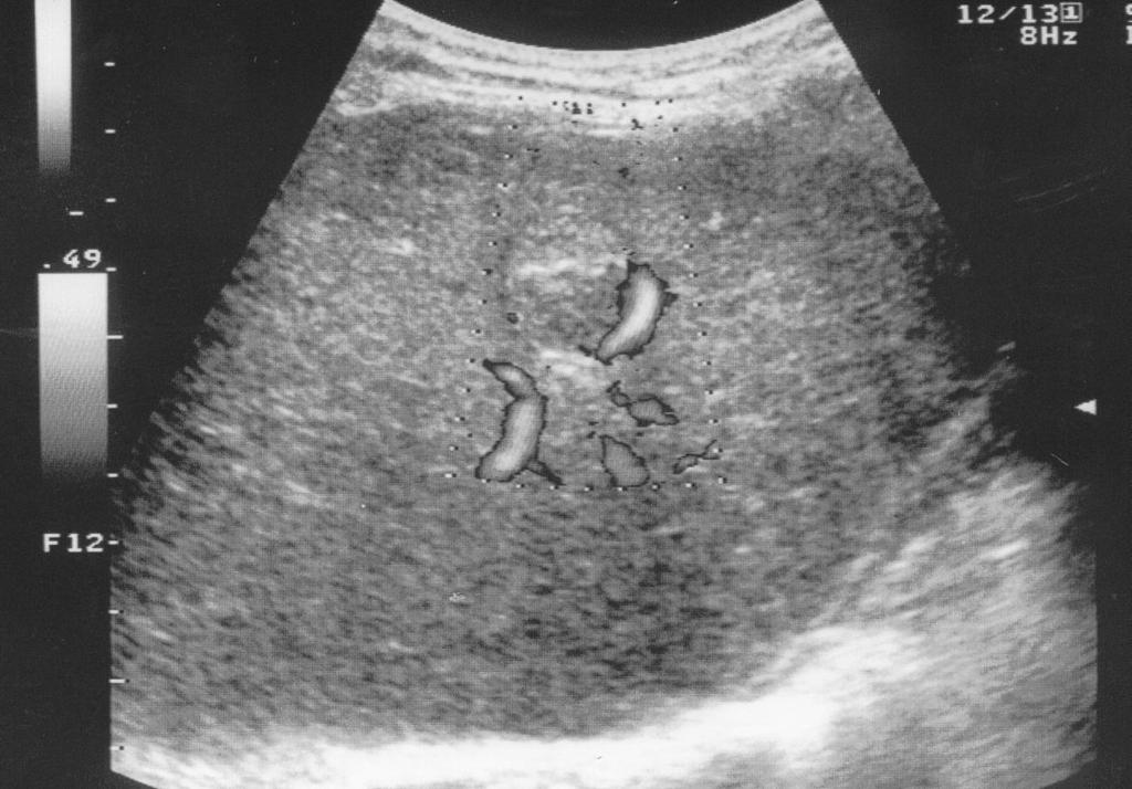
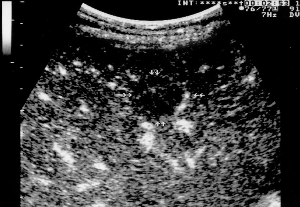
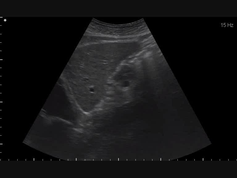
10 CEUS: Patterns HCC Focal liver lesions Comportamento Fase arteriosa sec. Fase portale sec Fase sinusoidale sec Regenerative nodule Focal fatty sparing Focal Avascolare fatty change Non Hemangioma visibile A. Giorgio AJR 2004
11
12
13
14
15
16
17 Comportamento
18 CEUS: Patterns HCC Focal liver lesions Comportamento Fase arteriosa sec. Fase portale sec Fase sinusoidale sec Ipervascolare Regenerative omogeneo nodule Ipervascolare Focal fatty sparing reticolare Focal fatty change Hemangioma

19
20
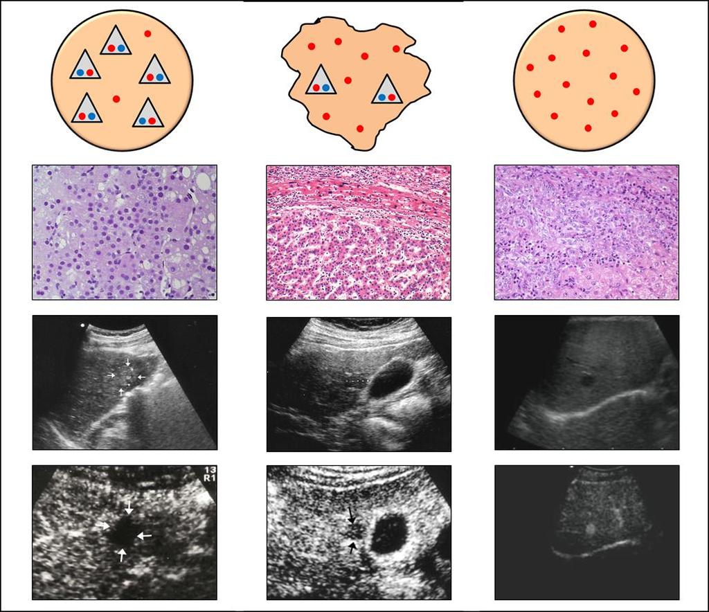
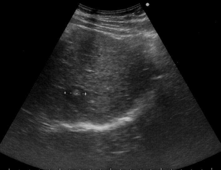
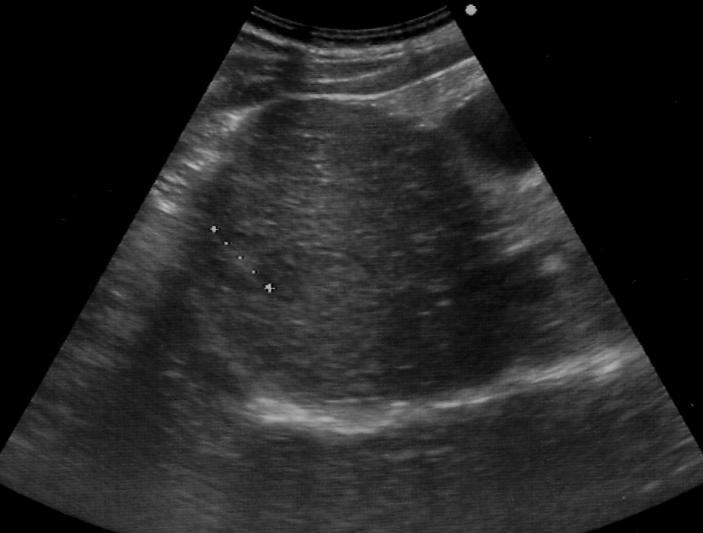
21 Small nodular lesions in cirrhotic livers: characterization with contrast enhanced ultrasound A. Giorgio et al, Anticancer Research, June 2011 aim to investigate the usefulness of CEUS in the characterization of dysplastic nodules (DN), early hepatocellular carcinoma (HCC) and overt HCC 2 cm during US surveillance in cirrhosis 36 consecutive pts with a single nodule 2 cm (9-20 mm) underwent CEUS, all 36 underwent biopsy histology 6 pts had low grade dysplastic nodule (LGDN) 5 had high grade dysplastic nodule (HGDN), 14 and 11 patients had early and overt HCC, respectively
22 Small nodular lesions in cirrhotic livers: characterization with contrast enhanced ultrasound A. Giorgio et al, Anticancer Research, June 2011 CEUS On CEUS, 3 LGDN were missed and 8 HGDN were avascular; 1 early HCC was missed and 1 was avascular, 12 early HCC showed the so called reticular pattern 10 overt HCC presented the typical rapid, intense and homogeneous enhancement on arterial phase and washout in the portal phase and 1 showed the reticular pattern
23 Small nodular lesions in cirrhotic livers: characterization with contrast enhanced ultrasound A. Giorgio et al, Anticancer Research, June 2011 conclusion CEUS is a useful technique in the characterization of small nodular lesions emerging during US surveillance of cirrhosis, with high sensitivity and high specificity CEUS is able to identify the unpaired arteries substituting portal tracts of early HCC which lead to well differentiated overt HCC On the basis of arterial hypervascularity, sensitivity of CEUS for early HCC was 87% and specificity 100%; for overt HCC sensitivity was 91% and specificity 98%
24
25 CEUS: Patterns HCC Focal liver lesions Comportamento Fase arteriosa sec. Fase portale sec Fase sinusoidale sec Regenerative Ipervascolare nodule Focal Ipovascolare fatty sparing Focal Avascolare fatty change Non Hemangioma visibile A. Giorgio et al AJR 2004
26
27 Cotrast-enhanced ultrasound of hepatocellular carcinoma: Correlation of washout time and angiogenesis Xia Y et al, Clin Hem Microcirc, 2011 The wash-out time was longer in well differentiated HCC pts compared to those with moderately poorly to differentiated HCC Evaluation of HCC by contrast enhanced sonography: correlation with pathologic differentiation Xu JF et al, J Ultrasound Med, 2011 The time to peak, contrast enhanced time, and wash-out time of the well differentiated HCC were longer than those of the moderately to poorly differentiated HCC
28 Cost-effectiveness analysis on the surveillance for HCC in liver cirrhosis pts using contrast enhanced ultrsonography Tanaka H et al, Hepatol Res, Jan 2012 CONCLUSION CEUS survellance for HCC is a cost effective strategy for cirrhotic and gains longest additional life years,with similar degree for Incremental Cost-Effectiveness Ratio in the US surveillance group. CEUS surveillance using Sonazoid is expected to be used not only in Japan, but also world-wide
29 CEUS and contrast enhanced CT Giorgio et al. AJR;2004 Gaiani et al. J Hepatol; 2004 Day et al. Hepatol Res; 2008 Pompili et al. Dig Liver Dis; 2009 Similar sensitivity and specificity
30 Diagnosis of hepatic nodules 20 mm or smaller in cirrhosis: prospective validation of the noninvasive diagnostic criteria for HCC. Forner A Llovet JM, Bruix J et al, Hepatology, 47 (1) 2008 AIM to evaluates the accuracy of CEUS and MRI for the diagnosis of nodules 20 mm or smaller detected US surveillance 89 cirrhotic pts without prior HCC in whom US detected a small solitary nodule (mean 14 mm) Methods MRI, CEUS and FNB were performed at baseline intense arterial contrast uptake followed by washout in the delayed/venous phase was registered as conclusive for HCC
31 Diagnosis of hepatic nodules 20 mm or smaller in cirrhosis: prospective validation of the noninvasive diagnostic criteria for HCC. Forner A, Llovet JM, Bruix J et al, Hepatology, 47 (1) 2008 results diagnosis of HCC 20 mm or smaller can be established without a positive biopsy if both CEUS and MRI are conclusive however, sensitivity of these noninvasive criteria is 33% and as occurs with biopsy, absence of a conclusive pattern does not rule out malignancy these results validate the American Association for the study of Liver Disease (AASLD) guidelines
32 Contrast-enhanced sonography in the characterization of small hepatocellular carcinomas in cirrhotic patients comparison with contrast-enhanced ultrafast magnetic resonance imaging A. Giorgio et al, AntiCancer Research (27); December 2007
33 Results 6 Concordance between CEUS and MRI Overall: 75.0% HCCs mm: 89.2% HCCs < 10 mm: 27.3%
34
35
36
37 Guidelines and Good Clinical Practice Recommendations for Contrast Enhanced Ultrasound (CEUS) Update 2008 the diagnostic of HCC for lesions >2 cm, newly emerged during surveillance in cirrhosis, can be established on CEUS alone in addition to CEUS, a confirmation of arterial hypervascularisation and subsequent wash out by CT/MR is requested to established the diagnostic of HCC in FLL 1-2 cm detected during surveillance, programs M. Claudon et al, European Journal of Ultrasound (29),2008
38 INTRAHEPATIC CHOLANGIOCARCINOMA
39 Intrahepatic Cholangiocarcinoma (ICC) US appearance
")
40 Intrahepatic Cholangiocarcinoma (ICC) US appearance
41 Enhancement patterns of intrahepatic cholangiocarcinoma: comparison between contrast-enhanced ultrasound and contrast-enhanced CT Chen LD et al. BJR 2008 ARTERIAL PHASE ENHANCEMENT PATTERNS OF ICC ON CEUS AND CECT Type 1 - Peripheral irregular rim-like hyperenhancement Irregular rim-like hyperenhancement at the peripheral portion of the lesion and inhomogeneous hypoenhancement at the central portion, with strip-like enhancement extending to the central portion of the lesion. Type 2 Diffuse heterogeneous hyperenhancement Heterogeneous hyperenhancementat at both the periphery and the central portion of the lesion Type 3 Diffuse homogeneous hyperenhancement Homogeneous hyperenhancementat at both the periphery and the central portion of the lesion Type 4 Diffuse heterogeneous hypoenhancement Heterogeneous hypoenhancement at both the periphery and the central portion of the lesion
42 Enhancement patterns of intrahepatic cholangiocarcinoma: comparison between contrast-enhanced ultrasound and contrast-enhanced CT Chen LD et al. BJR 2008 PORTAL PHASE (CEUS sec; CECT sec) - 97,5% of the lesions were hypoenhancing on CEUS - 62,5% of the lesions were hypoenhancing on CECT LATE PHASE (CEUS sec) 100% of the lesions were hypoenhancing on CEUS Intratumoral blood vessels were exhibited in - 50% of the lesions on CEUS - 22,5% of the lesions on CECT
43 Enhancement patterns of intrahepatic cholangiocarcinoma: comparison between contrast-enhanced ultrasound and contrast-enhanced CT Chen LD et al. BJR 2008 The different enhancement patterns during arterial phase may relate to different pathological components in the tumour: Rim-like hyperenhancement may be due to the presence of a rich tumour cellularity in the peripheral portion and fibrosis in the central portion Diffuse hyperenhancement may be the result of a rich cellularity in all the portions of the tumour Diffuse hypoenhancement may be due to the presence of abundant fibrous stroma in the tumour Smaller lesions tend to show homogeneous hyperenhancement
44 Enhancement patterns of intrahepatic cholangiocarcinoma: comparison between contrast-enhanced ultrasound and contrast-enhanced CT Chen LD et al. BJR 2008 CONCLUSIONS The enhancement patterns of ICC on CEUS were consistent with those on CECT in the arterial phase, whereas in the portal phase ICC faded out more obviously on CEUS than on CECT. CEUS had the same accuracy as CECT for diagnosing of ICC, and thus could be used as a new modality for the characterization of ICC
45
46
47

48 Cholangiocarcinoma in cirrhosis:absence of contrast washout in delayed phases by MRI avoids misdiagnosis of HCC Rimola J et al. Hepatology 2009 Arterial phase Portal phase Delayed phase A 57 year old woman with ICC in the right lobe of the liver
49 Arterial phase Enhancement pattern of ICC Intrahepatic Peripheral Cholangiocarcinoma in Cirrhosis Patients May Display a Vascular Pattern Similar to Hepatocellular Carcinoma on Contrast-Enhanced Ultrasound Vilana et al. Hepatology 2010 CEUS findings in our study 5 ICC displayed a homogeneous arterial contrast uptake followed by a rapid contrast washout in the portal phase (<60 sec), thus being fully indistiguishable from HCC Strict applications of the AASL guidelines would have led to a false HCC diagnosis in those 3 nodules larger than 2 cm if CEUS had been the only dynamic imaging technique applied MRI findings in all our patients an MRI scan was done for diagnosis and staging purposes. The most frequent finding was progressive contrast uptake through the different phases, and in no cases was a wash-out detected, allowing a clear differentiation with HCC


50 Intrahepatic Peripheral Cholangiocarcinoma in Cirrhosis Patients May Arterial Display a Vascular phase Enhancement Pattern Similar to Hepatocellular pattern of Carcinoma ICC on Contrast-Enhanced Ultrasound Vilana et al. Hepatology 2010 Homogeneous hyperenhancement Arterial phase Delayed phase CEUS MRI
51 Arterial phase Enhancement pattern of ICC Intrahepatic Peripheral Cholangiocarcinoma in Cirrhosis Patients May Display a Vascular Pattern Similar to Hepatocellular Carcinoma on Contrast-Enhanced Ultrasound Vilana et al. Hepatology 2010 About EFSUMB guidelines for CEUS: They suggested that the typical enhanced pattern for cholangiocarcinoma is a rim-like enhancement (or nonenhancement) during the portal and delayed phases However, these reccomendations are based on caseseries of limited number of patients, most of them noncirrhosis, with large tumors
52 Arterial phase Enhancement pattern of ICC Intrahepatic Peripheral Cholangiocarcinoma in Cirrhosis Patients May Display a Vascular Pattern Similar to Hepatocellular Carcinoma on Contrast-Enhanced Ultrasound Vilana et al. Hepatology 2010 CONCLUSIONS: The use of CEUS as the only imaging tool for noninvasive HCC diagnosis may be inappropriate and could not replace a dynamic MRI scan CEUS may establish the malignant nature of a hepatic nodule in a cirrhotic liver, but should not pretend to establish its final diagnosis

53 AASLD Practice Guidelines for HCC management 2005
54 AASLD Practice Guidelines for HCC management Update 2010
55 Giorgio A, CEUS and HCC. Are the 2008 EFSUMB guidelines still valid or has their wash-out already started? Ultraschall June 2011 But, there is one more thing: while it is well known that CEUS has become a complementary exam to the conventional US both in European and Asian countries as the first radiological tool in the characterization of focal liver lesions, it is also known that CEUS cannot assess the staging of intra or extra hepatic HCC -i.e. presence of a single or multiple hepatic nodules or presence of spread in other abdominal organs- because, using CEUS, it is possible to study a single nodule each time (for the short duration of the arterial phase seconds), and, therefore, an enhanced CT or MRI is necessary in the clinical practice to stage the disease, either for treatment strategies (especially surgical therapies-resection or transplantation-) and even for medical-legal reasons
56 Giorgio A, CEUS and HCC. Are the 2008 EFSUMB guidelines still valid or has their wash-out already started? Ultraschall June 2011 So, why eliminating such a valid and accurate technique in the evaluation of nodules > 1 cm arising in a cirrhotic liver? If enhanced MRI or CT examinations, anyway to be performed after CEUS, will confirm the CEUS wash out in the portal and delayed phases -and this will be the largest majority of cases-, the diagnosis of the HCC nodules will be established without biopsy; on the other hand, in cases with discordant findings between CEUS and enhanced MRI (or enhanced multislice CT), a biopsy with cutting needle will be necessary.
57 Giorgio A, CEUS and HCC. Are the 2008 EFSUMB guidelines still valid or has their wash-out already started? Ultraschall June 2011 As a result, the EFSUMB 2008 guidelines, in my opinion, could still remain valid: In addition to CEUS, a confirmation of arterial hypervascularization and subsequent wash out by CT/MRI is requested to establish the diagnosis of HCC in focal liver lesions from 1-2 cm large detecting during surveillance programs And, obviously, I would add also: in nodules >2 cm


58 Intrahepatic Arterial phase Cholangiocarcinoma Enhancement and Hepatocellular pattern of Carcinoma: ICC Differential diagnosis with Contrast-Enhanced Ultrasound Chen LD et al. Eur Radiol 2010 The CEUS enhancement patterns of 50 ICC were retrospectively analyzed and compared with 50 HCC Arterial phase Only 3% of ICC showed homogeneous hyperenhancement
59 Enhancement patterns of intrahepatic cholangiocarcinoma: comparison between contrast-enhanced ultrasound and contrast-enhanced CT Chen LD et al. BJR 2008 PORTAL PHASE (CEUS sec) - 97,5% of the lesions were hypoenhancing on CEUS LATE PHASE (CEUS sec) 100% of the lesions were hypoenhancing on CEUS Intratumoral blood vessels were exhibited in - 50% of the lesions on CEUS
60 Intrahepatic Cholangiocarcioma and Hepatocellular Carcinoma: Differential diagnosis with Contrast-Enhanced Ultrasound Chen LD et al. Eur Radiol 2010 Peripheral portion of the tumours Time-intensity curves of both the central and the peripheral portion revealed marked enhancement after contrast agent injection in HCC and a weak enhancement in ICC throughout the phases. ICC ICC HCC Central portion of the tumours HCC
61 Arterial Intrahepatic phase Cholangiocarcioma Enhancement and Hepatocellular pattern Carcinoma: of ICC Differential diagnosis with Contrast-Enhanced Ultrasound Chen LD et al. Eur Radiol 2010 The ICC washed out more thoroughly during the portal or late phase in comparison with HCC, both at the periphery and centre of the tumours, so that the intensity of ICC was lower than that of HCC. This phenomenon may indicate that more vessels that trapped microbulbbles were present in the HCC
62 Arterial Intrahepatic phase Cholangiocarcioma Enhancement and Hepatocellular pattern Carcinoma: of ICC Differential diagnosis with Contrast-Enhanced Ultrasound Chen LD et al. Eur Radiol 2010 In comparison with ICC, the abnormal artery supply of HCC was richer, and microvascularisation increased owing to tumour angiogenesis, as well as the presence of abnormal arterovenous shunts, so the intensity of HCC was higher than ICC in the arterial phase IN CONCLUSION: CEUS improves the diagnostic performance significantly in the differentiation between ICC and HCC
63

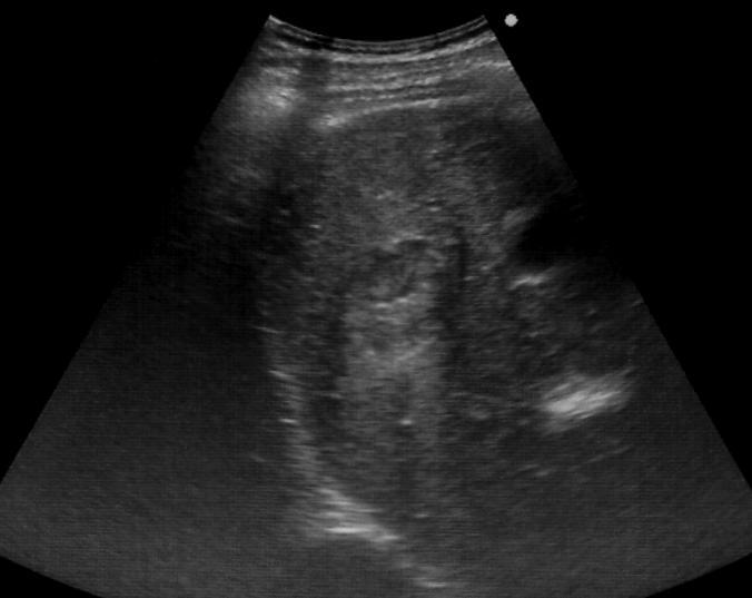
64 HCC B-mode 18 sec CEUS Arterial phase sec 60 sec Portal phase sec 230 sec Sinusoidal phase sec


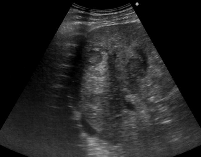
65 HCC B-mode 17 sec CEUS Arterial phase sec 53 sec Portal phase sec 240 sec Sinusoidal phase sec
66 Guidelines and good clinical Pratice Recommendation for CEUS in the liver Update 2012 M. Claudon et Al. Ultraschall 2012 Hyperhenancement in the arterial phase, followed by wash - out in the late phase corresponds to HCC in more than 97% of cases CCC and hepatic lymphoma comprice the remining 1-3% of cases.
67 EFSUMB guidelines 2012 FLL in cirrhotic liver Hyper-E in the arterial phase yes Hypo-E in late phase (=wash-out) Malignant Consider as HCC Iso-E in the portal and late phases Very suspicious for malignancy (positive predictive value >85% for HCC, usually well differentiated
LIVER IMAGING TIPS IN VARIOUS MODALITIES. M.Vlychou, MD, PhD Assoc. Professor of Radiology University of Thessaly
 LIVER IMAGING TIPS IN VARIOUS MODALITIES M.Vlychou, MD, PhD Assoc. Professor of Radiology University of Thessaly Hepatocellular carcinoma is a common malignancy for which prevention, screening, diagnosis,
Detection and Characterization of Hepatocellular Carcinoma by Imaging
 CLINICAL GASTROENTEROLOGY AND HEPATOLOGY 2005;3:S136 S140 Detection and Characterization of Hepatocellular Carcinoma by Imaging OSAMU MATSUI Department of Imaging Diagnosis and Interventional Radiology,
Contrast Enhanced Ultrasound of Parenchymal Masses in Children
 Contrast Enhanced Ultrasound of Parenchymal Masses in Children Sue C Kaste, DO On behalf of Beth McCarville, MD St. Jude Children s Research Hospital Memphis, TN Overview Share St. Jude experience with
Mædica - a Journal of Clinical Medicine
 Mædica - a Journal of Clinical Medicine ORIGINAL PAPERS How Often Hepatocellular Carcinoma Has a Typical Pattern in Contrast Enhanced Ultrasound? Alina MARTIE, MD; Ioan SPOREA, MD, PhD; Roxana SIRLI, MD,
The role of contrast enhanced ultrasound (ceus) in the assessment of liver nodules in patients with cirrhosis
 in the assessment of liver nodules in patients with cirrhosis") Review Medical Ultrasonography 2010, Vol. 12, no. 2, 145-149 The role of contrast enhanced ultrasound (ceus) in the assessment of liver nodules in patients with cirrhosis Mirela Dănilă, Ioan Sporea, Roxana
Review Medical Ultrasonography 2010, Vol. 12, no. 2, 145-149 The role of contrast enhanced ultrasound (ceus) in the assessment of liver nodules in patients with cirrhosis Mirela Dănilă, Ioan Sporea, Roxana
RICCARDO LENCIONI,CLOTILDE DELLA PINA, LAURA CROCETTI,DANIA CIONI. Chapter 1
 RICCARDO LENCIONI,CLOTILDE DELLA PINA, LAURA CROCETTI,DANIA CIONI Chapter 1 Impact of European Federation of Societies for Ultrasound in Medicine and Biology (EFSUMB) Guidelines on the Use of Contrast
Evangelos Chartampilas Bioclinic Hospital Thessaloniki, Greece
 Evangelos Chartampilas Bioclinic Hospital Thessaloniki, Greece Hepatospecificcontrast agents Gadobenate dimeglumine (Multihance) Gadoxeticacid (Primovist) 3-5% liver uptake 50% liver uptake Hepatobiliary
Innovations in HCC Imaging: MDCT/MRI
 Innovations in HCC Imaging: MDCT/MRI Anthony E. Cheng, M.D. Cardinal MRI Center Cardinal Santos Medical Center, Wilson Street, San Juan Innovations in HCC Imaging: Goals/Objectives MDCT/MRI Learn the diagnostic
*, Meng-Xia Yuan, Kuan-sheng Ma, Xiao-Wu Li, Chun-Lin Tang, Xiao-Hang Zhang, De-Yu Guo, Xiao-Chu Yan. Abstract. Introduction
 Detailed Analysis of Temporal Features on Contrast Enhanced Ultrasound May Help Differentiate Intrahepatic Cholangiocarcinoma from Hepatocellular Carcinoma in Cirrhosis Rui Li 1 1 2 2 1 1 *, Meng-Xia Yuan,
Simplifying liver assessment in internal medicine
 Ultrasound Customer story Simplifying liver assessment in internal medicine Philips Affiniti ultrasound for elastography and contrast-enhanced ultrasound (CEUS) Where Sonography Institute, Uster, Switzerland
Surveillance for Hepatocellular Carcinoma
 Surveillance for Hepatocellular Carcinoma Marion G. Peters, MD John V. Carbone, MD, Endowed Chair Professor of Medicine Chief of Hepatology Research University of California San Francisco Recorded on April
HEPATO-BILIARY IMAGING
 HEPATO-BILIARY IMAGING BY MAMDOUH MAHFOUZ MD PROF.OF RADIOLOGY CAIRO UNIVERSITY mamdouh.m5@gmail.com www.ssregypt.com CT ABDOMEN Indications Patient preparation Patient position Scanogram Fasting 4-6 hours
With the widespread use of hepatic imaging, liver masses
 2B: Liver Assessment of the Liver Mass: What Do You Need to Know? With the widespread use of hepatic imaging, liver masses are detected either unexpectedly or in the course of screening for liver cancer
Discrimination between neoplastic and non-neoplastic lesions in cirrhotic liver using contrast-enhanced ultrasound
 The British Journal of Radiology, 85 (2012), 1376 1384 Discrimination between neoplastic and non-neoplastic lesions in cirrhotic liver using contrast-enhanced ultrasound 1,2 H-X XU, MD, PhD, 2 M-D LU,
Evaluation of contrast-enhanced ultrasound for diagnosis of dysplastic nodules with a focus of hepatocellular carcinoma in liver cirrhosis patients
 Original Article Evaluation of contrast-enhanced ultrasound for diagnosis of dysplastic nodules with a focus of hepatocellular carcinoma in liver cirrhosis patients Wei Wu, Minhua Chen, Kun Yan, Yin Dai,
INTRODUCTION. Key Words: Contrast enhanced ultrasonography; Liver masses. ORiginal Article
 Gut and Liver, Vol. 8, No. 3, May 2014, pp. 292-297 ORiginal Article Clinically Useful Diagnostic Tool of Contrast Enhanced Ultrasonography for Focal Liver Masses: Comparison to Computed Tomography and
Contrast-Enhanced Ultrasound in Assessing Therapeutic Response in Ablative Treatments of Hepatocellular Carcinoma
 Contrast-Enhanced Ultrasound in Assessing Therapeutic Response in Ablative Treatments of Hepatocellular Carcinoma Zeno Sparchez 1, Pompilia Radu 1, Ofelia Anton 1, Mihai Socaciu 2, Radu Badea 1 1) 3 rd
Modern liver imaging techniques - A new era in liver ultrasound
 Modern liver imaging techniques - A new era in liver ultrasound Yuko Kono, M.D., Ph.D. Clinical Professor Departments of Medicine and Radiology University of California, San Diego San Diego, USA How to
Guidelines for the use of contrast-enhanced ultrasound in hepatocellular carcinoma
 EJC SUPPLEMENTS 6 (2008) 1 8 available at www.sciencedirect.com journal homepage: www.ejconline.com Guidelines for the use of contrast-enhanced ultrasound in hepatocellular carcinoma Riccardo Lencioni
Interesting Cases from Liver Tumor Board. Jeffrey C. Weinreb, M.D.,FACR Yale University School of Medicine
 Interesting Cases from Liver Tumor Board Jeffrey C. Weinreb, M.D.,FACR Yale University School of Medicine jeffrey.weinreb@yale.edu Common Liver Diseases Hemangioma Cyst FNH Focal Fat/Sparing THID Non-Cirrhotic
Malignant Focal Liver Lesions
 Malignant Focal Liver Lesions Other Than HCC Pablo R. Ros, MD, MPH, PhD Departments of Radiology and Pathology University Hospitals Cleveland Medical Center Case Western Reserve University Pablo.Ros@UHhospitals.org
Objectives. HCC Incidence and Mortality. Disclosure Statement HCC. Imaging of Hepatocellular Carcinoma. Treatment of Hepatocellular Carcinoma
 Imaging of Hepatocellular Carcinoma and the use of LI RADS Treatment of Hepatocellular Carcinoma Aaron D. Anderson, D.O. AOCR April 2015 Objectives Show how the use of LI RADS can simplify the diagnosis
Liver Tumors. Prof. Dr. Ahmed El - Samongy
 Liver Tumors Prof. Dr. Ahmed El - Samongy Objective 1. Identify the most important features of common benign liver tumors 2. Know the risk factors, diagnosis, and management of hepatocellular carcinoma
Hepatocellular carcinoma Cholangiocarcinoma. Jewels of hepatobiliary cancer imaging : what to look for? Imaging characteristics of HCC.
 Outline : Imaging Jewels Jewels of hepatobiliary cancer imaging : what to look for? Hepatocellular carcinoma Cholangiocarcinoma Surachate Siripongsakun, M.D. Chulabhorn Cancer Center Imaging characteristics
The Focal Hepatic Lesion: Radiologic Assessment
 The Focal Hepatic Lesion: Radiologic Assessment Kevin Kuo, Harvard Medical School Year III Our Patient: PS 67 y/o female w/ long history of alcohol use Drinking since age 18, up to one bottle of wine/day
HCC and mass effect. Hepatocellular cancer: what if the AFP is rising but no lesion seen on imaging? What you need to know about AFP.
 Hepatocellular cancer: what if the AFP is rising but no lesion seen on imaging? Arun J Sanyal M.B.B.S., M.D. Charles Caravati Professor of Medicine Virginia Commonwealth University Imaging features used
Surveillance for HCC Who, how Diagnosis of HCC Surveillance for HCC in Practice
 Surveillance for Hepatocellular Carcinoma Hashem B. El-Serag, MD, MPH Dan L. Duncan Professor of Medicine Chief, Gastroenterology and Hepatology Houston VA & Baylor College of Medicine Houston, TX Outline
MRI for HCC surveillance and reporting: LI-RADS. Donald G. Mitchell, M.D. Thomas Jefferson University Philadelphia, PA
 MRI for HCC surveillance and reporting: LI-RADS Donald G. Mitchell, M.D. Thomas Jefferson University Philadelphia, PA Cirrhotic Nodules Regenerative Nodule Atypical Nodule Hyperplastic Nodule Dysplastic
The Contribution of Contrast Enhanced Ultrasound for the characterization of benign liver lesions in clinical practice a monocentric experience
 Original papers Med Ultrason 2012, Vol. 14, no. 4, 283-287 The Contribution of Contrast Enhanced Ultrasound for the characterization of benign liver lesions in clinical practice a monocentric experience
HEPATOCYTE SPECIFIC CONTRAST MEDIA: WHERE DO WE STAND?
 HEPATOCYTE SPECIFIC CONTRAST MEDIA: WHERE DO WE STAND? Andrew T. Trout, MD @AndrewTroutMD Disclosures No relevant disclosures Outline Review of hepatocyte specific contrast media Review of hepatocellular
The UGent Institutional Repository is the electronic archiving and dissemination platform for
 biblio.ugent.be The UGent Institutional Repository is the electronic archiving and dissemination platform for all UGent research publications. Ghent University has implemented a mandate stipulating that
Contrast-enhanced ultrasound (CEUS) of focal liver lesions. A useful, rapid and accessible tool.
 of focal liver lesions. A useful, rapid and accessible tool.") Contrast-enhanced ultrasound (CEUS) of focal liver lesions. A useful, rapid and accessible tool. Poster No.: C-2329 Congress: ECR 2012 Type: Educational Exhibit Authors: S. Santamaria Jareño, J. Carrero
Contrast-enhanced ultrasound (CEUS) of focal liver lesions. A useful, rapid and accessible tool. Poster No.: C-2329 Congress: ECR 2012 Type: Educational Exhibit Authors: S. Santamaria Jareño, J. Carrero
LI-RADS v2016 Key Dates
 Key Dates April 2014: ACR CEUS LI-RADS working group was formed Chair: Yuko Kono, Co-chair: Andrej Lyshchik Members: David Cosgrove, Christoph Dietrich, Hyun-Jung Jang, Tae Kim, Fabio Piscaglia, Claude
Jesse Civan, M.D. Medical Director, Jefferson Liver Tumor Center
 Liver Tumors Jesse Civan, M.D. Medical Director, Jefferson Liver Tumor Center Differential Diagnosis Malignant Metastatic from non-hepatic primary Hepatocellular carcinoma Cholangiocarcinoma Biliary cystcarcinoma
IS THERE A DIFFERENCE IN LIVER CANCER RATES IN PATIENTS WHO RECEIVE TREATMENT FOR HEPATITIS?
 IS THERE A DIFFERENCE IN LIVER CANCER RATES IN PATIENTS WHO RECEIVE TREATMENT FOR HEPATITIS? Dr. Sammy Saab David Geffen School of Medicine, Los Angeles, USA April 2018 DISCLAIMER Please note: The views
State of the Art Imaging for Hepatic Malignancy: My Assignment
 State of the Art Imaging for Hepatic Malignancy: My Assignment CT vs MR vs MRCP Which one to choose for HCC vs Cholangiocarcinoma What special protocols to use for liver tumors Role of PET and Duplex US
Cirrhosis and Portal Hypertension Gastroenterology Teaching Project American Gastroenterological Association
 CIRRHOSIS AND PORTAL HYPERTENSION Cirrhosis and Portal Hypertension Gastroenterology Teaching Project American Gastroenterological Association WHAT IS CIRRHOSIS? What is Cirrhosis? DEFINITION OF CIRRHOSIS
Contrast-enhanced ultrasound for characterisation of hepatic lesions appearing non-hypervascular on CT in chronic liver diseases
 The British Journal of Radiology, 85 (2012), 351 357 Contrast-enhanced ultrasound for characterisation of hepatic lesions appearing non-hypervascular on CT in chronic liver diseases H MARUYAMA, MD, PhD,
The Diagnosis of Hypovascular Hepatic Lesions Showing Hypo-intensity in the Hepatobiliary Phase of Gd-EOB- DTPA-enhanced MR Imaging in High-risk
 2013 67 4 239 244 The Diagnosis of Hypovascular Hepatic Lesions Showing Hypo-intensity in the Hepatobiliary Phase of Gd-EOB- DTPA-enhanced MR Imaging in High-risk Patients for Hepatocellular Carcinoma
International Journal of Current Medical Sciences- Vol. 6, Issue,, pp , June, 2016 A B S T R A C T
 ISSN: 2320-8147 International Journal of Current Medical Sciences- Vol. 6, Issue,, pp. 122-126, June, 2016 COMPUTED TOMOGRAPHY IN HEPATIC METASTASES Ananthakumar P and Adaikkappan M., Available online
Hepatocellular Carcinoma HCC Updated November 2015 by: Dr. Mohammed Alghamdi (Medical Oncology Fellow, University of Calgary)
") Hepatocellular Carcinoma HCC Updated November 2015 by: Dr. Mohammed Alghamdi (Medical Oncology Fellow, University of Calgary) Staff Reviewers: Dr. Yoo Joung Ko (Medical Oncologist, Sunnybrook Odette Cancer
Hepatocellular Carcinoma HCC Updated November 2015 by: Dr. Mohammed Alghamdi (Medical Oncology Fellow, University of Calgary) Staff Reviewers: Dr. Yoo Joung Ko (Medical Oncologist, Sunnybrook Odette Cancer
Cosmin Caraiani, 2,3 Liliana Chiorean, 1 Radu Badea 1. Introduction
 Human & Veterinary Medicine International Journal of the Bioflux Society OPEN ACCESS Research Article Diagnosis of hepatocellular carcinoma usefulness of magnetic resonance T2-weighted images, diffusion
Evaluation of Liver Mass Lesions. American College of Gastroenterology 2013 Regional Postgraduate Course
 Evaluation of Liver Mass Lesions American College of Gastroenterology 2013 Regional Postgraduate Course Lewis R. Roberts, MB ChB, PhD Division of Gastroenterology and Hepatology Mayo Clinic College of
Intrahepatic cholangiocarcinoma (ICC) is a highly
 is a highly") Intrahepatic Peripheral Cholangiocarcinoma in Cirrhosis Patients May Display a Vascular Pattern Similar to Hepatocellular Carcinoma on Contrast-Enhanced Ultrasound Ramón Vilana, 1,2 * Alejandro Forner,
Intrahepatic Peripheral Cholangiocarcinoma in Cirrhosis Patients May Display a Vascular Pattern Similar to Hepatocellular Carcinoma on Contrast-Enhanced Ultrasound Ramón Vilana, 1,2 * Alejandro Forner,
Hepatocellular Carcinoma: Diagnosis and Management
 Hepatocellular Carcinoma: Diagnosis and Management Nizar A. Mukhtar, MD Co-director, SMC Liver Tumor Board April 30, 2016 1 Objectives Review screening/surveillance guidelines Discuss diagnostic algorithm
Contrast-enhanced ultrasound for assessing focal liver lesions.
 Contrast-enhanced ultrasound for assessing focal liver lesions. Poster No.: C-1455 Congress: ECR 2014 Type: Educational Exhibit Authors: A. Garcia Etxebarria, L. Atilano, M. Bringas veiga, A. Mera 1 3
Hepatocelluar nodules in liver cirrhosis: hemodynamic evaluation (angiographyassisted CT) with special reference to multi-step hepatocarcinogenesis
 with special reference to multi-step hepatocarcinogenesis") Abdominal Imaging ª The Author(s) 2011. This article is published with open access at Springerlink.com Published online: 26 January 2011 Abdom Imaging (2011) 36:264 272 DOI: 10.1007/s00261-011-9685-1 INVITED
Abdominal Imaging ª The Author(s) 2011. This article is published with open access at Springerlink.com Published online: 26 January 2011 Abdom Imaging (2011) 36:264 272 DOI: 10.1007/s00261-011-9685-1 INVITED
ACG Clinical Guideline: Diagnosis and Management of Focal Liver Lesions
 ACG Clinical Guideline: Diagnosis and Management of Focal Liver Lesions Jorge A. Marrero, MD, 1 Joseph Ahn, MD, FACG, 2 K. Rajender Reddy, MD, FACG 3 1 University of Texas at Southwestern, Dallas, Texas,
Hepatocellular Carcinoma. Markus Heim Basel
 Hepatocellular Carcinoma Markus Heim Basel Outline 1. Epidemiology 2. Surveillance 3. (Diagnosis) 4. Staging 5. Treatment Epidemiology of HCC Worldwide, liver cancer is the sixth most common cancer (749
CEUS LI-RADS: algorithm, implementation, and key differences from CT/MRI
 Abdominal Radiology ª Springer Science+Business Media, LLC 2017 Abdom Radiol (2017) DOI: 10.1007/s00261-017-1250-0 CEUS LI-RADS: algorithm, implementation, and key differences from CT/MRI Stephanie R.
Effective Health Care Program
 Comparative Effectiveness Review Number 143 Effective Health Care Program Techniques for the Diagnosis and Staging of Hepatocellular Carcinoma Executive Background and Objectives Hepatocellular carcinoma
Alice Fung, MD Oregon Health and Science University
 Alice Fung, MD Oregon Health and Science University Disclosure Comments The speaker Alice Fung, MD Has relevant financial relationships to disclose. Received honorarium from (Guerbet). This individual
Financial Disclosure
 Benign Liver Masses Adil Abdalla, MBBS Creighton University-CHI Health August 25, 2018 Financial Disclosure Nothing to disclose Financial Disclosure 1 Objectives To assess patients with benign liver tumors
Are we adequately screening at-risk patients for hepatocellular carcinoma in the outpatient setting?
 Rajani Sharma, PGY1 Geriatrics CRC Project, 12/19/13 Are we adequately screening at-risk patients for hepatocellular carcinoma in the outpatient setting? A. Study Purpose and Rationale Hepatocellular carcinoma
Dysplastic Nodules. Department of Pathology, Chonbuk National University Medical School. Woo Sung Moon. Introduction
 Dysplastic Nodules Department of Pathology, Chonbuk National University Medical School Woo Sung Moon 만성간질환에발생하는간의결절병변에는재생결절, 형성이상결절및간세포암종이있다. 여러인자에의해손상받은간세포는괴사와재생과정을반복하며재생결절, 형성이상결절을형성하는동안유전자의변이와 epigenetic
Technological advancements improve the sensitivity of CEUS diagnostics
 Technological advancements improve the sensitivity of CEUS diagnostics. Martegani, MD, L. iani, MD Department of Diagnostic Imaging, Valduce Hospital, Como, Italy Characterization with Ultrasound B C D
Diagnostics guidance Published: 29 August 2012 nice.org.uk/guidance/dg5
 SonoVue (sulphur hexafluoride microbubbles) contrast agent for contrast-enhanced ultrasound imaging of the liver Diagnostics guidance Published: 29 August 2012 nice.org.uk/guidance/dg5 NICE 2018. All rights
Radiological Reasoning: Incidentally Discovered Liver Mass
 AJR Integrative Imaging LIFELONG LEARNING FOR RADIOLOGY This Radiological Reasoning article is available for SAM credit and CME credits when completed with the additional educational material provided
2/21/2014. Disclosure statement CT, MRI, LI-RADS. CT and MRI as first-line modalities PLAN. CT- and MRI-based diagnosis. Role of CT and MRI
 Ancillary features that may favor HCC LR1 LR2 LR3 LR4 LR5 Ancillary features that may favor benignity,, LI-RADS An Tang, MD, MSc CHUM, University of Montreal CASL HCC Meeting: Consensus, Controversies
PROPOSTA DI UN NUOVO ALGORIMO PER LA DIAGNOSI ECOGRAFICA DELLE MALATTIE CRONICHE DEL FEGATO
 PROPOSTA DI UN NUOVO ALGORIMO PER LA DIAGNOSI ECOGRAFICA DELLE MALATTIE CRONICHE DEL FEGATO A. Giorgio Direttore del servizio di Ecografia Interventistica Istituto Clinico S.Rita -IRCCS -Atripalda (Avellino)
Objectives. LI-RADS v2017. Working Groups. Cynthia Santillan. Released October 2014 Diagnosis. Screening/ Surveillance. Diagnosis
 LR-NC Cynthia Santillan LR-TIV Objectives 1. To teach participants how to apply the Liver Imaging Reporting and Data System () to their interpretation of imaging studies for the evaluation of hepatocellular
Invited Re vie W. Analytical histopathological diagnosis of small hepatocellular nodules in chronic liver diseases
 Histol Histopathol (1 998) 13: 1077-1 087 http://www.ehu.es/histoi-histopathol Histology and Histopathology Invited Re vie W Analytical histopathological diagnosis of small hepatocellular nodules in chronic
US LI-RADS v2017 CORE
 US LI-RADS v2017 CORE Screening or surveillance US in patient at high risk for HCC US category US-1 US-2 US-3 Negative Subthreshold Positive Category Concept Definition US-1 Negative US-2 Subthreshold
Radiation burden of hepatocellular carcinoma screening program in hepatitis B virus patients should we recommend magnetic resonance imaging instead?
 Radiation burden of hepatocellular carcinoma program in hepatitis B virus patients should we recommend magnetic resonance imaging instead? Background: Current Hepatocellular Carcinoma (HCC) surveillance
Liver Cancer And Tumours
 Liver Cancer And Tumours What causes liver cancer? Many factors may play a role in the development of cancer. Because the liver filters blood from all parts of the body, cancer cells from elsewhere can
Utility of Adding Primovist Magnetic Resonance Imaging to Analysis of Hepatocellular Carcinoma by Liver Dynamic Computed Tomography
 CLINICAL GASTROENTEROLOGY AND HEPATOLOGY 2013;11:187 192 Utility of Adding Primovist Magnetic Resonance Imaging to Analysis of Hepatocellular Carcinoma by Liver Dynamic Computed Tomography YOUNG JOO JIN,*
Hepatocellular carcinoma (HCC) is a malignant liver neoplasm
 is a malignant liver neoplasm") Diagn Interv Radiol 2011; 17:328 333 Turkish Society of Radiology 2011 ABDOMINAL IMAGING ORIGINAL ARTICLE Correlation of dynamic multidetector CT findings with pathological grades of hepatocellular carcinoma
Diagn Interv Radiol 2011; 17:328 333 Turkish Society of Radiology 2011 ABDOMINAL IMAGING ORIGINAL ARTICLE Correlation of dynamic multidetector CT findings with pathological grades of hepatocellular carcinoma
Ultrasound screening for hepatocellular carcinoma in patients with advanced liver fibrosis. An overview.
 Review Med Ultrason 2014, Vol. 16, no. 2, 139-144 DOI: Ultrasound screening for hepatocellular carcinoma in patients with advanced liver fibrosis. An overview. Mirela Dănilă, Ioan Sporea Gastroenterology
MRI of Small Hepatocellular Carcinoma: Typical Features Are Less Frequent Below a Size Cutoff of 1.5 cm
 Gastrointestinal Imaging Original Research Choi et al. MRI of Small HCC Gastrointestinal Imaging Original Research Moon Hyung Choi 1 Joon-Il Choi 1 Young Joon Lee 1 Michael Yong Park 1 Sung Eun Rha 1 Chandana
PATHOLOGY OF LIVER TUMORS
 PATHOLOGY OF LIVER TUMORS Pathobasic, 31.05.2016 WHO Classification Approach to a Liver Mass Lesion in a patient with chronic liver disease? Lesion in a patient without chronic liver disease? Malignant
Trans-arterial radioembolisation (TARE) of unresectable HCC using Y-90 microspheres: is it dangerous in case of portal vein thrombosis?
 of unresectable HCC using Y-90 microspheres: is it dangerous in case of portal vein thrombosis?") Trans-arterial radioembolisation (TARE) of unresectable HCC using Y-90 microspheres: is it dangerous in case of portal vein thrombosis? Poster No.: C-1634 Congress: ECR 2014 Type: Authors: Keywords: DOI:
Trans-arterial radioembolisation (TARE) of unresectable HCC using Y-90 microspheres: is it dangerous in case of portal vein thrombosis? Poster No.: C-1634 Congress: ECR 2014 Type: Authors: Keywords: DOI:
Hepatocellular Carcinoma (HCC): Burden of Disease
: Burden of Disease") Hepatocellular Carcinoma (HCC): Burden of Disease Blaire E Burman, MD VM Hepatology Hepatocellular Carcinoma (HCC) Primary HCCs most often arise in the setting of chronic inflammation, liver damage, and
Hepatocellular Carcinoma (HCC): Burden of Disease Blaire E Burman, MD VM Hepatology Hepatocellular Carcinoma (HCC) Primary HCCs most often arise in the setting of chronic inflammation, liver damage, and
Ultrasound Imaging of Liver Tumors Current Clinical Applications
 Ultrasound Imaging of Liver Tumors Current Clinical Applications 5 R. Badea 1 and Simona Ioanitescu 2 1 Ultrasound Dept., Institute of Gastroenterology and Hepatology, Univ. of Medicine & Pharmacy Iuliu
Radiology of hepatobiliary diseases
 GI cycle - Lecture 14 436 Teams Radiology of hepatobiliary diseases Objectives 1. To Interpret plan x-ray radiograph of abdomen with common pathologies. 2. To know the common pathologies presentation.
CT/MRI LI-RADS v2017 CORE
 CT/MRI LI-RADS v2017 CORE Untreated observation without pathologic proof in patient at high risk for HCC If cannot be categorized due to image degradation or omission If definite tumor in vein (TIV) If
Multiphasic MDCT Enhancement Pattern of Hepatocellular Carcinoma Smaller Than 3 cm in Diameter: Tumor Size and Cellular Differentiation
 Gastrointestinal Imaging Original Research Yoon et al. MDCT of Hepatocellular Carcinoma Gastrointestinal Imaging Original Research Soon Ho Yoon 1 Jeong Min Lee 1,2 Young Ho So 1 Sung Hyun Hong 3 Soo Jin
HEPATOCELLULAR CARCINOMA: SCREENING, DIAGNOSIS, AND TREATMENT
 HEPATOCELLULAR CARCINOMA: SCREENING, DIAGNOSIS, AND TREATMENT INTRODUCTION: Hepatocellular carcinoma (HCC): Fifth most common cancer worldwide Third most common cause of cancer mortality In Egypt: 2.3%
Learning Objectives. After attending this presentation, participants will be able to:
 Learning Objectives After attending this presentation, participants will be able to: Describe HCV in 2015 Describe how to diagnose advanced liver disease and cirrhosis Identify the clinical presentation
Liver Ultrasound - Beyond the Basics. Pamela Parker Lead Sonographer
 Liver Ultrasound - Beyond the Basics Pamela Parker Lead Sonographer Aims Review what we know about the liver Reasons for imaging Focal lesions Diffuse disease Can we do more? The Liver The Liver The Liver
Enhancements in Hepatobiliary Imaging:
 Enhancements in Hepatobiliary Imaging: S. Channual 1, MD; A. Pahwa 2, MD; S. Raman 1, MD. 1 UCLA Medical Center, Department of Radiologic Sciences 2 Olive-View UCLA Medical Center, Department of Radiology
Newcastle HPB MDM updated radiology imaging protocol recommendations. Author Dr John Scott. Consultant Radiologist Freeman Hospital
 Newcastle HPB MDM updated radiology imaging protocol recommendations Author Dr John Scott. Consultant Radiologist Freeman Hospital This document is intended as a guide to aid radiologists and clinicians
Liver imaging reporting and data system (LI-RADS) version 2014: understanding and application of the diagnostic algorithm
 version 2014: understanding and application of the diagnostic algorithm") pissn 2287-2728 eissn 2287-285X Liver Imaging Clinical and Molecular Hepatology 2016;22:296-307 Liver imaging reporting and data system (LI-RADS) version 2014: understanding and application of the diagnostic
pissn 2287-2728 eissn 2287-285X Liver Imaging Clinical and Molecular Hepatology 2016;22:296-307 Liver imaging reporting and data system (LI-RADS) version 2014: understanding and application of the diagnostic
The incidence of hepatocellular carcinoma (HCC)
") HEPATOBILIARY MALIGNANCIES Cholangiocarcinoma in Cirrhosis: Absence of Contrast Washout in Delayed Phases by Magnetic Resonance Imaging Avoids Misdiagnosis of Hepatocellular Carcinoma Jordi Rimola, 1 *
HEPATOBILIARY MALIGNANCIES Cholangiocarcinoma in Cirrhosis: Absence of Contrast Washout in Delayed Phases by Magnetic Resonance Imaging Avoids Misdiagnosis of Hepatocellular Carcinoma Jordi Rimola, 1 *
Characterization of Focal Liver Lesions using Contrast Enhanced Ultrasound as a First Line Method: a Large Monocentric Experience
 ORIGINAL PAPER Characterization of Focal Liver Lesions using Contrast Enhanced Ultrasound as a First Line Method: a Large Monocentric Experience Ioan Sporea, Alina Martie, Simona Bota, Roxana Șirli, Alina
Gastrointestinal Imaging Original Research
 Gastrointestinal Imaging Original Research Jang et al. Multiphase CT in HCC Gastrointestinal Imaging Original Research Hyun-Jung Jang 1 Tae Kyoung Kim 1 Korosh Khalili 1 Leyla Yazdi 1 Ravi Menezes 1 Seong
Early detection and characterization of hepatocellular. Early Detection and Curative Treatment of Early-Stage Hepatocellular Carcinoma
 CLINICAL GASTROENTEROLOGY AND HEPATOLOGY 2005;3:S144 S148 Early Detection and Curative Treatment of Early-Stage MASATOSHI KUDO Department of Gastroenterology and Hepatology, Kinki University School of
Pitfalls in the diagnosis of well-differentiated hepatocellular lesions
 2013 Colorado Society of Pathology Pitfalls in the diagnosis of well-differentiated hepatocellular lesions Sanjay Kakar, MD University of California, San Francisco Outline Hepatocellular adenoma: new WHO
Small Liver Nodule Detection With a High-Frequency Transducer in Patients With Chronic Liver Disease
 SE SERIES Small Liver Nodule Detection With a High-Frequency Transducer in Patients With hronic Liver Disease Report of 3 cases nnemarie uadu, MD, Monique. Meyer, MD We report 3 cases in which small liver
Liver imaging takes a step forward with Ingenia
 Publication for the Philips MRI Community ISSUE 49 2013 / 2 Liver imaging takes a step forward with Ingenia Lyon South Hospital strives to move from several studies first CT, then MR or PET to using just
Contrast enhanced ultrasound (CEUS) in gallbladder and bile duct pathology: technique, interpretation and clinical applications
 in gallbladder and bile duct pathology: technique, interpretation and clinical applications") Contrast enhanced ultrasound (CEUS) in gallbladder and bile duct pathology: technique, interpretation and clinical applications Poster No.: C-2099 Congress: ECR 2011 Type: Scientific Exhibit Authors: E.
Contrast enhanced ultrasound (CEUS) in gallbladder and bile duct pathology: technique, interpretation and clinical applications Poster No.: C-2099 Congress: ECR 2011 Type: Scientific Exhibit Authors: E.
Hepatocellular Carcinoma in the Cirrhotic Liver: Evaluation Using Computed Tomography and Magnetic Resonance Imaging
 Hepatocellular Carcinoma in the Cirrhotic Liver: Evaluation Using Computed Tomography and Magnetic Resonance Imaging Mehmet Coskun Abstract Hepatocellular carcinoma is the fifth most common tumor in patients
Magnetic resonance imaging findings of hepatocellular carcinoma: typical and atypical findings
 Asian Biomedicine Vol. 4 No. 1 February 2010; 113-124 Clinical report Magnetic resonance imaging findings of hepatocellular carcinoma: typical and atypical findings Laddawan Vajragupta, Khanitha Kittisatra,
We are IntechOpen, the world s leading publisher of Open Access books Built by scientists, for scientists. International authors and editors
 We are IntechOpen, the world s leading publisher of Open Access books Built by scientists, for scientists 3,350 108,000 1.7 M Open access books available International authors and editors Downloads Our
A) PUBLIC HEALTH B) PRESENTATION & DIAGNOSIS
 PUBLIC HEALTH B) PRESENTATION & DIAGNOSIS") Hepatocellular Carcinoma HCC Updated November 2015 by: Dr. Mohammed Alghamdi (Medical Oncology Fellow, University of Calgary), April 2017 by Dr. Jenny Ko (Medical Oncologist, Abbotsford Centre, BC Cancer
Hepatocellular Carcinoma HCC Updated November 2015 by: Dr. Mohammed Alghamdi (Medical Oncology Fellow, University of Calgary), April 2017 by Dr. Jenny Ko (Medical Oncologist, Abbotsford Centre, BC Cancer
CTA/MRA of Pediatric Hepatic Masses Radiology-Pathology Correlation
 Acta Radiológica Portuguesa, Vol.XVIII, nº70, pág. 41-50, Abr.-Jun., 2006 CTA/MRA of Pediatric Hepatic Masses Radiology-Pathology Correlation Marilyn J. Siegel Mallinckrodt Institute of Radiology, Washington
Screening for Hepatoma and an Introduction to LIRADS
 Screening for Hepatoma and an Introduction to LIRADS Helena Gabriel, MD Associate Professor of Radiology Director, School of Ultrasound rthwestern University Feinberg School of Medicine Chicago, IL Overview
Guideline 11. Content. Abstract! List of abbreviations
 Guideline 11 Guidelines and Good Clinical Practice Recommendations for Contrast Enhanced Ultrasound (CEUS) in the Liver Update 2012 A WFUMB-EFSUMB Initiative in Cooperation With Representatives of AFSUMB,
PREVALENCE OF NAFLD & NASH
 - - PREVALENCE OF & USA Prevalence in Middle Age Patients San Antonio, Texas (Williams et al., Gastroenterology 2011; 140:124-31) Dallas Heart Study Prevalence Numbers (Browning et al., Hepatology 2004;40:1387-95)