Alice Fung, MD Oregon Health and Science University
|
|
|
- Fay Simmons
- 5 years ago
- Views:
Transcription

1 Alice Fung, MD Oregon Health and Science University
2 Disclosure Comments The speaker Alice Fung, MD Has relevant financial relationships to disclose. Received honorarium from (Guerbet). This individual s financial interest are not related to the content of the activity. The speaker/planner Atif Zaman, MD MPH have no relevant financial relationships to disclose. The speaker/planner Lauren Myers, MMSc, PA-C have no relevant financial relationships to disclose.
3 Ultrasound Computed Tomography Magnetic Resonance Imaging Nuclear Medicine

4 Uses high frequency sound waves and their echoes Advantages: Noninvasive Excellent screening tool Relatively inexpensive Disadvantages: Low sensitivity High FN rates for detection of liver metastases Operator dependent


5 Indications: RUQ pain Bile duct obstruction Gallstones Abnormal LFT s Assess for underlying liver disease, ie steatosis Suspected liver masses Cyst versus solid lesion Hepatic Vasculature Patency of hepatic and portal venous systems Flow direction Hepatopedal: toward the liver Hepatofugal: away from the liver Consider requesting dopplers when examining the liver

6 Ultrasound for HCC Screening Goal: detect HCC at early stage when amenable to curative therapy US every 6 months Sensitivity 65% US + AFP: 90% sensitivity for early stage HCC


7 Ultrasound Radiology Speak Patient Scanning Characteristics Hyperechoic and Heterogeneous hepatic parenchyma Fatty infiltration or fibrosis Hepatocellular dysfunction Hypo or Hyper-echoic lesions If not a cyst, then further work up is necessary! US LI-RADS
8 US Liver Imaging Reporting and Data System (LI-RADS) US Category US-1: Negative US-2: Subthreshold (<10 mm) US-3: Positive (>/= 10 mm) US Visualization Score US-A: No or minimal limitations US-B: Moderate limitations US-C: Severe limitations

9 Uses X-rays to acquire images displayed in axial, sagittal, and coronal planes Advantages: Quick exam with short breath-holds Relatively available High spatial resolution Disadvantages Ionizing radiation Iodinated IV contrast is mandatory Consider MRI if Iodinated IV contrast cannot be administered Except for specific scenarios, noncontrast CT to evaluate for liver pathologies is insufficient
10 Indications: Characterize lesion seen by ultrasound or single-phase CT Screening in patients whose ultrasounds are no longer diagnostic (cirrhotic patients) Treatment response Evaluation of degree of portal hypertension Ascites Varices Vascular patency, including TIPS Post-transplant complications


11 Hepatic Enhancement Noncontrast Arterial Phase (25 secs) Portal Venous Phase (70 secs) Equilibrium (Delayed) Phase


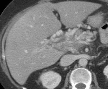
12 HCC Supplied by hepatic artery Enhance during the arterial phase before normal liver parenchyma Normal liver parenchyma Supplied by portal veins Maximally enhances during the portal venous phase

13 In contrast, hemangiomas exhibit slow, progressive enhancement.

14 Uses magnetic properties of protons Advantages: Spatial resolution High soft tissue contrast resolution Very sensitive No radiation or iodinated contrast Disadvantages Costly Not as readily available Longer exam times than CT Acquisition time Patient cooperation with exam Claustrophobia Longer breath-holds Gadolinium contrast
15 Indications Patients allergic to iodinated contrast Lesion detection & characterization Vascular patency Biliary tree assessment Similar indications as with CT Reliance on multiphase post contrast imaging and T1 and T2-weighted sequences for lesion detection and characterization


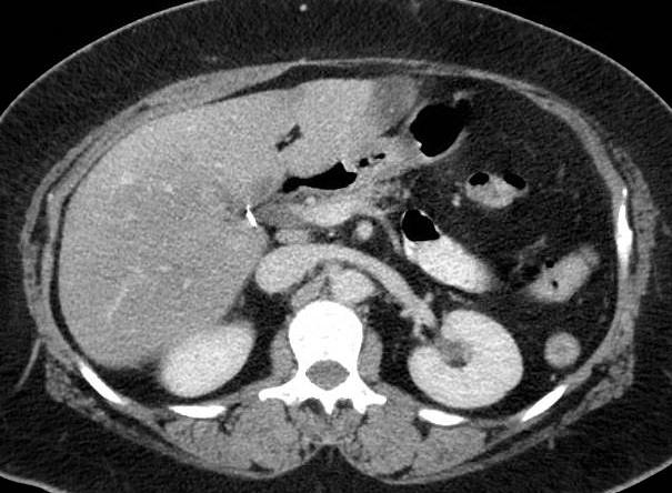
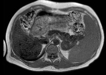
16 CT/MR Radiology Speak Appearance of cirrhosis Nodular hepatic contour Shrunken liver Left lateral segment/caudate lobe hypertrophy Expanded gallbladder fossa


17 Evidence of portal hypertension MPV diameter >12 mm Spleen size >12 cm Ascites Varices Recanalized paraumbilical vein Gallbladder wall edema often seen and not to be suggestive of cholecystitis Hypoalbuminemia Reactive to hepatitis


18 Perfusional abnormality/shunt Cavernous transformation of the portal vein Replaced hepatic arteries


19 Confluent fibrosis Bridging fibrosis
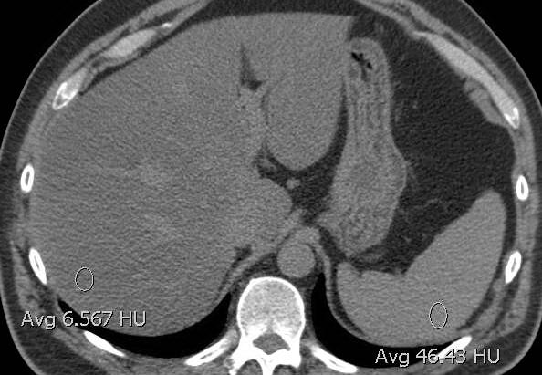
20 Geographic hepatic steatosis

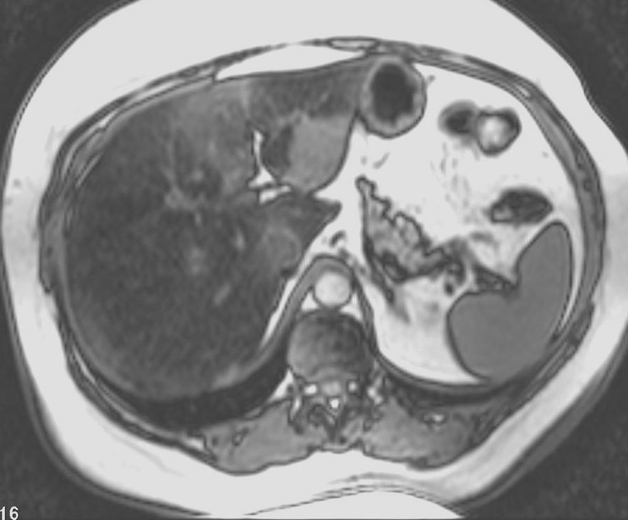
21 MRI and Fat Evaluation

22 MRI and Iron Evaluation
23 Liver Imaging Reporting and Data System (LI-RADS) Lesions versus observations Observations assigned category codes based on size and suspicion of HCC LR-NC: Noncategorized LR-TIV: Tumor in Vein, irregardless of type of tumor LR1: definitely benign LR2: probably benign LR3: intermediate probability of HCC LR4: probably HCC LR5: definitely HCC near 100% PPV LR-M: Malignancy, not specifically HCC
24 Thank You!
Hepatic Imaging: What Every Practitioner Should Know
 Hepatic Imaging: What Every Practitioner Should Know Shuchi K. Rodgers, MD Section Chief, Abdominal Imaging Director of Ultrasound Department of Radiology Einstein Medical Center rodgerss@einstein.edu
Radiology of hepatobiliary diseases
 GI cycle - Lecture 14 436 Teams Radiology of hepatobiliary diseases Objectives 1. To Interpret plan x-ray radiograph of abdomen with common pathologies. 2. To know the common pathologies presentation.
US LI-RADS v2017 CORE
 US LI-RADS v2017 CORE Screening or surveillance US in patient at high risk for HCC US category US-1 US-2 US-3 Negative Subthreshold Positive Category Concept Definition US-1 Negative US-2 Subthreshold
HEPATO-BILIARY IMAGING
 HEPATO-BILIARY IMAGING BY MAMDOUH MAHFOUZ MD PROF.OF RADIOLOGY CAIRO UNIVERSITY mamdouh.m5@gmail.com www.ssregypt.com CT ABDOMEN Indications Patient preparation Patient position Scanogram Fasting 4-6 hours
LIVER IMAGING TIPS IN VARIOUS MODALITIES. M.Vlychou, MD, PhD Assoc. Professor of Radiology University of Thessaly
 LIVER IMAGING TIPS IN VARIOUS MODALITIES M.Vlychou, MD, PhD Assoc. Professor of Radiology University of Thessaly Hepatocellular carcinoma is a common malignancy for which prevention, screening, diagnosis,
The Focal Hepatic Lesion: Radiologic Assessment
 The Focal Hepatic Lesion: Radiologic Assessment Kevin Kuo, Harvard Medical School Year III Our Patient: PS 67 y/o female w/ long history of alcohol use Drinking since age 18, up to one bottle of wine/day
Computed tomography. Department of Radiology, University Medical School, Szeged
 Computed tomography Department of Radiology, University Medical School, Szeged voxel +1-4 +2 +5 +3 +1 0-2 pixel -2 0 +1-4 -6 +5 +2 +1 Department of Radiology, University Medical School, Szeged
Newcastle HPB MDM updated radiology imaging protocol recommendations. Author Dr John Scott. Consultant Radiologist Freeman Hospital
 Newcastle HPB MDM updated radiology imaging protocol recommendations Author Dr John Scott. Consultant Radiologist Freeman Hospital This document is intended as a guide to aid radiologists and clinicians
Imaging of liver and pancreas
 Imaging of liver and pancreas.. Disease of the liver Focal liver disease Diffusion liver disease Focal liver disease Benign Cyst Abscess Hemangioma FNH Hepatic adenoma HCC Malignant Fibrolamellar carcinoma
State of the Art Imaging for Hepatic Malignancy: My Assignment
 State of the Art Imaging for Hepatic Malignancy: My Assignment CT vs MR vs MRCP Which one to choose for HCC vs Cholangiocarcinoma What special protocols to use for liver tumors Role of PET and Duplex US
Liver Ultrasound - Beyond the Basics. Pamela Parker Lead Sonographer
 Liver Ultrasound - Beyond the Basics Pamela Parker Lead Sonographer Aims Review what we know about the liver Reasons for imaging Focal lesions Diffuse disease Can we do more? The Liver The Liver The Liver
Modern liver imaging techniques - A new era in liver ultrasound
 Modern liver imaging techniques - A new era in liver ultrasound Yuko Kono, M.D., Ph.D. Clinical Professor Departments of Medicine and Radiology University of California, San Diego San Diego, USA How to
Portal Venous Thrombosis: Tumor VS Bland Thrombus
 June 2015 Portal Venous Thrombosis: Tumor VS Bland Thrombus SERGIO ALFARO, HARVARD MEDICAL SCHOOL YEAR III GILLIAN LIEBERMAN, MD Overview 2 Index Patient History Portal Venous Thrombosis (PVT) Imaging
IMAGING OF LIVER, BILIARY TREE, PANCREAS
 IMAGING OF LIVER, BILIARY TREE, PANCREAS Department of Radiology West China Hospital, Sichuan University Yao Jin Learning Points The methodology for imaging the LBP (liver, biliary tree, and pancreas )
Innovations in HCC Imaging: MDCT/MRI
 Innovations in HCC Imaging: MDCT/MRI Anthony E. Cheng, M.D. Cardinal MRI Center Cardinal Santos Medical Center, Wilson Street, San Juan Innovations in HCC Imaging: Goals/Objectives MDCT/MRI Learn the diagnostic
MRI of the Hepatobiliary System
 BODY APPLICATIONS OF MRI 51 Chapter Four C H A P T E R F O U R MRI of the Hepatobiliary System After completing this chapter, the reader will be able to: Develop a protocol for liver MRI Identify benign
Evaluation of Liver Mass Lesions. American College of Gastroenterology 2013 Regional Postgraduate Course
 Evaluation of Liver Mass Lesions American College of Gastroenterology 2013 Regional Postgraduate Course Lewis R. Roberts, MB ChB, PhD Division of Gastroenterology and Hepatology Mayo Clinic College of
Elastography in the. technically difficult patient. EPIQ ultrasound system. Ultrasound
 Ultrasound Elastography in the technically difficult patient EPIQ ultrasound system Chairman Department of Diagnostic Radiology Allegheny General Hospital Pittsburgh, PA, USA You can offer more information
Diseases of liver. Dr. Mohamed. A. Mahdi 4/2/2019. Mob:
 Diseases of liver Dr. Mohamed. A. Mahdi Mob: 0123002800 4/2/2019 Cirrhosis Cirrhosis is a complication of many liver disease. Permanent scarring of the liver. A late-stage liver disease. The inflammation
Acknowledgements. Update of Focal Liver Lesions Goals. Focal Liver Lesions. Imaging Choices For Liver Lesions. Focal Liver Lesions
 Acknowledgements Update of Focal Liver Lesions 2012 Giles Boland Massachusetts General Hospital Harvard Medical School No disclosures Dushyant Sahani Mukesh Harisinghani Goals Focal liver lesions Imaging
Liver Cancer And Tumours
 Liver Cancer And Tumours What causes liver cancer? Many factors may play a role in the development of cancer. Because the liver filters blood from all parts of the body, cancer cells from elsewhere can
Malignant Focal Liver Lesions
 Malignant Focal Liver Lesions Other Than HCC Pablo R. Ros, MD, MPH, PhD Departments of Radiology and Pathology University Hospitals Cleveland Medical Center Case Western Reserve University Pablo.Ros@UHhospitals.org
MRI OF FOCAL LESIONS IN
 Introduction MRI OF FOCAL LESIONS IN THE NON-CIRRHOTIC LIVER Ivan Pedrosa M.D. Ph.D. Associate Professor of Radiology and Advanced Imaging Research Center University of Texas Southwestern. Dallas, TX Incidental
Abdomen Sonography Examination Content Outline
 Abdomen Sonography Examination Content Outline (Outline Summary) # Domain Subdomain Percentage 1 2 3 Anatomy, Perfusion, and Function Pathology, Vascular Abnormalities, Trauma, and Postoperative Anatomy
Objectives. LI-RADS v2017. Working Groups. Cynthia Santillan. Released October 2014 Diagnosis. Screening/ Surveillance. Diagnosis
 LR-NC Cynthia Santillan LR-TIV Objectives 1. To teach participants how to apply the Liver Imaging Reporting and Data System () to their interpretation of imaging studies for the evaluation of hepatocellular
Liver imaging takes a step forward with Ingenia
 Publication for the Philips MRI Community ISSUE 49 2013 / 2 Liver imaging takes a step forward with Ingenia Lyon South Hospital strives to move from several studies first CT, then MR or PET to using just
Financial Disclosure
 Benign Liver Masses Adil Abdalla, MBBS Creighton University-CHI Health August 25, 2018 Financial Disclosure Nothing to disclose Financial Disclosure 1 Objectives To assess patients with benign liver tumors
Ascites Management. Atif Zaman, MD MPH Oregon Health & Science University Professor of Medicine Division of Gastroenterology and Hepatology
 Ascites Management Atif Zaman, MD MPH Oregon Health & Science University Professor of Medicine Division of Gastroenterology and Hepatology Disclosure 1. The speaker Atif Zaman, MD MPH have no relevant
Appendix 5. EFSUMB Newsletter. Gastroenterological Ultrasound
 EFSUMB Newsletter 87 Examinations should encompass the full range of pathological conditions listed below A log book listing the types of examinations undertaken should be kept Training should usually
MRI Abdomen Protocol Pancreas/MRCP with Contrast
 MRI Abdomen Protocol Pancreas/MRCP with Contrast Reviewed By: Brett Mollard, MD; Anna Ellermeier, MD Last Reviewed: July 2018 Contact: (866) 761-4200 Standard uses: 1. Characterization of cystic and solid
Liver Cancer (Hepatocellular Carcinoma or HCC) Overview
 Overview") Liver Cancer (Hepatocellular Carcinoma or HCC) Overview Recent advances in liver cancer care seek to address the rising incidence of liver cancer, which has steadily increased over the past three decades.
Liver Cancer (Hepatocellular Carcinoma or HCC) Overview Recent advances in liver cancer care seek to address the rising incidence of liver cancer, which has steadily increased over the past three decades.
Objectives. HCC Incidence and Mortality. Disclosure Statement HCC. Imaging of Hepatocellular Carcinoma. Treatment of Hepatocellular Carcinoma
 Imaging of Hepatocellular Carcinoma and the use of LI RADS Treatment of Hepatocellular Carcinoma Aaron D. Anderson, D.O. AOCR April 2015 Objectives Show how the use of LI RADS can simplify the diagnosis
Case Study: #3: Gallbladder Carcinoma?
 Case Study: #3: Gallbladder Carcinoma? By: Megan Wyatt K. SON Wyatt 225 2B1 RDMS, RVT Patient: Male 85 YOA Caucasian Indication: Elevated Alkaline Phosphatase History Annual physical showed elevated alkaline
Interesting Cases from Liver Tumor Board. Jeffrey C. Weinreb, M.D.,FACR Yale University School of Medicine
 Interesting Cases from Liver Tumor Board Jeffrey C. Weinreb, M.D.,FACR Yale University School of Medicine jeffrey.weinreb@yale.edu Common Liver Diseases Hemangioma Cyst FNH Focal Fat/Sparing THID Non-Cirrhotic
Anatomical and Functional MRI of the Pancreas
 Anatomical and Functional MRI of the Pancreas MA Bali, MD, T Metens, PhD Erasme Hospital Free University of Brussels Belgium mbali@ulb.ac.be Introduction The use of MRI to investigate the pancreas has
Imaging of common diseases of hepatobiliary and GI system
 Imaging of common diseases of hepatobiliary and GI system Natthaporn Tanpowpong, M.D. Diagnostic radiology Faculty of Medicine, Chulalongkorn University Normal plain radiograph A = Common bile duct
Vascular Imaging in the Pediatric Abdomen. Jonathan Swanson, MD
 Vascular Imaging in the Pediatric Abdomen Jonathan Swanson, MD Goals and Objectives To understand the imaging approach, appearance, and clinical manifestations of the common pediatric abdominal vascular
Complete Summary GUIDELINE TITLE. Liver lesion characterization. BIBLIOGRAPHIC SOURCE(S)
") Complete Summary GUIDELINE TITLE Liver lesion characterization. BIBLIOGRAPHIC SOURCE(S) Foley WD, Bree RL, Gay SB, Glick SN, Heiken JP, Huprich JE, Levine MS, Ros PR, Rosen MP, Shuman WP, Greene FL, Rockey
Complete Summary GUIDELINE TITLE Liver lesion characterization. BIBLIOGRAPHIC SOURCE(S) Foley WD, Bree RL, Gay SB, Glick SN, Heiken JP, Huprich JE, Levine MS, Ros PR, Rosen MP, Shuman WP, Greene FL, Rockey
CT & MRI of Benign Liver Neoplasms Srinivasa R Prasad
 CT & MRI of Benign Liver Neoplasms Srinivasa R Prasad No financial disclosures Acknowledgements Many thanks to Drs. Heiken, Narra & Menias (MIR) Dr. Sahani (MGH) for sharing images Benign Liver Tumors:
Liver Tumors. Prof. Dr. Ahmed El - Samongy
 Liver Tumors Prof. Dr. Ahmed El - Samongy Objective 1. Identify the most important features of common benign liver tumors 2. Know the risk factors, diagnosis, and management of hepatocellular carcinoma
Essentials of Clinical MR, 2 nd edition. 65. Benign Hepatic Masses
 65. Benign Hepatic Masses Pulse sequences acquired for abdominal MRI typically consist of fast acquisition schemes such as single-shot turbo spin echo (i.e. HASTE) and gradient echo schemes such as FLASH
Abdominal Ultrasound. Diane Hallinen, MD. Bloodroot
 Abdominal Ultrasound Diane Hallinen, MD Bloodroot Abdominal Ultrasound Vasculature Hepatobiliary Spleen Kidney Bladder Bowel Where to put the probe? Vasculature We are going to talk about Celiac Trunk
Simplifying liver assessment in internal medicine
 Ultrasound Customer story Simplifying liver assessment in internal medicine Philips Affiniti ultrasound for elastography and contrast-enhanced ultrasound (CEUS) Where Sonography Institute, Uster, Switzerland
GASTROINTESTINAL IMAGING STUDY GUIDE
 GASTROINTESTINAL IMAGING STUDY GUIDE Pharynx Diverticula Foreign bodies Trauma o Motility Disorders Esophagus Diverticula Trauma Esophagitis Barrett esophagus Rings, webs, and strictures Varices Benign
Liver MRI in 30 minutes
 X Liver MRI in 30 minutes SCBT/MR Annual Meeting Salt Lake City September 18, 2016 Scott B. Reeder, MD, PhD Department of Radiology University of Wisconsin Madison, WI Disclosures University of Wisconsin-Madison
Radiologic Imaging Magnetic Resonance Imaging (MRI)
") Radiologic Imaging X-ray has always been the golden rule in diagnosing and treating podiatric patients. Unfortunately, for some patients the diagnosis is not as evident. That is when we need to utilize
Radiologic Imaging X-ray has always been the golden rule in diagnosing and treating podiatric patients. Unfortunately, for some patients the diagnosis is not as evident. That is when we need to utilize
Hepatobiliary Ultrasound Rimon Bengiamin, MD, RDMS Assistant Clinical Professor Director of Emergency Ultrasound UCSF Fresno. Objectives. Why?
 Hepatobiliary Ultrasound Rimon Bengiamin, MD, RDMS Assistant Clinical Professor Director of Emergency Ultrasound UCSF Fresno Objectives Discuss the goals of point-of-care biliary ultrasound Review the
Diffuse Liver Disease:
 Diffuse Liver Disease: MRI Evaluation Fat Iron Cirrhosis Donald G. Mitchell, M.D. Thomas Jefferson University Philadelphia, PA Fatty Liver Disease Common, increasing» Diabetes» Diet, high fructose corn
Pediatric Hepatobiliary, Pancreatic & Splenic US
 Pediatric Hepatobiliary, Pancreatic & Splenic US Susan J. Back, MD Department of Radiology, The Children s Hospital of Philadelphia No Disclosures Objectives Normal Abnormal: cases and US advances Objectives
Hepato-Pancreatico-Biliary Surgery. Dr. Ankur J. Shah. MS, DNB, MNAMS, MRCSEd (UK), FRCS (UK)
, FRCS (UK)") Hepato-Pancreatico-Biliary Surgery Dr. Ankur J. Shah MS, DNB, MNAMS, MRCSEd (UK), FRCS (UK) Consultant Hepato-Pancreatico-Biliary and Liver Transplant Surgeon Ansh Liver Clinic Prevention to Cure Address
Hepato-Pancreatico-Biliary Surgery Dr. Ankur J. Shah MS, DNB, MNAMS, MRCSEd (UK), FRCS (UK) Consultant Hepato-Pancreatico-Biliary and Liver Transplant Surgeon Ansh Liver Clinic Prevention to Cure Address
Interventional Radiology in Liver Cancer. Nakarin Inmutto MD
 Interventional Radiology in Liver Cancer Nakarin Inmutto MD Liver cancer Primary liver cancer Hepatocellular carcinoma Cholangiocarcinoma Metastasis Interventional Radiologist Diagnosis Imaging US / CT
Abdomen and Retroperitoneum Ultrasound Protocols
 Abdomen and Retroperitoneum Ultrasound Protocols Reviewed By: Anna Ellermeier, MD Last Reviewed: March 2018 Contact: (866) 761-4200, Option 1 **NOTE for all examinations: 1. If documenting possible flow
RICCARDO LENCIONI,CLOTILDE DELLA PINA, LAURA CROCETTI,DANIA CIONI. Chapter 1
 RICCARDO LENCIONI,CLOTILDE DELLA PINA, LAURA CROCETTI,DANIA CIONI Chapter 1 Impact of European Federation of Societies for Ultrasound in Medicine and Biology (EFSUMB) Guidelines on the Use of Contrast
Biliary Ultrasonography Kathleen O Brien MD MPH RDMS Kaiser Permanente South Sacramento
 Biliary Ultrasonography Kathleen O Brien MD MPH RDMS Kaiser Permanente South Sacramento https://www.google.com/search?sa=g&hl=en&q=public+disclosure&tbm=isch&tbs=simg:caqsigeahwelekju2aqaaawlelcmpwgaygpgcamskpib_1qnza7ai
Hepatocellular Carcinoma: Diagnosis and Management
 Hepatocellular Carcinoma: Diagnosis and Management Nizar A. Mukhtar, MD Co-director, SMC Liver Tumor Board April 30, 2016 1 Objectives Review screening/surveillance guidelines Discuss diagnostic algorithm
iu22 Liver Shear Wave ElastPQ
 iu22 Liver Shear Wave ElastPQ Clinical Case Study Lucy Wang Clinical Application Specialist ASEAN Case Study: History: 58-year-old male patient, hepatitis B virus (HBV) carrier, with non clinical symptoms
Evaluation of Diffuse Liver Diseases Using Conventional Ultrasound
 IOSR Journal of Dental and Medical Sciences (IOSR-JDMS) e-issn: 2279-0853, p-issn: 2279-0861.Volume 16, Issue 6 Ver. VII (June. 2017), PP 70-74 www.iosrjournals.org Evaluation of Diffuse Liver Diseases
Jaundice. Agnieszka Dobrowolska- Zachwieja, MD, PhD
 Jaundice Agnieszka Dobrowolska- Zachwieja, MD, PhD Jaundice definition Jaundice, as in the French jaune, refers to the yellow discoloration of the skin. It arises from the abnormal accumulation of bilirubin
Normal Sonographic Anatomy
 hapter 2:The Liver DUNSTAN ABRAHAM Normal Sonographic Anatomy Homogeneous, echogenic texture (Figure 2-1) Measures approximately 15 cm in length and 10 12.5 cm anterior to posterior; measurement taken
Non-Invasive Testing for Liver Fibrosis
 NORTHWEST AIDS EDUCATION AND TRAINING CENTER Non-Invasive Testing for Liver Fibrosis John Scott, MD, MSc Associate Professor, University of Washington Associate Clinic Director, Hep/Liver Clinic, Harborview
IT 의료융합 1 차임상세미나 복부질환초음파 이재영
 IT 의료융합 1 차임상세미나 2013-4-3 복부질환초음파 이재영 나는오늘누구를위하여 종을울리나? 전통적의료 의사 공학설계자 의사 최첨단진단장비들 USG, CT, MRI 환자 환자 현대의료 사용자중심의사고 US in the Abdomen Detection DDx Look Behavior Response by external stimuli Guiding Tool
FOR CMS (MEDICARE) MEMBERS ONLY NATIONAL COVERAGE DETERMINATION (NCD) FOR MAGNETIC RESONANCE IMAGING:
 MEMBERS ONLY NATIONAL COVERAGE DETERMINATION (NCD) FOR MAGNETIC RESONANCE IMAGING:") National Imaging Associates, Inc. Clinical guidelines BONE MARROW MRI Original Date: July 2008 Page 1 of 5 CPT Codes: 77084 Last Review Date: September 2014 NCD 220.2 MRI Last Effective Date: July 2011
National Imaging Associates, Inc. Clinical guidelines BONE MARROW MRI Original Date: July 2008 Page 1 of 5 CPT Codes: 77084 Last Review Date: September 2014 NCD 220.2 MRI Last Effective Date: July 2011
Contrast Enhanced Ultrasound of Parenchymal Masses in Children
 Contrast Enhanced Ultrasound of Parenchymal Masses in Children Sue C Kaste, DO On behalf of Beth McCarville, MD St. Jude Children s Research Hospital Memphis, TN Overview Share St. Jude experience with
Cholangiocarcinoma (Bile Duct Cancer)
") Cholangiocarcinoma (Bile Duct Cancer) The Bile Duct System (Biliary Tract) A network of bile ducts (tubes) connects the liver and the gallbladder to the small intestine. This network begins in the liver
Cholangiocarcinoma (Bile Duct Cancer) The Bile Duct System (Biliary Tract) A network of bile ducts (tubes) connects the liver and the gallbladder to the small intestine. This network begins in the liver
Imaging iconography of gallbladder cancer. Assessment by CT.
 1 REVISTA DE IMAGENOLOGIA- EII / Vol. XVI / Num. 2 Imaging iconography of gallbladder cancer. Assessment by CT. Doctors Crisci, Alejandro (1); Landó, Fernando.(2). CASMU CT Department Hospital of Tacuarembó
Initial Evaluation for HCV Therapy. Hope McGratty PA-C, MPH
 Initial Evaluation for HCV Therapy Hope McGratty PA-C, MPH Conflict of Interest Disclosure Statement None Who are we talking about today? Treatment naïve Chronic infection This patient seems complicated
Body MRI from the Liver to the Bladder
 Body MRI from the Liver to the Bladder I Want You! Audience Participation Methodist Hospital Continuing Education Seminar Jordan Swensson, MD November 7, 2015 Objectives Observe the uses of MRI for organs
CTA/MRA of Pediatric Hepatic Masses Radiology-Pathology Correlation
 Acta Radiológica Portuguesa, Vol.XVIII, nº70, pág. 41-50, Abr.-Jun., 2006 CTA/MRA of Pediatric Hepatic Masses Radiology-Pathology Correlation Marilyn J. Siegel Mallinckrodt Institute of Radiology, Washington
Liver, Pancreas and Biliary System. Wirana Angthong, M.D.
 Liver, Pancreas and Biliary System Wirana Angthong, M.D. Objectives Outline Anatomy Imaging Techniques Common Diseases Outline Anatomy Imaging Techniques Common Diseases Liver anatomy Morphological anatomy:
My Patient Has Abdominal Pain PoCUS of the Biliary Tract and the Urinary Tract
 My Patient Has Abdominal Pain PoCUS of the Biliary Tract and the Urinary Tract Objectives PoCUS for Biliary Disease PoCUS for Renal Colic PoCUS for Urinary Retention Biliary Disease A patient presents
Hepatobiliary Malignancies Retrospective Study at Truman Medical Center
 Hepatobiliary Malignancies 206-207 Retrospective Study at Truman Medical Center Brandon Weckbaugh MD, Prarthana Patel & Sheshadri Madhusudhana MD Introduction: Hepatobiliary malignancies are cancers which
Gemstone Spectral Imaging quantifies lesion characteristics for a confident diagnosis
 GE Healthcare Gemstone Spectral Imaging quantifies lesion characteristics for a confident diagnosis CT clinical case study lesion characterization Desiree Morgan, MD Vice Chair of Clinical Research Professor
Abdominal Imaging - 9 Topics in 90 min
 Abdominal Imaging 9 topics in 90 min Antonio C. Westphalen, MD PhD Departments of Radiology and Biomedical Imaging, and Urology Liver Biliary tree Gallbladder Pancreas Kidneys Small bowel Colon Abscess?
Liver imaging the revolution
 Liver imaging the revolution Valérie Vilgrain Hôpital Beaujon, Paris, France PHC 2018 - www.aphc.info At the Beginning of the story Radiology in the 1970s US Garrett Radiology 1976 abscess Taylor Radiology
Recently role of non-invasive diagnostics methods
 CERTAIN ASPECTS OF NLS-DIAGNOSTICS OF LIVER FOCAL PATHOLOGY A.Y. Shvack, V.I. Nesterov, N.L. Ogluzdina This article contains information about NLS-graphy application in diagnostics of liver focal affections:
Liver Perfusion Analysis New Frontiers in Dynamic Volume Imaging. Case Study Brochure Chang Gung Memorial Hospital.
 New Frontiers in Dynamic Volume Imaging dynamic volume CT Case Study Brochure Chang Gung Memorial Hospital http://www.toshibamedicalsystems.com Toshiba Medical Systems Corporation 2010-2011. All rights
ACG Clinical Guideline: Diagnosis and Management of Focal Liver Lesions
 ACG Clinical Guideline: Diagnosis and Management of Focal Liver Lesions Jorge A. Marrero, MD, 1 Joseph Ahn, MD, FACG, 2 K. Rajender Reddy, MD, FACG 3 1 University of Texas at Southwestern, Dallas, Texas,
Chief Complain. Liver lesion found in routine health check 41 days ago
 Chief Complain Liver lesion found in routine health check 41 days ago Present Illness On 2005-7-26 at 台北署立醫院 he underwent a health check for the first time. Abdominal US showed suspicious of a 6*5 cm hepatoma,
Liver Specific MRI using Gd-EOB-DTPA Disodium (Primovist) Effects Change in Management of Indeterminate Liver Lesions.
 Effects Change in Management of Indeterminate Liver Lesions.") Liver Specific MRI using Gd-EOB-DTPA Disodium (Primovist) Effects Change in Management of Indeterminate Liver Lesions. Poster No.: C-1751 Congress: ECR 2012 Type: Authors: Keywords: DOI: Educational Exhibit
Liver Specific MRI using Gd-EOB-DTPA Disodium (Primovist) Effects Change in Management of Indeterminate Liver Lesions. Poster No.: C-1751 Congress: ECR 2012 Type: Authors: Keywords: DOI: Educational Exhibit
Soft Tissue Tumour & Sarcoma Imaging Guidelines 2012
 Soft Tissue Tumour & Sarcoma Imaging Guidelines 2012 Version Control This is a controlled document please destroy all previous versions on receipt of a new version. Date Approved: March 2011 reissued April
How to integrate surgery in the treatment of patients with liver-only metastatic disease
 How to integrate surgery in the treatment of patients with liver-only metastatic disease Luis Sabater Ortí MD, PhD Associate Professor University of Valencia European Board Surgical Qualification HBP (EBSQ-HPB)
Cirrhosis and Portal Hypertension Gastroenterology Teaching Project American Gastroenterological Association
 CIRRHOSIS AND PORTAL HYPERTENSION Cirrhosis and Portal Hypertension Gastroenterology Teaching Project American Gastroenterological Association WHAT IS CIRRHOSIS? What is Cirrhosis? DEFINITION OF CIRRHOSIS
Appropriate Imaging Tests Lead to Meaningful Results. Dr. Richard Wasley May 2011
 Appropriate Imaging Tests Lead to Meaningful Results Dr. Richard Wasley May 2011 Summarize the advantages and limitations of specific imaging tests and why clinical information is so important to radiologists
Ankur A. Gupta 1, Danny C. Kim, Glenn A. Krinsky, Vivian S. Lee
 Pictorial Essay T and MRI of irrhosis and its Mimics nkur. Gupta 1, Danny. Kim, Glenn. Krinsky, Vivian S. Lee irrhosis is among the leading causes of death in the western world. irrhosis and its associated
LI-RADS Reporting. Chapter 14. Primary Authors. Contributing Authors. Memorial Sloan Kettering Cancer Center
 Chapter 14 LI-RADS Primary Authors Victoria Chernyak Mustafa R. Bashir Montefiore Medical Center Duke University Medical Center Contributing Authors Richard K. Do Aya Kamaya Ania Z. Kielar Donald G. Mitchell
Guidelines, Policies and Statements D5 Statement on Abdominal Scanning
 Guidelines, Policies and Statements D5 Statement on Abdominal Scanning Disclaimer and Copyright The ASUM Standards of Practice Board have made every effort to ensure that this Guideline/Policy/Statement
Compute-aided Differentiation of Focal Liver Disease in MR Imaging
 1063 Compute-aided Differentiation of Focal Liver Disease in MR Imaging Xuejun Zhang a, Masayuki Kanematsu b, Hiroshi Fujita c, Takeshi Hara c, Hiroshi Kondo b, Xiangrong Zhou c, Wenguang Li a and Hiroaki
HCC and mass effect. Hepatocellular cancer: what if the AFP is rising but no lesion seen on imaging? What you need to know about AFP.
 Hepatocellular cancer: what if the AFP is rising but no lesion seen on imaging? Arun J Sanyal M.B.B.S., M.D. Charles Caravati Professor of Medicine Virginia Commonwealth University Imaging features used
RADIOLOGY TEACHING CONFERENCE
 RADIOLOGY TEACHING CONFERENCE John Athas, MD Monica Tadros, MD Columbia University, College of Physicians & Surgeons Department of Otolaryngology- Head & Neck Surgery September 27, 2007 CT SCAN IMAGING
Approach to the Patient with Liver Disease
 Approach to the Patient with Liver Disease Diagnosis of liver disease Careful history taking Physical examination Laboratory tests Radiologic examination and imaging studies Liver biopsy Liver diseases
Transarterial Chemoembolisation (TACE) with Drug-Eluting Beads
 with Drug-Eluting Beads") Transarterial Chemoembolisation (TACE) with Drug-Eluting Beads A minimally invasive treatment for liver cancer Provided as an educational service by Biocompatibles UK Ltd, a BTG International group company
Transarterial Chemoembolisation (TACE) with Drug-Eluting Beads A minimally invasive treatment for liver cancer Provided as an educational service by Biocompatibles UK Ltd, a BTG International group company
Jesse Civan, M.D. Medical Director, Jefferson Liver Tumor Center
 Liver Tumors Jesse Civan, M.D. Medical Director, Jefferson Liver Tumor Center Differential Diagnosis Malignant Metastatic from non-hepatic primary Hepatocellular carcinoma Cholangiocarcinoma Biliary cystcarcinoma
Workup of a Solid Liver Lesion
 Workup of a Solid Liver Lesion Joseph B. Cofer MD FACS Chief Quality Officer Erlanger Health System Affiliate Professor of Surgery UTHSC-Chattanooga I have no financial or other relationships with any
Radiological Reasoning: Incidentally Discovered Liver Mass
 AJR Integrative Imaging LIFELONG LEARNING FOR RADIOLOGY This Radiological Reasoning article is available for SAM credit and CME credits when completed with the additional educational material provided
Dr Claire Smith, Consultant Radiologist St James University Hospital Leeds
 Dr Claire Smith, Consultant Radiologist St James University Hospital Leeds Imaging in jaundice and 2ww pathway Image protocol Staging Limitations Pancreatic cancer 1.2.4 Refer people using a suspected
Surveillance for Hepatocellular Carcinoma
 Surveillance for Hepatocellular Carcinoma Marion G. Peters, MD John V. Carbone, MD, Endowed Chair Professor of Medicine Chief of Hepatology Research University of California San Francisco Recorded on April
LIVER CIRRHOSIS. The liver extracts nutrients from the blood and processes them for later use.
 LIVER CIRRHOSIS William Sanchez, M.D. & Jayant A. Talwalkar, M.D., M.P.H. Advanced Liver Disease Study Group Miles and Shirley Fiterman Center for Digestive Diseases Mayo College of Medicine Rochester,
Gastrointestinal tract
 Gastrointestinal tract Colloidal liver-spleen imaging Presented by: Jehad Felemban Introduction: To obtain better anatomic display of liver and spleen architecture, we use (CT Ultrasound). (Radionuclide
US in non-traumatic acute abdomen. Lalita, M.D. Radiologist Department of radiology Faculty of Medicine ChiangMai university
 US in non-traumatic acute abdomen Lalita, M.D. Radiologist Department of radiology Faculty of Medicine ChiangMai university Sagittal Orientation Transverse (Axial) Orientation Coronal Orientation Intercostal