HEPATOCYTE SPECIFIC CONTRAST MEDIA: WHERE DO WE STAND?
|
|
|
- Trevor Morris
- 5 years ago
- Views:
Transcription
1 HEPATOCYTE SPECIFIC CONTRAST MEDIA: WHERE DO WE STAND? Andrew T. Trout,
2 Disclosures No relevant disclosures
3 Outline Review of hepatocyte specific contrast media Review of hepatocellular carcinoma Review of focal nodular hyperplasia (FNH) Review of hepatocellular adenoma subtypes The trouble begins
4 Hepatocyte Specific Contrast Media Gadoxetate disodium [Eovist, Primovist] Initial extracellular phase followed by 50/50 biliary/renal excretion Gadobenate dimeglumine [MultiHance]
5 Blood Bile Hepatocyte Specific Contrast Media Uptake Organic anion transporting polypeptides OATP8 (OATP1B3) OATP-C (OATP1B1) Excretion Multidrug resistance-associated proteins MRP2 - canalicular MRP3 - sinusoidal OATP MRP3 MRP2
6 Blood Bile Hepatocyte Specific Contrast Media HBP uptake depends one or more factors: OATP expression MRP expression Presence of functioning bile ducts OATP MRP3 MRP2 Tsuboyama T, et al. Radiology Jun;255(3):824-33
7 Blood Bile Hepatocyte Specific Contrast Media HBP hyperintensity due to one or more factors: OATP expression MRP2 expression Mal-located MRP2 To pseudoglands (some HCC) Abnormal bile ducts OATP MRP3 MRP2 Tsuboyama T, et al. Radiology Jun;255(3): Yoneda N, et al. Jpn J Radiol Jul;30(6):
8 Hepatocyte Specific Contrast Media In general, bad things are dark on the hepatobiliary phase Non-functioning hepatocytes Decreased OATP8 expression w/ progression of hepatocarcinogenesis May precede neoangiogenesis In general, benign things are bright on the hepatobiliary phase Lots of exceptions
 Overlap between HBP and equilibrium phases may get pseudo-washout due to early")
9 Hepatocyte Specific Contrast Media Good article! Washout caution (gadoxetate disodium) Overlap between HBP and equilibrium phases may get pseudo-washout due to early background hepatocyte uptake Call washout only if unequivocal in portal venous phase
10 Hepatocellular Carcinoma Hepatocarcinogenesis (in the cirrhotic liver) generally a relatively scripted process Progressive neoangiogenesis Increasing numbers of unpaired arteries Gradual diversion of venous drainage from hepatic to portal veins Progressive loss of OATP transporters during process of hepatocarcinogenesis Process accounts for imaging appearance with increasing arterial hyperenhancement, PV washout and hypoenhancement in HBP Park HJ, et al. Liver Cancer Jun;6(3):


11 Focal Nodular Hyperplasia (FNH) Hyperplastic, hypercellular nodule Hepatocytes w/o atypia Abnormal duct proliferation Lack of mature bile ducts Central scar with large artery Normal background liver Believed to be hyperplastic response to vascular abnormality T2w Pre Art PV International Working Party. Hepatology Sep;22(3): HBP
12 FNH vs. FNH-like lesions FNH-like lesions Arising in abnormal liver FNH occur only in normal liver Can look like low-grade HCC (and adenoma) May just be inflammatory adenomas Choi JY, et al. J Gastroenterol Hepatol Jun;26(6): Quaglia A, et al. Histopathology Jan;42(1):14-21 Yoneda N, et al. Radiographics Nov-Dec;36(7):
13 Hepatocellular Adenomas Inflammatory Hepatocyte nuclear factor-1α (HNF-1α)- inactivated β-catenin activated Unclassified
14 Inflammatory Adenomas Used to be classified as a subtype (telangiectatic) of FNH Up to 50% of adenomas Risk factors: obesity, metabolic syndrome, EtOH Higher rate of malignant degeneration (esp. w/ β- catenin mutation) Higher rate of hemorrhage McInnes MD, et al. Radiology Nov;277(2): Thomeer MG, et al. J Magn Reson Imaging May;39(5):


15 HNF-1α-inactivated Adenomas Steatotic adenomas Majority show loss of signal on opposed-phase 35-50% of adenomas Most common in women on OCPs Commonly multiple IP T2w Art OP Pre PV Ba-Ssalamah A, et al. Radiology Oct;277(1): Yoneda N, et al. Radiographics Nov-Dec;36(7): HBP
16 β-catenin activated Adenomas More frequent in men than other adenomas Assoc w/ glycogen storage disease, familial adenomatous polyposis Greatest risk of malignant transformation Yoneda N, et al. Radiographics Nov-Dec;36(7):
17 Unclassified Adenoma Everything else. No clear mutational etiology No consistent imaging features
18 Exceptions to the bad things are dark, benign things are bright on HBP rule:

19 HBP Hyperintense Adenomas Agarwal et al. 7 patients, 24 inflammatory adenomas 11 lesions in 4 patients had classic imaging features of FNH Most likely adenoma subtype to be iso- to hyperintense on HBP Agarwal S, et al. AJR Am J Roentgenol Oct;203(4):W Thomeer MG, et al. J Magn Reson Imaging May;39(5):
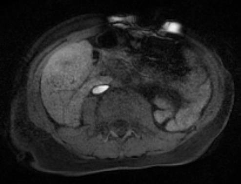
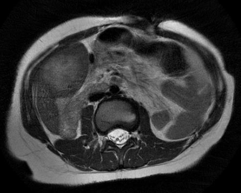
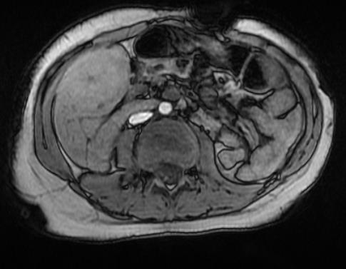
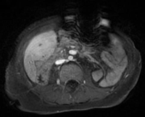
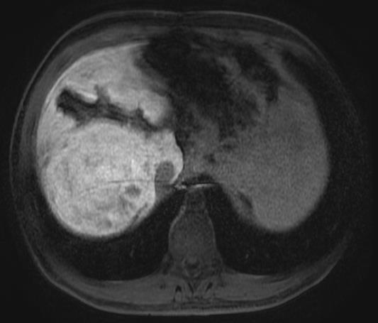
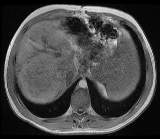
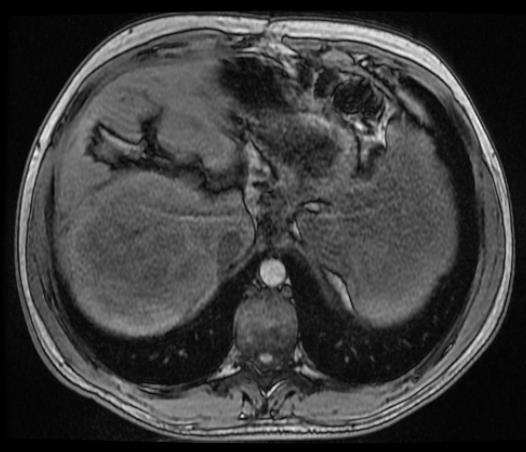
20 HBP Hyperintense Adenomas T2w Pre OP 9 year old w/ diminutive portal vein (Abernethy II) Inflammatory adenoma Art PV HBP

21 HBP Hyperintense Adenomas Yoneda et al. Case report of β- catenin activated adenoma Didn t look like benign lesion on any phase
22 HBP Hyperintense Adenoma T2w Pre IP OP 18 year old Art PV HBP Well differentiated hepatocellular neoplasm w/ β- catenin activation


23 HBP Hyperintense Adenomas Ba-Ssalamah et al. Review of 43 HCAs 6 β-catenin activated adenomas 5 / 6 HBP iso- to hyperintense Most β-catenin activated adenomas and 29% of inflammatory adenomas mimicked FNH (retained on HBP)


24 HBP Hyperintense Adenomas 11 year old T2w IP OP Inflammatory adenoma w/ β- catenin activation Pre Art PV HBP
25 6 studies, 309 patients 397 lesions: 164 adenomas, 233 FNH 257 w/ pathology Molecular subtyping in only 1 paper % sensitivity for dx of FNH 95% CI as low as 77% % specificity for dx of FNH 95% CI as low as 54% McInnes MD, et al. Radiology Nov;277(2):413-23
26 Major concern re: bias Study that did molecular subtyping had lowest specificity for FNH (87%) Suggests higher specificity in other studies due to misclassification of inflammatory adenoma as telangiectatic FNH Study that did molecular subtyping had highest rate of adenomas iso to hyperintense on HBP Range: 0-67% McInnes MD, et al. Radiology Nov;277(2):413-23
:780-9")
27 HBP Hyperintense HCC In general HCC appear hypointense on hepatocyte phase 6 15% are iso- or hyperintense Due to overexpression of OATP8 Kitao A, et al. Radiology Dec;265(3):780-9
28 Kitao et. al. Radiology 2012 Hyperintense HCC More differentiated Lower serum tumor marker levels (e.g. AFP) ± less fibrous capsule and hepatic vein invasion Less portal vein invasion Lower recurrence rate ± longer survival Kitao A, et al. Radiology Dec;265(3):780-9
29 Kitao et. al. Radiology 2012 Hyperintense HCC More differentiated LESS AGGRESSIVE Lower serum tumor marker levels (e.g. AFP) ± less fibrous capsule and hepatic vein invasion Less portal vein invasion Lower recurrence rate ± longer survival Kitao A, et al. Radiology Dec;265(3):780-9
30 What to do? If its dark on HBP, view w/ suspicion Except: cysts, venous malformations ( hemangiomas ) If imaging features aren t classic for a benign lesion, be skeptical e.g. the T1 bright FNH e.g. the heterogeneously HBP hyperintense FNH

31 What to do? Incorporate other information History OCPs, EtOH, Obesity Adenoma Background liver Growth Hypointense rim on HBP or internal mosaic architecture can be helpful signs of HCC Hope TA, et. al. Abdom Imaging Mar;40(3):613-25
32 Summary Hepatocyte specific contrast media can be a valuable tool in diagnosis of liver lesions Its not as simple as HBP bright = benign
33 SAM Question #1 Which of the following lesions is most likely to be iso- to hyperintense on the hepatobiliary phase when imaged with a hepatocyte specific contrast material? a) Hepatocellular carcinoma b) Inflammatory adenoma c) Metastasis d) HNF-1α inactivated adenoma
34 SAM Question #2 Hepatobiliary phase hypointensity of a lesion imaged with a hepatocyte specific contrast material can be explained by which of the following processes: a) Decreased OATP expression b) Abnormal background liver c) Rapid drainage of bile from the lesion d) Increased blood flow to the lesion
35 THANK YOU Andrew T. Trout,
Evangelos Chartampilas Bioclinic Hospital Thessaloniki, Greece
 Evangelos Chartampilas Bioclinic Hospital Thessaloniki, Greece Hepatospecificcontrast agents Gadobenate dimeglumine (Multihance) Gadoxeticacid (Primovist) 3-5% liver uptake 50% liver uptake Hepatobiliary
Evaluation of Liver Mass Lesions. American College of Gastroenterology 2013 Regional Postgraduate Course
 Evaluation of Liver Mass Lesions American College of Gastroenterology 2013 Regional Postgraduate Course Lewis R. Roberts, MB ChB, PhD Division of Gastroenterology and Hepatology Mayo Clinic College of
Paradoxical uptake of Gd-EOB-DTPA of focal hepatic nodule in the hepatobiliary phase
 Paradoxical uptake of Gd-EOB-DTPA of focal hepatic nodule in the hepatobiliary phase Poster No.: C-1869 Congress: ECR 2011 Type: Educational Exhibit Authors: S. M. Ha, C. Lee, K. A. Kim, J. Lee, Y.-S.
Enhancements in Hepatobiliary Imaging:
 Enhancements in Hepatobiliary Imaging: S. Channual 1, MD; A. Pahwa 2, MD; S. Raman 1, MD. 1 UCLA Medical Center, Department of Radiologic Sciences 2 Olive-View UCLA Medical Center, Department of Radiology
MRI OF FOCAL LESIONS IN
 Introduction MRI OF FOCAL LESIONS IN THE NON-CIRRHOTIC LIVER Ivan Pedrosa M.D. Ph.D. Associate Professor of Radiology and Advanced Imaging Research Center University of Texas Southwestern. Dallas, TX Incidental
Financial Disclosure
 Benign Liver Masses Adil Abdalla, MBBS Creighton University-CHI Health August 25, 2018 Financial Disclosure Nothing to disclose Financial Disclosure 1 Objectives To assess patients with benign liver tumors
Hepatocellular carcinoma Cholangiocarcinoma. Jewels of hepatobiliary cancer imaging : what to look for? Imaging characteristics of HCC.
 Outline : Imaging Jewels Jewels of hepatobiliary cancer imaging : what to look for? Hepatocellular carcinoma Cholangiocarcinoma Surachate Siripongsakun, M.D. Chulabhorn Cancer Center Imaging characteristics
LIVER IMAGING TIPS IN VARIOUS MODALITIES. M.Vlychou, MD, PhD Assoc. Professor of Radiology University of Thessaly
 LIVER IMAGING TIPS IN VARIOUS MODALITIES M.Vlychou, MD, PhD Assoc. Professor of Radiology University of Thessaly Hepatocellular carcinoma is a common malignancy for which prevention, screening, diagnosis,
Diagnostic Challenges and Pitfalls in MR Imaging with Hepatocyte-specific
 Note: This copy is for your personal non-commercial use only. To order presentation-ready copies for distribution to your colleagues or clients, contact us at www.rsna.org/rsnarights. ABDOMINAL AND GASTROINTESTINAL
Innovations in HCC Imaging: MDCT/MRI
 Innovations in HCC Imaging: MDCT/MRI Anthony E. Cheng, M.D. Cardinal MRI Center Cardinal Santos Medical Center, Wilson Street, San Juan Innovations in HCC Imaging: Goals/Objectives MDCT/MRI Learn the diagnostic
Acknowledgements. Update of Focal Liver Lesions Goals. Focal Liver Lesions. Imaging Choices For Liver Lesions. Focal Liver Lesions
 Acknowledgements Update of Focal Liver Lesions 2012 Giles Boland Massachusetts General Hospital Harvard Medical School No disclosures Dushyant Sahani Mukesh Harisinghani Goals Focal liver lesions Imaging
Pitfalls in the diagnosis of well-differentiated hepatocellular lesions
 2013 Colorado Society of Pathology Pitfalls in the diagnosis of well-differentiated hepatocellular lesions Sanjay Kakar, MD University of California, San Francisco Outline Hepatocellular adenoma: new WHO
Hepatobiliary Contrast Agents for Liver MRI
 Hepatobiliary Contrast Agents for Liver MRI Scott B. Reeder, MD, PhD International Society for Magnetic Resonance in Medicine Sociedad Mexicana de Radiologia e Imagen (SMRI) Mexico City June 4, 2014 Department
CTA/MRA of Pediatric Hepatic Masses Radiology-Pathology Correlation
 Acta Radiológica Portuguesa, Vol.XVIII, nº70, pág. 41-50, Abr.-Jun., 2006 CTA/MRA of Pediatric Hepatic Masses Radiology-Pathology Correlation Marilyn J. Siegel Mallinckrodt Institute of Radiology, Washington
Visualization of multistep hepatocarcinogenesis using various imaging biomarkers
 Visualization of multistep hepatocarcinogenesis using various imaging biomarkers Award: Certificate of Merit Poster No.: C-0120 Congress: ECR 2014 Type: Educational Exhibit Authors: S. Kobayashi, T. Gabata,
Jesse Civan, M.D. Medical Director, Jefferson Liver Tumor Center
 Liver Tumors Jesse Civan, M.D. Medical Director, Jefferson Liver Tumor Center Differential Diagnosis Malignant Metastatic from non-hepatic primary Hepatocellular carcinoma Cholangiocarcinoma Biliary cystcarcinoma
Approach to Liver Lesions. Anjana A. Pillai, MD Associate Professor of Medicine Director, Liver Tumor Clinic The University of Chicago Medical Center
 Approach to Liver Lesions Anjana A. Pillai, MD Associate Professor of Medicine Director, Liver Tumor Clinic The University of Chicago Medical Center Objectives Identify common clinical features and imaging
Essentials of Clinical MR, 2 nd edition. 65. Benign Hepatic Masses
 65. Benign Hepatic Masses Pulse sequences acquired for abdominal MRI typically consist of fast acquisition schemes such as single-shot turbo spin echo (i.e. HASTE) and gradient echo schemes such as FLASH
PATHOLOGY OF LIVER TUMORS
 PATHOLOGY OF LIVER TUMORS Pathobasic, 31.05.2016 WHO Classification Approach to a Liver Mass Lesion in a patient with chronic liver disease? Lesion in a patient without chronic liver disease? Malignant
Key Words: Adenoma, Liver, Hepatocellular carcinoma, Magnetic resonance imaging, Beta-catenin
 pissn: 2234-8646 eissn: 2234-8840 http://dx.doi.org/10.5223/pghn.2015.18.2.144 Pediatr Gastroenterol Hepatol Nutr 2015 June 18(2):144-148 Case Report PGHN Atypical β-catenin Activated Child Hepatocellular
Diagnostic efficacy of Gd-EOB-DTPA (Primovist)-enhanced MR imaging and CT for hepatocellular carcinoma
-enhanced MR imaging and CT for hepatocellular carcinoma") Diagnostic efficacy of Gd-EOB-DTPA (Primovist)-enhanced MR imaging and CT for hepatocellular carcinoma Poster No.: C-0124 Congress: ECR 2010 Type: Scientific Exhibit Topic: Abdominal Viscera (Solid Organs)
Diagnostic efficacy of Gd-EOB-DTPA (Primovist)-enhanced MR imaging and CT for hepatocellular carcinoma Poster No.: C-0124 Congress: ECR 2010 Type: Scientific Exhibit Topic: Abdominal Viscera (Solid Organs)
Liver Tumors. Prof. Dr. Ahmed El - Samongy
 Liver Tumors Prof. Dr. Ahmed El - Samongy Objective 1. Identify the most important features of common benign liver tumors 2. Know the risk factors, diagnosis, and management of hepatocellular carcinoma
MRI for HCC surveillance and reporting: LI-RADS. Donald G. Mitchell, M.D. Thomas Jefferson University Philadelphia, PA
 MRI for HCC surveillance and reporting: LI-RADS Donald G. Mitchell, M.D. Thomas Jefferson University Philadelphia, PA Cirrhotic Nodules Regenerative Nodule Atypical Nodule Hyperplastic Nodule Dysplastic
Objectives. HCC Incidence and Mortality. Disclosure Statement HCC. Imaging of Hepatocellular Carcinoma. Treatment of Hepatocellular Carcinoma
 Imaging of Hepatocellular Carcinoma and the use of LI RADS Treatment of Hepatocellular Carcinoma Aaron D. Anderson, D.O. AOCR April 2015 Objectives Show how the use of LI RADS can simplify the diagnosis
The Focal Hepatic Lesion: Radiologic Assessment
 The Focal Hepatic Lesion: Radiologic Assessment Kevin Kuo, Harvard Medical School Year III Our Patient: PS 67 y/o female w/ long history of alcohol use Drinking since age 18, up to one bottle of wine/day
Radiology of hepatobiliary diseases
 GI cycle - Lecture 14 436 Teams Radiology of hepatobiliary diseases Objectives 1. To Interpret plan x-ray radiograph of abdomen with common pathologies. 2. To know the common pathologies presentation.
Abdominal MRI Techniques in Pediatric Oncology
 Abdominal MRI Techniques in Pediatric Oncology Jonathan R. Dillman, M.D. Assistant Professor Departments of Radiology & Urology Section of Pediatric Radiology C.S. Mott Children s Hospital Disclosures
ACG Clinical Guideline: Diagnosis and Management of Focal Liver Lesions
 ACG Clinical Guideline: Diagnosis and Management of Focal Liver Lesions Jorge A. Marrero, MD, 1 Joseph Ahn, MD, FACG, 2 K. Rajender Reddy, MD, FACG 3 1 University of Texas at Southwestern, Dallas, Texas,
CT & MRI of Benign Liver Neoplasms Srinivasa R Prasad
 CT & MRI of Benign Liver Neoplasms Srinivasa R Prasad No financial disclosures Acknowledgements Many thanks to Drs. Heiken, Narra & Menias (MIR) Dr. Sahani (MGH) for sharing images Benign Liver Tumors:
Hepatocelluar nodules in liver cirrhosis: hemodynamic evaluation (angiographyassisted CT) with special reference to multi-step hepatocarcinogenesis
 with special reference to multi-step hepatocarcinogenesis") Abdominal Imaging ª The Author(s) 2011. This article is published with open access at Springerlink.com Published online: 26 January 2011 Abdom Imaging (2011) 36:264 272 DOI: 10.1007/s00261-011-9685-1 INVITED
Abdominal Imaging ª The Author(s) 2011. This article is published with open access at Springerlink.com Published online: 26 January 2011 Abdom Imaging (2011) 36:264 272 DOI: 10.1007/s00261-011-9685-1 INVITED
Review of Hepatobiliary Contrast Agents: Current Applications and Challenges
 REVIEW Review of Hepatobiliary Contrast Agents: Current Applications and Challenges Alex Frydrychowicz, M.D.*, The group of hepatobiliary contrast agents comprises two gadolinium-based contrast agents
Contrast Enhanced Ultrasound of Parenchymal Masses in Children
 Contrast Enhanced Ultrasound of Parenchymal Masses in Children Sue C Kaste, DO On behalf of Beth McCarville, MD St. Jude Children s Research Hospital Memphis, TN Overview Share St. Jude experience with
CT-MRI LI-RADS v2017: A Comprehensive Guide for Beginners
 Review Article CT-MRI LI-RADS v2017: A Comprehensive Guide for Beginners Francesca Patella 1, Filippo Pesapane* 1, Enrico Maria Fumarola 1, Ilaria Emili 1, Riccardo Spairani 1, Salvatore Alessio Angileri
Role of Gd-EOB-DTPA enhanced MR Imaging in the evaluation of the transplanted liver: Advantages and Limitations
 Role of Gd-EOB-DTPA enhanced MR Imaging in the evaluation of the transplanted liver: Advantages and Limitations Robinson Yu, MD, Amir A. Borhani, MD, Alessandro Furlan, MD, Matthew T. Heller, MD, Mitchell
Liver Specialty Evening Conference. Matthew M. Yeh, MD, PhD Professor of Pathology Adjunct Professor of Medicine University of Washington, Seattle
 Liver Specialty Evening Conference Matthew M. Yeh, MD, PhD Professor of Pathology Adjunct Professor of Medicine University of Washington, Seattle Case History A 65 year-old man presents with abdominal
HCC and mass effect. Hepatocellular cancer: what if the AFP is rising but no lesion seen on imaging? What you need to know about AFP.
 Hepatocellular cancer: what if the AFP is rising but no lesion seen on imaging? Arun J Sanyal M.B.B.S., M.D. Charles Caravati Professor of Medicine Virginia Commonwealth University Imaging features used
Hepatocellular Adenomas: Genetics & Imaging Update 2017
 No financial disclosures Hepatocellular Adenomas: Genetics & Imaging Update 2017 Srinivasa Prasad MD The UT MD Anderson Cancer Center Aims & Objectives To provide a current update on genetics & molecular
Liver Specific MRI using Gd-EOB-DTPA Disodium (Primovist) Effects Change in Management of Indeterminate Liver Lesions.
 Effects Change in Management of Indeterminate Liver Lesions.") Liver Specific MRI using Gd-EOB-DTPA Disodium (Primovist) Effects Change in Management of Indeterminate Liver Lesions. Poster No.: C-1751 Congress: ECR 2012 Type: Authors: Keywords: DOI: Educational Exhibit
Liver Specific MRI using Gd-EOB-DTPA Disodium (Primovist) Effects Change in Management of Indeterminate Liver Lesions. Poster No.: C-1751 Congress: ECR 2012 Type: Authors: Keywords: DOI: Educational Exhibit
Interesting Cases from Liver Tumor Board. Jeffrey C. Weinreb, M.D.,FACR Yale University School of Medicine
 Interesting Cases from Liver Tumor Board Jeffrey C. Weinreb, M.D.,FACR Yale University School of Medicine jeffrey.weinreb@yale.edu Common Liver Diseases Hemangioma Cyst FNH Focal Fat/Sparing THID Non-Cirrhotic
Consensus Report of the Fifth International Forum for Liver MRI
 Fifth International Forum for Liver MRI Gastrointestinal Imaging Commentary Gastrointestinal Imaging Commentary FOCUS ON: Christoph J. Zech 1,2 Carlo Bartolozzi 3 Paulette Bioulac-Sage 4 Pierce K. Chow
Hyperplasia / Hypertrophy, Cirrhosis, Diagnostic procedure, MR, CT-Angiography, CT, Liver, Abdomen /ecr2012/C-2202
 Hepatic nodules showing ring-like enhancement on hepatobiliary phase of Gd-EOB-DTPA enhanced MRI can be divided into two subtypes based on blood supply: FNH and NRH-like nodules Poster No.: C-2202 Congress:
Atypical Well differentiated Hepatocellular Neoplasms Cruising through the maze of criteria, terminology and risk assessment
 2016 Hans Popper Companion Meeting Atypical Well differentiated Hepatocellular Neoplasms Cruising through the maze of criteria, terminology and risk assessment Disclosure Dr. Kakar has nothing to Disclose
O Farrell Legacy UPDATE ON WHO NOMENCLATURE. World Health Organization, 2010 DISCLOSURES WITH EMPHASIS ON PROBLEM HEPATOCELLULAR TUMORS
 O Farrell Legacy UPDATE ON WHO NOMENCLATURE WITH EMPHASIS ON PROBLEM HEPATOCELLULAR TUMORS Linda Ferrell, MD University of California San Francisco Vice Chair, Director of Surgical Pathology World Health
MR imaging of primary sclerosing cholangitis (PSC) using the hepatobiliary specific contrast agent Gd-EOB-DTPA
 using the hepatobiliary specific contrast agent Gd-EOB-DTPA") MR imaging of primary sclerosing cholangitis (PSC) using the hepatobiliary specific contrast agent Gd-EOB-DTPA Poster No.: C-0019 Congress: ECR 2010 Type: Educational Exhibit Topic: Abdominal Viscera (Solid
MR imaging of primary sclerosing cholangitis (PSC) using the hepatobiliary specific contrast agent Gd-EOB-DTPA Poster No.: C-0019 Congress: ECR 2010 Type: Educational Exhibit Topic: Abdominal Viscera (Solid
Liver imaging reporting and data system (LI-RADS) version 2014: understanding and application of the diagnostic algorithm
 version 2014: understanding and application of the diagnostic algorithm") pissn 2287-2728 eissn 2287-285X Liver Imaging Clinical and Molecular Hepatology 2016;22:296-307 Liver imaging reporting and data system (LI-RADS) version 2014: understanding and application of the diagnostic
pissn 2287-2728 eissn 2287-285X Liver Imaging Clinical and Molecular Hepatology 2016;22:296-307 Liver imaging reporting and data system (LI-RADS) version 2014: understanding and application of the diagnostic
Detection and Characterization of Hepatocellular Carcinoma by Imaging
 CLINICAL GASTROENTEROLOGY AND HEPATOLOGY 2005;3:S136 S140 Detection and Characterization of Hepatocellular Carcinoma by Imaging OSAMU MATSUI Department of Imaging Diagnosis and Interventional Radiology,
Hepatobiliary Contrast Agents
 Hepatobiliary Contrast Agents SCBT/MR Annual Meeting Salt Lake City September 21, 2016 Scott B. Reeder, MD, PhD Department of Radiology University of Wisconsin Madison, WI Disclosures University of Wisconsin-Madison
With the widespread use of hepatic imaging, liver masses
 2B: Liver Assessment of the Liver Mass: What Do You Need to Know? With the widespread use of hepatic imaging, liver masses are detected either unexpectedly or in the course of screening for liver cancer
Invited Re vie W. Analytical histopathological diagnosis of small hepatocellular nodules in chronic liver diseases
 Histol Histopathol (1 998) 13: 1077-1 087 http://www.ehu.es/histoi-histopathol Histology and Histopathology Invited Re vie W Analytical histopathological diagnosis of small hepatocellular nodules in chronic
CT/MRI LI-RADS v2017 CORE
 CT/MRI LI-RADS v2017 CORE Untreated observation without pathologic proof in patient at high risk for HCC If cannot be categorized due to image degradation or omission If definite tumor in vein (TIV) If
CASE 1 11/1/2016 HEPATOBILIARY IMAGING CASE PRESENTATIONS DECLARATION. Dr. Chirag Patel ORGAN IMAGING yr old lady
 HEPATOBILIARY IMAGING CASE PRESENTATIONS DECLARATION No financial disclosures or affiliations with commercial organisations No discussion of investigational or off-label use of medical devices, products
Alastair Burt Newcastle University
 Alastair Burt Newcastle University Benign Hepatocellular adenoma 8170/0 Focal nodular hyperplasia Malignancy-associated and premalignant lesions Large cell change (formerly dysplasia ) Small cell change
INTRODUCTION. Yun Ku Cho 1, Ju Won Kim 1, Mi Young Kim 1, and Hyeon Je Cho 2
 Gut and Liver, Vol. 12,. 1, January 2018, pp. 79-85 ORiginal Article n-hypervascular Hypointense dules on Hepatocyte Phase Gadoxetic Acid-Enhanced MR Images: Transformation of MR Hepatobiliary Hypointense
Lewis R. Roberts, MB, ChB, PhD, FACG
 2B: Hot Topics in Liver Disease Evaluation of Liver Mass Lesions Lewis R. Roberts, MB, ChB, PhD, FACG Clinical Classification of Liver Mass Lesions It is helpful to subclassify lesions into three clinical
Benign liver tumors : Diagnosis and management
 4th International Hepatology Conference 2016 HEPATOLOGY SOCIETY, DHAKA, BANGLADESH Benign liver tumors : Diagnosis and management Pr Laurence Chiche Hepato biliary surgery and transplantation Bordeaux,
Pathological Classification of Hepatocellular Carcinoma
 3 rd APASL Single Topic Conference: HCC in 3D Pathological Classification of Hepatocellular Carcinoma Glenda Lyn Y. Pua, M.D. HCC Primary liver cancer is the 2 nd most common cancer in Asia HCC is the
Hepatocellular adenomas (HCAs) are uncommon primary benign tumours. They are constantly monoclonal tumours.
 are uncommon primary benign tumours. They are constantly monoclonal tumours.") Hepatocellular adenoma: Evaluation with contrast enhanced ultrasound, MRI And Correlation with pathologic and phenotypic classification. About 26 lesions. Poster No.: C-1561 Congress: ECR 2011 Type: Scientific
Hepatocellular adenoma: Evaluation with contrast enhanced ultrasound, MRI And Correlation with pathologic and phenotypic classification. About 26 lesions. Poster No.: C-1561 Congress: ECR 2011 Type: Scientific
HCC e CEUS. Prof. A. Giorgio. Direttore IX UOC di Malattie Infettive ad Indirizzo Ecointerventistico
 HCC e CEUS Prof. A. Giorgio Direttore IX UOC di Malattie Infettive ad Indirizzo Ecointerventistico The natural history of compensated cirrhosis due to hepatitis C virus: a 17 year cohort study of 214 patients
HEPATO-BILIARY IMAGING
 HEPATO-BILIARY IMAGING BY MAMDOUH MAHFOUZ MD PROF.OF RADIOLOGY CAIRO UNIVERSITY mamdouh.m5@gmail.com www.ssregypt.com CT ABDOMEN Indications Patient preparation Patient position Scanogram Fasting 4-6 hours
Gadoxetic Acid enhanced MRI of Hepatocellular Carcinoma: Value of Washout in Transitional and Hepatobiliary Phases
 ORIGINAL RESEARCH GASTROINTESTINAL IMAGING Gadoxetic Acid enhanced MRI of Hepatocellular Carcinoma: Value of Washout in Transitional and Hepatobiliary Phases Dong Hwan Kim, MD* Sang Hyun Choi, MD, PhD*
New developments in liver MR imaging
 Parallel symposium B. 간질환에대한영상검사및중재적시술 (What are new in imaging diagnosis and interventional treatment of liver diseases) 울산대학교의과대학서울아산병원영상의학과 New developments in liver MR imaging Hyung Jin Won, M.D. Department
Contrast-enhanced ultrasound (CEUS) of focal liver lesions. A useful, rapid and accessible tool.
 of focal liver lesions. A useful, rapid and accessible tool.") Contrast-enhanced ultrasound (CEUS) of focal liver lesions. A useful, rapid and accessible tool. Poster No.: C-2329 Congress: ECR 2012 Type: Educational Exhibit Authors: S. Santamaria Jareño, J. Carrero
Contrast-enhanced ultrasound (CEUS) of focal liver lesions. A useful, rapid and accessible tool. Poster No.: C-2329 Congress: ECR 2012 Type: Educational Exhibit Authors: S. Santamaria Jareño, J. Carrero
RICCARDO LENCIONI,CLOTILDE DELLA PINA, LAURA CROCETTI,DANIA CIONI. Chapter 1
 RICCARDO LENCIONI,CLOTILDE DELLA PINA, LAURA CROCETTI,DANIA CIONI Chapter 1 Impact of European Federation of Societies for Ultrasound in Medicine and Biology (EFSUMB) Guidelines on the Use of Contrast
MRI of Small Hepatocellular Carcinoma: Typical Features Are Less Frequent Below a Size Cutoff of 1.5 cm
 Gastrointestinal Imaging Original Research Choi et al. MRI of Small HCC Gastrointestinal Imaging Original Research Moon Hyung Choi 1 Joon-Il Choi 1 Young Joon Lee 1 Michael Yong Park 1 Sung Eun Rha 1 Chandana
Pancreatic Adenocarcinoma: Everything You Need to Know From Cross-Sectional Imaging to Treatment
 Pancreatic Adenocarcinoma: Everything You Need to Know From Cross-Sectional Imaging to Treatment Andrew W. Bowman, MD PhD Assistant Professor of Radiology Mayo Clinic Florida SCBT-MR Annual Meeting Nashville,
Pseudo Washout Sign in High-Flow Hepatic Hemangioma on Gadoxetic Acid Contrast-Enhanced MRI Mimicking Hypervascular Tumor
 Gastrointestinal Imaging Clinical Observations Doo et al. Pseudo Washout Sign on MRI of Hemangioma Gastrointestinal Imaging Clinical Observations Kyung Won Doo 1 Chang Hee Lee Jae Woong Choi Jongmee Lee
Rare primary liver tumors - MRI pictorial review
 Rare primary liver tumors - MRI pictorial review Poster No.: C-2293 Congress: ECR 2017 Type: Educational Exhibit Authors: R. Lameiras, J. Cruz, J. Felício Costa, F. D. Figueiredo, A. 1 1 1 1 2 1 1 1 2
ABDOMINAL DIFFUSION WEIGHTED MR
 ABDOMINAL DIFFUSION WEIGHTED MR Frank Miller, M.D. FACR Professor of Radiology Chief, Body Imaging Section Medical Director, MR Imaging Northwestern University Feinberg School of Medicine fmiller@northwestern.edu
Recent Advances in the Imaging Diagnosis of Hepatocellular Carcinoma: Value of Gadoxetic Acid-Enhanced MRI
 2235-1795/16/0051-0067$39.50/0 67 Imaging Diagnosis of HCC Recent Advances: Review Recent Advances in the Imaging Diagnosis of Hepatocellular Carcinoma: Value of Gadoxetic Acid-Enhanced MRI Ijin Joo a,
Evaluation of contrast-enhanced ultrasound for diagnosis of dysplastic nodules with a focus of hepatocellular carcinoma in liver cirrhosis patients
 Original Article Evaluation of contrast-enhanced ultrasound for diagnosis of dysplastic nodules with a focus of hepatocellular carcinoma in liver cirrhosis patients Wei Wu, Minhua Chen, Kun Yan, Yin Dai,
Gadoxetate Disodium Enhanced MRI to Differentiate Dysplastic Nodules and Grade of Hepatocellular Carcinoma: Correlation With Histopathology
 Gastrointestinal Imaging Original Research Channual et al. Gadoxetate Disodium Enhanced MRI of DNs and HCCs Gastrointestinal Imaging Original Research Stephanie Channual 1 Nelly Tan 1 Surachate Siripongsakun
ADRENAL MR: PEARLS AND PITFALLS
 ADRENAL MR: PEARLS AND PITFALLS Frank Miller, M.D. Lee F. Rogers MD Professor of Medical Education Chief, Body Imaging Section and Fellowship Medical Director, MR Imaging Professor of Radiology Northwestern
Characterization of Incidental Liver Lesions: Comparison of Multidetector CT versus Gd-EOB-DTPA-Enhanced MR Imaging
 : Comparison of Multidetector CT versus Gd-EOB-DTPA-Enhanced MR Imaging Yong Eun Chung, Myeong-Jin Kim, Yeo-Eun Kim, Mi-Suk Park, Jin Young Choi, Ki Whang Kim* Department of Radiology, Severance Hospital,
Magnetic resonance imaging findings of hepatocellular carcinoma: typical and atypical findings
 Asian Biomedicine Vol. 4 No. 1 February 2010; 113-124 Clinical report Magnetic resonance imaging findings of hepatocellular carcinoma: typical and atypical findings Laddawan Vajragupta, Khanitha Kittisatra,
Liver imaging takes a step forward with Ingenia
 Publication for the Philips MRI Community ISSUE 49 2013 / 2 Liver imaging takes a step forward with Ingenia Lyon South Hospital strives to move from several studies first CT, then MR or PET to using just
ADRENAL LESIONS 10/09/2012. Adrenal + lesion. Introduction. Common causes. Anatomy. Financial disclosure. Dr. Boraiah Sreeharsha. Nothing to declare
 ADRENAL LESIONS Financial disclosure Nothing to declare Dr. Boraiah Sreeharsha MBBS;FRCR;FRCPSC Introduction Adrenal + lesion Adrenal lesions are common 9% of the population Increase in the detection rate
ESUR 2018, Sept. 13 th.-16 th., 2018 Barcelona, Spain
 ESUR 2018, Sept. 13 th.-16 th., 2018 Barcelona, Spain OUR APPROACH Incidental adrenal nodule/mass Isaac R Francis, M.B;B.S University of Michigan, Ann Arbor, Michigan Disclosures None (in memory) M Korobkin,
INTRODUCTION. Key Words: Contrast enhanced ultrasonography; Liver masses. ORiginal Article
 Gut and Liver, Vol. 8, No. 3, May 2014, pp. 292-297 ORiginal Article Clinically Useful Diagnostic Tool of Contrast Enhanced Ultrasonography for Focal Liver Masses: Comparison to Computed Tomography and
Liver nodules mimicking metastatic disease
 Liver nodules mimicking metastatic disease Poster No.: C-1703 Congress: ECR 2011 Type: Educational Exhibit Authors: F. Vandenbroucke, B. Ilsen, B. Op de Beeck, J. de Mey ; 1 1 2 2 3 2 3 Brussels/BE, Brussel/BE,
Hepatic Lymphoma Representing Iso-Signal Intensity on Hepatobiliary Phase, in Gd-EOB-DTPA-Enhanced MRI: Case Report
 pissn 2384-1095 eissn 2384-1109 imri 2015;19:200-204 http://dx.doi.org/10.13104/imri.2015.19.3.200 Hepatic Lymphoma Representing Iso-Signal Intensity on Hepatobiliary Phase, in Gd-EOB-DTPA-Enhanced MRI:
Malignant Focal Liver Lesions
 Malignant Focal Liver Lesions Other Than HCC Pablo R. Ros, MD, MPH, PhD Departments of Radiology and Pathology University Hospitals Cleveland Medical Center Case Western Reserve University Pablo.Ros@UHhospitals.org
Usefulness of Gadobenate Dimeglumine - Enhanced Hepatobiliary Phase MR Imaging on Predicting Histological Grade of Hepatocellular Carcinoma
 Usefulness of Gadobenate Dimeglumine - Enhanced Hepatobiliary Phase MR Imaging on Predicting Histological Grade of Hepatocellular Carcinoma Sung Ho Park, Myeong-Jin Kim, Jin-Young Choi, Joon Seok Lim,
Multistep hepatocarcinogenesis is characterized by the following
 Early hepatocellular carcinoma with high-grade atypia in small vaguely nodular lesions Hidenori Ojima, 1 Yohei Masugi, 1 Hanako Tsujikawa, 1 Katsura Emoto, 1 Yoko Fujii-Nishimura, 1 Mami Hatano, 1 Miho
Liver Lesions: How to Evaluate?
 Liver Lesions: How to Evaluate? Disclosures Consulting: BMS, Bayer, BTG, Eisai Laura Kulik MD Professor of Medicine, Surgery and Interventional Radiology Northwestern University Finberg School of Medicine
Purpose. Background. Purpose
 Does diffusion-weighted imaging (DWI) add diagnostic confidence in discriminating between malignant and benign solid focal lesions if included in contrast-enhanced magnetic resonance imaging (MRI) of the
Hepatocellular Carcinoma in the Cirrhotic Liver: Evaluation Using Computed Tomography and Magnetic Resonance Imaging
 Hepatocellular Carcinoma in the Cirrhotic Liver: Evaluation Using Computed Tomography and Magnetic Resonance Imaging Mehmet Coskun Abstract Hepatocellular carcinoma is the fifth most common tumor in patients
Newcastle HPB MDM updated radiology imaging protocol recommendations. Author Dr John Scott. Consultant Radiologist Freeman Hospital
 Newcastle HPB MDM updated radiology imaging protocol recommendations Author Dr John Scott. Consultant Radiologist Freeman Hospital This document is intended as a guide to aid radiologists and clinicians
Liver Imaging Reporting and Data System LI-RADS v A Pictorial Review of the Categories on CT and MRI
 Liver Imaging Reporting and Data System LI-RADS v2014 - A Pictorial Review of the Categories on CT and MRI Poster No.: C-1584 Congress: ECR 2017 Type: Educational Exhibit Authors: M. El Hawari, K. Ali,
Intrahepatic Mass-Forming Cholangiocarcinoma: Enhancement Pattern on Gd-BOPTA-MRI with Emphasis on Hepatobiliary Phase
 Intrahepatic Mass-Forming Cholangiocarcinoma: Enhancement Pattern on Gd-BOPTA-MRI with Emphasis on Hepatobiliary Phase Poster No.: C-2590 Congress: ECR 2015 Type: Scientific Exhibit Authors: G. Mamone,
Liver Cancer. Su Jong Yu, M.D. Department of Internal Medicine, Liver Research Institute, Seoul National University College of Medicine
 Liver Cancer Su Jong Yu, M.D. Department of Internal Medicine, Liver Research Institute, Seoul National University College of Medicine Primary Liver Cancer Hepatocellular carcinoma (HCC) : > 80% Derived
Consensus Statements From a Multidisciplinary Expert Panel on the Utilization and Application of a Liver-Specific MRI Contrast Agent (Gadoxetic Acid)
") Gastrointestinal Imaging Review Jhaveri et al. Consensus Statement on the Use of Gadoxetic Acid for Liver MRI Gastrointestinal Imaging Review Kartik Jhaveri 1 Sean Cleary 2 Pascale Audet 3 Fady Balaa 4
Gastrointestinal Imaging Review Jhaveri et al. Consensus Statement on the Use of Gadoxetic Acid for Liver MRI Gastrointestinal Imaging Review Kartik Jhaveri 1 Sean Cleary 2 Pascale Audet 3 Fady Balaa 4
The Contribution of Contrast Enhanced Ultrasound for the characterization of benign liver lesions in clinical practice a monocentric experience
 Original papers Med Ultrason 2012, Vol. 14, no. 4, 283-287 The Contribution of Contrast Enhanced Ultrasound for the characterization of benign liver lesions in clinical practice a monocentric experience
New insights into fatty liver disease. Rob Goldin Centre for Pathology, Imperial College
 New insights into fatty liver disease Rob Goldin Centre for Pathology, Imperial College r.goldin@imperial.ac.uk Prevalence of NASH Global prevalence of NAFLD is 25% with highest prevalence in the Middle
Non-mass Enhancement on Breast MRI. Aditi A. Desai, MD Margaret Ann Mays, MD
 Non-mass Enhancement on Breast MRI Aditi A. Desai, MD Margaret Ann Mays, MD Breast MRI Important screening and diagnostic tool, given its high sensitivity for breast cancer detection Breast MRI - Indications
Major and ancillary magnetic resonance features of LI-RADS to assess HCC: an overview and update
 Granata et al. Infectious Agents and Cancer (2017) 12:23 DOI 10.1186/s13027-017-0132-y REVIEW Major and ancillary magnetic resonance features of LI-RADS to assess HCC: an overview and update Open Access
Liver imaging the revolution
 Liver imaging the revolution Valérie Vilgrain Hôpital Beaujon, Paris, France PHC 2018 - www.aphc.info At the Beginning of the story Radiology in the 1970s US Garrett Radiology 1976 abscess Taylor Radiology
Hypervascular Liver Lesions on MRI
 Residents Section Pattern of the Month Residents Section Pattern of the Month Residents inradiology aisal Khosa 1 tif N. Khan Ronald L. isenberg Khosa, Khan N, isenberg RL Keywords: lesions, liver, MRI
Workup of a Solid Liver Lesion
 Workup of a Solid Liver Lesion Joseph B. Cofer MD FACS Chief Quality Officer Erlanger Health System Affiliate Professor of Surgery UTHSC-Chattanooga I have no financial or other relationships with any
Liver Cancer And Tumours
 Liver Cancer And Tumours What causes liver cancer? Many factors may play a role in the development of cancer. Because the liver filters blood from all parts of the body, cancer cells from elsewhere can