Sulfur hexafluoride-filled microbubbles SonoVue 3-7microns diameter Blood pool agent
|
|
|
- Francis Newman
- 5 years ago
- Views:
Transcription

1
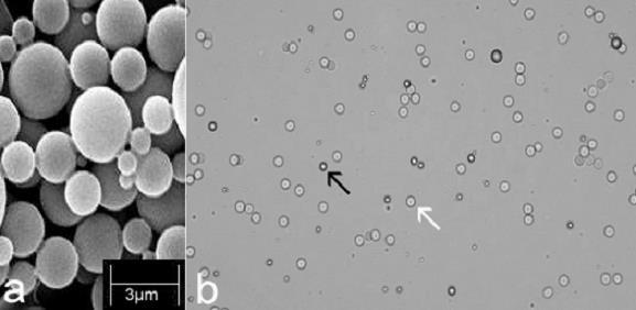
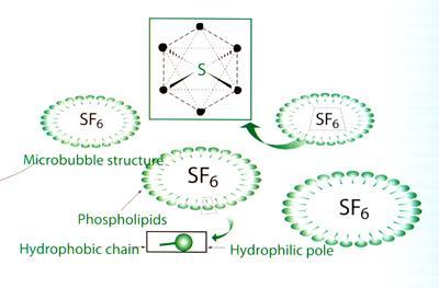
2 Sulfur hexafluoride-filled microbubbles SonoVue 3-7microns diameter Blood pool agent
3 Extremely good tolerance in clinical practice - No nephrotoxicity, - No thyroid interaction - No need of Blood test before IV Rare anaphylactoïd reaction ( Gd chelates) - incidence < 0,002% - no cross allergy with Iodine contrast Do not use in case of pregnancy and Breast feeding (precaution) => Can be used when Iodine and Gadolinium cannot
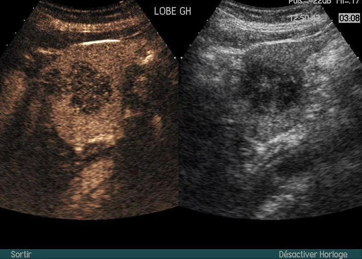
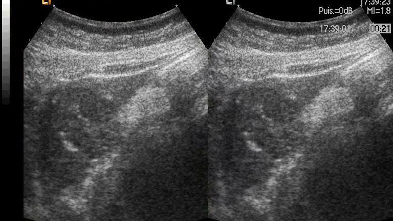
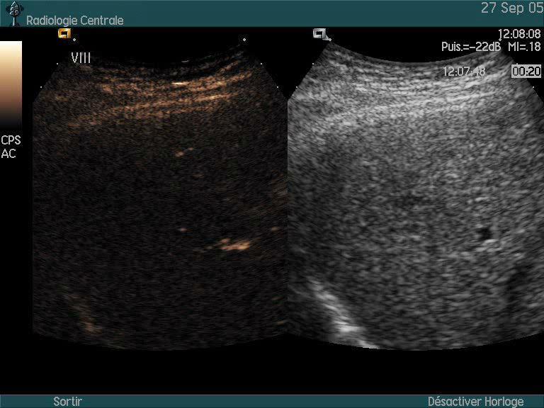
4 Early Phase Higher temporal resolution than CT or MRI Hemangiomas FNH Adenoma Mets HCC
5 Late phase Iodine/gado : extravascular leaking ++ if tumoral vessels Microbubbles : Wash-out if tumoral vessels Stagnation in the sinusoid capillaries or venous lakes Liver Hemangiomas FNH, Adenoma Well differentiated HCC Mets CHC
6 Early Phase Higher sensitivity to low amount of circulating contrast No enhancement means no (or almost no) circulating vessels

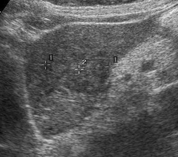
7 CEUS is already very useful in every day practice: to characterize FLL, kidney and pancreas to assess the effect of vascular destructive treatment like RFA or Chemoembolisation CEUS can be also interesting in more advanced research imaging in oncology To identify recurrences? Way?

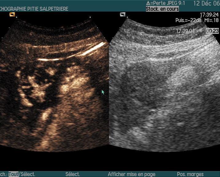
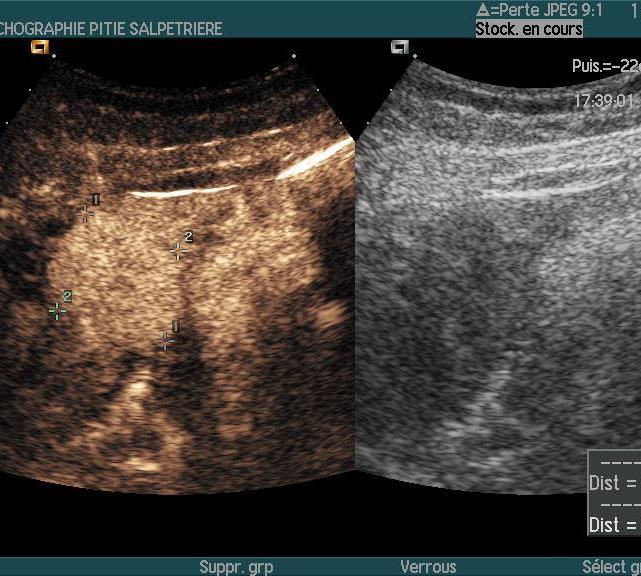
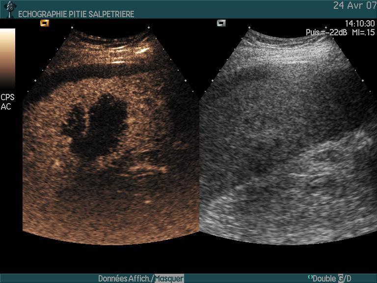
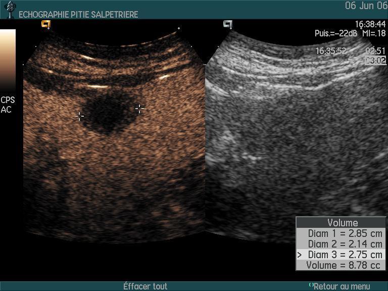
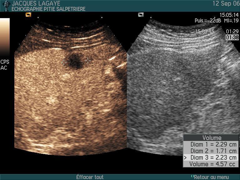
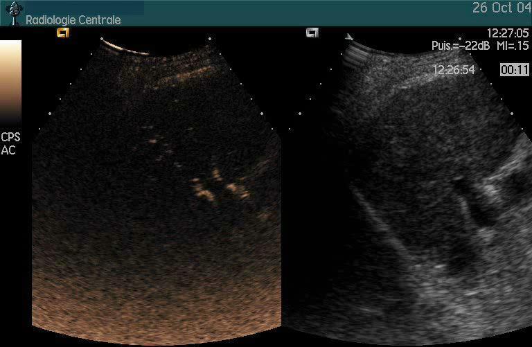
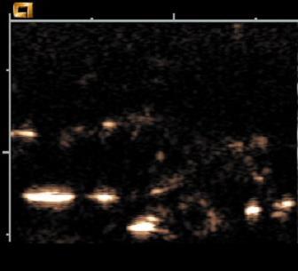
8 Sarcome utérin


9 Sarcome utérin
10
11
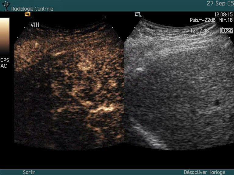
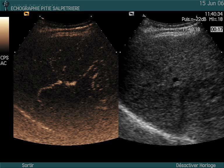
12
13 Percentage of correct diagnosis US without contrast : US with contrast : 1 Alrecht T. Eur Radiol Oct;14 Suppl 8:P Tranquart F. J Radiol 2009, 90: Trillaud H, World J Gastroenterol 2009; 15:

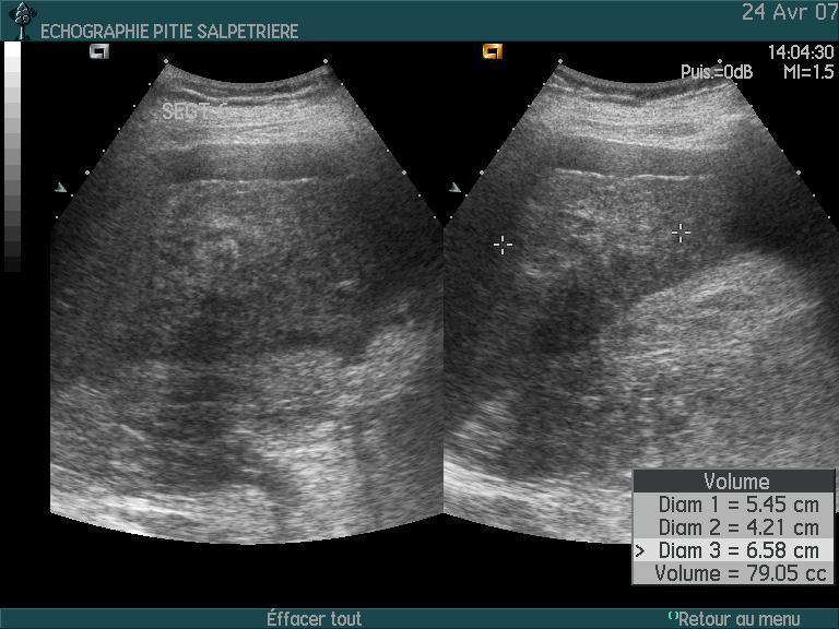
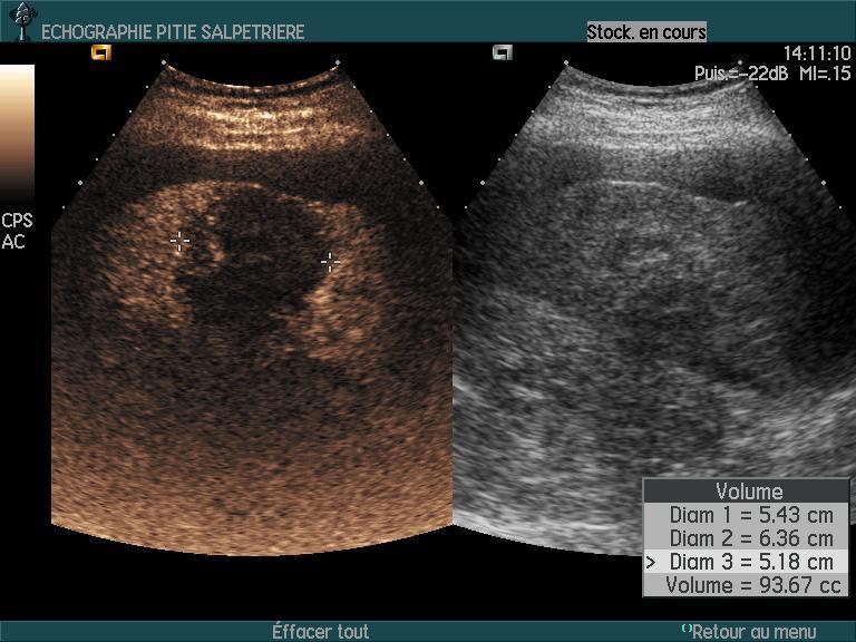
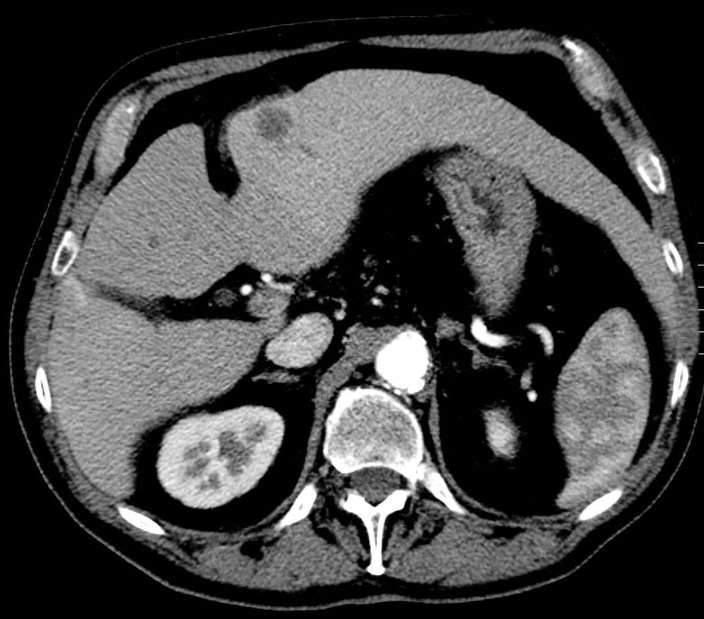
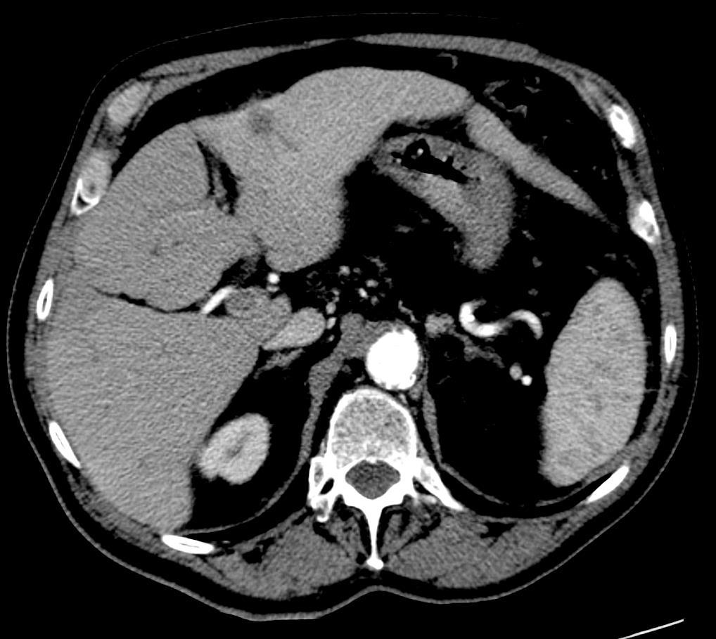
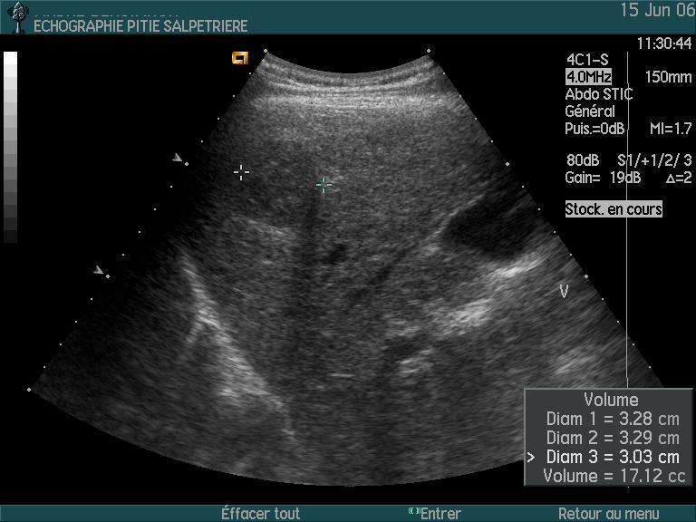
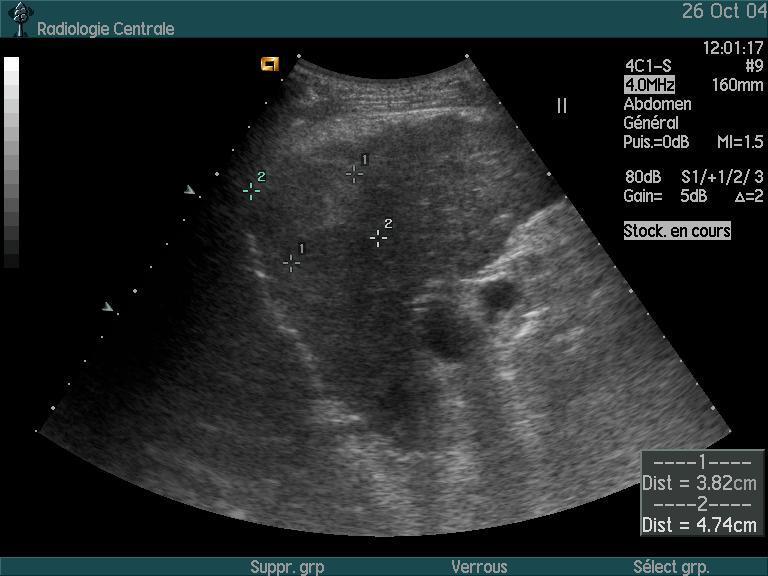
14 Sensitivity for malignancy Specificity for malignancy CEUS 95% 94% 6% 5% CECT 95% 93% 7% 5% Pooled estimates from the meta-analysis of 4 studies: Seitz K Ultraschall Med 2009;30: Li R, J Clin Ultrasound 2007;35: Catala V, Eur Radiol 2007;17: Solbiati L. Abstract D-55. European Congress of Radiology 2006 CEUS 90% 67% 13% 10% CEMRI 82% 63% 17% 18% Seitz K Ultraschall Med 2009;30:383 9 FP FN No significant difference in the accuracy of CEUS and CECT or CEMRI for the characterization (as malignant) of focal FLLs CEUS alone may be adequate to rule out liver malignancy in people with incidentally detected FLL
15 Early Phase Higher sensitivity to low amount of circulating contrast No enhancement means no (or almost no) circulating vessels


16 Courtesy Pr Correas, Necker Hosp, Paris
17
18 Unlike Liver : not used to differentiate solid tumors (same pattern) But: Characterization of complex cystic masses as benign, indeterminate or malignant (Recommendation level: A;1b) To make the distinction between hypovascular solid lesions on CT and atypical cysts (Recommendation level: B;2b)
19 Simple visual assessment To see if vessels are present or not Usefull for liver lesion treated with Tace, RFA, Vascular disruptive agents
20


21 1 month 3 months 6months
22 Mean devascularized Volume (cm3) D1-D8 W3-W6 M3-M6 M6-M9 Y1-Y ± 37.4 cc 11.3 ± 12.1 cc
23
24

25 1 mois post cryo Cryoablation
26 Cryoablation

27 Shiozawa et al, J Clin Ultrasound 2010;38: CEUS proposed alternately with CT or MRI to reduce irradiation or cost?

28 Pre TACE 48 h post TACE After TACE Before TACE
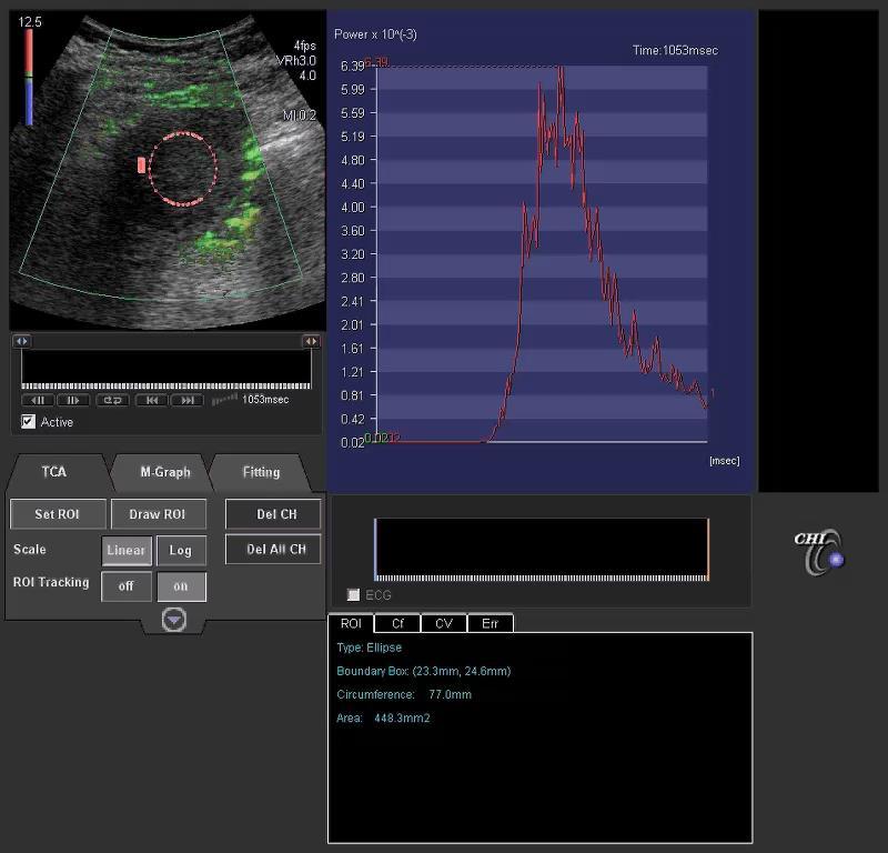
29
30
31
32 During the procedure to immediately assess the success of the procedure 1 CEUS seems to be more sensitive than dynamic CT in depicting the residual tumor blood supply to HCCs one week after TACE 2-3 No comparison between CEUS and MRI seems to be 1- Moshouris et al, Cardiovasc Intervent Radiol 2010;16:Ahead of print 2- Xia et al, Oncology 2008;75: Kim et al, j Ultrasound Med 2006;25:477-86
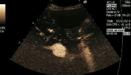
33 GIST
34
35
36 quantitative assessment of the vascular bed of the lesion Similar to DCE MRI or functional CT Variation of the local concentration of microbubbles as a function of time By measuring the effect on the images Pb or AIF
 Rabbit kidney")
37 Enhancement experiment. Theory time (sec) Rabbit kidney Continuous injection SonoVue
![[microbubbles] C( t) A 1 e t 1,2 A 0,8 A Fractional bld vol 0,4 :](/docs-images/94/119801966/images/38-0.jpg "Fraction of blood replaced per s 0 0 10 20 time (sec) A reflects the")
38 [microbubbles] C( t) A 1 e t 1,2 A 0,8 A Fractional bld vol 0,4 : Fraction of blood replaced per s time (sec) A reflects the relative BF
39 True blood pool agent => BV, BF, MTT > PS, Ktrans, Kep, VE
 Endothelial Cell Membrane Sensitivity + Sensitivity - PET")
40 Ligand specific for a selected marker Disease-specific marker expressed on the vessel wall (angiogenesis) Endothelial Cell Membrane Sensitivity + Sensitivity - PET Microbubbles MRI


41 phospholipid membrane phospholipid PEG Heterodimer peptide C 4 F 10 /N 2 BR38/InnoVue BR55 agent VEGFR2 Over expressed in tumoral vessels

42 Targeted imaging SonoVue BR55


43 Targeted imaging 40 sec 10 min SonoVue BR55


44 CT26 Bmode 14MHz 3LL 7 MHz IM = 0,1 00:00 00:22 10:10 baseline bolus pre destruction 10:23 post destruction

 (30mg/kg")


45 Targeted imaging Sunitinib (40mg/kg per day) (30mg/kg per day) Imatinib VEGFR 2 RET PDGFR ckit VEGFR 2 BCR -ABL PDGFR ckit 45 CT26

46 LIP : Targeted imaging Payen T, Ultrasound Med Biol Aug;41(8):
47 LIP : Targeted imaging: Main results Mice treated with Sunitinib => expression of VEGFR2 (significant after 24h to reach 40% of the initial level VEGFR2 expression of mice treated with Imatinib initialy and afterward but without significance VEGFR2 expression of mice treated with Placebo significantly (p<0.02) after 48h to reach 80% of the initial level VEGFR2 expression is significantly lower in the group Sunitinib compared to placebo after 24h (p<0.04) VEGFR2 expression is significantly lower in the group Sunitinib compared to Imatinib after 96h (p<0.003) VEGFR2 expression is significantly higher in the group Imatinib compared to Sunitinib after 96h(p<0.05)


 and is safe and")
48 Prostate cancer Immunostaining demonstrates moderate VEGFR2 expression in that PCa lesion BR55 Is able to bind to VEGFR2 in humans (prolonged enhancement >10 min) and is safe and well-tolerated
49 Very powerful technique to visualize the microcirculation Blood pool agent Very sensitive technique Recommended To characterize liver lesion, complex renal or pancreatic cysts To assess vascular bed destruction after RFA, Tace Great potential to quantify the microcirculation Preclinical research More and more in human New: targeted microbubbles

50 Thank you Merci
Contrast Enhanced Ultrasound of Parenchymal Masses in Children
 Contrast Enhanced Ultrasound of Parenchymal Masses in Children Sue C Kaste, DO On behalf of Beth McCarville, MD St. Jude Children s Research Hospital Memphis, TN Overview Share St. Jude experience with
The Contribution of Contrast Enhanced Ultrasound for the characterization of benign liver lesions in clinical practice a monocentric experience
 Original papers Med Ultrason 2012, Vol. 14, no. 4, 283-287 The Contribution of Contrast Enhanced Ultrasound for the characterization of benign liver lesions in clinical practice a monocentric experience
Mædica - a Journal of Clinical Medicine
 Mædica - a Journal of Clinical Medicine ORIGINAL PAPERS How Often Hepatocellular Carcinoma Has a Typical Pattern in Contrast Enhanced Ultrasound? Alina MARTIE, MD; Ioan SPOREA, MD, PhD; Roxana SIRLI, MD,
Imaging of liver and pancreas
 Imaging of liver and pancreas.. Disease of the liver Focal liver disease Diffusion liver disease Focal liver disease Benign Cyst Abscess Hemangioma FNH Hepatic adenoma HCC Malignant Fibrolamellar carcinoma
RICCARDO LENCIONI,CLOTILDE DELLA PINA, LAURA CROCETTI,DANIA CIONI. Chapter 1
 RICCARDO LENCIONI,CLOTILDE DELLA PINA, LAURA CROCETTI,DANIA CIONI Chapter 1 Impact of European Federation of Societies for Ultrasound in Medicine and Biology (EFSUMB) Guidelines on the Use of Contrast
Zoltan Harkanyi M.D., Ph.D. Department of Radiology, Heim Pal Children s Hospital, Budapest, Hungary
 Zoltan Harkanyi M.D., Ph.D. Department of Radiology, Heim Pal Children s Hospital, Budapest, Hungary CEUS expereince 10 years Department of Radiology, Heim Pal Children s Hospital, Budapest US N o 1 study
INTRODUCTION. Key Words: Contrast enhanced ultrasonography; Liver masses. ORiginal Article
 Gut and Liver, Vol. 8, No. 3, May 2014, pp. 292-297 ORiginal Article Clinically Useful Diagnostic Tool of Contrast Enhanced Ultrasonography for Focal Liver Masses: Comparison to Computed Tomography and
Perfusion Physics. ICMRI2018 March 29-31, 2018 Grand Hilton Hotel, Seoul, Korea. Asian Forum Ⅱ: Perfusion MRI SY24-1.
 SY24-1 Perfusion Physics Hiroyuki Kabasawa MR Collaborations and Development, GE Healthcare, Tokyo, Japan Perfusion is referred as the blood supply to micro capillary in tissue. Perfusion parameter such
INTERDISCIPLINARY DISCUSSIONS IN LOCALISED RCC DIAGNOSIS AND SURGICAL STRATEGIES FOR ATYPICAL RENAL CYSTIC LESIONS. Maria Cova
 INTERDISCIPLINARY DISCUSSIONS IN LOCALISED RCC DIAGNOSIS AND SURGICAL STRATEGIES FOR ATYPICAL RENAL CYSTIC LESIONS Maria Cova Radiology Department University of Trieste (IT) Eleventh European International
Modern liver imaging techniques - A new era in liver ultrasound
 Modern liver imaging techniques - A new era in liver ultrasound Yuko Kono, M.D., Ph.D. Clinical Professor Departments of Medicine and Radiology University of California, San Diego San Diego, USA How to
Role of DE-CT in Oncology
 Role of DE-CT in Oncology Dushyant Sahani, M.D Director of CT Associate Professor of Radiology Massachusetts General Hospital Harvard Medical School Email-dsahani@partners.org Disclosure Research Grant
The UGent Institutional Repository is the electronic archiving and dissemination platform for
 biblio.ugent.be The UGent Institutional Repository is the electronic archiving and dissemination platform for all UGent research publications. Ghent University has implemented a mandate stipulating that
Acknowledgements. Update of Focal Liver Lesions Goals. Focal Liver Lesions. Imaging Choices For Liver Lesions. Focal Liver Lesions
 Acknowledgements Update of Focal Liver Lesions 2012 Giles Boland Massachusetts General Hospital Harvard Medical School No disclosures Dushyant Sahani Mukesh Harisinghani Goals Focal liver lesions Imaging
Disclosures. Diffusion and Perfusion Imaging in the Head and Neck. Learning objectives ???
 Disclosures No relevant financial disclosures Diffusion and Perfusion Imaging in the Head and Neck Ashok Srinivasan, MD Associate Professor Director of Neuroradiology University of Michigan Health System
Innovations in HCC Imaging: MDCT/MRI
 Innovations in HCC Imaging: MDCT/MRI Anthony E. Cheng, M.D. Cardinal MRI Center Cardinal Santos Medical Center, Wilson Street, San Juan Innovations in HCC Imaging: Goals/Objectives MDCT/MRI Learn the diagnostic
Abdominal applications of DWI
 Postgraduate course, SPR San Antonio (Texas), May 14-15, 2013 Abdominal applications of DWI Rutger A.J. Nievelstein Wilhelmina Children s s Hospital, Utrecht (NL) Outline What is DWI? How to perform? Challenges
LIVER IMAGING TIPS IN VARIOUS MODALITIES. M.Vlychou, MD, PhD Assoc. Professor of Radiology University of Thessaly
 LIVER IMAGING TIPS IN VARIOUS MODALITIES M.Vlychou, MD, PhD Assoc. Professor of Radiology University of Thessaly Hepatocellular carcinoma is a common malignancy for which prevention, screening, diagnosis,
CT & MRI of Benign Liver Neoplasms Srinivasa R Prasad
 CT & MRI of Benign Liver Neoplasms Srinivasa R Prasad No financial disclosures Acknowledgements Many thanks to Drs. Heiken, Narra & Menias (MIR) Dr. Sahani (MGH) for sharing images Benign Liver Tumors:
S th US Contrast
 S3-1 Comparison of CEUS and CECT or CEMRI in Assessment of Tumor Vascularity and Response to Thermal Ablation in Patients with Hepatocellular Carcinoma: A Multi-centre Study in China Ming-De LU, 1 Xiao-Ling
Contrast-enhanced ultrasound (CEUS) of focal liver lesions. A useful, rapid and accessible tool.
 of focal liver lesions. A useful, rapid and accessible tool.") Contrast-enhanced ultrasound (CEUS) of focal liver lesions. A useful, rapid and accessible tool. Poster No.: C-2329 Congress: ECR 2012 Type: Educational Exhibit Authors: S. Santamaria Jareño, J. Carrero
Contrast-enhanced ultrasound (CEUS) of focal liver lesions. A useful, rapid and accessible tool. Poster No.: C-2329 Congress: ECR 2012 Type: Educational Exhibit Authors: S. Santamaria Jareño, J. Carrero
ABDOMINAL DIFFUSION WEIGHTED MR
 ABDOMINAL DIFFUSION WEIGHTED MR Frank Miller, M.D. FACR Professor of Radiology Chief, Body Imaging Section Medical Director, MR Imaging Northwestern University Feinberg School of Medicine fmiller@northwestern.edu
Functional aspects of anatomical imaging techniques
 Functional aspects of anatomical imaging techniques Nilendu Purandare Associate Professor & Consultant Radiologist Tata Memorial Centre Functional/metabolic/molecular imaging (radioisotope scanning) PET
 116 ( 3. 0 cm), 146 ( 3. 0 cm) 42 48 ; 48, 5 CT 38 (79. 2 %),, ;6 ;4 3 1. 5 cm, 1 2. 2 cm 27 (56. 0 %) ; 14 (29. 0 %) 2 4,17 1 2, 4, 42, 87. 5 %(42/ 48 ), ; CT, ; ; ; Early diagnosis of small hepatocellular
CTA/MRA of Pediatric Hepatic Masses Radiology-Pathology Correlation
 Acta Radiológica Portuguesa, Vol.XVIII, nº70, pág. 41-50, Abr.-Jun., 2006 CTA/MRA of Pediatric Hepatic Masses Radiology-Pathology Correlation Marilyn J. Siegel Mallinckrodt Institute of Radiology, Washington
Impact of reader's experience in contrast-enhanced ultrasonography (CEUS) management of patients with indeterminate liver lesions
 management of patients with indeterminate liver lesions") Impact of reader's experience in contrast-enhanced ultrasonography (CEUS) management of patients with indeterminate liver lesions Poster No.: C-1699 Congress: ECR 2011 Type: Scientific Paper Authors: E.
Impact of reader's experience in contrast-enhanced ultrasonography (CEUS) management of patients with indeterminate liver lesions Poster No.: C-1699 Congress: ECR 2011 Type: Scientific Paper Authors: E.
MEASUREMENT OF EFFECT SOLID TUMOR EXAMPLES
 MEASUREMENT OF EFFECT SOLID TUMOR EXAMPLES Although response is not the primary endpoint of this trial, subjects with measurable disease will be assessed by standard criteria. For the purposes of this
8/3/2016. Consultant for / research support from: Astellas Bayer Bracco GE Healthcare Guerbet Medrad Siemens Healthcare. Single Energy.
 U. Joseph Schoepf, MD Prof. (h.c.), FAHA, FSCBT-MR, FNASCI, FSCCT Professor of Radiology, Medicine, and Pediatrics Director, Division of Cardiovascular Imaging Consultant for / research support from: Astellas
Workup of a Solid Liver Lesion
 Workup of a Solid Liver Lesion Joseph B. Cofer MD FACS Chief Quality Officer Erlanger Health System Affiliate Professor of Surgery UTHSC-Chattanooga I have no financial or other relationships with any
Byung Ihn Choi, M.D. Department of Radiology Seoul National University Hospital
 Byung Ihn Choi, M.D. Department of Radiology Seoul National University Hospital CEUS & US Elastography : Contents CEUS Introduction Contrast agents & imaging Clinical application US Video WS Summary US
Hepatic Imaging: What Every Practitioner Should Know
 Hepatic Imaging: What Every Practitioner Should Know Shuchi K. Rodgers, MD Section Chief, Abdominal Imaging Director of Ultrasound Department of Radiology Einstein Medical Center rodgerss@einstein.edu
Liver Cancer (Hepatocellular Carcinoma or HCC) Overview
 Overview") Liver Cancer (Hepatocellular Carcinoma or HCC) Overview Recent advances in liver cancer care seek to address the rising incidence of liver cancer, which has steadily increased over the past three decades.
Liver Cancer (Hepatocellular Carcinoma or HCC) Overview Recent advances in liver cancer care seek to address the rising incidence of liver cancer, which has steadily increased over the past three decades.
Newcastle HPB MDM updated radiology imaging protocol recommendations. Author Dr John Scott. Consultant Radiologist Freeman Hospital
 Newcastle HPB MDM updated radiology imaging protocol recommendations Author Dr John Scott. Consultant Radiologist Freeman Hospital This document is intended as a guide to aid radiologists and clinicians
Characterization of Focal Liver Lesions using Contrast Enhanced Ultrasound as a First Line Method: a Large Monocentric Experience
 ORIGINAL PAPER Characterization of Focal Liver Lesions using Contrast Enhanced Ultrasound as a First Line Method: a Large Monocentric Experience Ioan Sporea, Alina Martie, Simona Bota, Roxana Șirli, Alina
Evangelos Chartampilas Bioclinic Hospital Thessaloniki, Greece
 Evangelos Chartampilas Bioclinic Hospital Thessaloniki, Greece Hepatospecificcontrast agents Gadobenate dimeglumine (Multihance) Gadoxeticacid (Primovist) 3-5% liver uptake 50% liver uptake Hepatobiliary
Hepatobiliary and Pancreatic Malignancies
 Hepatobiliary and Pancreatic Malignancies Gareth Eeson MD MSc FRCSC Surgical Oncologist and General Surgeon Kelowna General Hospital Interior Health Consultant, Surgical Oncology BC Cancer Agency Centre
Contrast Enhanced US
 Contrast Enhanced US c/o Dr Stephanie Wilson Deborah Rubens MD Professor of Imaging Sciences, Biomedical Engineering and Oncology University of Rochester Medical Center None Disclosures Thank You Dr Stephanie
Simplifying liver assessment in internal medicine
 Ultrasound Customer story Simplifying liver assessment in internal medicine Philips Affiniti ultrasound for elastography and contrast-enhanced ultrasound (CEUS) Where Sonography Institute, Uster, Switzerland
NATIONAL INSTITUTE FOR HEALTH AND CARE EXCELLENCE. Report for Guidance Executive
 NATIONAL INSTITUTE FOR HEALTH AND CARE EXCELLENCE Centre for Health Technology Evaluation Report for Guidance Executive Review of DG5: SonoVue (sulphur hexafluoride microbubbles) - contrast agent for contrast
What Radiologists do?
 Multimodality Imaging in Oncology 2018 March 5 th 9th Diagnostic Imaging in Oncology What Radiologists do? Chikako Suzuki, MD, PhD Department of Diagnostic Radiology, KS Solna Department of Molecular Medicine
Jon Trent, MD, PhD. Associate Professor Dept. of Sarcoma Medical Oncology The University of Texas, M. D. Anderson Cancer Center
 Gastrointestinal Stromal Tumor GISTS 2010: After Standard of Care Jon Trent, MD, PhD Associate Professor Dept. of Sarcoma Medical Oncology The University of Texas, M. D. Anderson Cancer Center jtrent@mdanderson.org
Multiparametric imaging in oncology
 Multiparametric imaging in oncology p1 T p2 p2 T T p3 p1 p3 T Marco Ravanelli Roberto Maroldi The goal of traditional imaging is high spatial and contrast resolution diagnosis, tumor extent treatment planning,
Essentials of Clinical MR, 2 nd edition. 65. Benign Hepatic Masses
 65. Benign Hepatic Masses Pulse sequences acquired for abdominal MRI typically consist of fast acquisition schemes such as single-shot turbo spin echo (i.e. HASTE) and gradient echo schemes such as FLASH
Contrast-enhanced ultrasound for assessing focal liver lesions.
 Contrast-enhanced ultrasound for assessing focal liver lesions. Poster No.: C-1455 Congress: ECR 2014 Type: Educational Exhibit Authors: A. Garcia Etxebarria, L. Atilano, M. Bringas veiga, A. Mera 1 3
Automatic detection of prostate cancer using quantitative perfusion parameters in contrast-enhanced ultrasound.
 Automatic detection of prostate cancer using quantitative perfusion parameters in contrast-enhanced ultrasound. Poster No.: C-1798 Congress: ECR 2016 Type: Scientific Exhibit Authors: M. Skendi, A. KHAIROUNE,
Imaging Work-Up of a Neck Mass - Adults & Children
 Disclosures Imaging Work-Up of a Neck Mass - Adults & Children I have nothing to disclose Christine M Glastonbury MBBS Professor of Radiology & Biomedical Imaging Otolaryngology-Head & Neck Surgery and
Technique, research and benefits
 Interface between technology, clinical, human and social science and public health: Case study interventional radiology Technique, research and benefits Nicolas Grenier, Bordeaux University Hospital Why
Case Report Contrast Enhanced Ultrasound of a Gallbladder Lesion in a Patient with a History of Renal Cell and Rectal Cancer
 Case Reports in Gastrointestinal Medicine Volume 2013, Article ID 538534, 4 pages http://dx.doi.org/10.1155/2013/538534 Case Report Contrast Enhanced Ultrasound of a Gallbladder Lesion in a Patient with
HCC e CEUS. Prof. A. Giorgio. Direttore IX UOC di Malattie Infettive ad Indirizzo Ecointerventistico
 HCC e CEUS Prof. A. Giorgio Direttore IX UOC di Malattie Infettive ad Indirizzo Ecointerventistico The natural history of compensated cirrhosis due to hepatitis C virus: a 17 year cohort study of 214 patients
Perfusion MRI. Youngkyoo Jung, PhD Associate Professor Radiology, Biomedical Engineering, and Clinical & Translational Science Institute
 Perfusion MRI Youngkyoo Jung, PhD Associate Professor Radiology, Biomedical Engineering, and Clinical & Translational Science Institute Perfusion The delivery of blood to a capillary bed in tissue Perfusion
Damian Dupuy, MD. Image Guided Intervention (IGI) Studies 10:25 11:05 AM
 Studies 10:25 11:05 AM") Damian Dupuy, MD Image Guided Intervention (IGI) Studies 10:25 11:05 AM Image Guided Intervention (IGI) Studies Damian E. Dupuy, M.D., FACR Professor of Diagnostic Imaging The Warren Alpert Medical School
Damian Dupuy, MD Image Guided Intervention (IGI) Studies 10:25 11:05 AM Image Guided Intervention (IGI) Studies Damian E. Dupuy, M.D., FACR Professor of Diagnostic Imaging The Warren Alpert Medical School
ESUR 2018, Sept. 13 th.-16 th., 2018 Barcelona, Spain
 ESUR 2018, Sept. 13 th.-16 th., 2018 Barcelona, Spain OUR APPROACH Incidental adrenal nodule/mass Isaac R Francis, M.B;B.S University of Michigan, Ann Arbor, Michigan Disclosures None (in memory) M Korobkin,
Thyroid Nodule. Disclosure. Learning Objectives P A P A P A 3/18/2014. Nothing to disclose.
 Thyroid Nodule Evaluating the patient with a thyroid nodule and some management options. Miguel V. Valdez PA C Disclosure Nothing to disclose. Learning Objectives Examination of thyroid gland Options for
Liver imaging the revolution
 Liver imaging the revolution Valérie Vilgrain Hôpital Beaujon, Paris, France PHC 2018 - www.aphc.info At the Beginning of the story Radiology in the 1970s US Garrett Radiology 1976 abscess Taylor Radiology
Radiological assessment of neoadjuvent chemotherapy for breast cancer
 XV th Balkan Congress of Radiology Budapest, Hungary, October 12 15, 2017 Radiological assessment of neoadjuvent chemotherapy for breast cancer V. Bešlagić C l i n i c o f R a d i o l o g y, U n i v e
Financial Disclosure
 Benign Liver Masses Adil Abdalla, MBBS Creighton University-CHI Health August 25, 2018 Financial Disclosure Nothing to disclose Financial Disclosure 1 Objectives To assess patients with benign liver tumors
Analysis of only 0-1 min clip or 1-4 min Clip for focal liver lesions during contrast-enhanced ultrasonography
 Tropical Journal of Pharmaceutical Research April 2016; 15 (4): 833-839 ISSN: 1596-5996 (print); 1596-9827 (electronic) Pharmacotherapy Group, Faculty of Pharmacy, University of Benin, Benin City, 300001
Hepatobiliary Contrast Agents
 Hepatobiliary Contrast Agents SCBT/MR Annual Meeting Salt Lake City September 21, 2016 Scott B. Reeder, MD, PhD Department of Radiology University of Wisconsin Madison, WI Disclosures University of Wisconsin-Madison
Fusion Ultrasound: Characterization of Abdominal Masses with MR, CT, PET, and Contrast Ultrasound
 Fusion Ultrasound: Characterization of Abdominal Masses with MR, CT, PET, and Contrast Ultrasound Mollie Rashid, MD Corinne Deurdulian, MD Hisham Tchelepi, MD Keck School of Medicine, University of Southern
ADRENAL LESIONS 10/09/2012. Adrenal + lesion. Introduction. Common causes. Anatomy. Financial disclosure. Dr. Boraiah Sreeharsha. Nothing to declare
 ADRENAL LESIONS Financial disclosure Nothing to declare Dr. Boraiah Sreeharsha MBBS;FRCR;FRCPSC Introduction Adrenal + lesion Adrenal lesions are common 9% of the population Increase in the detection rate
Technological advancements improve the sensitivity of CEUS diagnostics
 Technological advancements improve the sensitivity of CEUS diagnostics. Martegani, MD, L. iani, MD Department of Diagnostic Imaging, Valduce Hospital, Como, Italy Characterization with Ultrasound B C D
Contrast Enhanced US
 Contrast Enhanced US c/o Dr Stephanie Wilson Deborah Rubens MD Professor of Imaging Sciences, Biomedical Engineering and Oncology University of Rochester Medical Center None Disclosures Thank You Dr Stephanie
Liver surgery, acute GI tract bleeding
 Semmelweis University, Faculty of Medicine, 1 st Department of Surgery Liver surgery, acute GI tract bleeding Oszkár HAHN M.D. LIVER CYST US, CT, MRI Parasite (ELISA, eosinophil, anaphylaxy) Echinococcus
China Medical Technologies, Inc.
 China Medical Technologies, Inc. China Medical Technologies, Inc. (CMT) is a high-tech enterprise, trading on Nasdaq with the ticker CMED. We currently conduct our operations principally through our wholly-owned
Liver Cancer And Tumours
 Liver Cancer And Tumours What causes liver cancer? Many factors may play a role in the development of cancer. Because the liver filters blood from all parts of the body, cancer cells from elsewhere can
HEPATO-BILIARY IMAGING
 HEPATO-BILIARY IMAGING BY MAMDOUH MAHFOUZ MD PROF.OF RADIOLOGY CAIRO UNIVERSITY mamdouh.m5@gmail.com www.ssregypt.com CT ABDOMEN Indications Patient preparation Patient position Scanogram Fasting 4-6 hours
CRYOABLATION OF SOLID TUMORS
 Status Active Medical and Behavioral Health Policy Section: Surgery Policy Number: IV-05 Effective Date: 06/16/2014 Blue Cross and Blue Shield of Minnesota medical policies do not imply that members should
EJC SUPPLEMENTS 6 (2008) available at journal homepage:
 available at journal homepage:") EJC SUPPLEMENTS 6 (2008) 9 15 available at www.sciencedirect.com journal homepage: www.ejconline.com Role of contrast-enhanced ultrasound in the blinded assessment of focal liver lesions in comparison
EJC SUPPLEMENTS 6 (2008) 9 15 available at www.sciencedirect.com journal homepage: www.ejconline.com Role of contrast-enhanced ultrasound in the blinded assessment of focal liver lesions in comparison
Contrast-enhanced ultrasound (CEUS) in the evaluation and characterization of complex renal cysts
 in the evaluation and characterization of complex renal cysts") Contrast-enhanced ultrasound (CEUS) in the evaluation and characterization of complex renal cysts Poster No.: C-2812 Congress: ECR 2018 Type: Educational Exhibit Authors: J. A. Torres de Abreu Macedo,
Contrast-enhanced ultrasound (CEUS) in the evaluation and characterization of complex renal cysts Poster No.: C-2812 Congress: ECR 2018 Type: Educational Exhibit Authors: J. A. Torres de Abreu Macedo,
Imaging techniques to characterize spleen involvement in patients with Hodgkin lymphoma
 Imaging techniques to characterize spleen involvement in patients with Hodgkin lymphoma Marco Picardi, MD Ematologia, Azienda Ospedaliera Universitaria Federico II, Naples, Italy 5th International Workshop
Is renal cryoablation becoming an effective alternative to partial nephrectomy?
 Is renal cryoablation becoming an effective alternative to partial nephrectomy? J GARNON 1, G TSOUMAKIDOU 1, H LANG 2, A GANGI 1 1 department of interventional radiology 2 department of urology University
Developing a Statistical Method of Quantifying Vascular Response after Radiotherapy Co-supervised by Dr. Glenn Bauman and Dr.
 6 Week Project Developing a Statistical Method of Quantifying Vascular Response after Radiotherapy Co-supervised by Dr. Glenn Bauman and Dr. Slav Yartsev Michal Stankiewicz April 9, 2013 Medical Biophysics,
Metastatic pancreatic cancer, icd-10
 P ford residence southampton, ny Metastatic pancreatic cancer, icd-10 A cancer diagnosis can be overwhelming. Find out about coping with the emotional, practical and physical effects. Taxanes are conventional
Multislice computed tomography/contrastenhanced ultrasound image fusion as a tool for evaluating unclear renal cysts
 Multislice computed tomography/contrastenhanced ultrasound image fusion as a tool for evaluating unclear renal cysts Johannes Rübenthaler 1, Stephanie Wilson 2, Dirk-ndré Clevert 1 1 Department of Clinical
International Journal of Current Medical Sciences- Vol. 6, Issue,, pp , June, 2016 A B S T R A C T
 ISSN: 2320-8147 International Journal of Current Medical Sciences- Vol. 6, Issue,, pp. 122-126, June, 2016 COMPUTED TOMOGRAPHY IN HEPATIC METASTASES Ananthakumar P and Adaikkappan M., Available online
USGFNA of thyroid nodules
 US Guided FNA (USGFNA) of neck masses INTERVENTIONAL HEAD & NECK ULTRASOUND Brendan C. Stack, Jr., MD., FACS, FACE Professor Otolaryngology-Head and Neck Surgery Indications Technique Interpretation Results
Anatomical and Functional MRI of the Pancreas
 Anatomical and Functional MRI of the Pancreas MA Bali, MD, T Metens, PhD Erasme Hospital Free University of Brussels Belgium mbali@ulb.ac.be Introduction The use of MRI to investigate the pancreas has
International Course in Contrast Enhanced Ultrasound
 International Course in Contrast Enhanced Ultrasound Part I: Liver, Gallbladder, Pancreas & Kidney Course Director: HP Weskott 9th - 12th October 2014 Hannover - Germany Dear colleagues, Like no other
Guideline 11. Content. Abstract! List of abbreviations
 Guideline 11 Guidelines and Good Clinical Practice Recommendations for Contrast Enhanced Ultrasound (CEUS) in the Liver Update 2012 A WFUMB-EFSUMB Initiative in Cooperation With Representatives of AFSUMB,
Diagnostics guidance Published: 29 August 2012 nice.org.uk/guidance/dg5
 SonoVue (sulphur hexafluoride microbubbles) contrast agent for contrast-enhanced ultrasound imaging of the liver Diagnostics guidance Published: 29 August 2012 nice.org.uk/guidance/dg5 NICE 2018. All rights
Update on RECIST and Staging of Common Pediatric Tumors Ethan A. Smith, MD
 Update on RECIST and Staging of Common Pediatric Tumors Ethan A. Smith, MD Section of Pediatric Radiology C.S. Mott Children s Hospital University of Michigan ethans@med.umich.edu Disclosures No relevant
The role for contrast-enhanced ultrasonography outside of focal liver lesions
 The role for contrast-enhanced ultrasonography outside of focal liver lesions Paul S. Sidhu King s College Hospital, London, UK Introduction Contrast-enhanced ultrasonography (US) of focal liver lesions
Clinical indications for positron emission tomography
 Clinical indications for positron emission tomography Oncology applications Brain and spinal cord Parotid Suspected tumour recurrence when anatomical imaging is difficult or equivocal and management will
HCC and mass effect. Hepatocellular cancer: what if the AFP is rising but no lesion seen on imaging? What you need to know about AFP.
 Hepatocellular cancer: what if the AFP is rising but no lesion seen on imaging? Arun J Sanyal M.B.B.S., M.D. Charles Caravati Professor of Medicine Virginia Commonwealth University Imaging features used
Evaluation and Management of Thyroid Nodules. Nick Vernetti, MD, FACE Palm Medical Group Las Vegas, Nevada
 Evaluation and Management of Thyroid Nodules Nick Vernetti, MD, FACE Palm Medical Group Las Vegas, Nevada Disclosure Consulting Amgen Speaking Amgen Objectives Understand the significance of incidental
11/1/2014. Radiologic incidentalomas Ordering pitfalls Newer technology and applications
 Bilal Tahir, MD Gitasree Borthakur, MD Indiana University School of Medicine Department of Radiology & Imaging Sciences October 31, 2014 ACP 2014 Dr. V. Aaron Nuclear (vaaron@iupui.edu) Dr. S. Westphal
Jeffrey C. Weinreb, MD, FACR Yale School of Medicine Yale-New Haven Hospital
 Jeffrey C. Weinreb, MD, FACR Yale School of Medicine Yale-New Haven Hospital jeffrey.weinreb@yale.edu 1991 1997 Whole body MRI: multistation approach x z Isocenter: Table Move: Multiple Steps Whole body
ACG Clinical Guideline: Diagnosis and Management of Focal Liver Lesions
 ACG Clinical Guideline: Diagnosis and Management of Focal Liver Lesions Jorge A. Marrero, MD, 1 Joseph Ahn, MD, FACG, 2 K. Rajender Reddy, MD, FACG 3 1 University of Texas at Southwestern, Dallas, Texas,
Alice Fung, MD Oregon Health and Science University
 Alice Fung, MD Oregon Health and Science University Disclosure Comments The speaker Alice Fung, MD Has relevant financial relationships to disclose. Received honorarium from (Guerbet). This individual
Ultrasound contrast agents (USCA)
") Ultrasound contrast agents (USCA) Jean-Yves Meuwly, MD, University Hospital Lausanne, Switzerland Ultrasound contrast agents Initially developed in order to enhance the Doppler signal Increase in signal
Ultrasound contrast agents (USCA) Jean-Yves Meuwly, MD, University Hospital Lausanne, Switzerland Ultrasound contrast agents Initially developed in order to enhance the Doppler signal Increase in signal
Contrast Enhanced Voiding Urosonography (cevus): How we do it
: How we do it") Contrast Enhanced Voiding Urosonography (cevus): How we do it Susan J. Back, MD Department of Radiology, The Children s Hospital of Philadelphia No Disclosures cevus What it is What to do What not to do
Contrast Enhanced Voiding Urosonography (cevus): How we do it Susan J. Back, MD Department of Radiology, The Children s Hospital of Philadelphia No Disclosures cevus What it is What to do What not to do
12th & 13th May 2016 Weston Education Centre, Kings College Hospital London
 EUROSON SCHOOL CEUS Course How to Incorporate CEUS into your Imaging Practice 12th & 13th Weston Education Centre, Kings College Hospital London CPD points for two days: EFSUMB 16 CME points BMUS 12 CPD
Case Report Multimodality Fusion with MRI, CT, and Ultrasound Contrast for Ablation of Renal Cell Carcinoma
 Hindawi Publishing Corporation Case Reports in Urology Volume 2012, Article ID 390912, 5 pages doi:10.1155/2012/390912 Case Report Multimodality Fusion with MRI, CT, and Ultrasound Contrast for Ablation
T2, T2*, ute. Yeo Ju Kim. Radiology, Inha University Hospital, Incheon, Korea
 SY28-1 T2, T2*, ute Yeo Ju Kim Radiology, Inha University Hospital, Incheon, Korea T2 relaxation times relate to the rate of transverse magnetization decay, caused by the loss of phase coherence induced
Ultrasonography Unit, Imaging department
 Quantification of tumor perfusion under anti-angiogenic treatment: DCE-US, an ideal tool Nathalie Lassau MD, PhD Ultrasonography Unit, Imaging department UPRES EA 4040. Université Paris XI Institut Gustave-Roussy.
Whole-Body Dynamic Contrast- Enhanced (DCE) MR Imaging in patients with myeloma
 MR Imaging in patients with myeloma") Whole-Body Dynamic Contrast- Enhanced (DCE) MR Imaging in patients with myeloma Alain Rahmouni, Department of Medical Imaging, Mondor Academic Hospital : Centre Hospitalo-Universitaire Henri Mondor-Assistance
Whole-Body Dynamic Contrast- Enhanced (DCE) MR Imaging in patients with myeloma Alain Rahmouni, Department of Medical Imaging, Mondor Academic Hospital : Centre Hospitalo-Universitaire Henri Mondor-Assistance
Renal Tumors in Transplantation
 Renal Tumors in Transplantation Kevin Morrison Clinical-Pathological Correlation September 15 th, 2004 Overview Case reports Neoplasia in transplantation Renal allograft tumors Native kidney RCC Role of
Bone PET/MRI : Diagnostic yield in bone metastases and malignant primitive bone tumors
 Bone PET/MRI : Diagnostic yield in bone metastases and malignant primitive bone tumors Lars Stegger, Benjamin Noto Department of Nuclear Medicine University Hospital Münster, Germany Content From PET to
Measure #405: Appropriate Follow-up Imaging for Incidental Abdominal Lesions National Quality Strategy Domain: Effective Clinical Care
 Measure #405: Appropriate Follow-up Imaging for Incidental Abdominal Lesions National Quality Strategy Domain: Effective Clinical Care 2016 PQRS OPTIONS FOR INDIVIDUAL MEASURES: CLAIMS, REGISTRY DESCRIPTION:
Advances in Imaging Technology In The Management of Colorectal Cancer
 Advances in Imaging Technology In The Management of Colorectal Cancer Dushyant Sahani, M.D Director of CT Associate Professor Department of Radiology Massachusetts General Hospital Harvard Medical School