Partial characterization of a togavirus (LOVV) associated with histopathological changes of the lymphoid organ of penaeid shrimps
|
|
|
- Derek Davis
- 5 years ago
- Views:
Transcription
1 Vol. 14: DISEASES OF AQUATIC ORGANISMS Dis. aquat. Org. Published November 3 Partial characterization of a togavirus (LOVV) associated with histopathological changes of the lymphoid organ of penaeid shrimps ' Laboratoire de Pathologie Cornparbe, Universitb Montpellier, Sciences et Techniques du Languedoc, CP 104 Place Eugene Bataillon, F Montpellier Cedex 5, France Department of Veterinary Science, University of Arizona, Tucson, Arizona 85721, USA ABSTRACT: Histological and ultrastructural studies of abnormal lymphoid organs from a populat~on of cultured Penaeus vannamei (Crustacea: Decapoda) revealed the presence of a previously unreported virus infecting this group of marine invertebrate animals. Named LOW (lymphoid organ vacuolization virus), the virus was found in the cytoplasm of lymphoid organ cells which had a highly vacuolated cytoplasm and intracytoplasmic eosinophilic to pale basophilic, Feulgen negative, inclusion bodies. Many affected cells also possessed pyknotic or karyorrhectic nuclei. In some foci, affected lymphoid cells formed large multicellular spherical structures, termed spheroids, which lacked a central vessel. Icosahedral nucleocapsids averaging 30 nm in diameter were present in dense cytoplasn~ic aggregates, occasionally forming paracrystalline arrays, or as single rows of particles thatwere closely associated with host cell membranes where they acquired their host-membrane-derived envelope. Purified enveloped virions had a buoyant density of 1.23 g ml-' and a diameter of 52 to 54 nm, while nucleocapsids were icosahedral in shape. 30 to 31 nm in diameter, and exhibited a buoyant density of 1.32 g ml-' Constitutive polypeptides had a molecular weight of , 38 and 37 kda. Based on its size, structure, and virogenesis, LOVV is considered to be a member of the Togaviridae. INTRODUCTION Histopathological changes of the lymphoid organ of penaeid shrimps have been recognized by different authors in different species. A condition has been reported in Penaeus rnonodon and certain other Penaeus spp. in which the lymphoid or 'Oka organ' displayed hyperplasia, formation of 'spheroids', and metastasis of detached 'spheroids' (Lightner & Brock 1986, Lightner et al. 1987). The condition was considered as an idiopathic proliferative disease of the lymphoid organ (Brock & Lightner 1990). Such lesions were observed also in P. monodon and P. penicillatus cultured in Taiwan (Lightner et al. 1987) and in P. esculentus in Australia (Paynter et al. 1985); they have also been found in P. vannamei, P. stylirostris, P. chinensis (Lightner & Brock unpubl.) and P. merguiensis in Australia (Owens et al. 1991). Brock & Lightner Addressee for correspondence (1990) did not consider the lymphoid organ changes they studied to be associated with a specific disease syndrome, because abnormal lymphoid organs were found in apparently healthy shrimp as well as in ones that displayed clinical signs of other disease syndromes. In marked contrast were reports from Australia, where nearly identical lymphoid organ lesions were associated initially with a serious disease syndrome of adult P. merguiensis (Owens & Hall-Mendelin 1988) and subsequently with a presumed parvo-like virus infection in cultured and wild P. monodon, P. esculentus and P. rnerguiensis (Owens et al. 1991). Recent electron microscopic investigations of hypertrophied lymphoid organ cells have provided evidence of virus-like particles of 18 to 20 nm diameter in the nuclei of lymphoid organ (LO) cells in Australian penaeid shrimp (Owens et al. 1991) and 25 to 30 nm diameter in the cytoplasm of L0 cells in Penaeus vannamei from North America (Lightner unpubl.). However, in both instances either purification attempts were not made or they were unsuccessful. O Inter-Research 1992
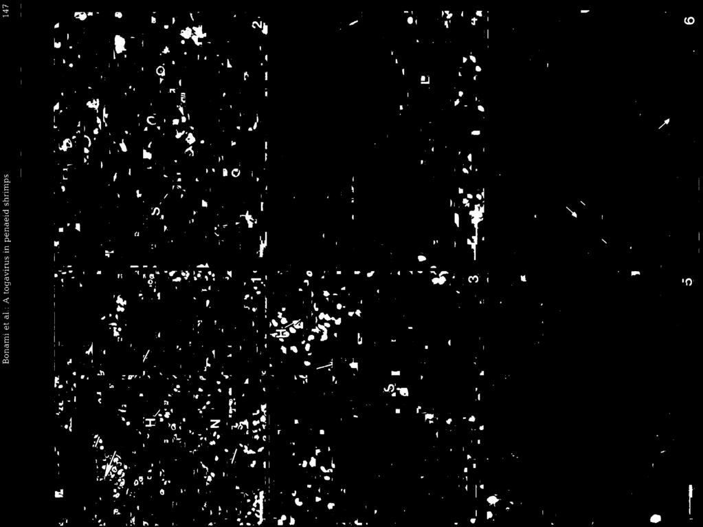
2 146 Dis. aquat. Org. 14: , 1992 We report here our results on the concentration, purification and partial characterization of a newly recognized virus that was found to be associated with lymphoid organ lesions in cultured Penaeus vannamei. For the purpose of this paper, we have named the virus LOW, which is abbreviated from 'lymphoid organ vacuolization virus', to reflect the principal lesion found to be associated with the virus. MATERIALS AND METHODS Experimental animals. The shrimp used in this study were subadult (ca 15 g average weight and 21 mo of age) Penaeus vannamei which had been reared from postlarvae obtained in June 1989 from a commercial source in Mazatlan, Sinaloa, Mexico. The shrimp were reared in isolated self-contained tanks. Each tank was eql5pped l..*?h its owr? bio!ogical fi!ter Artifjcial seawater in the tanks was circulated through a central stand pipe connected to a bottom drain Line, which in turn was connected to a biological filter. Water was returned to the rearing tank by use of air-lift pipes. Salinity was maintained at approximately 20 ppt by the addition of city water. Water temperature throughout the 21 mo growout period was maintained between 24 and 28 "C. Shrimp were fed twice daily with artificial pelleted shrimp feeds purchased from Rangen Feed Company (Buhl, ID, USA). Shrimp from this cultured population were preserved for histological and transmission electron nlicroscope (TEM) study, or frozen at -70 'C for use in later studies. Histopathology. Shrimp selected for histological study were preserved in Davidson's fixative (Humason 1979, Bell & Lightner 1988). Fixation was accomplished by first injecting ca 0.5 m1 of fixative directly into the hepatopancreas and adjacent areas of the cephalothorax of each live shrimp sampled. Then the cephalothorax was excised at its junction with the first abdominal segment, the carapace was slit just lateral to the dorsal midline using dissecting scissors, and the intact cephalothorax was immersed in fixative. After 48 h in Davldson's fixative, preserved shrimp cephalothoraces were transferred to 50 % ethanol, and then processed for routine histological examination using standard paraffin embedding procedures and hematoxylin and eosin staining methods (and other staining procedures as described in Bell & Lightner 1988). Electron microscopy. Specimens for electron microscopy were fixed live by injection into the hepatopancreas and adjacent tissues (using a 1 m1 tuberculin syringe) of cold (ca 4 "C) 6 O/O glutaraldehyde in 0.15 M Millonig's phosphate buffer (Pease 1964) supplemented with 1 % sodium chloride and 0.5 % sucrose to achieve a fixative osmolarity of 700 to 750 mosm. Subsequently, the abdomen was excised and the carapace was opened as described in the procedure for preserving shrimp with Davidson's fixative, and the shrimp were immersed whole in the same fixative. Specimens were stored in fixative at 4 "C until processed further. Lymphoid organs from the specimens were removed by dissection and postfixed in 1 % osmium tetroxide (in the same buffer as used for the glutaraldehyde fixative), dehydrated through graded alcohol, and embedded in Spurr's resin (Ladd Research Inc., Burlington, VT, USA). Ca 1 vm thick sections were prepared from each tissue block using glass knives and stained with 1 % toluidine blue (in 1 % aqueous sodium borate) for ca 1 min at 60 "C. Blocks showing potential lesions were then thin sectioned using a diamond knife on a Sorvall MT-2B ultramicrotome. Sections mounted on 200 mesh copper grids were stained with uranyl acetate and lead citrate and viewed in either an Hitachi HU-12 or H-500 electron microscope operating at 75 kv. Virus purification. Frozen shrimp which had been stored at -70 "C were used as the source of virus. Fig. 1 to 6. Penaeus vannamei infected by LOVV Plg Histological section of the lymphoid organ (LO) of a subadult shrimp displaying some essentially normal (N) areas and focal lesions. Normal areas consist of cords of L0 parenchyma1 cells which surround a prominent central hemolymph vessel (H). Areas with lesions lack a central vessel, and consist of cells which show karyomegaly and large prominent cytoplasmic vacuoles (arrows) and other inclusions. H&E stain. Scale bar = 50 pm. Higher magnification of a normal L0 cord with its central hemolymph vessel (H) and surrounding sheath cells (S). Adjacent to it are areas of L0 cells which lack a central vessel and which possess cells with prominent cytoplasmic vacuoles (V) and cytoplasmic inclusions that range from Feulgen negative, eosinophilic, and poorly defined to discrete basophilic Feulgen positive granules (G). Many affected cells are shown with slightly hypertrophied nuclei that possess marginated nuclear chromatin, leaving the nucleolus within the central portion of an otherwise hypochromatic nucleus. H&E stain. Scale bar = 40 gm. Flg. Spheroids (S), or spherical masses of L0 cells that lack a central vessel, form within focal L0 les~ons. Two are shown near a normal L0 cord with its central hemolymph vessel (H). Spheroids may contain foci of cells with hypertrophied nuclei and highly vacuolated cytoplasm (arrows). H&E stain. Scale bar = 50 pm. Fig. 4. Spheroids (S) detached from the somal L0 are shown in the hemocoel space between the L0 and a tubule of the antenna1 gland (AG]. H&E stain. Scale bar = 40 him. Fig TEM of an unorganized mass of 30 nm diameter LOW nucleocapsids. The tendency of the particles to associate with host cell membranes is somewhat evident (arrows). Scale bar = 200 nm. Fiq. 6. TEM of a cytoplasmic mass of LOW nucleocapsids which are closely associated with membranes (arrows) of host cell organelles. Scale bar = 200 nm


3
4 148 Dis. aquat. Org. 14: , 1992 Following rapid thawing, the carapace was removed prior to homogenizing the cephalothoraces in TN buffer (0.02 M Tris-HC1, 0.4 M NaC1, ph 7.4) using an Ultra-turrax tissue blender. The resultant suspension was clarified 3 times, at 2500, 6000 and rpm (5534 rotor), in runs of 10, 15 and 30 min, respectively. The first pellet obtained was resuspended in TN buffer, re-homogenized, and centrifuged 10 min at 2500 rpm, the resulting supernatant fluid was pooled with the first supernatant fluid obtalned previously, and the final supernatant fluid was pelleted 1.75 h at g. The pellet obtained was then extracted 3 or 4 times in freon (1,1,2-tnchloro-l, 2,2-trifluoroethane). This freonextracted suspension was layered on a 15 to 40 O/O (w/w) sucrose gradient and centrifuged at l l0 000 g for 3.75 h. Fractions containing presumptive virus particles were diluted in TN buffer and centrifuged at g for 1.75 h. Pellets were resuspended and subsamp!es were placed on coated grids, negatively stained with 2 % phosphotungstic acid (PTA) at ph 7.0, and evaluated for virus by TEM. Further purification of fractions containing virus was achieved on a preformed 15 to 45 O/O (w/w) CsCl gradient in 1 mm phosphate-buffered saline (PBS) ph 7.0 and run for 14.5 h at g. Virus bands were collected with a Buchler Auto-densiflow coupled to a Pharmacia fraction collector, and the absorbance of each fraction was recorded using a Pharmacia UV monitor set at a wavelength of 254 nm. Selected fractions were diluted in PBS buffer and pelleted at g for 2 h before being resuspended in a small volume of PBS for the subsequent analyses. Final preparations of purified virus were negatively stained and viewed by TEM as described previously. Virus density. The density of purified virions was determined by refractive index measurement using an Abbe refractometer of each fraction collected from a 15 to 45 % (w/w) CsCl gradient (in 1 mm PBS) after an isopycnic centrifugation at g for 15 h, and the density values plotted on the absorbance record of the gradient. SDS-PAGE. 12 O/O polyacrylamide vertical gels were run using the Laemmli (1970) buffer system (25 mm Tris, 192 mm glycine, ph 8.3) containing 0.1 % SDS. The run duration was 2 h at a constant current of 20 ma. Samples were diluted in gel sample buffer (2 % SDS, 5 M urea, 1 % 2-mercaptoethanol, 15 % glycerol, and % bromophenol blue) and heated to 100 "C for 3 min prior to loading of the samples onto the gel. Molecular weight (MW) markers were bovine albumin, egg albumin and trypsinogen, which have MWs of 66, 45 and 24 kda, respectively. After washing in fixative solution, the gels were stained by the silver stain method of Morrissey (1981). The MW of the polypeptides was estimated by measurement of the electrophoretic mobilities according to the method of Weber & Osborn (1969). RESULTS Experimental animals - history The population of Penaeus vannamei reported here showed no signs of disease or other abnormalities during the 21 mo they were cultured in a closed recirculating seawater system on the University of Arizona campus. Their 15 g average weight at the termination of the 21 mo growout is small for this age and for shrimp of this species when grown under ideal conditions. However, this final size is not unusual for shrimp reared at high density (ca 500 individuals per ) in closed recirculating systems. Histopathology The principal lesions in the lymphoid organs of these shrimp were multifocal degenerative lesions of L0 cords. Affected foci consisted of L0 parenchyma1 cells that displayed more basophilic staining characteristics (than adjacent areas with normal L0 cords) due to increased basophilia of the cytoplasm and to karyomegaly (Fig. 1). Affected L0 cords often lacked a central hemolymph vessel (Figs. 1 & 2), and in some cases formed 'spheroidal' structures composed of many cells (Figs. 3 & 4). Some affected areas contained a few pyknotic nuclei, while other nuclei were hypertrophied and possessed marginated nuclear chromatin, which left the nucleolus within the central portion of an otherwise hypochromatic nucleus (Fig. 2). Many L0 parenchymal cells contained cytoplasmic inclusion bodies that ranged from diffuse and eosinophilic to more discrete, basophilic, Feulgen positive, granular inclusions (Figs. 2 to 4). The size, location, and staining characteristics of these granules suggest that they may be either cell debris in phagosomes (with pyknotic or karyorrhectic nuclei) or virus inclusion bodies. Other Figs. 7 & 8. Flg. TEM of membrane-associated 30 nm diameter LOW nucleocapsids and LOW virions (arrows) which have acquired their envelope from the host cell membrane. Scale bar = 100 nm. Flg. TEM of purified LOVV Illustrated are full (Fn) ;~nd empty (En) nucleocaps~d showing a hexagonal profile; spherical enveloped virions (Ve); and markedly s\vollen enveloped vi.rions, several of which have their central nucleocapsids exposed (Vs). Scale bar = 150 nrn


5
6 150 Dis. aquat. Org. 14: , 1992 L0 cells contained large, very prominent cytoplasmic Buoyant density of virus particles vacuoles, some with basophilic bands of material closely associated with their membranes (Fig. 3). No After isopycnic centrifugation in CsCl gradient, the significant host inflammatory response was noted larger enveloped particles banded at a density of within or adjacent to these L0 lesions, and infiltrating g ml-l, while the smaller non-enveloped nucleocapsids hemocytes were typically not present in numbers grea- banded at g ml-' ter than in normal L0 tissue. No other lesions or abnormalities were noted in the shrimp examined. Size and structure of LOW Ultrastructure Transmission electron microscopy of selected LOS from 3 shrimp showed all three to possess cytoplasmic masses of ca 30 nm diameter virus particles (Figs. 5 & 6). The dense basophilic granular structures noted in histoiogical preparations (Figs. 2 to 4) were found to be electron-dense structures that contained no virus-like particles (not shown). Cells containing virus particles were typically vacuolated. Virus particles frequently occurred in dense membrane-bound aggregations, and sometimes showed a tendency to form crystalline arrays (Fig. 5). The virus was also found to be frequently closely associated with membranes within the infected cells (Fig. 6). In such areas, enveloped particles (measuring 50 to 60 nm in diameter) were found, with the viral nucleocapsids apparently acquiring their envelope by budding or by the invagination and fusion of membranes of the endoplasmic reticulum or Golgi around individual nucleocapsids (Fig. 7). The spherical enveloped particles, 52 to 54 nm in diameter, did not exhibit any peculiar structure, except small 7.5 nm subunits on the envelope surface (Fig. 10). The smaller hexagonal particles of 30 nm diameter exhibited 2 or 3 capsomeres on each edge of their icosahedral form, with 1 larger subunit at each of the vertices (Fig. 10). This subunit arrangement was most apparent on the vertices of empty particles (Figs. 8 to 10). The capsid, which was ca 4 nm thick, surrounded the inner area of ca 20 to 22 nm diameter. Most interesting were degraded spherical particles, which showed the 6-sided nucleocapsid through a more or less swollen envelope. Such swollen particles reached a diameter of 65 to 68 nm; on the largest swollen particles, the bilayered envelope seemed to consist of small spherical but empty subunits (Fig. 11). These virus-associated spherical subunits were also found free in the preparation and were interpreted as debris of degraded envelopes. Fig. l2 shotvs our interpretation of the structure of LOVV. Isolation and purification of LOW First attempts to purify the virus produced nonenveloped hexagonal full and empty particles of 30 to 32 nm diameter (Figs. 8 & 9). However, subsequent purification attempts ylelded virus preparations with 2 morphologically distinct particles: a 30 to 32 nm nonenveloped hexagonal particle and a spherical 50 nm diameter enveloped particle, which banded in CsCl gradients at 2 different densities. The same preparation exhibited possible evidence of a progression from one virus particle type to the other by the presence of particles showing different degrees of degradation of enveloped virions (Figs. 8, 10 & 11). Polypeptides of LOW SDS-PAGE of the 2 different virus fractions showed that the nucleocapsid contains at least 2 polypeptides, with molecular weights of 37 and 38 kda. The fractions containing enveloped particles and envelope debris show, in addition, at least a faint doublet at about 70 kda and one other polypeptide of 60 kda. In some preparations, 2 more additional bands (one of 45 kda and the other migrating with the dye, i.e. with a very low MW) were noted. These latter polypeptides are believed to be host cell contaminants rather than viral proteins. Figs. 9 to 12. Fig. 9. High-magnification TEM of full (Fn) and empty (En) ca 30 nm diameter nucleocapsids of LOVV A few particles show especially well the arrangement of capsomeres on their surfaces (arrows). Others show the presence of a large capsomere at each vertex (arrowheads) of the icosahedron, interspersed with 2 or 3 smaller subunits composing each face of the capsid. Scale bar = 25 nm. Fig. 10. High-magnification TEM of an enveloped LOW particle (Ve) showing the regularly arranged 7.5 nm diameter subunits projecting from the envelope surface (arrows). Also shown is an empty particle (En) that shows the presence of 2 or 3 capsomeres on each edge of the particle with larger subunits (arrowheads) visible at some of the vertices. Scale bar = 25 nm. Fig. 11. Enlargement of a swollen and degrading LOW virion. Subunits of the envelope are apparent (E), as is the now exposed nucleocapsid (N). Scale bar = 25 nm. Fig. 12. A graphical representation of the structure of LOVV, showing the inner 30 nm diameter icosahedral nucleocapsid within the enveloped 60 nm diameter mature virion


7
8 Dis. aquat. Org. 14: , 1992 DISCUSSION AND CONCLUSION Also acknowledged is the assistance by Tom Bell (Univ. of Arizona) in preparing the computer-generated image of LOW used as Fig. 12. Based on its size, shape, structure and virogenesis, LOVV can be related only to the Togaviridae family (Matthews 1982). Indeed, this agent develops in cyto- LITERATURE CITED plasm, where icosahedral nucleocapsids accumulate before they acquire the host-cell-derived envelope of Bell, T. A., Lghtner. D. V (1988). A handbook of normal the mature virion. Penaeid shrimp histology. World Aquaculture Society, Because so little virus-infected material was avail- Baton Rouge Brock, J. A., Lightner, able in this study, attempts to extract and characterize D. V. (1990). Diseases of Crustacea, Chap. 3.1, Diseases caused by microorganisms. In: knne, nucleic acid of LOW were unsuccessful. However, 0. (ed.) Diseases of marine animals. Vol. 111, Cephalopoda because no Feulgen-positive reaction (for DNA) associ- to Urocordata. Biologische Anstalt Helgoland, Hamburg. ated with cytoplasmic virus aggregates was detected in p histological sections of LOVV-associated L0 lesions, Humason, G. L. (1979). Animal hssue technique, 4th edn. Freeman, San Francisco we presume that the virus contains RNA. the Laemrnli, U. K. (1970). Cleavage of structural proteins during density values and the polypeptide analysis are in the assemblv of the head of bacteriovhaqe - T4. Nature, general agreement with the characteristics of the Lond. 227: Togaviridae (Matthews 1982). Hence, the discovery of Lightner. D. V., Brock, J. A. (1986). Proliferative changes of LOVV in Penaeus vannamei appears to be the first glandular and lymphoid tissues of the penaeid shrimps Penaeus monodon and P vannamei (Crustacea: report of a togavirus in any marine invertebrate animal. Decapoda) that possess characteristics of adenocarcinomas Despite the presence of detectable histopathology and lymphosarcomas. In: Samson, R. A.. Vlak, J. M,, only in the lymphoid organs of the affected Penaeus Peters, D. (eds.) Fundamental and applied aspects of vannamei, our successful attempts to purify the virus invertebrate pathology. Foundation of the Fourth International Colloquium of Invertebrate Pathology, Wageningen, from the other areas of the cephalothorax indicate the p. 469 agent is present in other cephalothoracic tissues, where Lightner, D. V., Hedrick, R. P,, Fryer. J. L., Chen. S. N., Liao. it apparently produces much more subtle histological I. C., Kou, G. H. (1987). A survey of cultured Penaeid changes. In the present study TEM examinations were shrimp in Taiwan for viral and other important diseases. Fish Path. 22: limited to lymphoid organs, and hence no evidence of Matthews, R. E. F. (1982). Class~f~cation and nomenclature of the presence of LOVV in other tissues was obtained. viruses. Intervirology 17: For the moment, nothing is known about the preva- Momssey, J. H. (1981). Silver stain for proteins in polyacrylence and pathogenicity of this virus in wild or cultured lamide gels: a modified procedure with enhanced uniform penaeid shrimp populations. However, the remarkable sensitivity. Analyt. Biochem. 117: Owens, L., De Beer, S., Smith, J. (1991). Lymphoidal parvosimilarity of histological changes noted in the lymphoid like virus in Australian prawns. Dis. aquat. Org. 11: organs of Penaeusmonodon (Lightner et al. 1987, Brock & Lightner 1990, Owens et al. 1991) suggests - - that LOVV Owens, L., Hall-Mendelin, S. (1988). Australian tropical (or a related agent) may occur as a common, if not penaeid diseases of importance to mariculture. In: perkins, F. O., Cheng, T C. (eds.) Abstracts of 3rd international significant, pathogen in Asian and Australian penaeids. colloquium on pathology in marine aquaculture. Virginia If this hypothesis is later found to be the case, it may be Institute of Marine Science, Gloucester Point. VA, p. 161 possible to purify more of the virus from Asian- and Paynter, J. L., Lightner, D. V., Lester, R. J. G. (1985). Prawn Australian-derived penaeid shrimp withlymphoid organ virus from juvenile Penaeus esculentus. In: Rothlisberg, lesions and subsequently to complete characterization of P C., Hill, B. J., Staples, D J (eds.) Second Australian natlonal prawn seminar. Nahonal Pratvn Seminar 2, this newly recognized type of virus in penaeid shrimp. Cleveland, Australia, p Weber, K., Osborn, M. (1969). The reliability of molecular Acknowledgements. This study was funded in part by the U.S. weight determination by dodecyl sulfate-polyacrylamide Department of Agriculture through grant number CSRS 88- gel electrophoresis. J. biol. Chem. 244: 440M412 Responsible Subject Editor: J E. Stewart, Dartmouth, N.S., Canada Manuscript first recezved: Apnl 16, 1992 Revised version accepted: June 30, 1992
Short communication. C W Tung, C S Wang and S N Chen
 Short communication Histological and electron microscopic study on Macrobrachium muscle virus (MMV) infection in the giant freshwater prawn, Macrobrachium rosenbergii (de Man), cultured in Taiwan C W Tung,
ELECTRON MICROSCOPIC STUDIES ON EQUINE ENCEPHALOSIS VIRUS
 Onderstepoort]. vet. Res. 40 (2), 53-58 (1973) ELECTRON MICROSCOPIC STUDIES ON EQUINE ENCEPHALOSIS VIRUS G. LECATSAS, B. J. ERASMUS and H. J. ELS, Veterinary Research Institute, Onderstepoort ABSTRACT
Ultrastructure of Mycoplasmatales Virus laidlawii x
 J. gen. Virol. (1972), I6, 215-22I Printed in Great Britain 2I 5 Ultrastructure of Mycoplasmatales Virus laidlawii x By JUDY BRUCE, R. N. GOURLAY, AND D. J. GARWES R. HULL* Agricultural Research Council,
(From The Rockefeller Institute) Materials and Methods. Observations with the Electron Microscope
 Materials and Methods. Observations with the Electron Microscope") ELECTRON MICROSCOPE STUDY OF THE DEVELOPMENT OF THE PAPILLOMA VIRUS IN THE SKIN OF THE RABBIT* BY ROBERT S. STONE,~ M.D., RICHARD E. SHOPE, M.D., DAN H. MOORE, P,~.D. (From The Rockefeller Institute) PLATES
ELECTRON MICROSCOPE STUDY OF THE DEVELOPMENT OF THE PAPILLOMA VIRUS IN THE SKIN OF THE RABBIT* BY ROBERT S. STONE,~ M.D., RICHARD E. SHOPE, M.D., DAN H. MOORE, P,~.D. (From The Rockefeller Institute) PLATES
psittaci by Silver-Methenamine Staining and
 JOURNAL OF BACTERIOLOGY, July 1972, p. 267-271 Copyright 1972 American Society for Microbiology Vol. 111, No. 1 Printed in U.S.A. Location of Polysaccharide on Chlamydia psittaci by Silver-Methenamine
ELECTRON MICROSCOPIC STUDY OF THE FORMATION OF BLUETONGUE VIRUS*
 Onderstepoort J. vet. Res. (1968), 35 (1), 139-150 Printed in the Repub. of S. Afr. by The Government Printer, Pretoria ELECTRON MICROSCOPIC STUDY OF THE FORMATION OF BLUETONGUE VIRUS* G. LECATSAS, Veterinary
New aspect of hepatic nuclear glycogenosis
 J. clin. Path. (1968), 21, 19 New aspect of hepatic nuclear glycogenosis in diabetes1 F. CARAMIA, F. G. GHERGO, C. BRANCIARI, AND G. MENGHINI From the Institute of General Pathology, University of Rome,
Electron Microscope Studies of HeLa Cells Infected with Herpes Virus
 244 STOKER, M. G. P., SMITH, K. M. & Ross, R. W. (1958). J. gen. Microbiol. 19,244-249 Electron Microscope Studies of HeLa Cells Infected with Herpes Virus BY M: G. P. STOKER, K. M. SMITH AND R. W. ROSS
Intercellular Matrix in Colonies of Candida
 JouRNAL OF BAcTEROLOGY, Sept. 1975, p. 1139-1143 Vol. 123, No. 3 Copyright 0 1975 American Society for Microbiology Printed in U.S.A. ntercellular Matrix in Colonies of Candida K. R. JOSH, J. B. GAVN,*
Electron Microscopic Evidence of Bacilliform Virus Infection in Kuruma Shrimp (Penaeus japonicus)
") Electron Microscopic Evidence Bacilliform Virus Infection in Kuruma Shrimp (Penaeus japonicus) Yukinori Takahashi*1, Toshiaki Itami*1, Masakazu Kondo*2, Minoru Maeda*1, Reiko Fujii*3, Susumu Tomonaga3,
Electron Microscopic Evidence Bacilliform Virus Infection in Kuruma Shrimp (Penaeus japonicus) Yukinori Takahashi*1, Toshiaki Itami*1, Masakazu Kondo*2, Minoru Maeda*1, Reiko Fujii*3, Susumu Tomonaga3,
A White Spot Disease - like syndrome in the Pacific Blue Shrimp (Litopenaeus stylirostris) as a form of bacterial shell disease
 as a form of bacterial shell disease") Aquaculture MARCH 2000; 183 (1-2) : 25-30 http://dx.doi.org/ 10.1016/S0044-8486(99)00284-7 @ 2004 Elsevier B.V. All rights reserved Archimer http://www.ifremer.fr/docelec/ Archive Institutionnelle de l
Aquaculture MARCH 2000; 183 (1-2) : 25-30 http://dx.doi.org/ 10.1016/S0044-8486(99)00284-7 @ 2004 Elsevier B.V. All rights reserved Archimer http://www.ifremer.fr/docelec/ Archive Institutionnelle de l
The Syndrome of Sea Cucumber (Apostichopus japonicus) Infected. by Virus and Bacteria *
 Infected. by Virus and Bacteria *") VIROLOGICA SINICA, February 2008, 23 (1):63-67 DOI 10.1007/s12250-008-2863-9 CLC number: Q938.8 Document code: A Article ID: 1674-0769 (2008) 01-0063-05 The Syndrome of Sea Cucumber (Apostichopus japonicus)
VIROLOGICA SINICA, February 2008, 23 (1):63-67 DOI 10.1007/s12250-008-2863-9 CLC number: Q938.8 Document code: A Article ID: 1674-0769 (2008) 01-0063-05 The Syndrome of Sea Cucumber (Apostichopus japonicus)
Taura Syndrome Virus Disease in Farm-Reared Penaeus monodon in Thailand
 Kasetsart J. (Nat. Sci.) 41 : 319-323 (2007) Taura Syndrome Virus Disease in Farm-Reared Penaeus monodon in Thailand Chalor Limsuwan and Niti Chuchird* ABSTRACT Taura syndrome virus (TSV) has caused major
Epstein-Barr Virus: Stimulation By 5 '-Iododeoxy uridine or 5 '-Brom odeoxy uridine in Human Lymphoblastoid Cells F ro m a Rhabdom yosarcom a*
 A n n a ls o f C l i n i c a l L a b o r a t o r y S c i e n c e, Vol. 3, No. 6 Copyright 1973, Institute for Clinical Science Epstein-Barr Virus: Stimulation By 5 '-Iododeoxy uridine or 5 '-Brom odeoxy
Ultrastructure of Connective Tissue Cells of Giant African Snails Achatina fulica (Bowdich)
") Kasetsart J. (Nat. Sci.) 36 : 285-290 (2002) Ultrastructure of Connective Tissue Cells of Giant African Snails Achatina fulica (Bowdich) Viyada Seehabutr ABSTRACT The connective tissue sheath of cerebral
Kasetsart J. (Nat. Sci.) 36 : 285-290 (2002) Ultrastructure of Connective Tissue Cells of Giant African Snails Achatina fulica (Bowdich) Viyada Seehabutr ABSTRACT The connective tissue sheath of cerebral
Yara Saddam. Amr Alkhatib. Ihsan
 1 Yara Saddam Amr Alkhatib Ihsan NOTE: Yellow highlighting=correction/addition to the previous version of the sheet. Histology (micro anatomy) :- the study of tissues and how they are arranged into organs.
STUDY ON RAINBOW TROUT NODULAR GILL DISEASE DETECTED IN POLAND
 Bull Vet Inst Pulawy 51, 547-551, 2007 STUDY ON RAINBOW TROUT NODULAR GILL DISEASE DETECTED IN POLAND JERZY ANTYCHOWICZ Department of Fish Diseases, National Veterinary Research Institute, 24-100 Pulawy,
Mammalian Melanosomal Proteins: Characterization by Polyacrylamide Gel Electrophoresis
 YALE JOURNAL OF BIOLOGY AND MEDICINE 46, 553-559 (1973) Mammalian Melanosomal Proteins: Characterization by Polyacrylamide Gel Electrophoresis VINCENT J. HEARING AND MARVIN A. LUTZNER Dermatology Branch,
Monodon baculovirus from Australia: ultrastructural observations
 DISEASES OF AQUATIC ORGANISMS Dis Aquat Org Published February 9 Monodon baculovirus from Australia: ultrastructural observations 'Department of Microbiology, 'Department of Parasitology and 3Centre for
Ultrastructural Study of Human Natural Killer CNK) Cell*)
 Cell*)") Hiroshima Journal of Medical Sciences Vol. 31, No. 1, March, 1982 HJIM 31-6 31 Ultrastructural Study of Human Natural Killer CNK) Cell*) Yoshinori KAWAGUCHI, Eishi KITTAKA, Yoshito TANAKA, Takeo TANAKA
Hiroshima Journal of Medical Sciences Vol. 31, No. 1, March, 1982 HJIM 31-6 31 Ultrastructural Study of Human Natural Killer CNK) Cell*) Yoshinori KAWAGUCHI, Eishi KITTAKA, Yoshito TANAKA, Takeo TANAKA
bacterial shell disease
 Ž. Aquaculture 183 2000 25 30 www.elsevier.nlrlocateraqua-online A white spot disease-like syndrome in the Pacific blue shrimp ž Litopenaeus stylirostris/ as a form of bacterial shell disease Cyrille Goarant
Reo-like virus in white shrimp Penaeus vannamei (Crustacea: Decapoda): CO-occurrence with Baculovirus penaei in experimental infections
: CO-occurrence with Baculovirus penaei in experimental infections") Vol. 8: 45-49, 1990 DISEASES OF AQUATIC ORGANISMS Dis. aquat. Org. Published March 6 Reo-like virus in white shrimp Penaeus vannamei (Crustacea: Decapoda): CO-occurrence with Baculovirus penaei in experimental
Vol. 8: 45-49, 1990 DISEASES OF AQUATIC ORGANISMS Dis. aquat. Org. Published March 6 Reo-like virus in white shrimp Penaeus vannamei (Crustacea: Decapoda): CO-occurrence with Baculovirus penaei in experimental
Electron Microscopy of Small Cells: Mycoplasma hominis
 JOURNAL of BAcTRiowOY, Dc. 1969, p. 1402-1408 Copyright 0 1969 American Society for Microbiology Vol. 100, No. 3 Printed In U.S.A. NOTES Electron Microscopy of Small Cells: Mycoplasma hominis JACK MANILOFF
Basophilic. Basophilic structures are stained by basic dyes: Mnemonic: Basophilic = Blue
 Cell Overview Basophilic Basophilic structures are stained by basic dyes: Basic dyes are positive Basophilic structures are negative (ex. DNA, RNA, ribosomes, RER) Mnemonic: Basophilic = Blue Acidophilic
Ultrastructural Comparison of a Virus from a Rhesus-Monkey Mammary Carcinoma with Four Oncogenic RNA Viruses
 Proc. Nat. Acad. Sci. USA Vol. 68, No. 7, pp. 1603-1607, July 1971 Ultrastructural Comparison of a Virus from a Rhesus-Monkey Mammary Carcinoma with Four Oncogenic RNA Viruses (primate cancer/murine mammary
Cell Overview. Hanan Jafar BDS.MSc.PhD
 Cell Overview Hanan Jafar BDS.MSc.PhD THE CELL is made of: 1- Nucleus 2- Cell Membrane 3- Cytoplasm THE CELL Formed of: 1. Nuclear envelope 2. Chromatin 3. Nucleolus 4. Nucleoplasm (nuclear matrix) NUCLEUS
FIRST DESCRIPTION OF AVIAN PAPILLOMAVIRUS INFECTION IN GYPS FULVUS, ITALY
 "Endangered large carnivores and scavenging raptors in Europe Faculty of Veterinary Medicine, Teramo 13-15 October2016 FIRST DESCRIPTION OF AVIAN PAPILLOMAVIRUS INFECTION IN GYPS FULVUS, ITALY Cristina
ON THE PRESENCE OF A CILIATED COLUMNAR EPITHELIAL CELL TYPE WITHIN THE BOVINE CERVICAL MUCOSA 1
 ON THE PRESENCE OF A CILIATED COLUMNAR EPITHELIAL CELL TYPE WITHIN THE BOVINE CERVICAL MUCOSA 1 R. I. Wordinger, 2 J. B. Ramsey, I. F. Dickey and I. R. Hill, Jr. Clemson University, Clemson, South Carolina
(Plates LXVIII-LXXI)
") [GANN, 54, 481-486; December, 1963] UDC 616.155.392-076.4:578.69 VIRUS-LIKE PARTICLES IN HUMAN CHLOROLEUKEMIA CELLS (Plates LXVIII-LXXI) Zensuke OTA, Shin-ya SUZUKI, and Satoru HIGASHI (Department of Internal
[GANN, 54, 481-486; December, 1963] UDC 616.155.392-076.4:578.69 VIRUS-LIKE PARTICLES IN HUMAN CHLOROLEUKEMIA CELLS (Plates LXVIII-LXXI) Zensuke OTA, Shin-ya SUZUKI, and Satoru HIGASHI (Department of Internal
Taura syndrome of marine penaeid shrimp: characterization of the viral agent
 Journal of General Virology (1997), 78, 313 319. Printed in Great Britain...... Taura syndrome of marine penaeid shrimp: characterization of the viral agent Jean-Robert Bonami, 1 Kenneth W. Hasson, 2 Jocelyne
Necrotizing Hepatopancreatitis
 [DRAFT] Necrotizing Hepatopancreatitis Pathogen information 1. Causative agent 1.1. Pathogen type Obligate intracellular rickettsial-like organism 1.2. Disease name and synonyms Necrotizing Hepatopancreatitis
DISEASES OF AQUATIC ORGANISMS Dis. aquat. Org. Lymphoidal parvovirus-like particles in Australian penaeid prawns
 Vol. 11: 129-134, 1991 DISEASES OF AQUATIC ORGANISMS Dis. aquat. Org. ' Published August 8 Lymphoidal parvovirus-like particles in Australian penaeid prawns Leigh Owens, Steve De Beer, Jan Smith Graduate
Evaluation of Chromatin Clumping and Myelination of the Spinal Cord of Pigs with Congenital Tremor
 Vet. Pathol. 12: 1-5 (1975) Evaluation of Chromatin Clumping and Myelination of the Spinal Cord of Pigs with Congenital Tremor C.H. LAMAR and D.C. VAN SICKLE School of Veterinary Medicine, Purdue University,
DISEASES OF AQUATIC ORGANISMS Dis. aquat. Org.
 Vol. 12: 229-233.1992 DISEASES OF AQUATIC ORGANISMS Dis. aquat. Org. Published April 23 NOTE Erythrocytic inclusion body syndrome: a light and electron microscopic study of infected erythrocytes of chinook
2.1.2 General Procedures for Electron Microscopy Applications in Diagnostic Virology
 2.1.2 General Procedures for Electron Microscopy Applications in Diagnostic Virology Jan Lovy 1 and Dorota W. Wadowska 2 1 N.J. Division of Fish & Wildlife, Office of Fish & Wildlife Health & Forensics,
Protocol for protein SDS PAGE and Transfer
 Protocol for protein SDS PAGE and Transfer According to Laemmli, (1970) Alaa El -Din Hamid Sayed, Alaa_h254@yahoo.com Serum Selection of a protein source cell cultures (bacteria, yeast, mammalian, etc.)
ELECTRON MICROSCOPY OF A SMALL PIGMENTED CUTANEOUS LESION*
 ELECTRON MICROSCOPY OF A SMALL PIGMENTED CUTANEOUS LESION* The description of the lesion in the title of this rcport is intentionally non-committal. Diagnosed clinically as a lentigo, it was removed as
Periodic occurrence of epithelial viral necrosis outbreaks in Penaeus vannamei in Ecuador
 DISEASES OF AQUATIC ORGANISMS Vol. 42: 91 99, Published August 31 Dis Aquat Org Periodic occurrence of epithelial viral necrosis outbreaks in Penaeus vannamei in Ecuador R. Jimenez*, R. Barniol, L. de
STRUCTURE, GENERAL CHARACTERISTICS AND REPRODUCTION OF VIRUSES
 STRUCTURE, GENERAL CHARACTERISTICS AND REPRODUCTION OF VIRUSES Introduction Viruses are noncellular genetic elements that use a living cell for their replication and have an extracellular state. Viruses
A Compact and a Dispersed Form of the Golgi Apparatus
 A Compact and a Dispersed Form of the Golgi Apparatus of Fish Liver 1 D. James Morre and Carole A. Lembi Department of Botany and Plant Pathology Purdue University, Lafayette, Indiana 47907, and H. H.
Yara shwabkeh. Osama Alkhader. Heba Kalbouneh
 2 Yara shwabkeh Osama Alkhader Heba Kalbouneh CELL OVERVIEW -Note ; the important thing is to know how the organelles appear under the microscope - the stains we usually use in Histology are composed of
The Microscopic World of Cells. The Microscopic World of Cells. The Microscopic World of Cells 9/21/2012
 Organisms are either: Single-celled, such as most prokaryotes and protists or Multicelled, such as plants, animals, and most fungi How do we study cells? Light microscopes can be used to explore the structures
ISOLATION OF A SARCOMA VIRUS FROM A SPONTANEOUS CHICKEN TUMOR
 ISOLATION OF A SARCOMA VIRUS FROM A SPONTANEOUS CHICKEN TUMOR Shigeyoshi ITOHARA, Kouichi HIRATA, Makoto INOUE, Masanori Veterinary Pathology, Faculty of Agriculture, Yamaguchi University* HATSUOKA, and
Expression of acid base transporters in the kidney collecting duct in Slc2a7 -/-
 Supplemental Material Results. Expression of acid base transporters in the kidney collecting duct in Slc2a7 -/- and Slc2a7 -/- mice. The expression of AE1 in the kidney was examined in Slc26a7 KO mice.
Penetration of Host Cell Membranes by Adenovirus 2
 JOURNAL OF VIROLOGY, Aug. 1973, p. 386-396 Copyright 1973 American Society for Microbiology Vol. 12, No. 2 Printed in U.S.A. Penetration of Host Cell Membranes by Adenovirus 2 DENNIS T. BROWN' AND BYRON
LESSON 1.4 WORKBOOK. Viral sizes and structures. Workbook Lesson 1.4
 Eukaryotes organisms that contain a membrane bound nucleus and organelles. Prokaryotes organisms that lack a nucleus or other membrane-bound organelles. Viruses small, non-cellular (lacking a cell), infectious
Thursday, October 16 th
 Thursday, October 16 th Good morning. Those of you needing to take the Enzymes and Energy Quiz will start very soon. Students who took the quiz Wednesday: Please QUIETLY work on the chapter 6 reading guide.
Some Observations on the Fine Structure of the Goblet Cells. Special Reference to the Well-Developed Agranular Endoplasmic Reticulum
 Okajimas Folia Anat. Jpn., 58(4-6) : 583-594, March 1982 Some Observations on the Fine Structure of the Goblet Cells in the Nasal Respiratory Epithelium of the Rat, with Special Reference to the Well-Developed
Polypeptides of Respiratory Syncytial Virus
 JOURNAL OF VIROLOGY, Jan. 1977, p. 427-431 Vol. 21, No. 1 Copyright C 1977 American Society for Microbiology Printed in U.S.A. Polypeptides of Respiratory Syncytial Virus SEYMOUR LEVINE Department ofimmunology
Microbiology Chapter 7 Viruses
 Microbiology Chapter 7 Viruses 7:1 Viral Structure and Classification VIRUS: a biological particle composed of genetic material (DNA or RNA) encased in a protein coat CAPSID: protein coat surrounding a
THE PREPARATION AND ULTRASTRUCTURE OF AVIAN ERYTHROCYTE NUCLEAR ENVELOPE ENCLOSED BY THE PLASMA MEMBRANE
 J. Cell Sci. 34, 81-90 (1978) 8l Printed in Great Britain Company of Biologists Limited igj8 THE PREPARATION AND ULTRASTRUCTURE OF AVIAN ERYTHROCYTE NUCLEAR ENVELOPE ENCLOSED BY THE PLASMA MEMBRANE JAMES
the structure of their ducts has been
 Tza JOURNAL 0? INVEa'riGATrVN DEBMATOLOOT Copyright t 1966 by The Williams & Wilkins Co. Vol. 46, No. I Printed in U.S.A. AN ELECTRON MICROSCOPIC STUDY OF THE ADULT HUMAN APOCRINE DUCT* KEN HASHIMOTO,
STUDIES OF THE HUMAN UNFERTILIZED TUBAL OVUM*t
 FERTILITY AND STERILITY Copyright @ 1973 by The Williams & Wilkins Co. Vol. 24, No.8, August 1973 Printed in U.S.A. STUDIES OF THE HUMAN UNFERTILIZED TUBAL OVUM*t C. NORIEGA, M.D., AND C. OBERTI, M.D.
The Viability of Taura Syndrome Virus in Low-salinity Water
 Kasetsart J. (Nat. Sci.) 39 : 406-410 (2005) The Viability of Taura Syndrome Virus in Low-salinity Water Niti Chuchird and Chalor Limsuwan ABSTRACT Taura syndrome virus (TSV) could survive up to 10 days
7'cA 5P 'Z /' IA.N 5 '7S abra'ry TECHNiCAL F:-'.. THE INSTITUTE OF PAPER CHEMISTRY, APPLETON, WISCONSIN IPC TECHNICAL PAPER SERIES NUMBER 16
 7'cA 5P 'Z /' IA.N 5 '7S abra'ry TECHNiCAL F:-'.. THE INSTITUTE OF PAPER CHEMISTRY, APPLETON, WISCONSIN IPC TECHNICAL PAPER SERIES NUMBER 16 DIFFERENTIATION OF TANNIN, LIPID, AND STARCH IN CULTURED PLANT
Determination of the Distribution of Cilia on the Surface of the Mantle of Cypraea caputserpentis utilizing Scanning Electron Microscopy
 Determination of the Distribution of Cilia on the Surface of the Mantle of Cypraea caputserpentis utilizing Scanning Electron Microscopy DURATION September 10, 1990- May 7, 1991 Tracie A. Yokoi Advisor
Notes Chapter 7 Cell Structure and Function Hooke looked at cork under a simple microscope and found tiny chambers he named cells.
 Notes Chapter 7 Cell Structure and Function 7.1 Cell discovery and Theory 1665 Hooke looked at cork under a simple microscope and found tiny chambers he named cells. Cells are the basic structural and
Further Observations on the Structure of Influenza Viruses A and C
 J. gen. ViroL (I969), 4, 365-370 With 2 plates Printed in Great Britain 365 Further Observations on the Structure of Influenza Viruses A and C By K. APOSTOLOV The Wellcome Research Laboratories, Beckenham,
WSC , Conference 9, Case 1. Tissue from a nyala.
 WSC 2009-2010, Conference 9, Case 1. Tissue from a nyala. MICROSCOPIC DESCRIPTION: Heart, atrium (1 pt.): Approximately 40% of the atrial myocardium is replaced by areas of fibrous connective tissue (1
Ultrastructural studies of human cutaneous nerve
 J. clin. Path. (1965), 18, 188 Ultrastructural studies of human cutaneous nerve with special reference to lamellated cell inclusions and vacuole-containing cells MARJORE J. EVANS, J. B. FNEAN, AND A. L.
Fine Structure of the Normal Trigeminal Ganglion in the Cat and Monkey*
 Fine Structure of the Normal Trigeminal Ganglion in the Cat and Monkey* DAVID S. MAXWELL, PH.D. Principal Contributor and Leader of Discussion HE inclusion of animal material m a y be justified as a means
Structural vs. nonstructural proteins
 Why would you want to study proteins associated with viruses or virus infection? Receptors Mechanism of uncoating How is gene expression carried out, exclusively by viral enzymes? Gene expression phases?
Acta Medica Okayama. Electron microscopic demonstration of a new virus isolated from a patient with SMON. Zensuke Ota DECEMBER 1970
 Acta Medica Okayama Volume 24, Issue 6 1970 Article 3 DECEMBER 1970 Electron microscopic demonstration of a new virus isolated from a patient with SMON Zensuke Ota Okayama University, Copyright c 1999
ELECTRON MICROSCOPIC STUDIES ON REOVIRUS TYPE I IN BHK 21 CELLS
 Onderstepoort J. vet. Res. (1968), 35 (1), 151-158 Printed in the Republic of S. Afr. by the Government Printer, Pretoria ELECTRON MICROSCOPIC STUDIES ON REOVIRUS TYPE I IN BHK 21 CELLS G. LECATSAS, Veterinary
ULTRASTRUCTURAL CHANGES IN THE INFECTIVE LARVAE OF NIPPOSTRONGYLUS BRASILIENSIS IN THE SKIN OF IMMUNE MICE
 ULTRASTRUCTURAL CHANGES IN THE INFECTIVE LARVAE OF NIPPOSTRONGYLUS BRASILIENSIS IN THE SKIN OF IMMUNE MICE by D. L. Lee ABSTRACT Infective stage larvae of Nippostrongylus brasiliensis are immobilized within
INVESTIGATIVE OPHTHALMOLOGY. Corneal and conjunctival changes in dysproteinemia
 August 1969 Volume 8, Number 4 INVESTIGATIVE OPHTHALMOLOGY Corneal and conjunctival changes in dysproteinemia 7?. M. H. Pinkerton and David M. Robertson A case of dysproteinemia with corneal and conjunctival
Chapter 7. (7-1 and 7-2) A Tour of the Cell
 A Tour of the Cell") Chapter 7 (7-1 and 7-2) A Tour of the Cell Microscopes as Windows to the World of Cells Cells were first described in 1665 by Robert Hooke. By the mid-1800s, the accumulation of scientific evidence led
Chapter 7 (7-1 and 7-2) A Tour of the Cell Microscopes as Windows to the World of Cells Cells were first described in 1665 by Robert Hooke. By the mid-1800s, the accumulation of scientific evidence led
Questions in Cell Biology
 Name: Questions in Cell Biology Directions: The following questions are taken from previous IB Final Papers on the subject of cell biology. Answer all questions. This will serve as a study guide for the
Published Online: 25 November, 1956 Supp Info: on November 16, 2018 jcb.rupress.org Downloaded from
 Published Online: 25 November, 1956 Supp Info: http://doi.org/10.1083/jcb.2.6.799 Downloaded from jcb.rupress.org on November 16, 2018 B~IEF NOrmS 799 Permanganate--A New Fixative for Electron Microscopy.*
THE FORM OF HAEMOGLOBIN IN THE ERYTHROCYTES OF THE COD, GADUS CALLARIAS
 J. Cell Set. 8, 407-412 (1971) 407 Printed in Great Britain THE FORM OF HAEMOGLOBIN IN THE ERYTHROCYTES OF THE COD, GADUS CALLARIAS N.W.THOMAS Department of Anatomy, Marischal College, Aberdeen, Scotland
Synthesis of Proteins in Cells Infected with Herpesvirus,
 Proceedings of the National Academy of Science8 Vol. 66, No. 3, pp. 799-806, July 1970 Synthesis of Proteins in Cells Infected with Herpesvirus, VI. Characterization of the Proteins of the Viral Membrane*
Chapter13 Characterizing and Classifying Viruses, Viroids, and Prions
 Chapter13 Characterizing and Classifying Viruses, Viroids, and Prions 11/20/2017 MDufilho 1 Characteristics of Viruses Viruses Minuscule, acellular, infectious agent having either DNA or RNA Cause infections
10/13/11. Cell Theory. Cell Structure
 Cell Structure Grade 12 Biology Cell Theory All organisms are composed of one or more cells. Cells are the smallest living units of all living organisms. Cells arise only by division of a previously existing
LYMPH GLAND. By : Group 1
 LYMPH GLAND By : Group 1 ANATOMY LYMPH NODE Lymphatic Organs Red bone marrow Thymus gland Lymph nodes Lymph nodules Spleen Primary organs Secondary organs Lymph Nodes Firm, smooth-surfaced, bean-shaped
Viral structure م.م رنا مشعل
 Viral structure م.م رنا مشعل Viruses must reproduce (replicate) within cells, because they cannot generate energy or synthesize proteins. Because they can reproduce only within cells, viruses are obligate
ENHANCEMENT OF THE GRANULATION OF ADRFNERGIC STORAGE VESICLES IN DRUG-FREE SOLUTION
 ENHANCEMENT OF THE GRANULATION OF ADRFNERGIC STORAGE VESICLES IN DRUG-FREE SOLUTION TAKASHI IWAYAMA and J. B. FURNESS. From the Department of Zoology, University of Melbourne, Victoria, Australia. Dr.
Eukaryotic cells are essentially two envelope systems. Nuclear materials are separated from cytoplasm by nuclear membrane. Complex structure Also
 Dr. Gugale Pritesh Ramanlal M.Sc., Ph.D., B.Ed., D.M.L.T. Email id - pritesh.gugale09@gmail.com Contact numbernumber- 8446475310 Eukaryotic cells are essentially two envelope systems. Nuclear materials
MYOFIBRILLAR STRUCTURAL CHANGES CAUSED BY MARINATION WITH CALCIUM PHOSPHATE OR CALCIUM CHLORIDE AND SODIUM PYROPHOSPHATE
 Cattlemen s Day 2002 MYOFIBRILLAR STRUCTURAL CHANGES CAUSED BY MARINATION WITH CALCIUM PHOSPHATE OR CALCIUM CHLORIDE AND SODIUM PYROPHOSPHATE T. E. Lawrence, A. T. Waylan, and C. L. Kastner Summary Ultrastructural
The Ultrastructure of Canine Cutaneous Papilloma1
 [CANCER RESEARCH 29, 2079-2084, November 1969] The Ultrastructure of Canine Cutaneous Papilloma1 A. M. Watrach College of Veterinary Medicine, University of Illinois, Urbana, Illinois 61801 SUMMARY The
BIOSC 041. v Today s lecture. v Today s lab. v Note- Monday is a holiday good time to do some reading!
 BIOSC 041 v Today s lecture Review questions Chapter 6, Cells More review questions v Today s lab Quick review of lab safety The Scientific Method start thinking about which environments you might want
Disorders of Cell Growth & Neoplasia. Histopathology Lab
 Disorders of Cell Growth & Neoplasia Histopathology Lab Paul Hanna April 2010 Case #84 Clinical History: 5 yr-old, West Highland White terrier. skin mass from axillary region. has been present for the
Cell Theory All living matter is composed of one or more The cell is the structural and functional unit of life All cells come from pre-existing cell
 Cell Theory All living matter is composed of one or more The cell is the structural and functional unit of life All cells come from pre-existing cell Prokeryotic Bacteria or archaea Cell wall, small circular
The Fine Structure of the Epithelial Cells of the Mouse Prostate* II. Ventral Lobe Epithelium
 Published Online: 1 June, 1960 Supp Info: http://doi.org/10.1083/jcb.7.3.511 Downloaded from jcb.rupress.org on September 28, 2018 The Fine Structure of the Epithelial Cells of the Mouse Prostate* II.
Medical Biology. Dr. Khalida Ibrahim
 Dr. Khalida Ibrahim Medical Biology MUSCLE TISSUE 1. Muscle tissue is characterized by its well-developed properties of contraction. 2. Muscle is responsible for the movements of the body and the various
VIRUS TAXONOMY AND REPLICATION
 VIRUS TAXONOMY AND REPLICATION Paulo Eduardo Brandão, PhD Department of Preventive Veterinary Medicine and Animal Health School of Veterinary Medicine University of São Paulo, Brazil I. VIRUS STRUCTURE
Structural biology of viruses
 Structural biology of viruses Biophysical Chemistry 1, Fall 2010 Coat proteins DNA/RNA packaging Reading assignment: Chap. 15 Virus particles self-assemble from coat monomers Virus Structure and Function
Avian Pathology. Bacterial diseases: histo slides. ECVP-ESVP Summer School 2012 Frédérique NGUYEN
 Avian Pathology Bacterial diseases: histo slides ECVP-ESVP Summer School 2012 Frédérique NGUYEN Bacterial diseases: histo slides B1. Turkey. Organs? Morphologic diagnosis? Special procedure? B2. Hen. Organ?
Title. Author(s)SONODA, Mitsuo; KOBAYASHI, Kosaku. CitationJapanese Journal of Veterinary Research, 18(3): 125- Issue Date DOI. Doc URL.
SONODA, Mitsuo; KOBAYASHI, Kosaku. CitationJapanese Journal of Veterinary Research, 18(3): 125- Issue Date DOI. Doc URL.") Title PLASMACYTOID CELLS OF CANINE PERIPHERAL BLOOD IN ELE Author(s)SONODA, Mitsuo; KOBAYASHI, Kosaku CitationJapanese Journal of Veterinary Research, 18(3): 125- Issue Date 1970-09 DOI 10.14943/jjvr.18.3.125
Title PLASMACYTOID CELLS OF CANINE PERIPHERAL BLOOD IN ELE Author(s)SONODA, Mitsuo; KOBAYASHI, Kosaku CitationJapanese Journal of Veterinary Research, 18(3): 125- Issue Date 1970-09 DOI 10.14943/jjvr.18.3.125
Detection of Hepatitis A Antigen in Human Liver
 INFECTION AND IMMUNITY, Apr. 1982, p. 320-324 0019-9567/82/040320-05$02.00/0 Vol. 36, No. 1 Detection of Hepatitis A Antigen in Human Liver YOHKO K. SHIMIZU,'* TOSHIO SHIKATA,' PAUL R. BENINGER,2 MICHIO
PHARMACEUTICAL MICROBIOLOGY JIGAR SHAH INSTITUTE OF PHARMACY NIRMA UNIVERSITY
 PHARMACEUTICAL MICROBIOLOGY JIGAR SHAH INSTITUTE OF PHARMACY NIRMA UNIVERSITY VIRUS - HISTORY In 1886, the Dutch Chemist Adolf Mayer showed TMD In 1892, the Russian Bactriologist Dimtri Iwanowski isolate
Dako IT S ABOUT TIME. Interpretation Guide. Agilent Pathology Solutions. ALK, ROS1 and RET IQFISH probes (Dako Omnis) MET IQFISH probe (Dako Omnis)
 MET IQFISH probe (Dako Omnis)") INTERPRETATION Dako Agilent Pathology Solutions IQFISH Interpretation Guide ALK, ROS1 and RET IQFISH probes (Dako Omnis) MET IQFISH probe (Dako Omnis) IT S ABOUT TIME For In Vitro Diagnostic Use ALK, ROS1,
INTERPRETATION Dako Agilent Pathology Solutions IQFISH Interpretation Guide ALK, ROS1 and RET IQFISH probes (Dako Omnis) MET IQFISH probe (Dako Omnis) IT S ABOUT TIME For In Vitro Diagnostic Use ALK, ROS1,
AET-treated normal red cells (PNH-like cells)
") J. clin. Path., 1971, 24, 677-684 Electron microscope study of PNH red cells and AET-treated normal red cells (PNH-like cells) S. M. LEWIS, G. LAMBERTENGHI, S. FERRONE, AND G. SIRCHIA From the Department
J. clin. Path., 1971, 24, 677-684 Electron microscope study of PNH red cells and AET-treated normal red cells (PNH-like cells) S. M. LEWIS, G. LAMBERTENGHI, S. FERRONE, AND G. SIRCHIA From the Department
The Zombies of the Scientific Community Viruses and Other Acellular Infectious Agents. Acellular Agents
 viruses protein and nucleic acid viroids RNA virusoids RNA prions proteins The Zombies of the Scientific Community Viruses and Other Acellular Infectious Agents Acellular Agents Viruses major cause of
APPENDIX 1 ETHICAL CLEARANCE
 APPENDIX 1 ETHICAL CLEARANCE 75 APPENDIX 2 76 PROCEDURE FOR PREPARING OF LIVER HISTOLOGY SLIDES Overview: Histology involves the use of a set of techniques to examine the morphology, architecture and composition
Cell Structure and Function
 Household pin w/ bactera Cell Structure and Function Chapter 4 Same bacteria on pinhead Fig. 4-1c, p.50 Review: Ionic Bonds Na has 11p and 10e making it (+) Cl has 18e and 17 p making it (-) The attraction
General Virology I. Dr Esam Ibraheem Azhar (BSc, MSc, Ph.D Molecular Medical Virology) Asst. Prof. Medical Laboratory Technology Department
 Asst. Prof. Medical Laboratory Technology Department") General Virology I Dr Esam Ibraheem Azhar (BSc, MSc, Ph.D Molecular Medical Virology) Asst. Prof. Medical Laboratory Technology Department ١ General Virology I Lecture Outline Introduction istory Definition
General Virology I Dr Esam Ibraheem Azhar (BSc, MSc, Ph.D Molecular Medical Virology) Asst. Prof. Medical Laboratory Technology Department ١ General Virology I Lecture Outline Introduction istory Definition
Explain the reason for this difference in resolving power.
 1. (a) An electron microscope has a much greater resolving power than an optical microscope. (i) Explain the meaning of the term resolving power. Explain the reason for this difference in resolving power.
Title. Author(s)SUGIMURA, Makoto; YAMADA, Junzo. CitationJapanese Journal of Veterinary Research, 18(1): Issue Date DOI. Doc URL.
SUGIMURA, Makoto; YAMADA, Junzo. CitationJapanese Journal of Veterinary Research, 18(1): Issue Date DOI. Doc URL.") Title BURSA OF FABRICIUS CONTAINING VIRUS-LIKE PARTICLES A OBSERVATION Author(s)SUGIMURA, Makoto; YAMADA, Junzo CitationJapanese Journal of Veterinary Research, 18(1): 31-3 Issue Date 1970-03 DOI 10.14943/jjvr.18.1.31
Title BURSA OF FABRICIUS CONTAINING VIRUS-LIKE PARTICLES A OBSERVATION Author(s)SUGIMURA, Makoto; YAMADA, Junzo CitationJapanese Journal of Veterinary Research, 18(1): 31-3 Issue Date 1970-03 DOI 10.14943/jjvr.18.1.31
Chapter 7 Notes. Section 1
 Chapter 7 Notes Section 1 Cells Cells remained out of sight during most of human history until the invention of the first microscopes. It was not until the mid 1600s that scientists began to use microscopes
Astrovirus-associated gastroenteritis in children
 Journal of Clinical Pathology, 1978, 31, 939-943 Astrovirus-associated gastroenteritis in children C. R. ASHLEY, E. 0. CAUL, AND W. K. PAVER1 From the Public Health Laboratory, Myrtle Road, Bristol BS2